These may be the last glasses you will ever need to buy

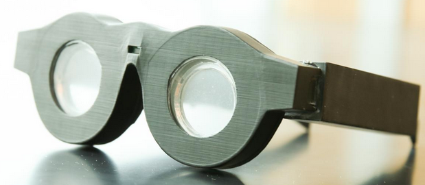
Early prototype of “smart glasses” with liquid-based lenses that can automatically adjust the focus on what a person is seeing, whether it’s far away or close up. The battery-powered frames can automatically adjust the focal length. Researchers expect to have smaller, lighter frames and packaged technology within three years. (credit: Dan Hixson/University of Utah College of Engineering)

Don’t throw away your bifocals or multiple glasses yet, but those days might soon be over. A team led by University of Utah engineers has created “smart glasses” with liquid-based lenses that can automatically adjust the focus on what you’re seeing, at any distance.

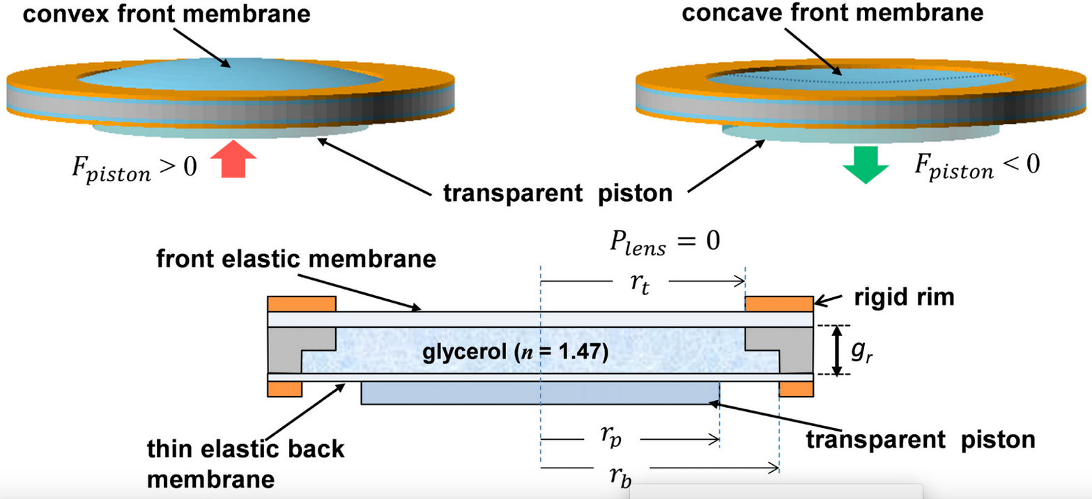
They’ve created eyeglass lenses made of glycerin, a thick colorless liquid, enclosed by flexible rubber-like membranes in the front and back. The rear membrane in each lens is connected to a series of three mechanical actuators that push the membrane back and forth like a transparent piston, changing the curvature of the liquid lens and therefore the focal length between the lens and the eye.

Simplified schematic of soft-membrane liquid lens (excluding actuators). The lens optical power is adjusted by vertically displacing the fluid with a transparent piston, deflecting the top membrane and changing its curvature. (credit: Nazmul Hasan et al./Optics Express)

In the bridge of the glasses is a distance meter that measures the distance from the glasses to an object via pulses of near-infrared light. When the wearer looks at an object, the meter instantly measures the distance and tells the actuators how to curve the lenses. If the user then sees another object that’s closer, the distance meter readjusts and tells the actuators to reshape the lens for farsightedness.

The lenses can change focus from one object to another in 14 milliseconds (faster than human reaction time). A rechargeable battery in the frames could last more than 24 hours per charge, according to electrical and computer engineering professor Carlos Mastrangelo, senior author of an open-access paper in a special edition of the journal Optics Express.

Before putting them on for the first time, users would input their eyeglasses prescription into an accompanying smartphone app, which then calibrates the lenses automatically via Bluetooth. Users only need to do that once, except for when their prescription changes over time. Theoretically, eyeglass wearers will never have to buy another pair again since these glasses would constantly adjust to their eyesight.

A startup company, Sharpeyes LLC, has been created to commercialize the glasses. The project was funded with a grant from the National Institutes of Health and the National Institute of Biomedical Imaging and Bioengineering.


University of Utah | Smart glasses that automatically focus on whatever you look at


Abstract of Tunable-focus lens for adaptive eyeglasses

We demonstrate the implementation of a compact tunable-focus liquid lens suitable for adaptive eyeglass application. The lens has an aperture diameter of 32 mm, optical power range of 5.6 diopter, and electrical power consumption less than 20 mW. The lens inclusive of its piezoelectric actuation mechanism is 8.4 mm thick and weighs 14.4 gm. The measured lens RMS wavefront aberration error was between 0.73 µm and 0.956 µm.

Scientists discover precise DNA sequence code critical for turning genes on

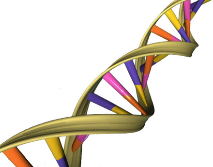
DNA sequence signal for the activation of human genes. Each tiny human cell contains about six feet of DNA, a double-helical molecular chain containing four types of several billion chemical nucleotides — adenine (A), cytosine (C), guanine (G) and thymine (T) — arranged in a specific sequence, or code, that when transcribed guide the cell into producing specific proteins. (credit: University of California — San Diego)

Molecular biologists at the University of California, San Diego (UC San Diego) have discovered a short sequence of DNA that is essential for turning on (expressing proteins) more than half of all human genes — an achievement that should provide scientists with a better understanding of how human genes are regulated.

Knowing what turns on genes is important. Each human cell contains about six feet of DNA, a double-helical molecular chain containing several billion chemical nucleotides — adenine (A), cytosine (C), guanine (G) and thymine (T) — arranged in a specific sequence, or code. Active genes undergo a process called transcription, in which the nucleotide sequence in DNA is read and converted into a sister language called RNA for processing. The processed RNA sequence then guides the cell to produce specific proteins that are essential for normal cellular functions.

A depiction of the double helical structure of DNA. Its four nucleotide coding units (A, T, C, G) are color-coded in pink, orange, purple and yellow. (credit: NHGRI)

“In these six feet of DNA, there are tens of thousands of genes, which are segments of DNA that direct specific functions, such as the production of a hormone or an enzyme,” explains James T. Kadonaga, PhD, a molecular biology professor at UC San Diego who headed the team of researchers. “It is essential for the cell to control the activity of each of its tens of thousands of genes, because the improper control of gene activity can lead to adverse outcomes such as cell death or the formation of a cancer cell.”

The “human Initiator”

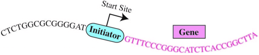
Enter the “Initiator.” The initiation of gene expression often occurs at a critical DNA sequence code called the “human Initiator.” This small piece of DNA helps gene expression machinery locate exactly where to begin transcribing. Although the concept of the Initiator has been known since the 1980s, the precise DNA sequence comprising the Initiator had eluded scientists.*

“There are many sequence signals that control gene activity in human cells and the Initiator is the most commonly occurring sequence at the start sites of genes,” Kadonaga said. Kadonaga and his team employed emerging genomic techniques and devised novel computational strategies to unlock the exact DNA sequence code for the human Initiator.

They also discovered that this sequence is located precisely at the start site of more than half of all human genes, underlining the importance of the human Initiator in the human genome. “The solution of the human Initiator code will enable us to explore new frontiers in gene regulation,” said Kadonaga. “In the future, it will be possible to use the code to identify other regulatory signals and in this way gain a more complete understanding of how human genes are turned on and off.”

“The authors verified the Initiator sequence in multiple cell lines, which is an impressive finding,” a scientist not involved in the studies told KurzweilAI. “However, none of these cell lines reflect normal human biology — they are essentially cancer cells proliferating in a dish. I would have liked to see this Initiator sequence verified in normal human cells from healthy patients.”

The research, now online and to be detailed in the February 10 print issue of the journal Genes & Development, was supported by grants from the National Institutes of Health.

* First observed by Pierre Chambon and his colleagues in Strasbourg, France in 1980, the human Initiator and its role in gene activation were articulated in 1989 by two MIT biologists, Stephen Smale and David Baltimore at MIT, who later revealed the approximate sequence code of the Initiator. Since then, however, other scientists had proposed a number of different sequences for the human Initiator, but none of them were found to be consistently associated with the start sites of human genes. As a result, the true Initiator sequence code remained a mystery — until now.


Abstract of The human initiator is a distinct and abundant element that is precisely positioned in focused core promoters

DNA sequence signals in the core promoter, such as the initiator (Inr), direct transcription initiation by RNA polymerase II. Here we show that the human Inr has the consensus of BBCA+1BW at focused promoters in which transcription initiates at a single site or a narrow cluster of sites. The analysis of 7678 focused transcription start sites revealed 40% with a perfect match to the Inr and 16% with a single mismatch outside of the CA+1 core. TATA-like sequences are underrepresented in Inr promoters. This consensus is a key component of the DNA sequence rules that specify transcription initiation in humans.

A deep learning algorithm outperforms some board-certified dermatologists in diagnosis of skin cancer

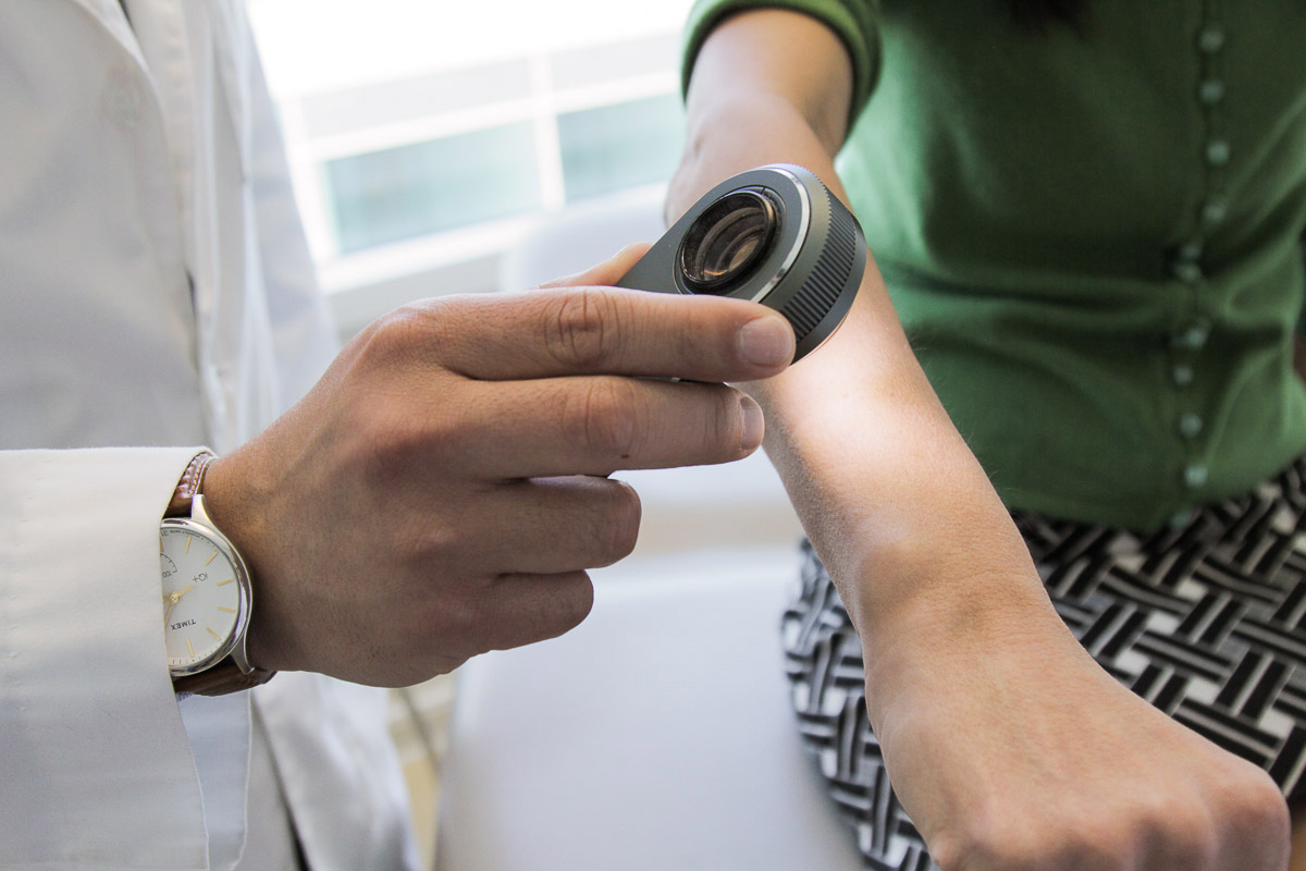
A dermatologist uses a dermatoscope, a type of handheld microscope, to look at skin. Stanford AI scientists have created a deep convolutional neural network algorithm for skin cancer that matched the performance of board-certified dermatologists. (credit: Matt Young)

Deep learning has been touted for its potential to enhance the diagnosis of diseases, and now a team of researchers at Stanford has developed a deep-learning algorithm that may make this vision a reality for skin cancer.*

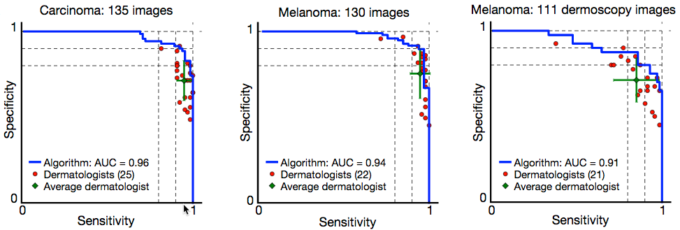
The researchers, led by Dr. Sebastian Thrun, an adjunct professor at the Stanford Artificial Intelligence Laboratory, reported in the January 25 issue of Nature that their deep convolutional neural network (CNN) algorithm performed as well or better than 21 board-certified dermatologists at diagnosing skin cancer. (See “Skin cancer classification performance of the CNN (blue) and dermatologists (red)” figure below.)

Diagnosing skin cancer begins with a visual examination. A dermatologist usually looks at the suspicious lesion with the naked eye and with the aid of a dermatoscope, which is a handheld microscope that provides low-level magnification of the skin. If these methods are inconclusive or lead the dermatologist to believe the lesion is cancerous, a biopsy is the next step. This deep learning algorithm may help dermatologists decide which skin lesions to biopsy.

“My main eureka moment was when I realized just how ubiquitous smartphones will be,” said Stanford Department of Electrical Engineering’s Andre Esteva, co-lead author of the study. “Everyone will have a supercomputer in their pockets with a number of sensors in it, including a camera. What if we could use it to visually screen for skin cancer? Or other ailments?”

It is projected that there will be 6.3 billion smartphone subscriptionst by the year 2021, according to Ericsson Mobility Report (2016), which could potentially provide low-cost universal access to vital diagnostic care.

Creating the deep convolutional neural network (CNN) algorithm

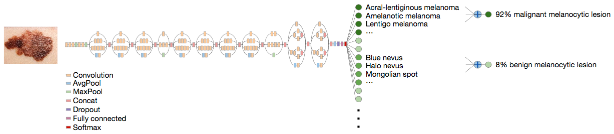
Deep CNN classification technique. Data flow is from left to right: an image of a skin lesion (for example, melanoma) is sequentially warped into a probability distribution over clinical classes of skin disease using Google Inception v3 CNN architecture pretrained on the ImageNet dataset (1.28 million images over 1,000 generic object classes) and fine-tuned on the team’s own dataset of 129,450 skin lesions comprising 2,032 different diseases. (credit: Andre Esteva et al./Nature)

Rather than building an algorithm from scratch, the researchers began with an algorithm developed by Google that was already trained to identify 1.28 million images from 1,000 object categories. It was designed primarily to be able to differentiate cats from dogs, but the researchers needed it to differentiate benign and malignant lesions. So they collaborated with dermatologists at Stanford Medicine, as well as Helen M. Blau, professor of microbiology and immunology at Stanford and co-author of the paper.

The algorithm was trained with nearly 130,000 images representing more than 2,000 different diseases with an associated disease label, allowing the system to overcome variations in angle, lighting, and zoom. The algorithm was then tested against 1,942 images of skin that were digitally annotated with biopsy-proven diagnoses of skin cancer. Overall, the algorithm identified the vast majority of cancer cases with accuracy rates that were similar to expert clinical dermatologists.

However, during testing, the researchers used only high-quality, biopsy-confirmed images provided by the University of Edinburgh and the International Skin Imaging Collaboration Project that represented the most common and deadliest skin cancers — malignant carcinomas and malignant melanomas.

Skin cancer classification performance of the CNN (blue) and dermatologists (red).** (credit: Andre Esteva et al./Nature)

The 21 dermatologists were asked whether, based on each image, they would proceed with biopsy or treatment, or reassure the patient. The researchers evaluated success by how well the dermatologists were able to correctly diagnose both cancerous and non-cancerous lesions in more than 370 images.***

However, Susan Swetter, professor of dermatology and director of the Pigmented Lesion and Melanoma Program at the Stanford Cancer Institute and co-author of the paper, notes that “rigorous prospective validation of the algorithm is necessary before it can be implemented in clinical practice, by practitioners and patients alike.”

* Every year there are about 5.4 million new cases of skin cancer in the United States, and while the five-year survival rate for melanoma detected in its earliest states is around 97 percent, that drops to approximately 14 percent if it’s detected in its latest stages.

** “Skin cancer classification performance of the CNN and dermatologists. The deep learning CNN outperforms the average of the dermatologists at skin cancer classification using photographic and
dermoscopic images. Our CNN is tested against at least 21 dermatologists at keratinocyte carcinoma and melanoma recognition. For each test, previously unseen, biopsy-proven images of lesions are displayed, and dermatologists are asked if they would: biopsy/treat the lesion or reassure the patient. Sensitivity, the true positive rate, and specificity, the true negative rate, measure performance. A dermatologist outputs a single prediction per image and is thus represented by a single red point. The green points are the average of the dermatologists for each task, with error bars denoting one standard deviation.” — Andre Esteva et al./Nature

*** The algorithm’s performance was measured through the creation of a sensitivity-specificity curve, where sensitivity represented its ability to correctly identify malignant lesions and specificity represented its ability to correctly identify benign lesions. It was assessed through three key diagnostic tasks: keratinocyte carcinoma classification, melanoma classification, and melanoma classification when viewed using dermoscopy. In all three tasks, the algorithm matched the performance of the dermatologists with the area under the sensitivity-specificity curve amounting to at least 91 percent of the total area of the graph. An added advantage of the algorithm is that, unlike a person, the algorithm can be made more or less sensitive, allowing the researchers to tune its response depending on what they want it to assess. This ability to alter the sensitivity hints at the depth and complexity of this algorithm. The underlying architecture of seemingly irrelevant photos —  including cats and dogs — helps it better evaluate the skin lesion images.


Abstract of Dermatologist-level classification of skin cancer with deep neural networks

Skin cancer, the most common human malignancy, is primarily diagnosed visually, beginning with an initial clinical screening and followed potentially by dermoscopic analysis, a biopsy and histopathological examination. Automated classification of skin lesions using images is a challenging task owing to the fine-grained variability in the appearance of skin lesions. Deep convolutional neural networks (CNNs) show potential for general and highly variable tasks across many fine-grained object categories. Here we demonstrate classification of skin lesions using a single CNN, trained end-to-end from images directly, using only pixels and disease labels as inputs. We train a CNN using a dataset of 129,450 clinical images—two orders of magnitude larger than previous datasets—consisting of 2,032 different diseases. We test its performance against 21 board-certified dermatologists on biopsy-proven clinical images with two critical binary classification use cases: keratinocyte carcinomas versus benign seborrheic keratoses; and malignant melanomas versus benign nevi. The first case represents the identification of the most common cancers, the second represents the identification of the deadliest skin cancer. The CNN achieves performance on par with all tested experts across both tasks, demonstrating an artificial intelligence capable of classifying skin cancer with a level of competence comparable to dermatologists. Outfitted with deep neural networks, mobile devices can potentially extend the reach of dermatologists outside of the clinic. It is projected that 6.3 billion smartphone subscriptions will exist by the year 2021 and can therefore potentially provide low-cost universal access to vital diagnostic care.

A 3D bioprinter that prints fully functional human skin

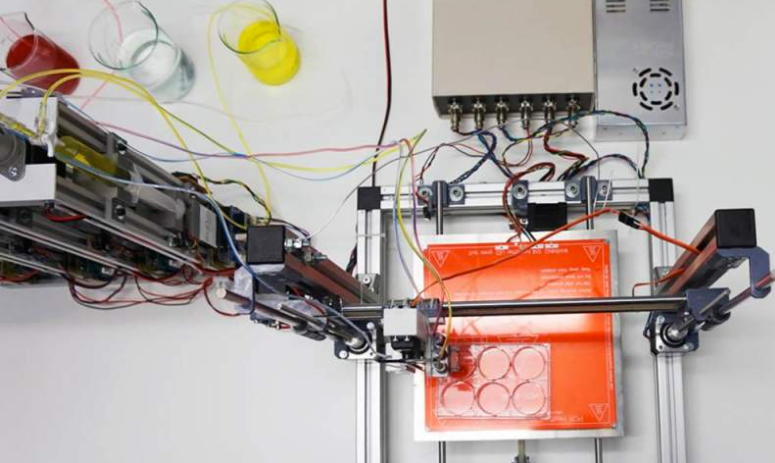
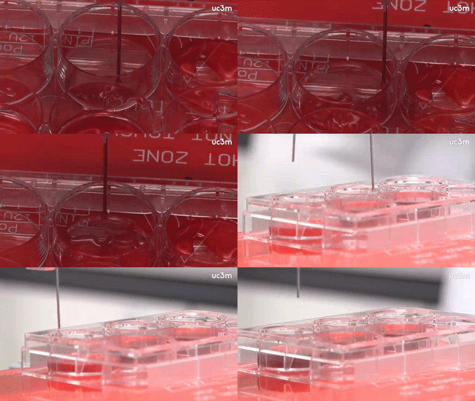
Skin-producing bioprinter (credit: Universidad Carlos III de Madrid)

A prototype 3D bioprinter that can create totally functional human skin has been developed by scientists from Universidad Carlos III de Madrid (UC3M) and BioDan Group in Spain. The skin has been used to treat burns as well as traumatic and surgical wounds in a large number of patients in Spain, according to the scientists.

The system provides two processes.

Autologous skin (from the patient’s own cells to generate human collagen) for therapeutic use, such as in the treatment of severe burns, instead of the animal collagen used in other methods.  The researchers have applied for approval by various European regulatory agencies to guarantee that the skin that is produced is adequate for use in transplants on burn patients and on those with other skin problems.

3D skin bioprinter in operation (credit: Universidad Carlos III de Madrid)

The 3D-printed skin replicates human bilayered skin, using “bioinks” (biological components) containing human plasma as well as primary human fibroblasts and keratinocytes obtained from skin biopsies. These are controlled by a computer, which deposits them on a print bed in an orderly manner to then produce the skin.

The researchers were able to generate 100 cm2 of printed skin in less than 35 minutes (including the 30 min required for fibrin gelation).

Allogeneic skin (from a stock of cells), done on a large scale for industrial processes. This skin can be used to test pharmaceutical products, cosmetics, and consumer chemical products where current regulations require testing that does not use animals.

“This method of bioprinting allows skin to be generated in a standardized, automated way, and the process is less expensive than manual production,” says Alfredo Brisac, CEO of BioDan Group, the Spanish bioengineering firm specializing in regenerative medicine that is collaborating on this research and commercializing this technology.

The research was published online in the journal Biofabrication.


UC3M | Científicos españoles crean una bioimpresora 3D de piel humana


Abstract of 3D bioprinting of functional human skin: production and in vivo analysis

Significant progress has been made over the past 25 years in the development of in vitro-engineered substitutes that mimic human skin, either to be used as grafts for the replacement of lost skin, or for the establishment of in vitro human skin models. In this sense, laboratory-grown skin substitutes containing dermal and epidermal components offer a promising approach to skin engineering. In particular, a human plasma-based bilayered skin generated by our group, has been applied successfully to treat burns as well as traumatic and surgical wounds in a large number of patients in Spain. There are some aspects requiring improvements in the production process of this skin; for example, the relatively long time (three weeks) needed to produce the surface required to cover an extensive burn or a large wound, and the necessity to automatize and standardize a process currently performed manually. 3D bioprinting has emerged as a flexible tool in regenerative medicine and it provides a platform to address these challenges. In the present study, we have used this technique to print a human bilayered skin using bioinks containing human plasma as well as primary human fibroblasts and keratinocytes that were obtained from skin biopsies. We were able to generate 100 cm2, a standard P100 tissue culture plate, of printed skin in less than 35 min (including the 30 min required for fibrin gelation). We have analysed the structure and function of the printed skin using histological and immunohistochemical methods, both in 3D in vitro cultures and after long-term transplantation to immunodeficient mice. In both cases, the generated skin was very similar to human skin and, furthermore, it was indistinguishable from bilayered dermo-epidermal equivalents, handmade in our laboratories. These results demonstrate that 3D bioprinting is a suitable technology to generate bioengineered skin for therapeutical and industrial applications in an automatized manner.

AI system performs better than 75 percent of American adults on standard visual intelligence test

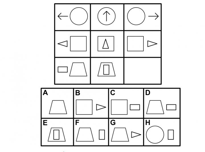
An example question from the Raven’s Progressive Matrices standardized fluid-intelligence test.* (credit: Ken Forbus)

A Northwestern University team has developed a new visual problem-solving computational model that performs in the 75th percentile for American adults on a standard intelligence test.

The research is an important step toward making artificial-intelligence systems that see and understand the world as humans do, says Northwestern Engineering’s Ken Forbus, Walter P. Murphy Professor of Electrical Engineering and Computer Science at Northwestern’s McCormick School of Engineering. The research was published online in January 2017 in the journal Psychological Review.

The new computational model** is built on CogSketch, an AI platform previously developed in Forbus’ laboratory. It can solve visual problems and understand sketches to give immediate, interactive feedback. CogSketch also incorporates a computational model of analogy, based on Northwestern psychology professor Dedre Gentner’s structure-mapping engine.

The ability to solve complex visual problems is one of the hallmarks of human intelligence. Developing artificial intelligence systems that have this ability provides new evidence for the importance of symbolic representations and analogy in visual reasoning, and it could potentially shrink the gap between computer and human cognition, the researchers suggest.

A nonverbal fluid-intelligence test

The researchers tested the AI system on Raven’s Progressive Matrices, a nonverbal standardized test that measures abstract reasoning.*** All of the test’s problems consist of a matrix with one image missing. The test taker is given six to eight choices for completing the matrix.

“The problems that are hard for people are also hard for the model, providing additional evidence that its operation is capturing some important properties of human cognition,” said Forbus.

“The Raven’s test is the best existing predictor of what psychologists call ‘fluid intelligence,’ or the general ability to think abstractly, reason, identify patterns, solve problems, and discern relationships,” said co-author Andrew Lovett, now a researcher at the U.S. Naval Research Laboratory. “Our results suggest that the ability to flexibly use relational representations, comparing and reinterpreting them, is important for fluid intelligence.”

“Most artificial intelligence research today concerning vision focuses on recognition, or labeling what is in a scene rather than reasoning about it,” Forbus said. “But recognition is only useful if it supports subsequent reasoning. Our research provides an important step toward understanding visual reasoning more broadly.”

* The test taker should choose answer D because the relationships between it and the other elements in the bottom row are most similar to the relationships between the elements of the top rows.

** “The reader may download (Windows) the computational model and run it on example problems,” the authors note in their Psychological Review paper.

** Raven’s Progressive Matrices (RPM) is an intelligence test that “requires that participants compare images in a (usually) 3×3 matrix, identify a pattern across the matrix, and solve for the missing image.” … Designed to measure a subject’s fluid intelligence, “it has remained popular for decades because it is highly successful at predicting a subject’s performance on other ability tests — not just visual tests, but verbal and mathematical as well,” the authors suggest in their Psychological Review paper.


Abstract of Modeling visual problem solving as analogical reasoning

We present a computational model of visual problem solving, designed to solve problems from the Raven’s Progressive Matrices intelligence test. The model builds on the claim that analogical reasoning lies at the heart of visual problem solving, and intelligence more broadly. Images are compared via structure mapping, aligning the common relational structure in 2 images to identify commonalities and differences. These commonalities or differences can themselves be reified and used as the input for future comparisons. When images fail to align, the model dynamically rerepresents them to facilitate the comparison. In our analysis, we find that the model matches adult human performance on the Standard Progressive Matrices test, and that problems which are difficult for the model are also difficult for people. Furthermore, we show that model operations involving abstraction and rerepresentation are particularly difficult for people, suggesting that these operations may be critical for performing visual problem solving, and reasoning more generally, at the highest level. (PsycINFO Database Record (c) 2016 APA, all rights reserved)

Magnetic brain stimulation improves a precise type of memory

An individual receiving noninvasive rTMS brain stimulation via electromagnetic coil (credit: Northwestern University)

Non-invasive magnetic brain stimulation can be used to precisely improve a specific type of memory — remembering highly precise contextual and spatial information — Northwestern Medicine scientists shown for the first time.

The new research could help in developing new treatments for people with brain injuries or dementia, said Joel Voss, assistant professor of medical social sciences at Northwestern University Feinberg School of Medicine, senior author of a paper published Jan. 19 in Current Biology.

Blue indicates where electromagnetic pulses were applied to modify the specific “core” (red) of a precise memory network in the hippocampus.  (credit: Northwestern University)

Precise memory (known as “recollection precision”), rather than general memory, is critical for knowing details such as the specific color, shape, and location of a building you are looking for, rather than simply knowing the part of town it’s in. This type of memory is crucial for normal functioning, and it is often lost in people with serious memory disorders.

Improving the spatial-precision-memory part of the brain

“We show that it is possible to target the portion of the brain responsible for this type of memory and to improve it,” said Voss. “People with brain injuries have problems with precise memory as do individuals with dementia, and so our findings could be useful in  for these conditions.”

The scientists first used MRI to identify brain networks related to spatial precision memory. Then they stimulated the specific brain network responsible for spatial memory* with repetitive transcranial magnetic stimulation (rTMS), using powerful electromagnets. That allowed the scientists to improve the precision of people’s memory for identifying locations. This benefit lasted a full 24 hours after receiving stimulation and corresponded to changes in brain activity.

Most previous studies using noninvasive magnetic brain stimulation have found only very general and short-lived effects on thinking abilities, rather than highly specific and long-lasting effects on an ability such as precise memory. The scientists used detailed memory tests to show that this procedure actually improved spatial-precision memory; they used EEG to show that these memory improvements corresponded to indicators of improved brain network function.

The research was supported by grants from the National Institute of Mental Health, the National Institute on Aging and the National Institute of Neurological Disorders and Stroke, all of the National Institutes of Health.

* The “hippocampal posterior-medial (HPM) network” (for which evidence has previously been mostly indirect, derived from correlative measures such as EEG neural-activity recordings).


Abstract of Stimulation of the Posterior Cortical-Hippocampal Network Enhances Precision of Memory Recollection

Episodic memory is thought to critically depend on interaction of the hippocampus with distributed brain regions [ 1–3 ]. Specific contributions of distinct networks have been hypothesized, with the hippocampal posterior-medial (HPM) network implicated in the recollection of highly precise contextual and spatial information [ 3–6 ]. Current evidence for HPM specialization is mostly indirect, derived from correlative measures such as neural activity recordings. Here we tested the causal role of the HPM network in recollection using network-targeted noninvasive brain stimulation in humans, which has previously been shown to increase functional connectivity within the HPM network [ 7 ]. Effects of multiple-day electromagnetic stimulation were assessed using an object-location memory task that segregated recollection precision from general recollection success. HPM network-targeted stimulation produced lasting (∼24 hr) enhancement of recollection precision, without effects on general success. Canonical neural correlates of recollection [ 8–10 ] were also modulated by stimulation. Late-positive evoked potential amplitude and theta-alpha oscillatory power were reduced, suggesting that stimulation can improve memory through enhanced reactivation of detailed visuospatial information at retrieval. The HPM network was thus specifically implicated in the processing of fine-grained memory detail, supporting functional specialization of hippocampal-cortical networks. These findings demonstrate that brain networks can be causally linked to distinct and specific neurocognitive functions and suggest mechanisms for long-lasting changes in memory due to network-targeted stimulation.

Microbiologists make big leap in developing ‘green’ electronics


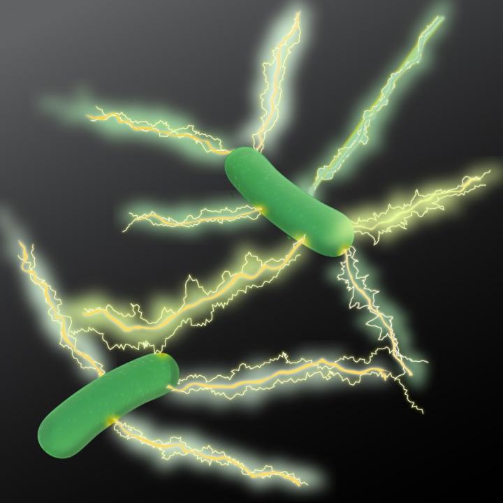
An artist’s rendition of Geobacter expressing electrically conductive nanowires. Microbiologists at UMass Amherst have discovered a new type of natural wire produced by bacteria that could greatly accelerate the development of sustainable “green” conducting materials for the electronics industry. (credit: UMass Amherst)

UMass Amherst research finds microbe yields better electronic material

Microbiologists at the University of Massachusetts Amherst report that they have discovered a new type of microbial nanowire produced by bacteria that could greatly accelerate the development of sustainable “green” conducting materials for the electronics industry.

The study by Derek Lovley and colleagues appears this week in an open-access paper in mBio, the American Society of Microbiology’s premier journal.

A bacterium known as Geobacter sulfurreducens uses the protein filaments naturally to make electrical connections with other microbes or minerals.

As Lovley explains, “Microbial nanowires are a revolutionary electronic material with substantial advantages over man-made materials. Chemically synthesizing nanowires in the lab requires toxic chemicals, high temperatures and/or expensive metals. The energy requirements are enormous. By contrast, natural microbial nanowires can be mass-produced at room temperature from inexpensive renewable feedstocks in bioreactors with much lower energy inputs. And the final product is free of toxic components.”

Confocal scanning laser micrographs of G. sulfurreducens anode biofilms harvested on day 10. Bar, 25 µm. (credit: Yang Tan et al./mBio)

The Microbial nanowires offer an unprecedented potential for developing novel electronic devices and sensors for diverse applications with a new environmentally friendly technology, Lovely says. “This is an important advance in microbial nanowire technology. The approach we outline in this paper demonstrates a rapid method for prospecting in nature to find better electronic materials.”

When his lab began looking at the protein filaments of other Geobacter species, they were surprised to find a wide range in conductivities. For example, one species recovered from uranium-contaminated soil produced poorly conductive filaments. However, another species, Geobacter metallireducens produced nanowires 5,000 times more conductive than the G. sulfurreducens wires. Lovley recalls, “I isolated metallireducens from mud in the Potomac River 30 years ago, and every couple of years it gives us a new surprise.”

In their new study supported by the U.S. Office of Naval Research, they did not study the G. metallireducens strain directly. Instead, they took the gene for the protein that assembles into microbial nanowires from it and inserted this into G. sulfurreducens. The result is a genetically modified G. sulfurreducens that expresses the G. metallireducens protein, making nanowires much more conductive than G. sulfurreducens would naturally produce.

Further, Lovley says, “We have found that G. sulfurreducens will express filament genes from many different types of bacteria. This makes it simple to produce a diversity of filaments in the same microorganism and to study their properties under similar conditions.”

The high conductivity of the G. metallireducens nanowires suggests that they may be an attractive material for the construction of conductive materials, electronic devices ,and sensors for medical or environmental applications. The authors say discovering more about the mechanisms of nanowire conductivity “provides important insight into how we might make even better wires with genes that we design ourselves.”


Abstract of Expressing the Geobacter metallireducens PilA in Geobacter sulfurreducens Yields Pili with Exceptional Conductivity

The electrically conductive pili (e-pili) of Geobacter sulfurreducens serve as a model for a novel strategy for long-range extracellular electron transfer. e-pili are also a new class of bioelectronic materials. However, the only other Geobacter pili previously studied, which were from G. uraniireducens, were poorly conductive. In order to obtain more information on the range of pili conductivities in Geobacter species, the pili of G. metallireducens were investigated. Heterologously expressing the PilA gene of G. metallireducens in G. sulfurreducens yielded a G. sulfurreducens strain, designated strain MP, that produced abundant pili. Strain MP exhibited phenotypes consistent with the presence of e-pili, such as high rates of Fe(III) oxide reduction and high current densities on graphite anodes. Individual pili prepared at physiologically relevant pH 7 had conductivities of 277 ± 18.9 S/cm (mean ± standard deviation), which is 5,000-fold higher than the conductivity of G. sulfurreducens pili at pH 7 and nearly 1 million-fold higher than the conductivity of G. uraniireducens pili at the same pH. A potential explanation for the higher conductivity of the G. metallireducens pili is their greater density of aromatic amino acids, which are known to be important components in electron transport along the length of the pilus. The G. metallireducens pili represent the most highly conductive pili found to date and suggest strategies for designing synthetic pili with even higher conductivities.

A ‘smart’ patch that automatically delivers insulin when needed

Tiny, painless microneedles on a patch can deliver insulin in response to rising glucose levels (credit: American Chemical Society)

A team of scientists has invented a replacement for daily glucose-level finger-pricking and insulin shots: a painless “smart” patch that monitors blood glucose and releases insulin when levels climb too high.

The report on the device, which has only been tested on mice so far, appears in the journal ACS Nano.

People with Type 1 diabetes don’t make insulin — a hormone that regulates blood glucose (sugar). Those with Type 2 diabetes can’t use insulin effectively. Either way, glucose builds up in the blood, which can lead to a host of health problems, including heart disease, stroke, blindness and amputation of toes, feet or legs.

To avoid these outcomes, people with Type 1 or advanced Type 2 diabetes regularly prick their fingers to measure blood-sugar levels, and some patients must inject themselves with insulin when needed. But sometimes, despite a person’s vigilance, glucose levels can still get out of whack.

A skin patch with painless microneedles

Self-assembly of block copolymer into vesicles loaded with insulin and glucose oxidase. The vesicles (engineered pouches) are dissociated to release insulin in the presence of a hyperglycemic state. (credit: American Chemical Society)

So Zhen Gu and colleagues* decided to invent a simpler, more effective, shot-free way to manage diabetes: a skin patch covered in painless microneedles that are loaded with tiny insulin-carrying pouches. The pouches (vesicles) are engineered to break apart rapidly and release the insulin in response to rising glucose levels.

Diabetic mice wearing the patch maintained consistent concentrations of insulin in their blood. When these mice received a shot of glucose, their blood sugar levels spiked initially, but then fell to normal levels within two hours.

Another automated approach: Insulin Pump Therapy system: 1. Insulin pump. 2: Flexible tubing delivers insulin from the pump reservoir to the infusion set. 3. A tiny tube called a cannula is inserted under your skin to deliver insulin. 4. Insulin in the blood (credit: Medtronics)

The authors acknowledge funding from the American Diabetes Association, National Institutes of Health, and the National Science Foundation.

* Affiliated with the University of North Carolina at Chapel Hill, North Carolina State University, and State Key Laboratory of Polymer Chemistry and Physics, Changchun Institute of Applied Chemistry, Chinese Academy of Sciences.


Abstract of H2O2-Responsive Vesicles Integrated with Transcutaneous Patches for Glucose-Mediated Insulin Delivery

A self-regulated “smart” insulin administration system would be highly desirable for diabetes management. Here, a glucose-responsive insulin delivery device, which integrates H2O2-responsive polymeric vesicles (PVs) with a transcutaneous microneedle-array patch was prepared to achieve a fast response, excellent biocompatibility, and painless administration. The PVs are self-assembled from block copolymer incorporated with polyethylene glycol (PEG) and phenylboronic ester (PBE)-conjugated polyserine (designated mPEG-b-P(Ser-PBE)) and loaded with glucose oxidase (GOx) and insulin. The polymeric vesicles function as both moieties of the glucose sensing element (GOx) and the insulin release actuator to provide basal insulin release as well as promote insulin release in response to hyperglycemic states. In the current study, insulin release responds quickly to elevated glucose and its kinetics can be modulated by adjusting the concentration of GOx loaded into the microneedles. In vivo testing indicates that a single patch can regulate glucose levels effectively with reduced risk of hypoglycemia.

Woman dies from antibiotic-resistant bacteria when no antibiotics worked

Carbapenem-resistant Enterobacteriaceae (CRE) bacteria (credit: Melissa Brower/CDC)

The death of a hospitalized patient in Reno Nevada for whom no available antibiotics worked highlights what World Health Organization and other public-health experts have been warning: antibiotic resistance is a serious threat and has gone global.

The patient — a female in her 70s — was admitted in for an infection and died in September 2016 from septic shock the CDC announced on Jan. 13. The patient had been treated for multiple infections in India before traveling to the United States. The infection that led to her hospitalization in Reno was caused by a strain of carbapenem-resistant Enterobacteriaceae (CRE)* bacteria known as Klebsiella pneumoniae. Although not all strains of Klebsiella pneumonia are CRE, the strain that infected this patient was resistant to all available antibiotics, according to the CDC. (Carbapeneum is a “drug of last resort.”)

In a paper in The Lancet in October, researchers reported that more than a third of blood infections in newborn babies involving Klebsiella pneumoniae and similar bacteria were resistant to multiple drugs to the point they were virtually untreatable and “threaten the return of a pre-antibiotic era in Indian neonatal intensive care units,” the study’s authors warned.

Spreading more widely and stealthily that thought

Highlighting the importance of this report, a new NIH-funded study published online January 16, 2017 in PNAS (Proceedings of the National Academy of Sciences) from Harvard T.H. Chan School of Public Health and the Broad Institute of MIT and Harvard found that the CRE family of highly drug-resistant and potentially deadly bacteria may be spreading more widely — and more stealthily — than previously thought.

The researchers examined CRE bacteria that were isolated from patients with infections at four U.S. hospitals. They found a wide variety of CRE . They also found a diverse array of genetic traits enabling CRE to resist antibiotics, and found that these traits are transferring easily among various CRE species.

The findings suggest that CRE may well be transmitting from person to person asymptomatically, and that genomic surveillance of this dangerous bacteria should be increased.

CRE are a class of bacteria that is resistant to multiple antibiotics, including carbapenems, which are considered last-resort drugs when other antibiotics have failed. CRE, which tend to spread in hospitals and long-term care facilities, cause an estimated 9,300 infections and 600 deaths in the U.S. each year, according to the CDC — and incidence is on the rise. CDC director Tom Frieden has called these “nightmare bacteria” because they are resistant to some of the last-ditch treatments available to doctors battling resistant infections.

A type of ultraviolet light called UVC could aid hospitals in the ongoing battle to keep drug-resistant bacteria from lingering in patient rooms and causing new infections. A CDC-NIH-funded study led by Duke Health and published in The Lancet finds use of UVC machines can cut transmission of four major superbugs by a cumulative 30 percent. (credit: Shawn Rocco/Duke Health)

Unobserved transmissions

“While the typical focus has been on treating sick patients with CRE-related infections, our new findings suggest that CRE is spreading beyond the obvious cases of disease,” said William Hanage, associate professor of epidemiology at Harvard Chan School and senior author of the study. “We need to look harder for this unobserved transmission within our communities and healthcare facilities if we want to stamp it out.”

The researchers looked at about 250 samples of CRE from hospitalized patients from three Boston-area hospitals and from one California hospital.

The researchers found what Hanage termed a “riot of diversity,” both among CRE species and among carbapenem resistance genes. They also found that resistance genes are moving easily from species to species, contributing to a continually evolving threat from CRE.

In addition, the researchers found that some CRE bacteria employ uncharacterized resistance mechanisms—implying that there are more to be discovered. The finding highlights the need for vigilance in searching for as yet unknown forms of resistance as they evolve and emerge.

What if there were no new antibacterial drugs?

But what’s also needed is new antibiotics. “Meaningful incentives for pharmaceutical companies have been suggested to encourage them to re-enter this area of drug development; these incentives include models to push companies to develop these drugs or by making it economically viable so as to pull companies back to this area as well as alternative routes of funding,” notes Prof Laura J V Piddock, Antimicrobial Agents Research Group, School of Immunity and Infection, College of Medical and Dental Sciences, University of Birmingham in “The crisis of no new antibiotics—what is the way forward?” in Lancet Infect Dis. in 2012.

“Also, few data exist on the human and economic costs of antibacterial resistance or the potential costs of inaction (i.e., what would the social effects be if no new antibacterial drugs were available and fatal infections became the norm?). To assess such costs, involvement of governmental agencies that recommend use of drugs is needed (i.e., FDA, European Medicines Agency, and in individual countries such as UK, The National Institute for Health and Clinical Excellence).”

* “Carbapenems are one of few antimicrobials that have been effective against multidrug-resistant bacteria, but their utility is threatened by the emergence of carbapenem-resistant Enterobacteriaceae (CRE),” according to an open-access PLoS ONE paper published in 2014. “Klebsiella pneumoniae is the most common CRE species in the United States, typically encountered as a hospital-acquired infection with high morbidity and mortality, and resistant to nearly all available antibiotics.” In this case, metallo-β-lactamase NDM-1 (New Delhi metallo-beta-lactamase or NDM), a version of Klebsiella pneumoniae, was found.

Wearable sensors can alert you when you are getting sick, Stanford study shows

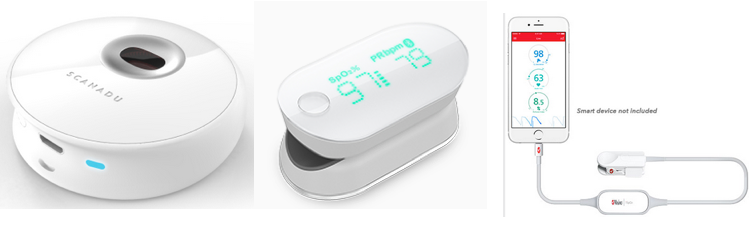
Current versions of three of the devices used for heart-rate and peripheral capillary oxygen saturation measurements in the study (credits left to right: Scanadu, iHealth, and Masimo)

Fitness monitors and other wearable biosensors can tell when your heart rate, activity, skin temperature, and other measures are abnormal, suggesting possible illness, including the onset of infection, inflammation, and even insulin resistance, according to a study by researchers at the Stanford University School of Medicine.

The team collected nearly 2 billion measurements from 60 people, including continuous data from each participant’s wearable biosensor devices* and periodic data from laboratory tests of their blood chemistry, gene expression, and other measures, and related this data to a range of normal (baseline) values for each person in the study compared to when they were ill.**

Participants wore between one and seven commercially available activity monitors and other monitors that collected more than 250,000 measurements a day. The team collected data on weight; heart rate; oxygen in the blood; skin temperature; activity, including sleep, steps, walking, biking and running; calories expended; acceleration; and even exposure to gamma rays and X-rays.

There were three participant groups: Participant #1 wore seven portable devices for large segments of this study; 43 individuals wore Intel’s Basis to measure activity (steps), heart rate, sleep, and skin temperature, with data securely uploaded to the cloud; and 16 individuals wore either iHealth or Masimo finger devices for sensing heart rate and SpO2 (peripheral capillary oxygen saturation).

Identifying health problems in advance

Wearable devices used by participant 1 (credit: Xiao Li/PLOS Biology)

The study, led by Michael Snyder, PhD, professor and chair of genetics, and senior author of the study, was published online Jan. 12 in open-access PLOS Biology. It demonstrated that, given a baseline range of values for each person, it is possible to monitor deviations from normal and associate those deviations with environmental conditions, illness, or other factors that affect health. Distinctive patterns of deviation from normal seem to correlate with particular health problems. Algorithms designed to pick up on these patterns of change could potentially contribute to clinical diagnostics and research.

The results of the current study raise the possibility of identifying inflammatory disease in individuals who may not even know they are getting sick.

For example, in several participants, higher-than-normal readings for heart rate and skin temperature correlated with increased levels of C reactive protein in blood tests. C reactive protein is an immune system marker for inflammation and often indicative of infection, autoimmune diseases, developing cardiovascular disease or even cancer. Snyder’s own data revealed four separate bouts of illness and inflammation, including a Lyme disease infection and another that he was unaware of until he saw his sensor data and an increased level of C reactive protein.

The wearable devices could also help distinguish participants with insulin resistance, a precursor for Type 2 diabetes. Of 20 participants who received glucose tests, 12 were insulin-resistant. The team designed and tested an algorithm combining participants’ daily steps, daytime heart rate and the difference between daytime and nighttime heart rate. The algorithm was able to process the data from just these few simple measures to predict which individuals in the study were likely to be insulin-resistant.

The study also revealed that declines in blood-oxygen levels during airplane flights were correlated with fatigue. Fortunately, the study showed that people tend to adapt on long flights; oxygen levels in their blood go back up, and they generally feel less fatigued as the hours go by.

The future of wearable devices: monitoring human health continuously

During a visit to the doctor, patients normally have their blood pressure and body temperature measured, but such data is typically collected only every year or two and often ignored unless the results are outside of normal range for entire populations. But biomedical researchers envisage a future in which human health is monitored continuously.

“We have more sensors on our cars than we have on human beings,” said Snyder. In the future, he said, he expects the situation will be reversed and people will have more sensors than cars do. Already, consumers have purchased millions of wearable devices, including more than 50 million smart watches and 20 million other fitness monitors. Most monitors are used to track activity, but they could easily be adjusted to more directly track health measures, Snyder said.

The work is an example of Stanford Medicine’s focus on “precision health,” whose goal is to anticipate and prevent disease in the healthy and to precisely diagnose and treat disease in the ill. With a precision health approach, every person could know his or her normal baseline for dozens of measures. Automatic data analysis could spot patterns of outlier data points and flag the onset of ill health, providing an opportunity for intervention, prevention or cure.

Researcher Elizabeth Colbert, of the Veterans Affairs Palo Alto Health Care System, is also a co-author. This research was funded by the National Institutes of Health, a gift from Bert and Candace Forbes, and Stanford’s Department of Genetics.

* “After evaluating more than 400 available wearable devices at the beginning of the study, we selected [seven] for participants to use. The criteria for selection [were] (1) ability to access the raw data from the manufacturer, (2) cost, (3) overlap in measurement of at least one component with another device to assist in reproducibility, and (4) ease of use, reasonable accuracy, and had a direct interface for raw data. These devices collectively measure (a) three physiological parameters, including heart rate, peripheral capillary oxygen saturation, and skin temperature, (b) six activity-related parameters, including sleep, steps, walking, biking, running, calories, and acceleration forces caused by movement, (c) weight, and (d) total gamma and X-ray radiation exposure.” — PLOS Biology paper authors

** “In this work, we investigate the use of portable devices to (1) easily and accurately record physiological measurements in individuals in real time (or at high frequency), (2) quantify daily patterns and reveal interesting physiological responses to different circadian cycles and environmental conditions, (3) identify personalized baseline norms and differences among individuals, (4) detect differences in health states among individuals (e.g., people with diabetes versus people without diabetes), and (5) detect inflammatory responses and assist in medical diagnosis at the early phase of disease development, thereby potentially impacting medical care.” — PLOS Biology paper authors


Abstract of Digital Health: Tracking Physiomes and Activity Using Wearable Biosensors Reveals Useful Health-Related Information

A new wave of portable biosensors allows frequent measurement of health-related physiology. We investigated the use of these devices to monitor human physiological changes during various activities and their role in managing health and diagnosing and analyzing disease. By recording over 250,000 daily measurements for up to 43 individuals, we found personalized circadian differences in physiological parameters, replicating previous physiological findings. Interestingly, we found striking changes in particular environments, such as airline flights (decreased peripheral capillary oxygen saturation [SpO2] and increased radiation exposure). These events are associated with physiological macro-phenotypes such as fatigue, providing a strong association between reduced pressure/oxygen and fatigue on high-altitude flights. Importantly, we combined biosensor information with frequent medical measurements and made two important observations: First, wearable devices were useful in identification of early signs of Lyme disease and inflammatory responses; we used this information to develop a personalized, activity-based normalization framework to identify abnormal physiological signals from longitudinal data for facile disease detection. Second, wearables distinguish physiological differences between insulin-sensitive and -resistant individuals. Overall, these results indicate that portable biosensors provide useful information for monitoring personal activities and physiology and are likely to play an important role in managing health and enabling affordable health care access to groups traditionally limited by socioeconomic class or remote geography.