Immune cells in covering of brain discovered; may play critical role in battling neurological diseases

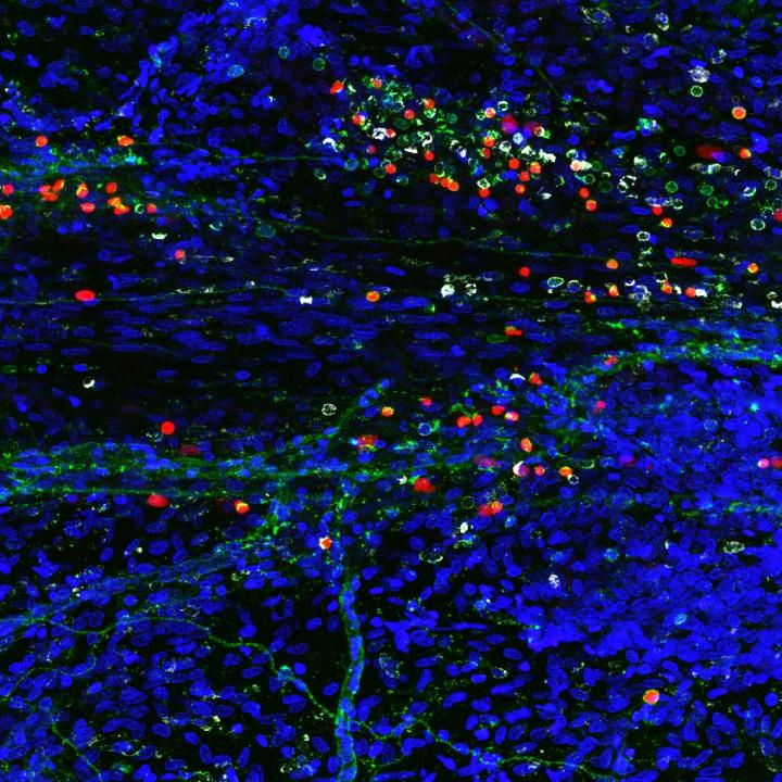
A composite image showing newly discovered immune cells in the brain (credit: Sachin Gadani | University of Virginia School of Medicine)

University of Virginia School of Medicine researchers have discovered a rare and powerful type of immune cell in the meninges (protective covering) of the brain that are activated in response to central nervous system injury — suggesting that these cells may play a critical role in battling Alzheimer’s, multiple sclerosis, meningitis, and other neurological diseases, and in supporting healthy mental functioning.

By harnessing the power of the cells, known as “type 2 innate lymphocytes” (ILC2s), doctors may be able to develop new treatments for neurological diseases, traumatic brain injury, and spinal cord injuries, as well as migraines, the researchers suggest. They also suspect the cells may be the missing link connecting the brain and the microbiota in our guts, a relationship that has been shown to be important in the development of Parkinson’s disease.

Important immune roles

ILC2 cells have previously been found in the gut, lung, and skin, the body’s barriers to disease. Their discovery by UVA researcher Jonathan Kipnis, PhD, in the meninges, the membranes surrounding the brain, comes as a surprise. They were found along the same vessels discovered by the Kipnis lab last year, which showed that the brain and the immune system are directly connected.

“This all comes down to immune system and brain interaction,” said Kipnis, chairman of UVA’s Department of Neuroscience. These where previously believed to be not communicating, but not only are these [immune] cells present in the areas near the brain, they are integral to its function, Kipnis said.

Immune cells play several important roles within the body, including guarding against pathogens, triggering allergic reactions, and responding to spinal cord injuries. But its their role in the gut that makes Kipnis suspect they may also be serving as a vital communicator between the brain’s immune response and our microbiomes (microbes in the body). That could be very important, because our intestinal flora is critical for maintaining our health and well being.

“These cells are potentially the mediator between the gut and the brain. They are the main responder to microbiota changes in the gut,” Kipnis said. “They may go from the gut to the brain, or they may just produce something that will impact those cells. We know the brain responds to things happening in the gut. Is it logical that these will be the cells that connect the two? Potentially.”

The findings have been published online by the Journal of Experimental Medicine. The work was supported by a National Institutes of Health grant.


Abstract of Characterization of meningeal type 2 innate lymphocytes and their response to CNS injury

The meningeal space is occupied by a diverse repertoire of immune cells. Central nervous system (CNS) injury elicits a rapid immune response that affects neuronal survival and recovery, but the role of meningeal inflammation remains poorly understood. Here, we describe type 2 innate lymphocytes (ILC2s) as a novel cell type resident in the healthy meninges that are activated after CNS injury. ILC2s are present throughout the naive mouse meninges, though are concentrated around the dural sinuses, and have a unique transcriptional profile. After spinal cord injury (SCI), meningeal ILC2s are activated in an IL-33–dependent manner, producing type 2 cytokines. Using RNAseq, we characterized the gene programs that underlie the ILC2 activation state. Finally, addition of wild-type lung-derived ILC2s into the meningeal space of IL-33R−/− animals partially improves recovery after SCI. These data characterize ILC2s as a novel meningeal cell type that responds to SCI and could lead to new therapeutic insights for neuroinflammatory conditions.
Apple’s first AI paper focuses on creating ‘superrealistic’ image recognition

Apple’s first paper on artificial intelligence, published Dec. 22 on arXiv (open access), describes a method for improving the ability of a deep neural network to recognize images.

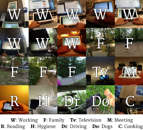
To train neural networks to recognize images, AI researchers have typically labeled (identified or described) each image in a dataset. For example, last year, Georgia Institute of Technology researchers developed a deep-learning method to recognize images taken at regular intervals on a person’s wearable smartphone camera.

Example images from dataset of 40,000 egocentric images with their respective labels (credit: Daniel Castro et al./Georgia Institute of Technology)

The idea was to demonstrate that deep-learning can “understand” human behavior and the habits of a specific person, and based on that, the AI system could offer suggestions to the user.

The problem with that method is the huge amount of time required to manually label the images (40,000 in this case). So AI researchers have turned to using synthetic images (such as from a video) that are pre-labeled (in captions, for example).

Creating superrealistic image recognition

But that, in turn, also has limitations. “Synthetic data is often not realistic enough, leading the network to learn details only present in synthetic images and fail to generalize well on real images,” the authors explain.

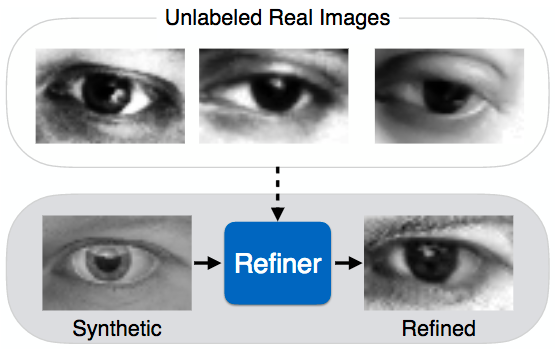
Simulated+Unsupervised (S+U) learning (credit: Ashish Shrivastava et al./arXiv)

So instead, the researchers have developed a new approach called “Simulated+Unsupervised (S+U) learning.”

The idea is to still use pre-labeled synthetic images (like the “Synthetic” image in the above illustration), but refine their realism by matching synthetic images to unlabeled real images (in this case, eyes) — thus creating a “superrealistic” image (the “Refined” image above), allowing for more accurate, faster image recognition, while preserving the labeling.

To do that, the researchers used a relatively new method (created in 2014) called Generative Adversarial Networks (GANs), which uses two neural networks that sort of compete with each other to create a series of superrealistic images.*


A visual Turing test

“To quantitatively evaluate the visual quality of the refined images, we designed a simple user study where subjects were asked to classify images as real or refined synthetic. Each subject was shown a random selection of 50 real images and 50 refined images in a random order, and was asked to label the images as either real or refined. The subjects found it very hard to tell the difference between the real images and the refined images.” — Ashish Shrivastava et al./arXiv


So will Siri develop the ability to identify that person whose name you forgot and whisper it to you in your AirPods, or automatically bring up that person’s Facebook page and latest tweet? Or is that getting too creepy?

* Simulated+Unsupervised (S+U) learning is “an interesting variant of adversarial gradient-based methods,” Jürgen Schmidhuber, Scientific Director of IDSIA (Swiss AI Lab), told KurzweilAI.

“An image synthesizer’s output is piped into a refiner net whose output is classified by an adversarial net trained to distinguish real from fake images. The refiner net tries to convince the classifier that it’s output is real, while being punished for deviating too much from the synthesizer output. Very nice and rather convincing applications!”

Schmidhuber also briefly explained his 1991 paper [1] that introduced gradient-based adversarial networks for unsupervised learning “when computers were about 100,000 times slower than today. The method was called Predictability Minimization (PM).

“An encoder network receives real vector-valued data samples (such as images) and produces codes thereof across so-called code units. A predictor network is trained to predict each code component from the remaining components. The encoder, however, is trained to maximize the same objective function minimized by the predictor.

“That is, predictor and encoder fight each other, to motivate the encoder to achieve a ‘holy grail’ of unsupervised learning, namely, a factorial code of the data, where the code components are statistically independent, which makes subsequent classification easy. One can attach an optional autoencoder to the code to reconstruct data from its code. After perfect training, one can randomly activate the code units in proportion to their mean values, to read out patterns distributed like the original training patterns, assuming the code has become factorial indeed.

“PM and Generative Adversarial Networks (GANs) may be viewed as symmetric approaches. PM is directly trying to map data to its factorial code, from which patterns can be sampled that are distributed like the original data. While GANs start with a random (usually factorial) distribution of codes, and directly learn to decode the codes into ‘good’ patterns. Both PM and GANs employ gradient-based adversarial nets that play a minimax game to achieve their goals.”

[1] J.  Schmidhuber. Learning factorial codes by predictability minimization. Technical Report CU-CS-565-91, Dept. of Comp. Sci., University of Colorado at Boulder, December 1991. Later also published in Neural Computation, 4(6):863-879, 1992. More: http://people.idsia.ch/~juergen/ica.html

Nanoarray sniffs out and distinguishes ‘breathprints’ of multiple diseases

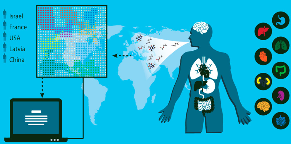
Schematic representation of the concept and design of the study, which involved collection of breath samples from 1404 patients diagnosed with one of 17 different diseases. One breath sample obtained from each subject was analyzed with the artificially intelligent nanoarray for disease diagnosis and classification (represented by patterns in the illustration), and a second sample was analyzed with gas chromatography–mass spectrometry to explore its chemical composition. (credit: Morad K. Nakhleh et al./ACS Nano)

An international team of 63 scientists in 14 clinical departments have identified a unique “breathprint” for 17 diseases with 86% accuracy and have designed a noninvasive, inexpensive, and miniaturized portable device that screens breath samples to classify and diagnose several types of diseases, they report in an open-access paper in the journal ACS Nano.

As far back as around 400 B.C., doctors diagnosed some diseases by smelling a patient’s exhaled breath, which contains nitrogen, carbon dioxide, oxygen, and a small amount of more than 100 other volatile chemical components. Relative amounts of these substances vary depending on the state of a person’s health. For example, diabetes creates a sweet breath smell. More recently, several teams of scientists have developed experimental breath analyzers, but most of these instruments focus on one disease, such as diabetes and melanoma, or a few diseases.

Detecting 17 diseases

The researchers developed an array of nanoscale sensors to detect the individual components in thousands of breath samples collected from 1404 patients who were either healthy or had one of 17 different diseases*, such as kidney cancer or Parkinson’s disease.

The team used mass spectrometry to identify the breath components associated with each disease. By analyzing the results with artificial intelligence techniques (binary classifiers), the team found that each disease produces a unique breathprint, based on differing amounts of 13 volatile organic chemical (VOC) components. They also showed that the presence of one disease would not prevent the detection of others — a prerequisite for developing a practical device to screen and diagnose various diseases.

Based on the research, the team designed an organic layer that functions as a sensing layer (recognition element) for adsorbed VOCs and an electrically conductive nanoarray based on resistive layers of molecularly modified gold nanoparticles and a random network of single-wall carbon nanotubes. The nanoparticles and nanotubes have different electrical conductivity patterns associated with different diseases.**

The authors received funding from the ERC and LCAOS of the European Union’s Seventh Framework Programme for Research and Technological Development, the EuroNanoMed Program under VOLGACORE, and the Latvian Council of Science.

* Lung cancer, colorectal cancer, head and neck cancer, ovarian cancer, bladder cancer, prostate cancer, kidney cancer, gastric cancer, Crohn’s disease, ulcerative colitis, irritable bowel syndrome, idiopathic Parkinson’s, atypical Parkinsonism, multiple sclerosis, pulmonary arterial hypertension, pre-eclampsia, and chronic kidney disease.

** During exposure to breath samples, interaction between the VOC components and the organic sensing layer changes the electrical resistance of the sensors. The relative change of sensor’s resistance at the peak (beginning), middle, and end of the exposure, as well as the area under the curve of the whole measurement were measured. All breath samples identified by the AI nanoarray were also examined using an independent lab-based analytical technique: gas chromatography linked with mass spectrometry.


Abstract of Diagnosis and Classification of 17 Diseases from 1404 Subjects via Pattern Analysis of Exhaled Molecules

We report on an artificially intelligent nanoarray based on molecularly modified gold nanoparticles and a random network of single-walled carbon nanotubes for noninvasive diagnosis and classification of a number of diseases from exhaled breath. The performance of this artificially intelligent nanoarray was clinically assessed on breath samples collected from 1404 subjects having one of 17 different disease conditions included in the study or having no evidence of any disease (healthy controls). Blind experiments showed that 86% accuracy could be achieved with the artificially intelligent nanoarray, allowing both detection and discrimination between the different disease conditions examined. Analysis of the artificially intelligent nanoarray also showed that each disease has its own unique breathprint, and that the presence of one disease would not screen out others. Cluster analysis showed a reasonable classification power of diseases from the same categories. The effect of confounding clinical and environmental factors on the performance of the nanoarray did not significantly alter the obtained results. The diagnosis and classification power of the nanoarray was also validated by an independent analytical technique, i.e., gas chromatography linked with mass spectrometry. This analysis found that 13 exhaled chemical species, called volatile organic compounds, are associated with certain diseases, and the composition of this assembly of volatile organic compounds differs from one disease to another. Overall, these findings could contribute to one of the most important criteria for successful health intervention in the modern era, viz. easy-to-use, inexpensive (affordable), and miniaturized tools that could also be used for personalized screening, diagnosis, and follow-up of a number of diseases, which can clearly be extended by further development.

Method discovered to remove damaging amyloid plaques found in Alzheimer’s disease

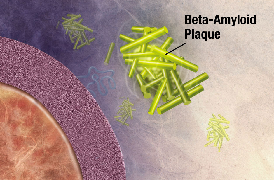
Illustration of formation of beta-amyloid plaque. Enzymes act on the APP (amyloid precursor protein) and cut it into fragments. The beta-amyloid fragment is crucial in the formation of senile plaques in Alzheimer’s disease. (credit: National Institute on Aging/NIH)

German scientists have discovered a strategy for removing amyloid plaques — newly forming clumps in a brain with Alzheimer’s disease that are created by misfolded proteins that clump together and damage nerve cells.

The scientists from the German Center for Neurodegenerative Diseases (DZNE) in Munich and the Ludwig Maximilians University (LMU) Munich took aged microglia cells (the  scavenger cells of the brain’s immune system) from a mouse model of Alzheimer’s disease and co-cultured them with microglia tissue from younger mouse brains. Surprisingly, within a few days, they found that the older amyloid plaques started to rejuvenate, resume cell division, and clear the brain from plaques by engulfing them. The amyloid plaque clearance potential of aged Alzheimer’s disease microglia could be fully restored, despite the continuous loss of neurons and astrocytes.

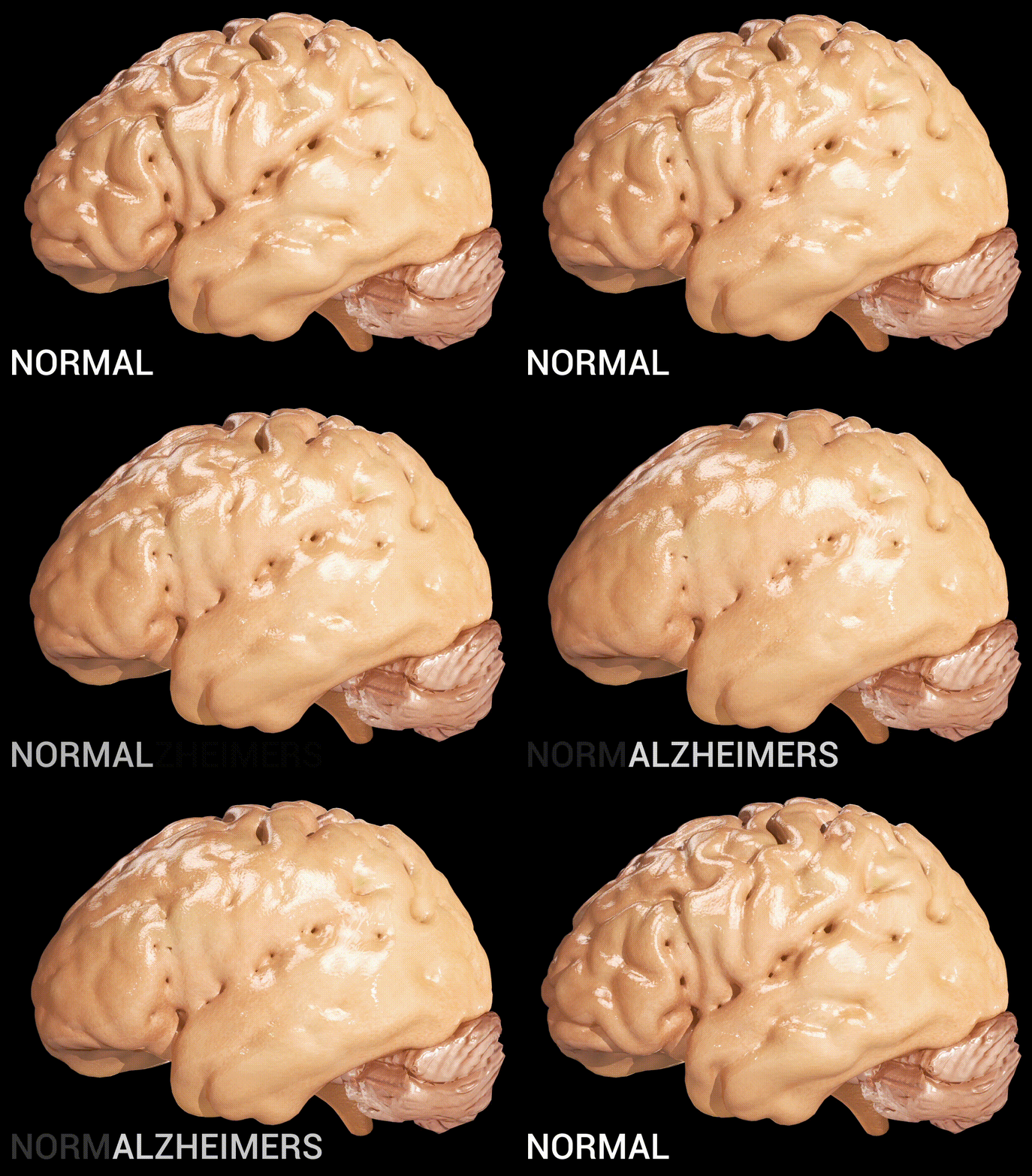
Cortical atrophy in Alzheimer’s Disease, associated with loss of gyri and sulci in the temporal lobe and parietal lobe, and parts of the frontal cortex and cingulate gyrus (credit: Doctor Jana/CC via Wikipedia)

They also found that the young microglia cells were secreting factors that helped the old microglia rejuvenate — especially a protein called “granulocyte-macrophage colony stimulating factor” (GM-CSF)* — which alone could do the job. Depletion of either old or young microglial cells prevented amyloid plaque clearance, indicating a synergistic effect of both populations.

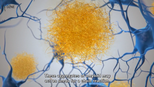
Click the image to view a DZNE video that describes the role of dysfunctional proteins in creating amyloid plaque in early Alzheimer’s disease, starting at 2:25. (credit: DZNE)

GM-CSF has previously been reported to reduce plaques and improve cognition in a mouse model of Alzheimer’s disease, but it’s not yet known if GM-CSF could potentially work as a new drug for Alzheimer’s disease in humans. Also, microglia secrete small proteins that induce inflammatory reactions and may harm neurons, the researchers say.

The researchers suggest that the new model system can be explored further to search for additional factors that enhance the clearance of amyloid plaques.

The work was published this week in The EMBO Journal.


National Institute on Aging | Inside the Brain: Unraveling the Mystery of Alzheimer’s Disease

* GM-CSF is manufactured using recombinant DNA technology. It is marketed as a protein therapeutic alled molgramostim or, when the protein is expressed in yeast cells, sargramostim. It is used as a medication to stimulate the production of white blood cells and thus prevent neutropenia  (loss of neutrophils, a type of white blood cell) following chemotherapy. (Patients with neutropenia are more susceptible to bacterial infections and, without prompt medical attention, the condition may become life-threatening.)


Abstract of Young microglia restore amyloid plaque clearance of aged microglia

Alzheimer′s disease (AD) is characterized by deposition of amyloid plaques, neurofibrillary tangles, and neuroinflammation. In order to study microglial contribution to amyloid plaque phagocytosis, we developed a novel ex vivo model by co‐culturing organotypic brain slices from up to 20‐month‐old, amyloid‐bearing AD mouse model (APPPS1) and young, neonatal wild‐type (WT) mice. Surprisingly, co‐culturing resulted in proliferation, recruitment, and clustering of old microglial cells around amyloid plaques and clearance of the plaque halo. Depletion of either old or young microglial cells prevented amyloid plaque clearance, indicating a synergistic effect of both populations. Exposing old microglial cells to conditioned media of young microglia or addition of granulocyte‐macrophage colony‐stimulating factor (GM‐CSF) was sufficient to induce microglial proliferation and reduce amyloid plaque size. Our data suggest that microglial dysfunction in AD may be reversible and their phagocytic ability can be modulated to limit amyloid accumulation. This novel ex vivo model provides a valuable system for identification, screening, and testing of compounds aimed to therapeutically reinforce microglial phagocytosis.

Using graphene to detect brain cancer cells

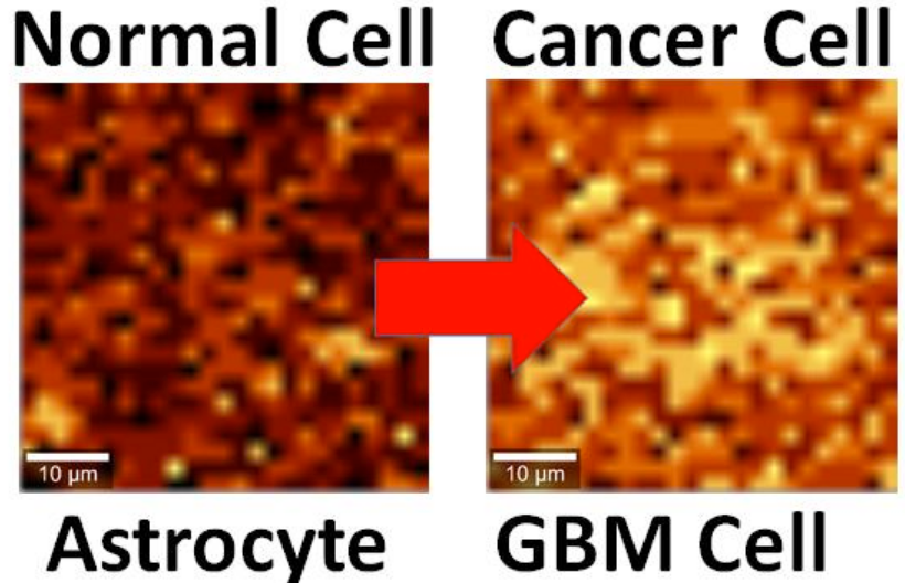
Brain cell culture. Left: Normal astrocyte brain cell; Right: cancerous Glioblastoma Multiforme (GBM) version, imaged by Raman spectrography. (credit: B. Keisham et al./ACS Appl. Mater. Interfaces)

By interfacing brain cells with graphene, University of Illinois at Chicago researchers have differentiated a single hyperactive Glioblastoma Multiforme cancerous astrocyte cell from a normal cell in the lab — pointing the way to developing a simple, noninvasive tool for early cancer diagnosis.

In the study, reported in the journal ACS Applied Materials & Interfaces, the researchers looked at lab-cultured human brain astrocyte cells taken from a mouse model. They compared normal astrocytes to their cancerous counterpart, highly malignant brain tumor glioblastoma multiforme.

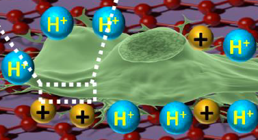
Illustration showing an astrocyte cell taken from a mouse brain draped over graphene (credit: B. Keisham et al./ACS Appl. Mater. Interfaces)

In a lab analysis, the cell is draped over graphene, explains Vikas Berry, associate professor and head of chemical engineering at UIC, who led the research along with Ankit Mehta, assistant professor of clinical neurosurgery in the UIC College of Medicine.

“The electric field around the cancer cell pushes away electrons in graphene’s electron cloud,” he said, which changes the vibration energy of the carbon atoms [in the graphene]. The change in vibration energy (resulting from the cancerous condition) can be pinpointed by Raman spectroscopy with a resolution of 300 nanometers, allowing for determining the activity of a single cell. (Raman spectroscopy is a highly sensitive method commonly used in chemistry to identify molecules by how they scatter laser light.)

“Graphene is the thinnest known material and is very sensitive to whatever happens on its surface,” Berry said. The nanomaterial is composed of a single layer of carbon atoms linked in a hexagonal chicken-wire pattern, and all the atoms share a cloud of electrons moving freely about the surface.

Patient biopsies planned

The technique is now being studied in a mouse model of cancer, with results that are “very promising,” Berry said. Experiments with patient biopsies would be further down the road. “Once a patient has brain tumor surgery, we could use this technique to see if the tumor relapses,” Berry said. “For this, we would need a cell sample we could interface with graphene and look to see if cancer cells are still present.”

The same technique may also work to differentiate between other types of cells or the activity of cells. “We may be able to use it with bacteria to quickly see if the strain is Gram-positive or Gram-negative,” Berry said. “We may be able to use it to detect sickle cells.”

Earlier this year, Berry and other coworkers introduced nanoscale ripples in graphene, causing it to conduct differently in perpendicular directions, useful for electronics. They wrinkled the graphene by draping it over a string of rod-shaped bacteria, then vacuum-shrinking the germs. “We took the earlier work and sort of flipped it over,” Berry said. “Instead of laying graphene on cells, we laid cells on graphene and studied graphene’s atomic vibrations.”

Funding was provided by UIC.


Abstract of Cancer Cell Hyperactivity and Membrane Dipolarity Monitoring via Raman Mapping of Interfaced Graphene: Toward Non-Invasive Cancer Diagnostics

Ultrasensitive detection, mapping, and monitoring of the activity of cancer cells is critical for treatment evaluation and patient care. Here, we demonstrate that a cancer cell’s glycolysis-induced hyperactivity and enhanced electronegative membrane (from sialic acid) can sensitively modify the second-order overtone of in-plane phonon vibration energies (2D) of interfaced graphene via a hole-doping mechanism. By leveraging ultrathin graphene’s high quantum capacitance and responsive phononics, we sensitively differentiated the activity of interfaced Glioblastoma Multiforme (GBM) cells, a malignant brain tumor, from that of human astrocytes at a single-cell resolution. GBM cell’s high surface electronegativity (potential ∼310 mV) and hyperacidic-release induces hole-doping in graphene with a 3-fold higher 2D vibration energy shift of approximately 6 ± 0.5 cm–1 than astrocytes. From molecular dipole-induced quantum coupling, we estimate that the sialic acid density on the cell membrane increases from one molecule per ∼17 nm2 to one molecule per ∼7 nm2. Furthermore, graphene phononic response also identified enhanced acidity of cancer cell’s growth medium. Graphene’s phonon-sensitive platform to determine interfaced cell’s activity/chemistry will potentially open avenues for studying activity of other cancer cell types, including metastatic tumors, and characterizing different grades of their malignancy.

How to enable soft robots to better mimick biological motions

Researchers used mathematical modeling to optimize the design of an actuator to perform biologically inspired motions (credit: Harvard SEAS)

Harvard researchers have developed a method for automatically designing actuators that enable fingers and knees in a soft robot to move more organically, a robot arm to move more smoothly along a path, or a wearable robot or exoskeleton to help a patient move a limb more naturally.

Designing such actuators is currently a complex design challenge, requiring a sequence of actuator segments, each performing a different motion. “Rather than designing these actuators empirically, we wanted a tool where you could plug in a motion and it would tell you how to design the actuator to achieve that motion,” said Katia Bertoldi, the John L. Loeb Associate Professor of the Natural Sciences and coauthor of the paper.

Designing an actuator that replicates a complex input motion. (A) Analytical models of actuator segments that can extend, expand, twist, or bend are the first input to the design tool. (B) The second input to the design tool is the kinematics of the desired motion. (C) The design tool outputs the optimal segment lengths and fiber angles for replicating the input motion. (credit: Fionnuala Connoll et al./PNAS)

The method developed by the team uses mathematical modeling of fluid-powered, fiber-reinforced actuators that can produce a wide range of motions. It optimizes the actuator design for performing specific motions (kinematics), different geometries, material properties, and pressure required for extending, expanding, twisting, and bending.

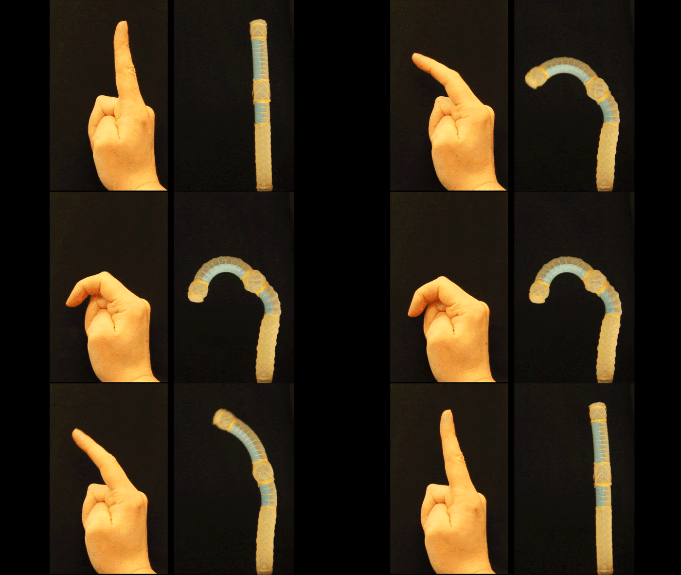
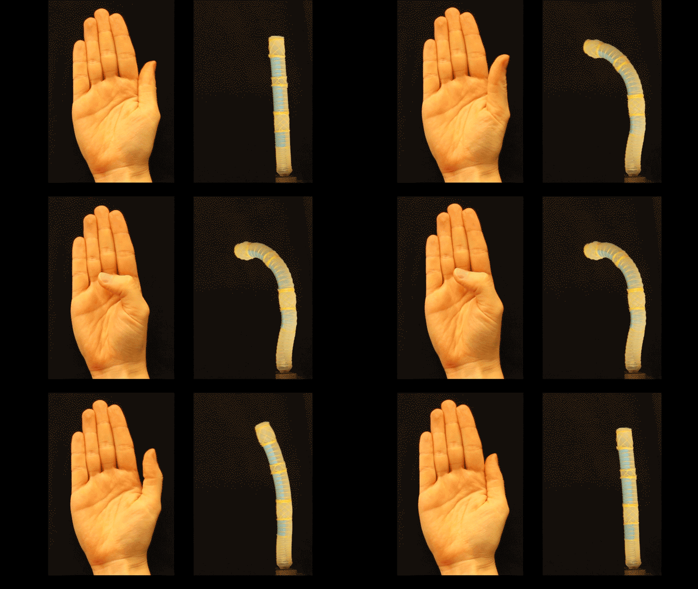
This soft actuator twists like a thumb when powered by a single pressure source (credit: Harvard SEAS)

The researchers from the Harvard John A. Paulson School of Engineering and Applied Sciences (SEAS) and the Wyss Institute for Biologically Inspired Engineering tested the model by designing a soft robot that bends like an index finger and twists like a texting thumb when powered by a single pressure source.

The research was published this week in the journal Proceedings of the National Academy of Sciences. “Future work will also focus on developing a model that combines bending with other motions, to increase the versatility of the algorithm,” the authors note in the paper.

In a future robot design, the model could conceivably be integrated with Cornell University’s design for soft, stretchable optoelectronic sensors in fingers that detect shape and texture.

The new methodology will be included in the Soft Robotic Toolkit, an online, open-source resource developed at SEAS to assist researchers, educators and budding innovators to design, fabrication, model, characterize and control their own soft robots.


Abstract of Automatic design of fiber-reinforced soft actuators for trajectory matching

Soft actuators are the components responsible for producing motion in soft robots. Although soft actuators have allowed for a variety of innovative applications, there is a need for design tools that can help to efficiently and systematically design actuators for particular functions. Mathematical modeling of soft actuators is an area that is still in its infancy but has the potential to provide quantitative insights into the response of the actuators. These insights can be used to guide actuator design, thus accelerating the design process. Here, we study fluid-powered fiber-reinforced actuators, because these have previously been shown to be capable of producing a wide range of motions. We present a design strategy that takes a kinematic trajectory as its input and uses analytical modeling based on nonlinear elasticity and optimization to identify the optimal design parameters for an actuator that will follow this trajectory upon pressurization. We experimentally verify our modeling approach, and finally we demonstrate how the strategy works, by designing actuators that replicate the motion of the index finger and thumb.

Cellular reprogramming turns back the aging clock in mice

This cartoon depicts turning back the aging clock through cellular regeneration of progeria mice (credit: Juan Carlos Izpisua Belmonte Lab/Salk Institute)

Salk Institute scientists have extended the average lifespan of live mice by 30 percent, according to a study published December 15 in Cell. They did that by rolling back the “aging clock” to younger years, using cellular reprogramming.

The finding suggests that aging is reversible by winding back an animal’s biological clock to a more youthful state and that lifespan can be extended. While the research does not yet apply directly to humans, it promises to lead to improved understanding of human aging and the possibility of rejuvenating human tissues.

To achieve this, the scientists worked with “progeria” mouse models — mice that had been genetically modified to carry a mutation that leads to premature aging (allowing the aging effects to be isolated and studied).*

Rather than attempting to correct the genetic mutations that cause premature aging (a difficult challenge), the Salk team instead focused on restoring the epigenome (a system of chemical marks on the genome that control which genes are or are not expressed as proteins)**.

Partial reprogramming avoids tumors and aging effects

To wind back the clock, the scientists first reprogrammed progeria mouse adult cells (such as skin cells) into an induced pluripotent stem (iPS) cells*** — an early embryonic state in which the cell is not yet specialized (“differentiated”) and can be programmed to perform specific functions, such as becoming part of the skin or heart.

To generate iPS cells, fibroblasts (or another type of adult somatic cell) are transduced with retroviruses encoding four pluripotency factors (SOX2, KLF4, c-MYC and OCT4). These differentiated cells can be used in disease models (such as progeria) for studying the molecular basis of a broad range of human diseases that are otherwise difficult to study. (credit: Yamanaka S. et al./Nature)

The scientists did that by inducing (modifying) the expression of four specific “epigenetic marks” called “Yamanaka factors” in the iPS cells, with the goal of allowing the cells to grow into adult cells without defects.

Researchers have previously done that successfully with cells in vitro (in a test tube), allowing these factors to be expressed for the needed 2 to 3 weeks for cells. But when they tried that in vivo (in live mice), it resulted in mice that later either died immediately or developed extensive tumors.

Partial cellular reprogramming with the four Yamanaka factors (Oct4, Sox2, Klf4, c-Myc) erases cellular markers of aging in progeria mouse cells (credit: Alejandro Ocampo et al./Cell)

So instead, the Salk team used partial in vivo cellular reprogramming. They induced expression of Yamanaka factors for just 2 to 4 days instead of 2 to 3 weeks. Instead of resulting in tumors or death, that prolonged the mice lifespan. An in vitro experiment also showed reduced DNA damage accumulation and restored nuclear structure.

This image depicts the discovery by Salk Institute researchers that partial cellular reprogramming reversed cellular signs of aging such as accumulation of DNA damage. (Left) Progeria mouse fibroblast cells; (Right) Progeria mouse fibroblast cells rejuvinated by partial reprogramming. (credit: Juan Carlos Izpisua Belmonte Lab/Salk Institute)

“These changes are the result of epigenetic remodeling in the cell,” notes lead investigator Juan Carlos Izpisua Belmonte, a professor in the Salk Institute of Biological Science’s Gene Expression Laboratory.

Several organs improved. For instance, tissue from skin, spleen, kidney and stomach all had improved appearance when inspected under the microscope. The cardiovascular system, which often fails and causes early death in these prematurely aging mice, also showed improvements in structure and function.

This image shows the discovery by researchers that induction of partial cellular reprogramming improved muscle regeneration in aged mice. (Left) Impaired muscle repair in aged mice; (Right) Improved muscle regeneration in aged mice subjected to reprogramming. (credit: Juan Carlos Izpisua Belmonte Lab/Salk Institute)

The team also tested partial reprogramming in mouse models that had injury, resulting in enhanced regeneration of muscle tissue and pancreas beta cells (which store and release insulin).

Next steps will involve learning more about how the epigenome changes during partial reprogramming. “We need to go back and explore which marks are changing and driving the aging process,” says Belmonte.

This study was supported by the G. Harold and Leila Y. Mathers Charitable Foundation, The Leona M. and Harry B. Helmsley Charitable Trust, The Glenn Foundation, Universidad Católica San Antonio de Murcia (UCAM), and Fundación Dr. Pedro Guillen.

* Mice do not perfectly mimic human aging. Researchers generate and use mouse models of premature aging by introducing mutations responsible for these human diseases in mouse.

** Epigenetic marks in the DNA regulate and protect the genome. Some marks turn on specialized functions, such as skin cell machinery in a skin cell; others turn off mechanisms that aren’t needed, such as liver cell machinery in the skin. They are modified over a lifetime in response to environmental changes, and have been proposed as drivers of aging.

*** Induced pluripotent stem cells (iPS cells or iPSCs) are a type of pluripotent stem cell that can be generated directly from adult cells. The iPSC technology was pioneered by Shinya Yamanaka’s lab in Kyoto, Japan, who showed in 2006 that the introduction of four specific genes encoding transcription factors could convert adult cells into pluripotent stem cells. He was awarded the 2012 Nobel Prize in Physiology or Medicine with Sir John Gurdon.


Salk Institute | Salk scientists reverse signs of aging in mice


Abstract of In Vivo Amelioration of Age-Associated Hallmarks by Partial Reprogramming

Aging is the major risk factor for many human diseases. In vitro studies have demonstrated that cellular reprogramming to pluripotency reverses cellular age, but alteration of the aging process through reprogramming has not been directly demonstrated in vivo. Here, we report that partial reprogramming by short-term cyclic expression of Oct4, Sox2, Klf4, and c-Myc (OSKM) ameliorates cellular and physiological hallmarks of aging and prolongs lifespan in a mouse model of premature aging. Similarly, expression of OSKM in vivo improves recovery from metabolic disease and muscle injury in older wild-type mice. The amelioration of age-associated phenotypes by epigenetic remodeling during cellular reprogramming highlights the role of epigenetic dysregulation as a driver of mammalian aging. Establishing in vivo platforms to modulate age-associated epigenetic marks may provide further insights into the biology of aging.

How diabetes drug metformin prevents, suppresses cancer growth

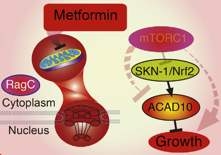
Metformin growth inhibition process (credit: Lianfeng Wu et al./Cell)

A team of Massachusetts General Hospital (MGH) and Harvard Medical School investigators has identified a pathway that appears to underlie the apparent ability of the diabetes drug metformin to both block the growth of human cancer cells and extend the lifespan of the C.elegans roundworm.

That finding implies that this single genetic pathway may play an important role in a wide range of organisms — including humans.

“We found that metformin reduces the traffic of molecules into and out of the nucleus — the ‘information center’ of the cell,” says Alexander Soukas, MD, PhD, of the MGH Center for Human Genetic Research, senior author of the study, published in the Thursday, Dec. 15 issue of Cell.

“Reduced nuclear traffic translates into the ability of the drug to block cancer growth and, remarkably, is also responsible for metformin’s ability to extend lifespan,” he said. “By shedding new light on metformin’s health-promoting effects, these results offer new potential ways that we can think about treating cancer and increasing healthy aging.”

Several studies have suggested that individuals taking metformin have a reduced risk of developing certain cancers and of dying from cancers that do develop. Current clinical trials are testing the impact of metformin on cancers of the breast, prostate and pancreas; and several research groups are working to identify its molecular targets.

How Metformin lowers blood glucose — and growth

Metformin’s ability to lower blood glucose in patients with type 2 diabetes appears to result from the drug’s effects on the liver. The drug reduces the liver’s ability to produce glucose for release into the bloodstream. Metformin has been thought to block the activity of mitochondria — structures that serve as the powerhouse of the cell.

But Soukas says more recent information suggests the mechanism is more complex. The Soukas team found a genetic pathway that slows the growth and extends the lifespan of C.elegans roundworms. That suggests that the roundworm could serve as a model for investigating the drug’s effects on cancer.*

“Amazingly, this pathway operates identically, whether in the worm or in human cancer cells,” says Soukas, who is an assistant professor of Medicine at Harvard Medical School. “Determining exactly how [it] slows cell growth will provide additional insights into novel therapeutic targets for cancer and possibly ways to manipulate the pathway to promote healthy aging.”**

Support for this study includes National Institutes of Health grants, a Broad Institute SPARC Grant, and the Ellison Medical Foundation New Scholar in Aging Award.

Metformin both suppresses cancer cell growth and promotes organismal longevity through a key transcriptional target that is induced through inhibition of mitochondrial respiration and modulation of mTOR signaling. (credit: Lianfeng Wu et al./Cell)

* The Soukas team’s experiments found that metformin’s action against cancer relies on two elements of a single genetic pathway – the nuclear pore complex, which allows the passage of molecules into and out of the nucleus, and an enzyme called ACAD10.

Basically, metformin’s suppression of mitochondrial activity reduces cellular energy, restricting the traffic of molecules through the nuclear pore. This shuts off an important cellular growth molecule called mTORC1, resulting in activation of ACAD10, which both slows the growth and extends the lifespan of C.elegans.

In human melanoma and pancreatic cancer cells, the investigators confirmed that application of drugs in the metformin family induced ACAD10 expression, an effect that depended on the function of the nuclear pore complex.

Without the complete signaling pathway – from mitochondrial suppression, through nuclear pore restriction to ACAD10 expression – cancer cells were no longer sensitive to the effects of metformin-like drugs.

** “Our experiments showed two very important things: if we force the nuclear pore to remain open or if we permanently shut down ACAD10, metformin can no longer block the growth of cancer cells. That suggests that the nuclear pore and ACAD10 may be manipulated in specific circumstances to prevent or even treat certain cancers.”

The essential contribution of ACAD10 to metformin’s anticancer action is intriguing, Soukas adds, because the only published study on ACAD10 function tied a variant in the gene to the increased risk of type 2 diabetes in Pima Indians, suggesting that ACAD10 also has a role in the drug’s antidiabetes action. “What ACAD10 does is a great mystery that we are greatly interested in solving,” he says. 


Abstract of An Ancient, Unified Mechanism for Metformin Growth Inhibition in C. elegans and Cancer

Metformin has utility in cancer prevention and treatment, though the mechanisms for these effects remain elusive. Through genetic screening in C. elegans, we uncover two metformin response elements: the nuclear pore complex (NPC) and acyl-CoA dehydrogenase family member-10 (ACAD10). We demonstrate that biguanides inhibit growth by inhibiting mitochondrial respiratory capacity, which restrains transit of the RagA-RagC GTPase heterodimer through the NPC. Nuclear exclusion renders RagC incapable of gaining the GDP-bound state necessary to stimulate mTORC1. Biguanide-induced inactivation of mTORC1 subsequently inhibits growth through transcriptional induction of ACAD10. This ancient metformin response pathway is conserved from worms to humans. Both restricted nuclear pore transit and upregulation of ACAD10 are required for biguanides to reduce viability in melanoma and pancreatic cancer cells, and to extend C. elegans lifespan. This pathway provides a unified mechanism by which metformin kills cancer cells and extends lifespan, and illuminates potential cancer targets.

A robotic hand with a human’s delicate sense of touch

A soft, sensitive robotic hand mounted on a robotic arm (credit: Cornell University)

Cornell University engineers have invented a new kind of robotic hand with a human’s delicate sense of touch.

Scenario: you lost part of your arm in a car accident. That artificial arm and hand you got from the hospital lets you feel and pick up things — even type on a keyboard. But not with the same sensitivity as a real hand. Now, an artificial prosthesis can even let you feel which one of three tomatoes is ripe (as shown in the video below).

The engineers’ trick was to use soft, stretchable optoelectronic (light + electronics) sensors in the fingers to detect shape and texture. (The sensors in existing prosthetic and robot hands use cruder tactile, or touch, sensors with bulky, rigid motors to measure strain.) The new prosthetic hand is a lot more sensitive. It can measure softness or hardness, how much the material stretches when touched, and how much force needs to be supplied to make the material deform.

How to make an (almost) human hand

A Cornell group led by Robert Shepherd, assistant professor of mechanical and aerospace engineering and principal investigator of Organic Robotics Lab, has published a paper describing this research in the debut edition of the journal Science Robotics (open access until Jan. 31).

Schematic of prosthetic hand structure and components (credit: Cornell University)

Unlike existing prosthetic and robot sensors, these sensors are integrated within the hand (instead of on the surface), so they can actually detect forces being transmitted through the thickness of the robot hand — simulating how a human hand feels. The more the prosthetic hand deforms (as it touches an object), the more light is lost. That variable loss of light, as detected by the photodiode, is what allows the prosthetic hand to “sense” its surroundings with high sensitivity and discrimination.*

This work was supported by a grant from Air Force Office of Scientific Research, and made use of the Cornell NanoScale Science and Technology Facility and the Cornell Center for Materials Research, both of which are supported by the National Science Foundation.

* The optoelectronic sensors are based on novel elastomeric optical waveguides, using a 3D-printed mold and a soft-lithography process to create a fluidically powered, stretchable material. Each high-precision waveguide incorporates an LED to generate light and a photodiode to measure the amount of light lost (which depends dynamically on the curvature, elongation, and force of the prosthetic hand).

To make the hand, Shepherd’s group used a four-step soft lithography process to produce the inside of the hand (core) and the cladding (outer surface of the waveguide), which also houses the LED (light-emitting diode) and the photodiode (light detector).


CNBC International | Scientists build a robotic hand with a soft touch | CNBC International


Abstract of Optoelectronically innervated soft prosthetic hand via stretchable optical waveguides

Because of their continuous and natural motion, fluidically powered soft actuators have shown potential in a range of robotic applications, including prosthetics and orthotics. Despite these advantages, robots using these actuators require stretchable sensors that can be embedded in their bodies for sophisticated functions. Presently, stretchable sensors usually rely on the electrical properties of materials and composites for measuring a signal; many of these sensors suffer from hysteresis, fabrication complexity, chemical safety and environmental instability, and material incompatibility with soft actuators. Many of these issues are solved if the optical properties of materials are used for signal transduction. We report the use of stretchable optical waveguides for strain sensing in a prosthetic hand. These optoelectronic strain sensors are easy to fabricate, are chemically inert, and demonstrate low hysteresis and high precision in their output signals. As a demonstration of their potential, the photonic strain sensors were used as curvature, elongation, and force sensors integrated into a fiber-reinforced soft prosthetic hand. The optoelectronically innervated prosthetic hand was used to conduct various active sensation experiments inspired by the capabilities of a real hand. Our final demonstration used the prosthesis to feel the shape and softness of three tomatoes and select the ripe one.

Dear President Trump: Here’s How to Make Space Great Again

(Credit: NASA Innovative Advanced Concepts)

By Brent Ziarnick, Peter Garretson, Everett Dolman, and Coyote Smith

President-elect Donald Trump often says that Americans no longer dream and must do so again. Nowhere can dreams be more inspiring and profitable than in space. But today, expanding space enterprise is not foremost on the minds of Americans or military strategists. As a recent CNN special showed, defense thinkers feel embattled in space, focused on protecting our existing investments rather than developing new ones that seize strategic advantage.


Major Brent Ziarnick, Lieutenant Colonel Peter Garretson, Everett Dolman, and Coyote Smith are members of the United States Air Force’s Space Horizons team. Space Horizons is a research group chartered by the Air University to explore the future of American space activity. The opinions herein are those of the authors alone and are not necessarily the views of Air University, the U.S. Air Force, or the U.S. government.


The first step to make space great again is for the United States to offer a constructive vision that can satisfy many American space needs, including defense. The Trump administration has an opportunity to transcend pessimism in space and focus America where it thrives: aggressive yet peaceful competition. Interested readers can view our complete recommendations, but a new Trump national space policy should declare:

The U.S. will be the first nation to mine an asteroid. The trillions of dollars in mineral wealth from asteroids can fuel a vibrant in-space economy capable of lifting up all humankind. America must lead this process.

The U.S. will be the first nation to extract resources from Earth’s moon to operate a commercial transportation service to and from the lunar surface. Our moon offers vast resources and tremendous logistical advantages for the development of that in-space economy. The U.S. will conduct research and establish public-private partnerships to advance the technology and the development of self-sustaining commercial services. The U.S. should also commit to being an early customer of such services, and it should take a leadership role in helping private industry develop businesses based on lunar exploration.

The U.S. will be the first nation to operate a propellant depot and on-orbit refueling service. Being able to refuel on orbit is key to an agile and fully reusable space transportation system. The United States will be the first to prove this technology and offer it as a commercial service to others.

The U.S. will be the first nation to operate a private space station. A thriving space economy must provide broad, affordable access to space across society, and it must have ordinary citizens living and working there permanently. As someone deeply knowledgeable about the hotel industry, the president-elect might understand the value of a U.S.-branded orbital tower.

The U.S. will operate the first fleet of fully reusable launch vehicles. Central to assured access for our citizenry is the ability to come and go to space with aircraft-like operations. A fully reusable architecture, technically feasible but never championed by the government, makes private spaceflight and even greater projects possible. America will provide the transportation system that fuels the larger global ecosystem of innovation.

The U.S. will build the first profitable solar power satellite. No single innovation in space could be as transformational as unlocking the vast potential of space-based solar energy generation to power Earth’s electrical needs; that could provide the hundreds of terawatts of renewable energy necessary to provide first-world living standards to the entire planet in a green and environmentally sustainable manner. The logistics system to create this space-power grid would require moving millions of metric tons of satellites to geostationary orbit and, consequently, will be orders of magnitude larger than any envisioned government-centric space program.

The U.S. will build the first comprehensive system to defend Earth from hazardous asteroids and comets. This planetary defense capability will initially start small, providing adequate defense against both 50-meter and 300-meter diameter objects with years of advance warning, and be built to provide comprehensive protection against extinction-level events. The United States will design, construct, and seek to test this capability in the current administration, and aim to maintain a standby global defense capability soon thereafter.

The U.S. will fly the first mission to another star. Interstellar spaceflight will be the ultimate expression of humanity mastering space travel. The American people must be the first to be ready.

This list of goals sounds audacious, perhaps outrageous, but it is entirely within the capability and character of the people who built the Transcontinental Railroad, the Hoover Dam, and conquered a continent. Americans are leaders in every one of these fields. It is only necessary for the new President to unleash America’s potential — once unleashed, American innovators will move these dreams toward reality faster than anyone can imagine.