Electric current and antiobiotic kill multidrug-resistant bacteria in biofilms

Washington State University researchers have successfully used a mild electric current to kill drug-resistant bacterial infections, a technology that may eventually be used to treat chronic wound infections. Red arrow indicates cell showing a stressed membrane. (credit: Washington State University)

A Washington State University research team has successfully used a mild electric current combined with an antibiotic to kill multidrug-resistant pseudomonas aeruginosa PAO1 bacteria in a lab-cultured biofilm.

These bacteria are responsible for chronic and serious infections in people with lung diseases, such as cystic fibrosis, and in chronic wounds. They also often cause pneumonia for people who are on ventilators and infections in burn victims.*

Schematic of experimental setup for the treatment of biofilm exposed to an e-scaffold, with an illustration of electrochemical production of hydrogen peroxide. The electrodes are connected to a potentiostat (600 mV source; not shown in figure). (credit: Sujala T. Sultana et al./Scientific Reports)

The researchers used an “e-scaffold” (electrochemical structure), a sort of electronic band-aid made out of conductive carbon fabric, which uses an electrical current to produce a low, constant concentration of hydrogen peroxide, an effective disinfectant, at the e-scaffold surface.

The hydrogen peroxide disrupts the biofilm (slime layer) matrix and damages the bacteria cell walls and DNA, which then allows better antibiotic penetration and efficacy against the bacteria. Bacteria that form biofilms are more difficult to kill because antibiotics only partially penetrate this protective layer. Subpopulations of “persister” cells survive treatment and are able to grow and multiply, resulting in chronic infections.

Tuning electrical stimulation to kill bacteria

Researchers have previously tried electrical stimulation, a method used to kill bacteria, for more than a century, but with limited results. In this study, the researchers determined the conditions necessary for the electrochemical reaction to produce hydrogen peroxide. The current has to be carefully controlled to assure the correct reaction at an exact rate to stop the bacteria from developing resistance, while not damaging surrounding tissue.**

The research to develop the e-scaffold actually came out of a failed attempt to improve fuel cells, according to research team leader Haluk Beyenal, the Paul Hohenschuh Distinguished Professor in WSU’s Gene and Linda Voiland School of Chemical Engineering and Bioengineering. When the researchers figured out they could only produce a small amount of electric current for their fuel cell cathode, they decided to see if they could use the process for a different purpose.

The technology may eventually be used to treat chronic wound infections, the researchers say. The researchers have filed a patent application and are working to commercialize the process. They hope to begin conducting clinical tests.

The work, reported in the online edition of npj Biofilms and Microbiomes, was supported by Beyenal’s National Science Foundation CAREER award.

* Bacterial resistance is a growing problem around the world. While antibiotics were a miracle drug of the 20th century, their widespread use has led to drug-resistant strains of bacteria. In the U.S., at least 2 million infections and 23,000 deaths are attributable to antibiotic-resistant bacteria each year, according to the U.S. Centers for Disease Control.

** As the researchers note in the paper, alternative antimicrobial treatments such as silver or mannitol, or in combination with conventional antibiotics, have toxic side effects at adequately high concentrations, and at low concentrations they often decompose before completely eliminating biofilm communities. In addition, persister cells can regrow and form biofilms with potentially enhanced tolerance to antibiotics.


Abstract of Eradication of Pseudomonas aeruginosa biofilms and persister cells using an electrochemical scaffold and enhanced antibiotic susceptibility

Biofilms in chronic wounds are known to contain a persister subpopulation that exhibits enhanced multidrug tolerance and can quickly rebound after therapeutic treatment. The presence of these “persister cells” is partly responsible for the failure of antibiotic therapies and incomplete elimination of biofilms. Electrochemical methods combined with antibiotics have been suggested as an effective alternative for biofilm and persister cell elimination, yet the mechanism of action for improved antibiotic efficacy remains unclear. In this work, an electrochemical scaffold (e-scaffold) that electrochemically generates a constant concentration of H2O2 was investigated as a means of enhancing tobramycin susceptibility in pre-grown Pseudomonas aeruginosa PAO1 biofilms and attacking persister cells. Results showed that the e-scaffold enhanced tobramycin susceptibility in P. aeruginosa PAO1 biofilms, which reached a maximum susceptibility at 40 µg/ml tobramycin, with complete elimination (7.8-log reduction vs control biofilm cells, P ≤ 0.001). Moreover, the e-scaffold eradicated persister cells in biofilms, leaving no viable cells (5-log reduction vs control persister cells, P ≤ 0.001). It was observed that the e-scaffold induced the intracellular formation of hydroxyl free radicals and improved membrane permeability in e-scaffold treated biofilm cells, which possibly enhanced antibiotic susceptibility and eradicated persister cells. These results demonstrate a promising advantage of the e-scaffold in the treatment of persistent biofilm infections.

Smart skin patch releases blood thinners in closed-loop control system

A painless microneedle skin patch for delivering the right amount of a blood-thinning drug (credit: Yuqi Zhang)

North Carolina researchers have developed a smart skin patch designed to monitor a patient’s blood and release a blood-thinning drug, as needed, to prevent thrombosis (dangerous blood clots).

Thrombosis — one of the leading causes of cardiovascular mortalities and morbidities worldwide — occurs when blood clots disrupt the normal flow of blood in the body, which can cause severe health problems such as pulmonary embolism, heart attack, or stroke.

Current treatments often rely on the use of blood thinners, such as heparin, requiring patients to test their blood on a regular basis to ensure proper dosages. Dosage is iffy; too large a dose can cause problems such as spontaneous hemorrhaging, while doses that are too small may not prevent thrombosis.

So researchers at North Carolina State University and the University of North Carolina at Chapel Hill developed a feedback-controlled anti-coagulant smart skin patch to release just the needed level of heparin.

Schematic of skin patch that releases blood-thining heparin (to prevent blood clots) in response to thrombin (an enzyme that initiates clotting in the blood) (credit: Yuqi Zhang)

The patch incorporates an array of painless microneedles made of a material that is responsive to thrombin (an enzyme that initiates clotting in the blood). When elevated levels of thrombin enzymes in the bloodstream come into contact with the microneedle, the enzymes cause the thrombin-responsive material in the microneedles to release the right amount (and no more) of blood-thining heparin into the bloodstream to prevent blood clots.*

The patch was successfully tested in a mouse model.

“Our goal was to generate a patch that can monitor a patient’s blood and release additional drugs when necessary — effectively, a self-regulating system,” says Zhen Gu, co-corresponding author on a paper published Nov. 25, 2016 in Advanced Materials describing the work. Gu is an associate professor in the joint biomedical engineering program at NC State and UNC.

“This paper represents a good first step, and we’re now looking for funding to perform additional preclinical testing,” Gu said.

The work was supported by the Alfred P. Sloan Foundation, NIH’s Clinical and Translational Science Awards, and the National Science Foundation.

* Closed-loop heparin delivery system

A peptide is introduced as a linker during the conjugation of Heparin to the main chain of hyaluronic acid (which has been UV-modified to be sensitive to thrombin. The presence of thrombin (top) causes the peptide to be cleaved (cut), triggering heparin drug release (right). The released heparin then inhibits the coagulation activation by inactivating thrombin (bottom). In a feedback loop, that process then suppresses further release of heparin (left), minimizing the risk of undesirable spontaneous hemorrhage. (credit: Yuqi Zhang et al./Advanced Materials)


Abstract of Thrombin-Responsive Transcutaneous Patch for Auto-Anticoagulant Regulation

A thrombin-responsive closed-loop patch is developed for prolonged heparin delivery in a feedback-controlled manner. This microneedle-based patch can sense activated thrombin and subsequently releases heparin to prevent coagulation in the blood flow. This “smart” heparin patch can be transcutaneously inserted into skin without drug leakage and can sustainably regulate blood coagulation in response to thrombin.

First glimpse of new concepts developing in the brain

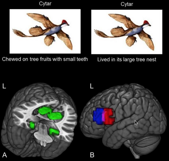
This image illustrates how the study participants learned about the habitat and the diet of eight animals, such as the cytar (not its real zoological name) and shows the set of habitat brain regions (A-green) and diet (B-red and blue) regions where the new knowledge was stored. (L refers to left hemisphere of the brain.) (credit: Carnegie Mellon University)

Carnegie Mellon University (CMU) scientists have for the first time documented the actual formation of newly learned concepts inside the brain.

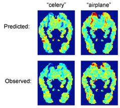
Predicting the unique brain activation patterns associated with names (credit: CMU)

Thanks to recent advances in brain imaging technology at CMU and elsewhere, it is now known how specific concrete objects are coded in the brain — neuroscientists can identify which object, such as a house or a banana, someone is thinking about from its functional magnetic resonance imaging (fMRI) brain signature.

Taking the next step, the researchers decided to observe the actual formation of these signatures in an experiment.

Neuroscientist Marcel Just, the D.O. Hebb University Professor of Cognitive Neuroscience in CMU’s Dietrich College of Humanities and Social Sciences, and Andrew Bauer, a Ph.D. student in psychology, taught 16 study participants diet and dwelling information about extinct animals to monitor the growth of the neural representations of eight new animal concepts in the participants’ brains.

Drawing on the previous research findings, the research team knew “where” to expect the new knowledge to emerge in the brains of their participants. Information about dwellings and information about eating have each been shown to reside in their own set of brain regions, regions that are common across people.

Over the course of an hour, the study participants were given a zoology mini-tutorial on the diets and habitats of the animals, while the scientists used fMRI to monitor the emergence of the concepts in the participants’ brains. As the new properties were taught, the activation levels in the eating regions and the dwelling regions changed.

Concept signatures

One important result was that after the zoology tutorial, each one of the eight animal concepts developed its own unique activation signature. This made it possible for a computer program to determine which of the eight animals a participant was thinking about at a given time. In effect, the program was reading their minds as they contemplated a brand-new thought.

Even though the animals had unique activation signatures, the animals that shared similar properties (such as a similar habitat) had similar activation signatures. That is, a resemblance between the properties of two animals resulted in a resemblance between their activation signatures. This finding shows that the activation signatures are not just arbitrary patterns, but are meaningful and interpretable.

“The activation signature of a concept is a composite of the different types of knowledge of the concept that a person has stored, and each type of knowledge is stored in its own characteristic set of regions,” Just said.

Another important result was that once a property of an animal was learned, it remained intact in the brain, even after other properties of the animal had been learned. This finding indicates the relative neural durability of what we learn.

Implications for learning and brain disorders

“Each time we learn something, we permanently change our brains in a systematic way,” said Bauer, the study’s lead author. “It was exciting to see our study successfully implant the information about extinct animals into the expected locations in the brain’s filing system.”

Just believes that the study provides a foundation for brain researchers to trace how a new concept makes its way into the brain from the words and graphics used to teach it. That suggests that it may be possible to assess the progress in learning a complicated concept like those in a high-school physics lesson. fMRI pattern analyses could diagnose which aspects of a concept students misunderstand (or lack), in a way that could guide the next iteration of instruction.

The results from this study also indicate that it may be possible to use a similar approach to understand the “loss” of knowledge in various brain disorders, such as dementia or Alzheimer’s disease, or due to brain injuries. The loss of a concept in the brain may be the reverse of the process that the study observed.


Abstract of Monitoring the growth of the neural representations of new animal concepts

Although enormous progress has recently been made in identifying the neural representations of individual object concepts, relatively little is known about the growth of a neural knowledge representation as a novel object concept is being learned. In this fMRI study, the growth of the neural representations of eight individual extinct animal concepts was monitored as participants learned two features of each animal, namely its habitat (i.e., a natural dwelling or scene) and its diet or eating habits. Dwelling/scene information and diet/eating-related information have each been shown to activate their own characteristic brain regions. Several converging methods were used here to capture the emergence of the neural representation of a new animal feature within these characteristic, a priori-specified brain regions. These methods include statistically reliable identification (classification) of the eight newly acquired multivoxel patterns, analysis of the neural representational similarity among the newly learned animal concepts, and conventional GLM assessments of the activation in the critical regions. Moreover, the representation of a recently learned feature showed some durability, remaining intact after another feature had been learned. This study provides a foundation for brain research to trace how a new concept makes its way from the words and graphics used to teach it, to a neural representation of that concept in a learner’s brain.

Caltech scientists use bacterial protein to merge silicon and carbon and create new organosilicon compounds

Artist rendering of organosilicon-based life (credit: Lei Chen and Yan Liang (BeautyOfScience.com) for Caltech)

Scientists at Caltech have “bred” a bacterial protein with the ability to make silicon-carbon bonds, with applications in several industries — something only chemists could do before. The research was published in the Nov. 24 issue of the journal Science.

Molecules with silicon-carbon (organosilicon) compounds are found in pharmaceuticals and many other products, including agricultural chemicals, paints, semiconductors, and computer and TV screens. Currently, these products are made synthetically, since silicon-carbon bonds are not found in nature.

The new research demonstrates that biology can be used to manufacture these bonds in ways that are more environmentally friendly and potentially much less expensive, according to the researchers.


Caltech | Bringing Silicon to Life: Scientists Persuade Nature to Make Silicon-Carbon Bonds

Directed evolution

The key to this research involves deliberate messing with nature: a method called directed evolution* pioneered in the early 1990s by Frances Arnold, Caltech’s Dick and Barbara Dickinson Professor of Chemical Engineering, Bioengineering and Biochemistry, and principal investigator of this project.

An example of directed evolution with comparison to natural evolution. The inner cycle indicates the 3 stages of the directed evolution cycle with the natural process being mimicked in parentheses. The outer circle demonstrates steps a typical experiment. The red symbols indicate functional variants, the pale symbols indicate variants with reduced function. (credit: Thomas Shafee CC)

Directed evolution has been used for years to make enzymes for household products, like detergents; and for “green” sustainable routes to making pharmaceuticals, agricultural chemicals, and fuels.

In directed evolution, new and better enzymes are created in labs by artificial selection, similar to the way that breeders modify corn, cows, or cats. Enzymes are a class of proteins that catalyze, or facilitate, chemical reactions. The directed evolution process begins with an enzyme that scientists want to enhance. The DNA coding for the enzyme is mutated in more-or-less random ways, and the resulting enzymes are tested for a desired trait. The top-performing enzyme is then mutated again, and the process is repeated until an enzyme that performs much better than the original is created.

Going where no enzyme has gone before

In the new study, the goal was not just to improve an enzyme’s biological function but to actually persuade it to do something that it had not done before. The researchers’ first step was to find a suitable candidate, an enzyme showing potential for making the silicon-carbon bonds.

“It’s like breeding a racehorse,” says Arnold, who is also the director of the Donna and Benjamin M. Rosen Bioengineering Center at Caltech. “A good breeder recognizes the inherent ability of a horse to become a racer and has to bring that out in successive generations. We just do it with proteins.”

An Icelandic hot spring (credit: Nordic Visitor)

The ideal candidate turned out to be a protein from a bacterium, Rhodothermus marinus, that grows in hot springs in Iceland. That protein, called cytochrome c, normally shuttles electrons to other proteins, but the researchers found that it also happens to act like an enzyme to create silicon-carbon bonds at low levels. The scientists then mutated the DNA coding for that protein within a region that specifies an iron-containing portion of the protein thought to be responsible for its silicon-carbon bond-forming activity. Next, they tested these mutant enzymes for their ability to make organosilicon compounds better than the original.

cytochrome c (credit: Caltech)

After only three rounds, they had created an enzyme that can selectively make silicon-carbon bonds 15 times more efficiently than the best catalyst invented by chemists. Furthermore, the enzyme is highly selective, which means that it makes fewer unwanted byproducts that have to be chemically separated out.

“This iron-based, genetically encoded catalyst is nontoxic, cheaper, and easier to modify compared to other catalysts used in chemical synthesis,” says Jennifer Kan, a postdoctoral scholar in Arnold’s lab and lead author of the new study. “The new reaction can also be done at room temperature and in water.”

The synthetic process for making silicon-carbon bonds often uses precious metals and toxic solvents, and requires extra processing to remove unwanted byproducts, all of which add to the cost of making these compounds.

Could life on Earth (or elsewhere) have evolved based on silicon-carbon?

The study is the first to show that nature can adapt to incorporate silicon into carbon-based molecules, the building blocks of life.

Carbon and silicon are chemically very similar, and silicon is the second most abundant element in Earth’s crust. They can both form bonds to four atoms simultaneously, making them well suited to form the long chains of molecules found in life, such as proteins and DNA. Science-fiction authors have imagined alien worlds with silicon-based life, like the lumpy Horta creatures portrayed in an episode of the 1960s TV series Star Trek.

“This study shows how quickly nature can adapt to new challenges,” says Arnold. “The DNA-encoded catalytic machinery of the cell can rapidly learn to promote new chemical reactions when we provide new reagents and the appropriate incentive in the form of artificial selection.”

However, no living organism is known [yet] to put silicon-carbon bonds together, even though silicon is so abundant, all around us, in rocks and all over the beach,” says Kan.

What about other planets (Mars has both silicon and carbon, for example) and asteroids? And could alien life have evolved silicon-carbon semiconductor brains? It would also be interesting to see if such a lifeform could be invented on Earth.

This research is funded by the National Science Foundation, the Caltech Innovation Initiative program, and the Jacobs Institute for Molecular Engineering for Medicine at Caltech.

* Not to be confused with a transhumanist concept for controlling human evolution.


Abstract of Directed evolution of cytochrome c for carbon–silicon bond formation: Bringing silicon to life

Enzymes that catalyze carbon–silicon bond formation are unknown in nature, despite the natural abundance of both elements. Such enzymes would expand the catalytic repertoire of biology, enabling living systems to access chemical space previously only open to synthetic chemistry. We have discovered that heme proteins catalyze the formation of organosilicon compounds under physiological conditions via carbene insertion into silicon–hydrogen bonds. The reaction proceeds both in vitro and in vivo, accommodating a broad range of substrates with high chemo- and enantioselectivity. Using directed evolution, we enhanced the catalytic function of cytochrome c from Rhodothermus marinus to achieve more than 15-fold higher turnover than state-of-the-art synthetic catalysts. This carbon–silicon bond-forming biocatalyst offers an environmentally friendly and highly efficient route to producing enantiopure organosilicon molecules.

New neural-network algorithm learns directly from human instructions instead of examples

Conventional neural-network image-recognition algorithm trained to recognize human hair (left), compared to the more precise heuristically trained algorithm (right) (credit: Wenzhangzhi Guo and Parham Aarabi/IEEE Trans NN & LS)

A new machine learning algorithm designed by University of Toronto researchers Parham Aarabi and Wenzhi Guo learns directly from human instructions, rather than an existing set of examples, as in traditional neural networks. In tests, it outperformed existing neural networks by 160 per cent.

Their “heuristically trained neural networks” (HNN) algorithm also outperformed its own training by nine per cent — it learned to recognize hair in pictures with greater reliability than that enabled by the training.

Aarabi and Guo trained their HNN algorithm to identify people’s hair in photographs, a challenging task for computers. “Our algorithm learned to correctly classify difficult, borderline cases — distinguishing the texture of hair versus the texture of the background,” says Aarabi. “What we saw was like a teacher instructing a child, and the child learning beyond what the teacher taught her initially.”

Heuristic training

Humans conventionally “teach” neural networks by providing a set of labeled data and asking the neural network to make decisions based on the samples it’s seen. For example, you could train a neural network to identify sky in a photograph by showing it hundreds of pictures with the sky labeled.

With HNN, humans provide direct instructions that are used to pre-classify training samples rather than a set of fixed examples. Trainers program the algorithm with guidelines such as “Sky is likely to be varying shades of blue,” and “Pixels near the top of the image are more likely to be sky than pixels at the bottom.”

Their work is published in the journal IEEE Transactions on Neural Networks and Learning Systems.

This heuristic-training approach addresses one of the biggest challenges for neural networks: making correct classifications of previously unknown or unlabeled data, the researchers say. This is crucial for applying machine learning to new situations, such as correctly identifying cancerous tissues for medical diagnostics, or classifying all the objects surrounding and approaching a self-driving car.

“Applying heuristic training to hair segmentation is just a start,” says Guo. “We’re keen to apply our method to other fields and a range of applications, from medicine to transportation.”


Abstract of Hair Segmentation Using Heuristically-Trained Neural Networks

We present a method for binary classification using neural networks (NNs) that performs training and classification on the same data using the help of a pretraining heuristic classifier. The heuristic classifier is initially used to segment data into three clusters of high-confidence positives, high-confidence negatives, and low-confidence sets. The high-confidence sets are used to train an NN, which is then used to classify the low-confidence set. Applying this method to the binary classification of hair versus nonhair patches, we obtain a 2.2% performance increase using the heuristically trained NN over the current state-of-the-art hair segmentation method.

Battery breakthrough charges in seconds, lasts for a week

Illustration representing the novel design of a hybrid supercapacitor, showing bundles of nanowires (blue) coated with 2D energy-storage materials (yellow) (credit: University of Central Florida)

University of Central Florida researchers have developed a radical new supercapacitor design that could one day replace lithium-ion batteries, allowing users to charge a mobile phone in a few seconds and with a charge that lasts a week, according to the researchers. The new battery would be flexible and a fraction of the size of a lithium-ion battery.

The proof-of-concept design is based on a hybrid supercapacitor composed of a core with millions of highly conductive nanowires coated with shells of two-dimensional materials.* It combines fast charging and discharging (high power density) and high storage capacity (high energy density).

Supercapacitor design: an array of electrically conductive nanowires (orange) with metal current-collector covering (blue) (credit: Nitin Choudhary et al./ACS Nano)

Optical image of core/shell nanowires on a tungsten foil under mechanical bending (left). Corresponding SEM image (right) shows high-density, well-aligned nanowires along with their faceted surface (inset). The scale bar in the inset is 500 nm. (credit: Nitin Choudhary et al./ACS Nano)

Another advantage would be “cyclic stability” (how many times a battery can be charged, drained and recharged before beginning to degrade). A lithium-ion battery can be recharged fewer than 1,500 times without significant failure, compared to recently developed supercapacitors based on two-dimensional materials, which can be recharged more than 30,000 times.

Supercapacitor prototype showing flexible design (credit: (credit: University of Central Florida)

Electric vehicles could also benefit from longer-range operation and sudden bursts of power and speed. The flexible material could mean a significant advancement in wearable tech, according to the researchers, and would also avoid the risk of overheating and explosion with lithium-ion batteries.

Hee-Suk Chung of Korea Basic Science Institute was also involved in the research, which was published recently in the journal ACS Nano.

* The core nanowire material is tungsten trioxide (WO3) and the two-dimensional shell material is a transition-metal dichalcogenide, tungsten disulfide (WS2).

Ragone plot to compare the performances of various technologies with the core/shell nanowires supercapacitor in this study (credit: Nitin Choudhary et al./ACS Nano)

Galvanostatic Charging/Discharging (GCD) various current densities in the voltage range of 0.3 and 0.5 V, showing nearly symmetrical voltage curves, indicating highly reversible and fast responses. (credit: Nitin Choudhary et al./ACS Nano)


Abstract of High-Performance One-Body Core/Shell Nanowire Supercapacitor Enabled by Conformal Growth of Capacitive 2D WS2 Layers

Two-dimensional (2D) transition-metal dichalcogenides (TMDs) have emerged as promising capacitive materials for supercapacitor devices owing to their intrinsically layered structure and large surface areas. Hierarchically integrating 2D TMDs with other functional nanomaterials has recently been pursued to improve electrochemical performances; however, it often suffers from limited cyclic stabilities and capacitance losses due to the poor structural integrity at the interfaces of randomly assembled materials. Here, we report high-performance core/shell nanowire supercapacitors based on an array of one-dimensional (1D) nanowires seamlessly integrated with conformal 2D TMD layers. The 1D and 2D supercapacitor components possess “one-body” geometry with atomically sharp and structurally robust core/shell interfaces, as they were spontaneously converted from identical metal current collectors via sequential oxidation/sulfurization. These hybrid supercapacitors outperform previously developed any stand-alone 2D TMD-based supercapacitors; particularly, exhibiting an exceptional charge–discharge retention over 30,000 cycles owing to their structural robustness, suggesting great potential for unconventional energy storage technologies.

Google’s new multilingual Neural Machine Translation System can translate between language pairs even though it has never been taught to do so

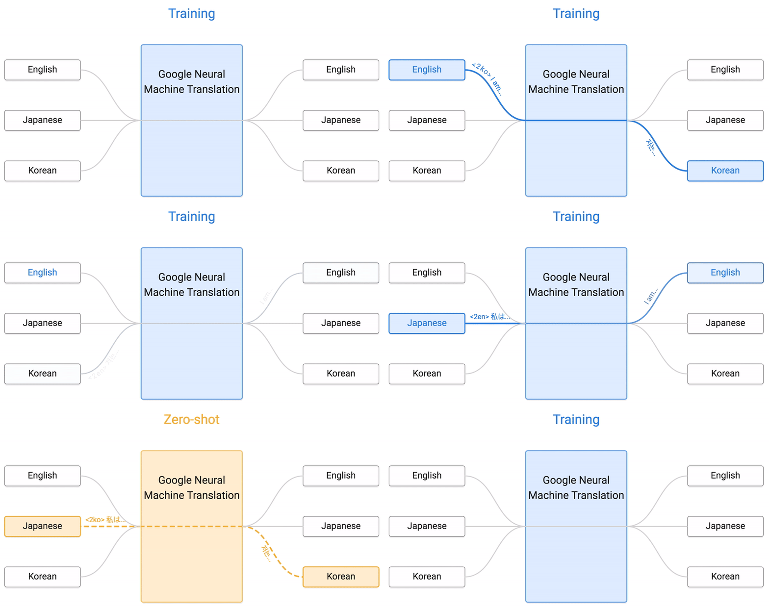
Google Neural Machine Translation (credit: Google)

Google researchers have announced they have implemented a neural machine translation system in Google Translate that improves translation quality and enables “Zero-Shot Translation” — translation between language pairs never seen explicitly by the system.

For example, in the animation above, the system was trained to translate bidirectionally between English and Japanese and between English and Korean. But the new system can also translate between Japanese and Korean — even though it has never been taught to do so.

Google calls this “zero-shot” translation — shown by the yellow dotted lines in the animation. “To the best of our knowledge, this is the first time this type of transfer learning has worked in machine translation,” the researchers say.

Neural networks for machine translation

What drove this development was the fact that in the last ten years, Google Translate has grown from supporting just a few languages to 103, translating more than 140 billion words every day. That required building and maintaining many different systems to translate between any two languages — incurring significant computational cost.

That meant the researchers needed to rethink the technology behind Google Translate.

As a result, the Google Brain Team and Google Translate team announced the first step in September: a new system called Google Neural Machine Translation (GNMT), which learns from millions of examples.

“GNMT reduces translation errors by more than 55%–85% on several major language pairs measured on sampled sentences from Wikipedia and news websites with the help of bilingual human raters,” the researchers note. “However, while switching to GNMT improved the quality for the languages we tested it on, scaling up to all the 103 supported languages presented a significant challenge.”

So as explained in a paper, “Google’s Multilingual Neural Machine Translation System: Enabling Zero-Shot Translation” (open access), the researchers extended the GNMT system, allowing for a single system to translate between multiple languages. “Our proposed architecture requires no change in the base GNMT system,” the authors explain, “instead it uses an additional ‘token’ at the beginning of the input sentence to specify the required target language to translate to.”

An interlingua

But the success of the zero-shot translation raised another important question: Is the system learning a common representation in which sentences with the same meaning are represented in similar ways regardless of language — i.e. an “interlingua”?

To find out, the researchers used a 3-dimensional representation of internal network data, allowing them to take a peek into the system as it translated a set of sentences between all possible pairs of the Japanese, Korean, and English languages, for example.

Developing an “interlinqua” (credit: Google)

Part (a) from the figure above shows an overall geometry of these translations. The points in this view are colored by the meaning; a sentence translated from English to Korean with the same meaning as a sentence translated from Japanese to English share the same color. From this view we can see distinct groupings of points, each with their own color. Part (b) zooms in to one of the groups, and the colors in part (c) show the different source languages.

Within a single group, we see a sentence with the same meaning (“The stratosphere extends from about 10km to about 50km in altitude”), but from three different languages, the researchers explain. “This means the network must be encoding something about the semantics [meaning] of the sentence, rather than simply memorizing phrase-to-phrase translations. We interpret this as a sign of existence of an interlingua in the network.”

The new Multilingual Google Neural Machine Translation system is running in production today for all Google Translate users, according to the researchers.


Abstract of Google’s Multilingual Neural Machine Translation System: Enabling Zero-Shot Translation

We propose a simple, elegant solution to use a single Neural Machine Translation (NMT) model to translate between multiple languages. Our solution requires no change in the model architecture from our base system but instead introduces an artificial token at the beginning of the input sentence to specify the required target language. The rest of the model, which includes encoder, decoder and attention, remains unchanged and is shared across all languages. Using a shared wordpiece vocabulary, our approach enables Multilingual NMT using a single model without any increase in parameters, which is significantly simpler than previous proposals for Multilingual NMT. Our method often improves the translation quality of all involved language pairs, even while keeping the total number of model parameters constant. On the WMT’14 benchmarks, a single multilingual model achieves comparable performance for English→French and surpasses state-of-the-art results for English→German. Similarly, a single multilingual model surpasses state-of-the-art results for French→English and German→English on WMT’14 and WMT’15 benchmarks respectively. On production corpora, multilingual models of up to twelve language pairs allow for better translation of many individual pairs. In addition to improving the translation quality of language pairs that the model was trained with, our models can also learn to perform implicit bridging between language pairs never seen explicitly during training, showing that transfer learning and zero-shot translation is possible for neural translation. Finally, we show analyses that hints at a universal interlingua representation in our models and show some interesting examples when mixing languages.

New unique brain ‘fingerprint’ method can identify a person with nearly 100% accuracy

A research team led by Carnegie Mellon University used diffusion MRI to measure the local connectome of 699 brains from five data sets. The local connectome comprises the point-by-point connections along all of the white matter pathways in the brain, as opposed to the connections between brain regions. To create a fingerprint, they used diffusion MRI data to calculate the distribution of water diffusion along the cerebral white matter’s fibers. (credit: Carnegie Mellon University)

Researchers have “fingerprinted” the white matter of the human brain using a new diffusion MRI method, mapping the brain’s connections (the connectome) at a more detailed level than ever before. They confirmed that structural connections in the brain are unique to each individual person and the connections were able to identify a person with nearly 100% accuracy.

The new method could provide biomarkers to help researchers determine how factors such as disease, the environment, genetic and social factors, and different experiences impact the brain and change over time.

“This means that many of your life experiences are somehow reflected in the connectivity of your brain,” said Timothy Verstynen, an assistant professor of psychology at Carnegie Mellon University and senior author of the study, published in open-access PLOS Computational Biology.

The local connectome: a personal biomarker

Demonstrating the level of detail, one local connectome fingerprint is shown in different zoom-in resolutions. A local connectome fingerprint has a total of 513,316 entries of scalar values. (credit: Fang-Cheng Yeh et al./PLoS Comput Biol)

For the study, the researchers used diffusion MRI to measure the local connectome of 699 brains from five data sets. The local connectome is the point-by-point connections along all of the white matter pathways in the brain, as opposed to the connections between brain regions. To create a fingerprint for each person, they used the diffusion MRI data to calculate the distribution of water diffusion along the cerebral white matter’s fibers.*

The measurements revealed the local connectome is highly unique to an individual and can be used as a personal biomarker for human identity. To test the uniqueness, the team ran more than 17,000 identification tests. With nearly 100 percent accuracy, they were able to tell whether two local connectomes, or brain “fingerprints,” came from the same person or not.

Curiously, they discovered that identical twins only share about 12 percent of structural connectivity patterns and the brain’s unique local connectome is sculpted over time, changing at an average rate of 13 percent every 100 days.

Decoding unexplored connectome data

“The most exciting part is that we can apply this new method to existing data and reveal new information that is already sitting there unexplored. The higher specificity allows us to reliably study how genetic and environmental factors shape the human brain over time, thereby opening a gate to understand how the human brain functions or dysfunctions,” said Fang-Cheng (Frank) Yeh, the study’s first author and now an assistant professor of neurological surgery at the University of Pittsburgh.

So we can start to look at how shared experiences — for example, poverty, or people who have the same pathological disease — are reflected in their brain connections, which could lead to new medical biomarkers for certain health concerns.

The team included researchers at the U.S. Army Research Laboratory, the University of Pittsburgh, the National Taiwan University, and the University of California, Santa Barbara. The Army Research Laboratory funded this research, which was supported by NSF BIGDATA, WU-Minn Consortium, the Ruentex Group, the Ministry of Economic Affairs, Taiwan, and National Institutes of Health.

* The local connectome is defined as the degree of connectivity between adjacent voxels within a white matter fascicle measured by the density of the diffusing water. A collection of these density measurements provides a high-dimensional feature vector that can describe the unique configuration of the structural connectome within an individual, providing a novel approach for comparing differences and similarities between individuals as pairwise distances. To evaluate the performance of this approach, the researchers used four independently collected diffusion MRI datasets with repeat scans at different time intervals (ranging from the same day to a year) to examine whether local connectome fingerprints can reliably distinguish the difference between within-subject and between-subject scans.


Abstract of Quantifying Differences and Similarities in Whole-Brain White Matter Architecture Using Local Connectome Fingerprints

Quantifying differences or similarities in connectomes has been a challenge due to the immense complexity of global brain networks. Here we introduce a noninvasive method that uses diffusion MRI to characterize whole-brain white matter architecture as a single local connectome fingerprint that allows for a direct comparison between structural connectomes. In four independently acquired data sets with repeated scans (total N = 213), we show that the local connectome fingerprint is highly specific to an individual, allowing for an accurate self-versus-others classification that achieved 100% accuracy across 17,398 identification tests. The estimated classification error was approximately one thousand times smaller than fingerprints derived from diffusivity-based measures or region-to-region connectivity patterns for repeat scans acquired within 3 months. The local connectome fingerprint also revealed neuroplasticity within an individual reflected as a decreasing trend in self-similarity across time, whereas this change was not observed in the diffusivity measures. Moreover, the local connectome fingerprint can be used as a phenotypic marker, revealing 12.51% similarity between monozygotic twins, 5.14% between dizygotic twins, and 4.51% between none-twin siblings, relative to differences between unrelated subjects. This novel approach opens a new door for probing the influence of pathological, genetic, social, or environmental factors on the unique configuration of the human connectome.

This tiny electronic device applied to the skin can pick up heart and speech sounds

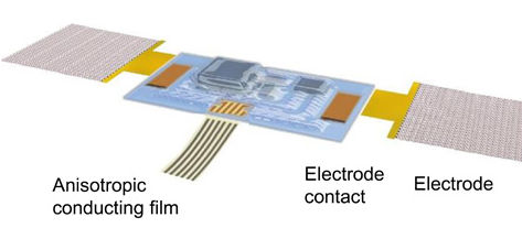
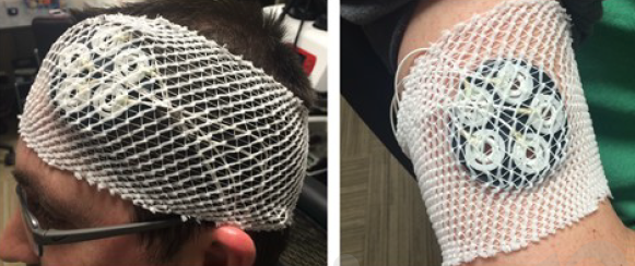
Illustration of the assembled acoustic sensor device and its interface with soft electrophysiology measurement electrodes and flexible cable for power supply and data acquisition (credit: Yuhao Liu et al./Science Advances)

Researchers from the University of Colorado Boulder and Northwestern University have developed a tiny, soft, wearable acoustic sensor that measures vibrations in the human body and can be used to monitor human heart health and recognize spoken words.

The stretchable Band-aid-like device attaches to the skin on nearly any surface of the body, using “epidermal electronics” to capture sound signals from the body.

It’s a sort of tiny, wearable stethoscope. As described in an open-access paper published Nov. 16 in Science Advances, a sister journal of Science, it can detect things like heart murmurs in cardiac patients and lung problems, and can monitor ventricular assist devices. It can also be used to pick up speech sounds (for automated speech recognition or controlling video games and other machines), and even movements in gastrointestinal tracts.

Listening in on the body

The sensor can also integrate electrodes that can record electrocardiogram (ECG) signals that measure the electrical activity of the heart as well electromyogram (EMG) signals that measure the electrical activity of muscles at rest and during contraction.

While the sensor was wired to an external data acquisition system for the tests, it can easily be converted into a wireless device, said CU Boulder Assistant Professor Jae-Woong Jeong, a lead author. Such sensors could be of use in remote, noisy places — including battlefields — producing quiet, high-quality cardiology or speech signals that can be read in real time at distant medical facilities.

Using the data from these sensors, a doctor at a hospital far away from a patient would be able to make a fast, accurate diagnosis, said Jeong.

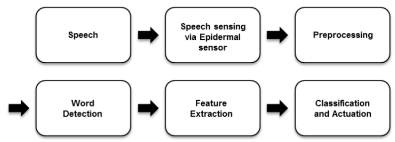
Process loop for a speech-based human-machine interface (credit: Yuhao Liu et al./Science Advances)

Vocal cord vibration signals also could be used by the military personnel or civilians to control robots, vehicles or drones. The speech recognition capabilities of the sensor also have implications for improving communication for people suffering from speech impairments, he said.

As part of the study, the team used the device to measure cardiac acoustic responses and ECG activity — including the detection of heart murmurs — in a group of elderly volunteers.

The researchers also were able to detect the acoustical signals of blood clots in a related lab experiment, said Jeong.


University of Colorado Boulder | CU Science Story — Wearable Tech


Abstract of Epidermal mechano-acoustic sensing electronics for cardiovascular diagnostics and human-machine interfaces

Physiological mechano-acoustic signals, often with frequencies and intensities that are beyond those associated with the audible range, provide information of great clinical utility. Stethoscopes and digital accelerometers in conventional packages can capture some relevant data, but neither is suitable for use in a continuous, wearable mode, and both have shortcomings associated with mechanical transduction of signals through the skin. We report a soft, conformal class of device configured specifically for mechano-acoustic recording from the skin, capable of being used on nearly any part of the body, in forms that maximize detectable signals and allow for multimodal operation, such as electrophysiological recording. Experimental and computational studies highlight the key roles of low effective modulus and low areal mass density for effective operation in this type of measurement mode on the skin. Demonstrations involving seismocardiography and heart murmur detection in a series of cardiac patients illustrate utility in advanced clinical diagnostics. Monitoring of pump thrombosis in ventricular assist devices provides an example in characterization of mechanical implants. Speech recognition and human-machine interfaces represent additional demonstrated applications. These and other possibilities suggest broad-ranging uses for soft, skin-integrated digital technologies that can capture human body acoustics.

Test of transcranial direct current stimulation (tDCS) of the brain shows improved multitasking performance

Placement of five anode electrodes (left) over the dorsolateral prefrontal cortex and the cathode (right) over the right shoulder (to avoid spurious cognitive effects from cortical excitability) (credit: Justin Nelson et al./ Front. Hum. Neurosci.)

In an experiment at the Air Force Research Laboratory, Wright-Patterson Air Force Base in Ohio, researchers* have found that transcranial direct-current stimulation (tDCS) of the brain can improve people’s multitasking skills and help avoid the drop in performance that comes with information overload.

The study was reported in a pre-publication paper in the open-access journal Frontiers of Human Neuroscience. It was motivated by the observation that various Air Force operations such as remotely piloted and manned aircraft operations require a human operator to monitor and respond to multiple events simultaneously over a long period of time. “With the monotonous nature of these tasks, the operator’s performance may decline shortly after their work shift commences,” according to the researchers.

The study set out to determine at what baud rate (difficulty level) improved multitasking throughput capacity kicks in and whether tDCS can improve performance, resulting in higher throughput capacity. The researchers used a common stimulation site for augmenting cognitive function via tDCS: the left dorsolateral prefrontal cortex (DLPFC), which has been associated with working memory, attention, vigilance, planning, and reasoning.**

Multitasking test

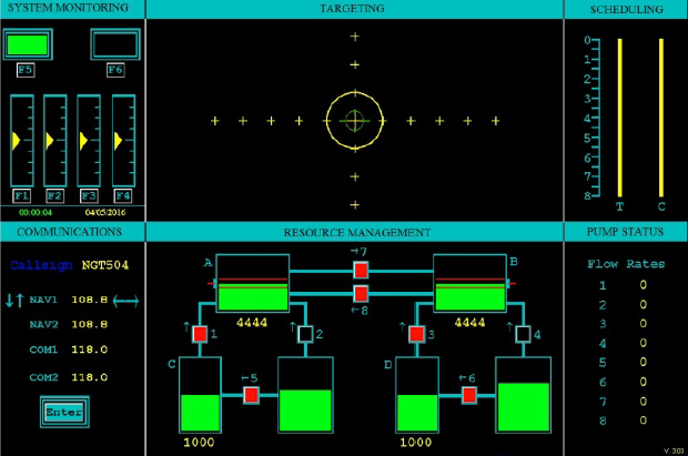
User interface of the multitasking Multi-Attribute Task Battery (MATB) (credit: Justin Nelson et al./ Front. Hum. Neurosci.)

The Air Force multi-attribute task battery (AF-MATB), based on the MATB multitasking test developed by NASA, requires the human operator (or experimental subject) to simultaneously monitor and respond to four independent tasks — systems monitoring, communication, targeting and resource management — on one computer screen.

There were 20 participants in the test, split evenly between experimental and control groups. The experimental group received 2mA of tDCS for a duration of 30 minutes. To emulate the subjective skin sensations present in active tDCS, the control group received sham stimulation at 2mA for only 30 seconds.

For example, the systems monitoring task (upper left) involved monitoring the two lights (rectangles), keeping the left light in the on status (displaying green) and the right light in the off status (displaying black). The triangular yellow markers shifted randomly towards the top or the bottom of the dial and began oscillating around a new location. When this event occurred, participants were to select the corresponding F1–F4 keys to reset the dials.

Augmented multitasking

The total input began at 0.6 bits/s and increased to 2.2 bits/s by a factor of 0.2 bits/s every four minutes. The study found that with tDCS stimulation, the subject’s multitasking throughput (or channel capacity) plateaued (maxed out) at 2.0 bits/s input, whereas the control group (no stimulation) plateaued near 1.3 bits/s, showing that tDCS had the ability to augment and enhance multitasking throughput by a factor of 1.5 in this experiment.

The finding potentially has important implications for high-workload environments that provide information to the operator via a wide range of stimuli. Additional research may be conducted to evaluate the robustness of these observed effects.

However, as reported by KurzweilAI, a recent open letter by 39 neuroscience researchers, published in the Annals of Neurology, warns that “outcomes of tDCS can be unpredictable … the benefits that are seen after tDCS in certain mental abilities may come at the expense of others.”

* Human factors engineers at the Wright State University Department of Biomedical, Industrial and Human Factors Engineering were also involved in the research.

** The left dorsolateral was selected as the stimulation site because this region of the brain is associated with sustained attention, working memory, decision making, planning and reasoning which are all directly involved with multitasking. The left DLPFC was chosen instead of the right DLPFC to enhance performance by right-handed participants, but stimulation of the right DLPFC may also influence working memory performance, according to the researchers.