
Turning hands and packages into displays

The SkinHaptics device sends ultrasound waves through the hand, focused at a precise point on the palm. (credit: Sri Subramanian / University of Sussex)

Imagine using your hand as an interactive touch-screen display. Sounds like science fiction, but Nokia Research Centre and the European Research Council have funded a study, led by the University of Sussex, to develop such a device, which could be used as a display for the next generation of smartwatches and other smart devices.

Called SkinHaptics, the device (still in the lab) sends ultrasonic pulses to your palm from the back of your hand, creating tactile sensations (similar to the iPhone 6s and Apple Watch “3D Touch” feature, which senses the amount of pressure your finger applies to the screen and can trigger various actions).
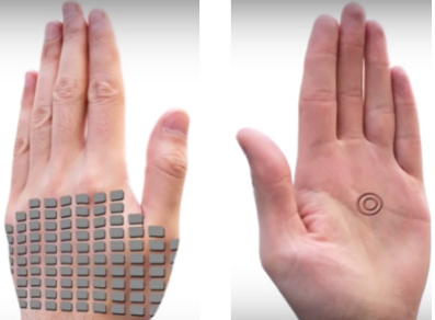
SkinHaptics ultrasound transmitter (left) and display (right) (credit: University of Sussex)

SkinHaptics is based on haptics, the science of applying touch sensation and control to interaction with computers and technology. It uses “time-reversal” processing, make the waves converge and become more targeted as they travel through the hand (rather than being dispersed), and ending at a precise point on the palm.

Professor Sriram Subramanian, who leads the research team at the University of Sussex, says technologies will need to engage other senses, such as touch, as we enter what designers are calling an “eye-free” age of technology. “If you imagine you are on your bike and want to change the volume control on your smartwatch, the interaction space on the watch is very small. So companies are looking at how to extend this space to the hand of the user.”

The findings were presented at the IEEE Haptics Symposium 2016 in Philadelphia.


Interact Lab | SkinHaptics: Ultrasound Focused in the Hand Creates Tactile Sensations

Package displays

In another innovative display concept, scientists at the University of Sheffield and technology company Novalia have developed technology for affixing an interactive display to packaging to display a promotion or an instructional label, for example.

This technology could be used in greetings cards or on products that allow a customer to receive a simple message or even ask questions.
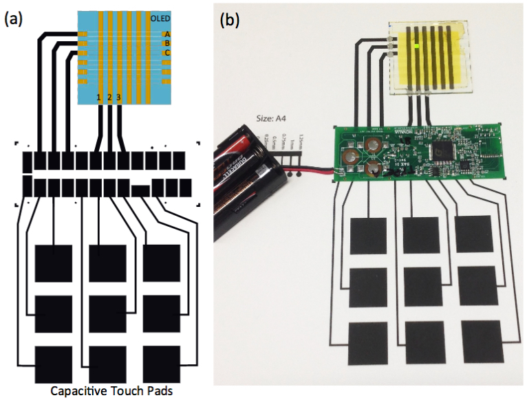
(a) Schematic of the capacitive touch-pad array, electrical circuit and LED array. (b) Photo of the integrated electronic system on the printed paper substrate. (credit: University of Sheffield)

In a paper published in the IEEE Journal of Display Technology, the team explains that the process involves printing electronic tracks onto paper, using a low-cost conductive graphite ink, to create capacitive touch pads and then affixing low-cost CMOS electronics and a polymer LED display to the paper, using an adhesive that conducts electricity.

The scientists also designed and constructed a touch-pad keyboard on paper that allows a user to selectively turn LEDs in the display on or off. The process could potentially be printed on other surfaces.

The research has been funded by the Engineering and Physical Sciences Research Council (EPSRC).


Abstract of SkinHaptics: Ultrasound Focused in the Hand Creates Tactile Sensations

Recent developments in on-body interfaces have extended the interaction space of physical devices to the skin of our hands. While these interfaces can easily project graphical elements on the bare hand, they cannot give tactile feedback. Here we present a technology that could help to expand the output capability of on-body interfaces to provide tactile feedback without restricting the skin as an interaction surface. SkinHaptics works by focusing ultrasound in the hand using a phased array of ultrasound transmitters and the acoustic time reversal signal processing technique. We present experimental results that show that this device can steer and focus ultrasound on the skin through the hand, which provides the basis for the envisioned technology. We then present results of a study that show that the focused energy can create sensations that are perceived under the skin and in the hand. We demonstrate the potential of SkinHaptics and discuss how our proof-of-concept device can be scaled beyond the prototype.

Abstract of Polymer Light Emitting Diodes Powered via Paper-mounted Electronics

We have interfaced an array of polymer light emitting diodes (OLEDs) fabricated onto a glass substrate to a sheet of paper via a pressure sensitive conducting adhesive. By screen-printing a series of capacitive touch pads and connecting tracks onto paper using a low-cost conductive graphite ink, we are able to drive individual pixels in the OLED array via CMOS-based electronics that are also attached to the paper. Three AA batteries are used to power the CMOS electronics, touch-pads and the OLED array, with pixels in the array operating at a brightness of up to 210 cd/m2. The work highlights a practical interface between plastic- and paper-based electronics.
Microscope uses nanosecond-speed laser and deep learning to detect cancer cells more efficiently

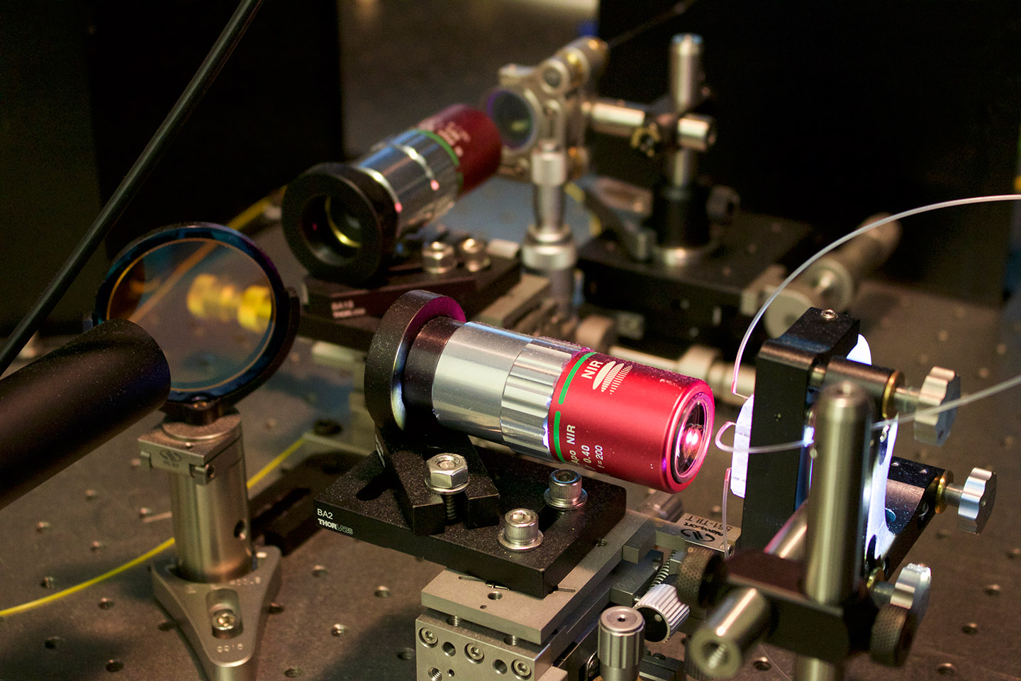
This microscope uses specially designed optics that boost image clarity and slows them enough to be detected and digitized at a rate of 36 million images per second. It then uses deep learning to distinguish cancer cells from healthy white blood cells. (credit: Tunde Akinloye/CNSI)

Scientists at the California NanoSystems Institute at UCLA have developed a new technique for identifying cancer cells in blood samples faster and more accurately than the current standard methods.

In one common approach to testing for cancer, doctors add biochemicals to blood samples. Those biochemicals attach biological “labels” to the cancer cells, and those labels enable instruments to detect and identify them. However, the biochemicals can damage the cells and render the samples unusable for future analyses. There are other current techniques that don’t use labeling but can be inaccurate because they identify cancer cells based only on one physical characteristic.

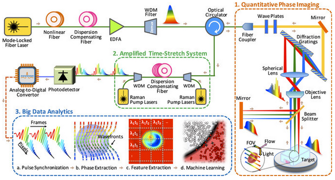
Time-stretch quantitative phase imaging (TS-QPI) and analytics system

The new technique images cells without destroying them and can identify 16 physical characteristics — including size, granularity and biomass — instead of just one.

Time-stretch quantitative phase imaging (TS-QPI) and analytics system (credit: Claire Lifan Chen et al./Nature Scientific Reports)

The new technique combines two components that were invented at UCLA:

  • A “photonic time stretch” microscope, which is capable of quickly imaging cells in blood samples. Invented by Barham Jalali, professor and Northrop-Grumman Optoelectronics Chair in electrical engineering, it works by taking pictures of flowing blood cells using laser bursts (similar to how a camera uses a flash). Each flash only lasts nanoseconds (billionths of a second) to avoid damage to cells, but that normally means the images are both too weak to be detected and too fast to be digitized by normal instrumentation. The new microscope overcomes those challenges by using specially designed optics that amplify and boost the clarity of the images, and simultaneously slow them down enough to be detected and digitized at a rate of 36 million images per second.
  • A deep learning computer program, which identifies cancer cells with more than 95 percent accuracy. Deep learning is a form of artificial intelligence that uses complex algorithms to extract patterns and knowledge from rich multidimenstional datasets, with the goal of achieving accurate decision making.

The study was published in the open-access journal Nature Scientific Reports. The researchers write in the paper that the system could lead to data-driven diagnoses by cells’ physical characteristics, which could allow quicker and earlier diagnoses of cancer, for example, and better understanding of the tumor-specific gene expression in cells, which could facilitate new treatments for disease.

The research was supported by NantWorks, LLC.


Abstract of Deep Learning in Label-free Cell Classification

Label-free cell analysis is essential to personalized genomics, cancer diagnostics, and drug development as it avoids adverse effects of staining reagents on cellular viability and cell signaling. However, currently available label-free cell assays mostly rely only on a single feature and lack sufficient differentiation. Also, the sample size analyzed by these assays is limited due to their low throughput. Here, we integrate feature extraction and deep learning with high-throughput quantitative imaging enabled by photonic time stretch, achieving record high accuracy in label-free cell classification. Our system captures quantitative optical phase and intensity images and extracts multiple biophysical features of individual cells. These biophysical measurements form a hyperdimensional feature space in which supervised learning is performed for cell classification. We compare various learning algorithms including artificial neural network, support vector machine, logistic regression, and a novel deep learning pipeline, which adopts global optimization of receiver operating characteristics. As a validation of the enhanced sensitivity and specificity of our system, we show classification of white blood T-cells against colon cancer cells, as well as lipid accumulating algal strains for biofuel production. This system opens up a new path to data-driven phenotypic diagnosis and better understanding of the heterogeneous gene expressions in cells.

​Clothes that receive and transmit digital information

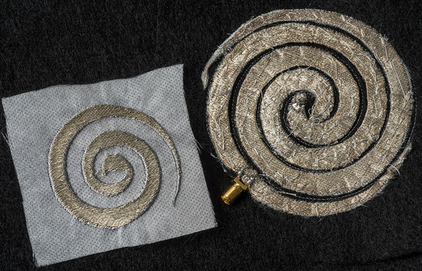
Researchers at The Ohio State University are developing embroidered antennas and circuits with 0.1 mm precision — the perfect size to integrate electronic components such as communication devices, sensors, and computer memory devices into clothing.  (credit: Photo by Jo McCulty, courtesy of The Ohio State University)

Ohio State University researchers have taken a key step in the design of  “functional textiles” — clothes that gather, store, or transmit digital information. They’ve developed a breakthrough method of weaving electronic components into fabric with 0.1mm precision — small enough to integrate components such as sensors and computer memory devices into clothing.

Imagine shirts that act as antennas for your smart phone or tablet, workout clothes that monitor your fitness level, sports equipment that monitors athletes’ performance, a bandage that tells your doctor how well the tissue beneath is healing, or a flexible fabric cap that senses or stimulates activity in the brain (eliminating restrictive tethered external wiring on the patient’s body).

“A revolution is happening in the textile industry,” said John Volakis, director of the ElectroScience Laboratory and the Roy & Lois Chope Chair Professor of Electrical Engineering at Ohio State. “We believe that functional textiles are an enabling technology for communications and sensing — and one day, even medical applications like imaging and health monitoring.”

Recently, he and research scientist Asimina Kiourti refined their patented fabrication method to create prototype wearables at a fraction of the cost and in half the time compared to two years ago. They published the new results in the journal IEEE Antennas and Wireless Propagation Letters.

Cell-phone antennas

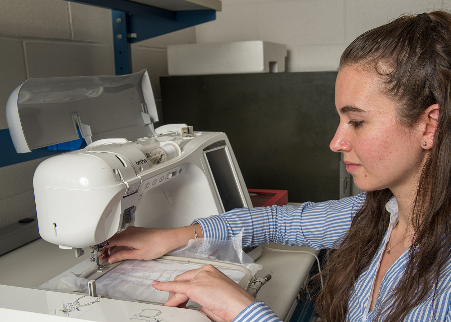
Asimina Kiourti, a research scientist at The Ohio State University, demonstrates the embroidery technique she and John Volakis invented for integrating electronics into clothing (photo credit: Jo McCulty/The Ohio State University)

In Volakis’ lab, the functional textiles, also called “e-textiles,” are created in part on a typical tabletop sewing machine. Like other modern sewing machines, it embroiders thread into fabric automatically based on a pattern loaded from a computer file. The researchers substitute the thread with fine silver metal wires that, once embroidered, feel the same as traditional thread to the touch.

“We started with a technology that is very well known — machine embroidery — and we asked: how can we functionalize embroidered shapes? How do we make them transmit signals at useful frequencies, like for cell phones or health sensors?” Volakis said. “Now, for the first time, we’ve achieved the accuracy of printed metal circuit boards, so our new goal is to take advantage of the precision to incorporate receivers and other electronic components.”

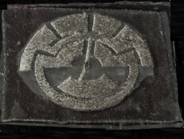
Broadband antenna constructed from multiple embroidered geometric shapes, each resonating at a different frequency (photo credit: Jo McCulty/The Ohio State University)

The shape of the embroidery determines the frequency of operation of the antenna or circuit, explained Kiourti. The shape of one broadband antenna, for instance, consists of more than half a dozen interlocking geometric shapes, each a little bigger than a fingernail, that form an intricate circle a few inches across. Each piece of the circle transmits energy at a different frequency, so that they cover a broad spectrum of energies when working together — achieving a “broadband” capability of the antenna for cell phone and Internet access.

In another design, tests showed that an embroidered spiral antenna (top photo) measuring approximately six inches across transmitted signals at frequencies of 1 to 5 GHz with near-perfect efficiency,  well-suited to broadband Internet and cellular communication.


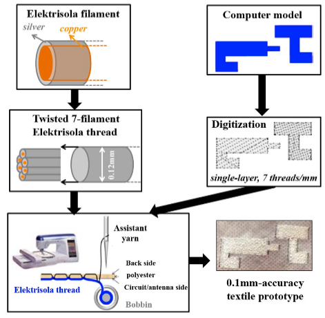
Proposed embroidery process to achieve 0.1mm geometrical precision (credit: Asimina Kiourti et al./IEEE Antennas Wireless Propag. Lett.)

On problem they had was that fine wires couldn’t provide as much surface conductivity as thick wires. So they had to find a way to work the fine thread into embroidery densities and shapes that would boost the surface conductivity and, thus, the antenna/sensor performance.

The new threads have a 0.1-mm diameter, made with only seven filaments. Each filament is copper at the center, enameled with pure silver. They purchase the wire by the spool at a cost of 3 cents per foot; Kiourti estimated that embroidering a single broadband antenna like the one mentioned above consumes about 10 feet of thread, for a material cost of around 30 cents per antenna. That’s 24 times less expensive than when Volakis and Kiourti created similar antennas in 2014.

In part, the cost savings comes from using less thread per embroidery. The researchers previously had to stack the thicker thread in two layers, one on top of the other, to make the antenna carry a strong enough electrical signal. But by refining the technique that she and Volakis developed, Kiourti was able to create the new, high-precision antennas in only one embroidered layer of the finer thread. So now the process takes half the time: only about 15 minutes for the broadband antenna mentioned above.

She’s also incorporated some techniques common to microelectronics manufacturing to add parts to embroidered antennas and circuits. One prototype antenna looks like a spiral and can be embroidered into clothing to improve cell phone signal reception. Another prototype, a stretchable antenna with an integrated RFID (radio-frequency identification) chip embedded in rubber, takes the applications for the technology beyond clothing — for tires, in this case.

The work fits well with Ohio State’s role as a founding partner of the Advanced Functional Fabrics of America Institute, a national manufacturing resource center for industry and government. The new institute, which joins some 50 universities and industrial partners, was announced earlier this month by U.S. Secretary of Defense Ashton Carter.

Syscom Advanced Materials in Columbus provided the threads used in Volakis and Kiourti’s initial work. The finer threads used in this study were purchased from Swiss manufacturer Elektrisola. The research is funded by the National Science Foundation, and Ohio State will license the technology for further development.


Abstract of Fabrication of Textile Antennas and Circuits With 0.1 mm Precision

We present a new selection of E-fibers (also referred to as E-threads) and associated embroidery process. The new E-threads and process achieve a geometrical precision down to 0.1 mm. Thus, for the first time, accuracy of typical printed circuit board (PCB) prototypes can be achieved directly on textiles. Compared to our latest embroidery approach, the proposed process achieves: 1) 3 × higher geometrical precision; 2) 24 × lower fabrication cost; 3) 50% less fabrication time; and 4) equally good RF performance. This improvement was achieved by employing a new class of very thin, 7-filament, Elektrisola E-threads ( diameter ≈ 0.12 mm, almost 2 × thinner than before). To validate our approach, we “printed” and tested a textile spiral antenna operating between 1-5 GHz. Measurement results were in good agreement with simulations. We envision this textile spiral to be integrated within a cap and unobtrusively acquire neuropotentials from wireless fully-passive brain implants. Overall, the proposed embroidery approach brings forward new possibilities for a wide range of applications.

Autonomous vehicles might have to be test-driven tens or hundreds of years to demonstrate their safety

A Lexus RX450h retrofitted by Google for its driverless car fleet (credit: Steve Jurvetson/CC)

Autonomous vehicles would have to be driven hundreds of millions of miles or even hundreds of billions of miles over tens and even hundreds of years (under some scenarios) to create enough data to statistically demonstrate their safety, when compared to the rate at which injuries and fatalities* occur in human-controlled cars and trucks, according to a new open-access RAND report.**

Although the total number of crashes, injuries and fatalities from human drivers is high**, the rate of these failures (77 injuries per 100 million miles driven) is low in comparison with the number of miles that people drive.

The solution is to find more practical testing methods, such as virtual testing and simulators, mathematical modeling, scenario testing, and pilot studies.

Another report, from the University of Michigan’s Transportation Research Institute in October 2015, found that even though they haven’t been at fault, self-driving test cars are involved in crashes at five times the rate of conventional cars per mile and four times the injury rate (although minor) — or twice as high, when figures are adjusted to take into account the fact that many accidents involving conventional cars go unreported.

* According to the Center for Disease Control and Prevention, motor vehicle accidents are a leading cause of premature death in the United States and are responsible for over $80 billion annually in medical care and lost productivity due to injuries. Autonomous vehicles hold enormous potential for managing this crisis and researchers say autonomous vehicles could significantly reduce the number of accidents caused by human error. According to the National Highway Traffic Safety Administration, more than 90 percent of automobile crashes are caused by human errors such as driving too fast, as well as alcohol impairment, distraction and fatigue. Autonomous vehicles are never drunk, distracted or tired; these factors are involved in 41 percent, 10 percent and 2.5 percent of all fatal crashes, respectively.

** As of March 2016, Google self-driving cars in “autonomous mode” have driven 1,498,214 miles. As of Feb. 29, 2016, only one Google self-driving car has caused an accident, according to Google (on Feb. 14, a Google self-driving car, traveling at 2 mph, pulled out in front of a public bus going 15 mph, according to the California DMV).

Chinese team genetically modifies human embryo, using CRISPR gene-editing technique

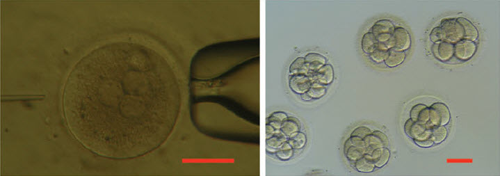
Photos showing injection of a human 3PN zygote (left) and development to 8 to 16-cell stage in vitro (right). Scale bar = 100 μm. (credit: Xiangjin Kang et al./Journal of Assisted Reproduction and Genetics)

Chinese researchers have genetically modified a human embryo using CRISPR/Cas9, the gene editing technique, using embryos that carried an extra set of chromosomes (so they were not viable) — hoping to learn more about the possibility of producing human babies that would be immune to HIV.

The Chinese team reports in the Journal of Assisted Reproduction and Genetics that they obtained 213 fertilized eggs from a fertility clinic, which had been deemed unsuitable for in vitro therapy.* They used the eggs to study a mutation that causes damage to an immune cell gene called CCR5 (this type of cell, when damaged naturally, has been found to lead to HIV resistance).

Aside from a previous study in China last year that involved editing human embryo genes (see Researchers in China have created genetically modified human embryos), most of the world has decided to hold off doing this controversial research. “We believe that any attempt to generate genetically modified humans through the modification of early embryos needs to be strictly prohibited until we can resolve both ethical and scientific issues,” the researchers say in their paper.

Failed experiment

The team reports that just 4 out of 26 of the embryos that were edited were modified successfully — some still contained genes that had not been modified, and others had resulted in unexpected gene mutations.

“This paper doesn’t look like it offers much more than anecdotal evidence that it works in human embryos, which we already knew,” George Daley, a stem-cell biologist at Children’s Hospital Boston, told Nature. “It’s certainly a long way from realizing the intended potential” — a human embryo with all its copies of CCR5 inactivated.

“It just emphasizes that there are still a lot of technical difficulties to doing precision editing in human embryo cells,” Xiao-Jiang Li, a neuroscientist at Emory University, also said in the Nature article . He thinks that researchers should work out these kinks in non-human primates, for example, before continuing to modify the genomes of human embryos using techniques such as CRISPR.

* The women who had donated the eggs all gave permission for the embryos to be used for genetic research, on condition that the embryos would not be allowed to mature into a human being (all of the embryos were destroyed after three days).


Abstract of Introducing precise genetic modifications into human 3PN embryos by CRISPR/Cas-mediated genome editing

Purpose

As a powerful technology for genome engineering, the CRISPR/Cas system has been successfully applied to modify the genomes of various species. The purpose of this study was to evaluate the technology and establish principles for the introduction of precise genetic modifications in early human embryos.

Methods

3PN zygotes were injected with Cas9 messenger RNA (mRNA) (100 ng/μl) and guide RNA (gRNA) (50 ng/μl). For oligo-injections, donor oligo-1 (99 bp) or oligo-2 (99 bp) (100 ng/μl) or dsDonor (1 kb) was mixed with Cas9 mRNA (100 ng/μl) and gRNA (50 ng/μl) and injected into the embryos.

Results

By co-injecting Cas9 mRNA, gRNAs, and donor DNA, we successfully introduced the naturally occurring CCR5Δ32 allele into early human 3PN embryos. In the embryos containing the engineeredCCR5Δ32 allele, however, the other alleles at the same locus could not be fully controlled because they either remained wild type or contained indel mutations.

Conclusions

This work has implications for the development of therapeutic treatments of genetic disorders, and it demonstrates that significant technical issues remain to be addressed. We advocate preventing any application of genome editing on the human germline until after a rigorous and thorough evaluation and discussion are undertaken by the global research and ethics communities.

‘Breakthrough Starshot’ aims to reach Alpha Centauri 20 years after launch

Alpha Centauri (credit: ESO Online Digitized Sky Survey)

Internet investor and science philanthropist Yuri Milner and physicist Stephen Hawking announced Tuesday a $100 million research and engineering program, Breakthrough Starshot, aiming to demonstrate proof of concept for light-propelled “nanocrafts” that could travel to Alpha Centauri, our nearest star system.

The nanocrafts would fly at 20 percent of light speed and capture images of possible planets and other scientific data, arriving in just over 20 years after their launch.

“Earth is a wonderful place, but it might not last forever,” said Hawking. “Sooner or later, we must look to the stars. Breakthrough Starshot is a very exciting first step on that journey.”

Breakthrough Starshot program

L-R) Yuri Milner, Breakthrough Prize and DST Global Founder; Stephen Hawking, CH, CBE, FRS, Dennis Stanton Avery and Sally Tsui Wong-Avery Director of Research, University of Cambridge; Freeman Dyson, Emeritus Professor, Princeton Institute for Advanced Study; Ann Druyan, Producer, Co-Founder and CEO of Cosmos Studios; Avi Loeb, Frank B. Baird, Jr. Professor of Science at Harvard University; Mae Jamison, Nasa Astronaut, Principal 100 Year Starship Foundation; and Peter Worden, Chairman, Breaktrough Prize Foundation, Former NASA Director speak on stage as Yuri Milner And Stephen Hawking host press conference to announce Breakthrough Starshot, a new space exploration initiative, at One World Observatory on April 12, 2016 in New York City. (Photo by Bryan Bedder/Getty Images for Breakthrough Prize Foundation)

The Breakthrough Starshot program will be led by Pete Worden, the former director of NASA AMES Research Center, and advised by a committee of world-class scientists and engineers. The board will consist of Stephen Hawking, Yuri Milner, and Mark Zuckerberg.

Nanocrafts

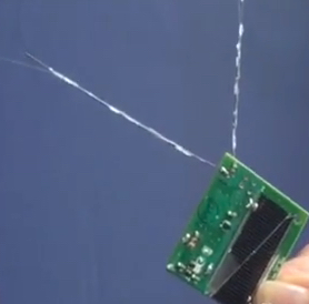
Nanocraft, comprising Lightsail and StarChip powered by Light Beamer (credit: Breakthrough Initiatives)

The Alpha Centauri star system is 25 trillion miles (4.37 light years) away. With today’s fastest spacecraft, it would take about 30,000 years to get there. The program aims to establish whether a gram-scale nanocraft on a sail pushed by a light beam can fly more than 1,000 times faster.

Nanocrafts are gram-scale robotic spacecrafts, comprising:

Starchip (credit: Breakthrough Initiatives)

Starchip: a gram-scale wafer carrying cameras, photon thrusters, power supply, navigation, and communication equipment, and constituting a fully functional space probe.

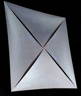
Lightsail (credit: Breakthrough Initiatives)

Lightsail: Advances in nanotechnology are producing increasingly thin and lightweight materials, promising to enable the fabrication of meter-scale sails no more than a few hundred atoms thick and at gram-scale mass.

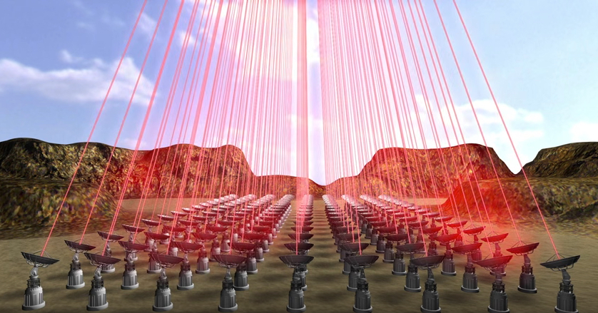
Light Beamer (credit: Breakthrough Initiatives)

Light Beamer. The rising power and falling cost of lasers has led to significant advances in light-beaming technology. Phased arrays of lasers (the “light beamer”) could potentially be scaled up to the 100 gigawatt level.

Breakthrough Starshot aims to bring economies of scale to the astronomical scale. The StarChip can be mass-produced at the cost of an iPhone and be sent on missions in large numbers to provide redundancy and coverage. The light beamer is modular and scalable. Once it is assembled and the technology matures, the cost of each launch is expected to fall to a few hundred thousand dollars.


Breakthrough Starshot

Path to the stars

The research and engineering phase is expected to last a number of years. Following that, development of the ultimate mission to Alpha Centauri would require a budget comparable to the largest current scientific experiments, and would involve building a ground-based kilometer-scale light beamer at high altitude in dry conditions, powered by a few gigawatt hours of energy per launch.

The plan is to launching a “mothership” carrying thousands of nanocrafts to a high-altitude orbit, taking advantage of adaptive optics technology in real time to compensate for atmospheric effects. Focusing the light beam on the lightsail would accelerate individual nanocrafts to the target speed within minutes.

The goal is to capture images of a planet and other scientific data and transmitting them back to Earth using a compact on-board laser communications system, using the same light beamer that launched the nanocrafts to receive data from them over 4 years later.

The organizers acknowledge that these and other system requirements represent significant engineering challenges, but are based on technology either already available or likely to be attainable in the near future under reasonable assumptions, the organizers say.

Kilometer-scale telescope

The proposed light propulsion system is at a scale that calls for global cooperation and support. Clearance for launches would be required from all the appropriate government and international organizations.

As the technology required for interstellar travel matures, a number of additional opportunities will emerge, including contributions to solar system exploration, using the light beamer as a kilometer-scale telescope for astronomical observations, and detection of Earth-crossing asteroids at large distances.

Astronomers estimate that there is a reasonable chance of an Earth-like planet existing in the “habitable zones” of Alpha Centauri’s three-star system. A number of scientific instruments, ground-based and space-based, are being developed and enhanced, which will soon identify and characterize planets around nearby stars. A separate Breakthrough Initiative will support some of these projects.

Getting it off the ground

The Breakthrough Starshot initiative is based entirely on research in the public domain. It will be dedicated to full transparency and open access, and open to experts in all relevant fields as well as the public to contribute ideas through its online forum.  The initiative will establish a research grant program, and will make available other funding to support relevant scientific and engineering research and development.

“We take inspiration from Vostok, Voyager, Apollo. and the other great missions,” said Pete Worden. “It’s time to open the era of interstellar flight, but we need to keep our feet on the ground to achieve this.”

First transistors made entirely of nanocrystal ‘inks’ in simplified process

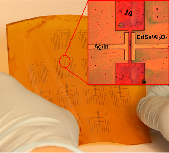
Because this process works at relatively low temperatures, many transistors can be made on a flexible backing at once. (credit: University of Pennsylvania)

University of Pennsylvania engineers have developed a simplified new approach for making transistors by sequentially depositing their components in the form of liquid nanocrystal “inks.” The new process open the door for transistors and other electronic components to be built into flexible or wearable applications. It also avoids the highly complex current process for creating transistors, which requires high-temperature, high-vacuum equipment. Also, the new lower-temperature process is compatible with a wide array of materials and can be applied to larger areas.

Transistors patterned on plastic backing

The researchers’ nanocrystal-based field effect transistors were patterned onto flexible plastic backings using spin coating, but could eventually be constructed by additive manufacturing systems, like 3D printers.

Published in the journal Science,  the study was lead by Cherie Kagan, the Stephen J. Angello Professor in the School of Engineering and Applied Science, and Ji-Hyuk Choi, then a member of her lab, now a senior researcher at the Korea Institute of Geoscience and Mineral Resources. Researchers at Korea University Korea’s Yonsei University were also involved.

Kagan’s group developed four nanocrystal inks that comprise the transistor, then deposited them on a flexible backing. (credit: University of Pennsylvania)

The researchers began by dispersing a specific type of nanocrystals in a liquid, creating nanocrystal inks. They developed a library of four of these inks: a conductor (silver), an insulator (aluminum oxide), a semiconductor (cadmium selenide), and a conductor combined with a dopant (a mixture of silver and indium). (“Doping” the semiconductor layer of a transistor with impurities controls whether the device creates a positive or negative charge.)

“These materials are colloids just like the ink in your inkjet printer,” Kagan said, “but you can get all the characteristics that you want and expect from the analogous bulk materials, such as whether they’re conductors, semiconductors or insulators.” Although the electrical properties of several of these nanocrystal inks had been independently verified, they had never been combined into full devices. “Our question was whether you could lay them down on a surface in such a way that they work together to form functional transistors.”

Laying down patterns in layers

Such a process entails layering or mixing them in precise patterns.

First, the conductive silver nanocrystal ink was deposited from liquid on a flexible plastic surface that was treated with a photolithographic mask, then rapidly spun to draw it out in an even layer. The mask was then removed to leave the silver ink in the shape of the transistor’s gate electrode.

The researchers followed that layer by spin-coating a layer of the aluminum oxide nanocrystal-based insulator, then a layer of the cadmium selenide nanocrystal-based semiconductor and finally another masked layer for the indium/silver mixture, which forms the transistor’s source and drain electrodes. Upon heating at relatively low temperatures, the indium dopant diffused from those electrodes into the semiconductor component.

“The trick with working with solution-based materials is making sure that, when you add the second layer, it doesn’t wash off the first, and so on,” Kagan said. “We had to treat the surfaces of the nanocrystals, both when they’re first in solution and after they’re deposited, to make sure they have the right electrical properties and that they stick together in the configuration we want.”

Because this entirely ink-based fabrication process works at lower temperatures than existing vacuum-based methods, the researchers were able to make several transistors on the same flexible plastic backing at the same time.

The inks’ specialized surface chemistry allowed them to stay in configuration without losing their electrical properties. (credit: University of Pennsylvania)

“Making transistors over larger areas and at lower temperatures have been goals for an emerging class of technologies, when people think of the Internet of things, large area flexible electronics and wearable devices,” Kagan said. “We haven’t developed all of the necessary aspects so they could be printed yet, but because these materials are all solution-based, it demonstrates the promise of this materials class and sets the stage for additive manufacturing.”

Because this entirely ink-based fabrication process works at lower temperatures than existing vacuum-based methods, the researchers were able to make several transistors on the same flexible plastic backing at the same time.

3D-printing transistors for wearables

“This is the first work,” Choi said, “showing that all the components, the metallic, insulating, and semiconducting layers of the transistors, and even the doping of the semiconductor, could be made from nanocrystals.”

“Making transistors over larger areas and at lower temperatures have been goals for an emerging class of technologies, when people think of the Internet of things, large area flexible electronics and wearable devices,” Kagan said. “We haven’t developed all of the necessary aspects so they could be printed yet, but because these materials are all solution-based, it demonstrates the promise of this materials class and sets the stage for additive manufacturing.”

The research was supported by the National Science Foundation, the U.S. Department of Energy, the Office of Naval Research, and the Korea Institute of Geoscience and Mineral Resources funded by the Ministry of Science, ICT, and Future Planning of Korea.


Abstract of Exploiting the colloidal nanocrystal library to construct electronic devices

Synthetic methods produce libraries of colloidal nanocrystals with tunable physical properties by tailoring the nanocrystal size, shape, and composition. Here, we exploit colloidal nanocrystal diversity and design the materials, interfaces, and processes to construct all-nanocrystal electronic devices using solution-based processes. Metallic silver and semiconducting cadmium selenide nanocrystals are deposited to form high-conductivity and high-mobility thin-film electrodes and channel layers of field-effect transistors. Insulating aluminum oxide nanocrystals are assembled layer by layer with polyelectrolytes to form high–dielectric constant gate insulator layers for low-voltage device operation. Metallic indium nanocrystals are codispersed with silver nanocrystals to integrate an indium supply in the deposited electrodes that serves to passivate and dope the cadmium selenide nanocrystal channel layer. We fabricate all-nanocrystal field-effect transistors on flexible plastics with electron mobilities of 21.7 square centimeters per volt-second.

Best textile manufacturing methods for creating human tissues with stem cells

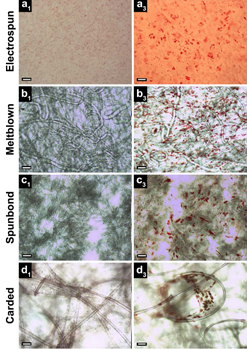
All four textile manufacturing processes and corresponding scaffold (structure) types studied exhibited the presence of lipid vacuoles (small red spheres, right column, indicating stem cells undergoing random differentiation), compared to control (left). Electrospun scaffolds (row a) exhibited only a monolayer of lipid vacuoles in a single focal plane, while meltblown, spunbond, and carded scaffolds (rows b, c, d) exhibited vacuoles in multiple planes throughout the fabric thickness. Scale bars: 100 μm (credit: S. A. Tuin et al./Biomedical Materials)

Elizabeth Loboa, dean of the Missouri University College of Engineering, and her team have tested new tissue- engineering methods (based on textile manufacturing) to find ones that are most cost-effective and can be produced in larger quantities.

Tissue engineering is a process that uses novel biomaterials seeded with stem cells to grow and replace missing tissues. When certain types of materials are used, the “scaffolds” that are created to hold stem cells eventually degrade, leaving natural tissue in its place. The new tissues could help patients suffering from wounds caused by diabetes and circulation disorders, patients in need of cartilage or bone repair, and women who have had mastectomies by replacing their breast tissue. The challenge is creating enough of the material on a scale that clinicians need to treat patients.

Comparing textile manufacturing techniques

Electrospinning experiment: nanofibers are collected into an ethanol bath and removed at predefined time intervals (credit: J. M. Coburn et al./The Johns Hopkins University/PNAS)

In typical tissue engineering approaches that use fibers as scaffolds, non-woven materials are often bonded together using an electrostatic field. This process, called electrospinning (see Nanoscale scaffolds and stem cells show promise in cartilage repair and Improved artificial blood vessels), creates the scaffolds needed to attach to stem cells.

However, large-scale production with electrospinning is not cost-effective. “Electrospinning produces weak fibers, scaffolds that are not consistent, and pores that are too small,” Loboa said. “The goal of ‘scaling up’ is to produce hundreds of meters of material that look the same, have the same properties, and can be used in clinical settings. So we investigated the processes that create textiles, such as clothing and window furnishings like drapery, to scale up the manufacturing process.”

The group published two papers using three industry-standard, high-throughput manufacturing techniques — meltblowing, spunbonding, and carding — to determine if they would create the materials needed to mimic native tissue.

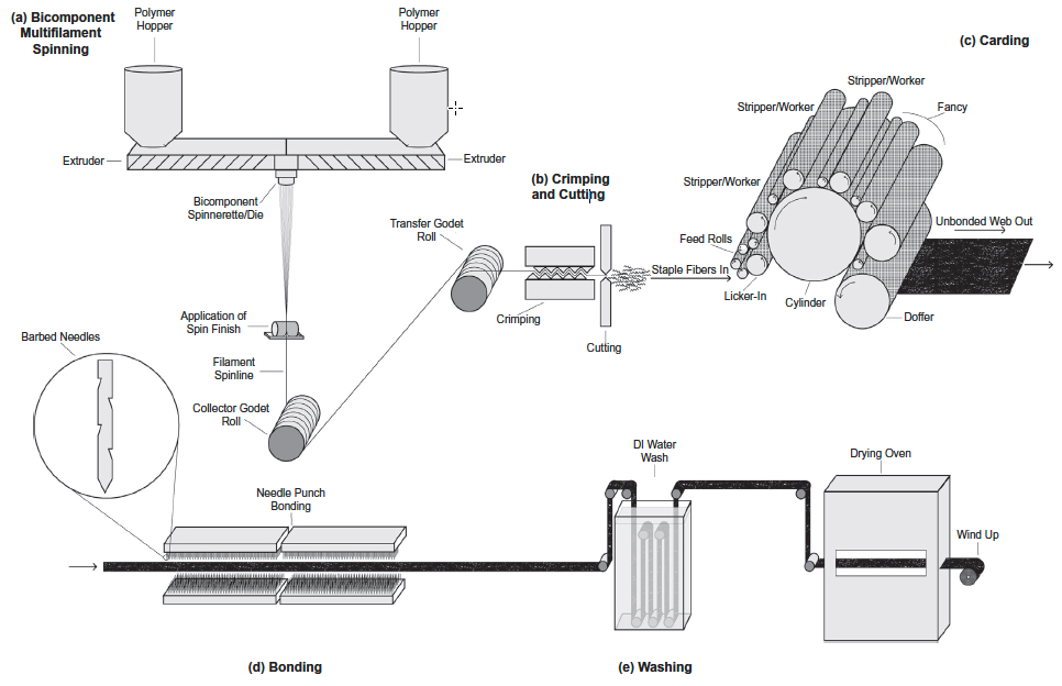
Meltblowing is a technique during which nonwoven materials are created using a molten polymer to create continuous fibers. Spunbond materials are made much the same way but the fibers are drawn into a web while in a solid state instead of a molten one. Carding involves the separation of fibers through the use of rollers, forming the web needed to hold stem cells in place.

Schematic of gilled fiber multifilament spinning and carded scaffold fabrication (credit: Stephen A. Tuin et al./Acta Biomaterialia)

Cost-effective methods

Loboa and her colleagues tested these techniques to create polylactic acid (PLA) scaffolds (a Food and Drug Administration-approved material used as collagen fillers), seeded with human stem cells. They then spent three weeks studying whether the stem cells remained healthy and if they began to differentiate into fat and bone pathways, which is the goal of using stem cells in a clinical setting when new bone and/or new fat tissue is needed at a defect site. Results showed that the three textile manufacturing methods proved as viable if not more so than electrospinning.

“These alternative methods are more cost-effective than electrospinning,” Loboa said. “A small sample of electrospun material could cost between $2 to $5. The cost for the three manufacturing methods is between $.30 to $3.00; these methods proved to be effective and efficient. Next steps include testing how the different scaffolds created in the three methods perform once implanted in animals.”

Researchers at North Carolina State University and the University of North Carolina at Chapel Hill were also involved in the two studies, which were published in Biomedical Materials (open access) and Acta Biomaterialia. The National Science Foundation, the National Institutes of Health, and the Nonwovens Institute provided funding for the studies.


Abstract of Creating tissues from textiles: scalable nonwoven manufacturing techniques for fabrication of tissue engineering scaffolds

Electrospun nonwovens have been used extensively for tissue engineering applications due to their inherent similarities with respect to fibre size and morphology to that of native extracellular matrix (ECM). However, fabrication of large scaffold constructs is time consuming, may require harsh organic solvents, and often results in mechanical properties inferior to the tissue being treated. In order to translate nonwoven based tissue engineering scaffold strategies to clinical use, a high throughput, repeatable, scalable, and economic manufacturing process is needed. We suggest that nonwoven industry standard high throughput manufacturing techniques (meltblowing, spunbond, and carding) can meet this need. In this study, meltblown, spunbond and carded poly(lactic acid) (PLA) nonwovens were evaluated as tissue engineering scaffolds using human adipose derived stem cells (hASC) and compared to electrospun nonwovens. Scaffolds were seeded with hASC and viability, proliferation, and differentiation were evaluated over the course of 3 weeks. We found that nonwovens manufactured via these industry standard, commercially relevant manufacturing techniques were capable of supporting hASC attachment, proliferation, and both adipogenic and osteogenic differentiation of hASC, making them promising candidates for commercialization and translation of nonwoven scaffold based tissue engineering strategies.


Abstract of Fabrication of novel high surface area mushroom gilled fibers and their effects on human adipose derived stem cells under pulsatile fluid flow for tissue engineering applications

The fabrication and characterization of novel high surface area hollow gilled fiber tissue engineering scaffolds via industrially relevant, scalable, repeatable, high speed, and economical nonwoven carding technology is described. Scaffolds were validated as tissue engineering scaffolds using human adipose derived stem cells (hASC) exposed to pulsatile fluid flow (PFF). The effects of fiber morphology on the proliferation and viability of hASC, as well as effects of varied magnitudes of shear stress applied via PFF on the expression of the early osteogenic gene marker runt related transcription factor 2 (RUNX2) were evaluated. Gilled fiber scaffolds led to a significant increase in proliferation of hASC after seven days in static culture, and exhibited fewer dead cells compared to pure PLA round fiber controls. Further, hASC-seeded scaffolds exposed to 3 and 6 dyn/cm2 resulted in significantly increased mRNA expression of RUNX2 after one hour of PFF in the absence of soluble osteogenic induction factors. This is the first study to describe a method for the fabrication of high surface area gilled fibers and scaffolds. The scalable manufacturing process and potential fabrication across multiple nonwoven and woven platforms makes them promising candidates for a variety of applications that require high surface area fibrous materials.

Statement of Significance

We report here for the first time the successful fabrication of novel high surface area gilled fiber scaffolds for tissue engineering applications. Gilled fibers led to a significant increase in proliferation of human adipose derived stem cells after one week in culture, and a greater number of viable cells compared to round fiber controls. Further, in the absence of osteogenic induction factors, gilled fibers led to significantly increased mRNA expression of an early marker for osteogenesis after exposure to pulsatile fluid flow. This is the first study to describe gilled fiber fabrication and their potential for tissue engineering applications. The repeatable, industrially scalable, and versatile fabrication process makes them promising candidates for a variety of scaffold-based tissue engineering applications.

Berkeley Lab captures first high-res 3D images of DNA segments

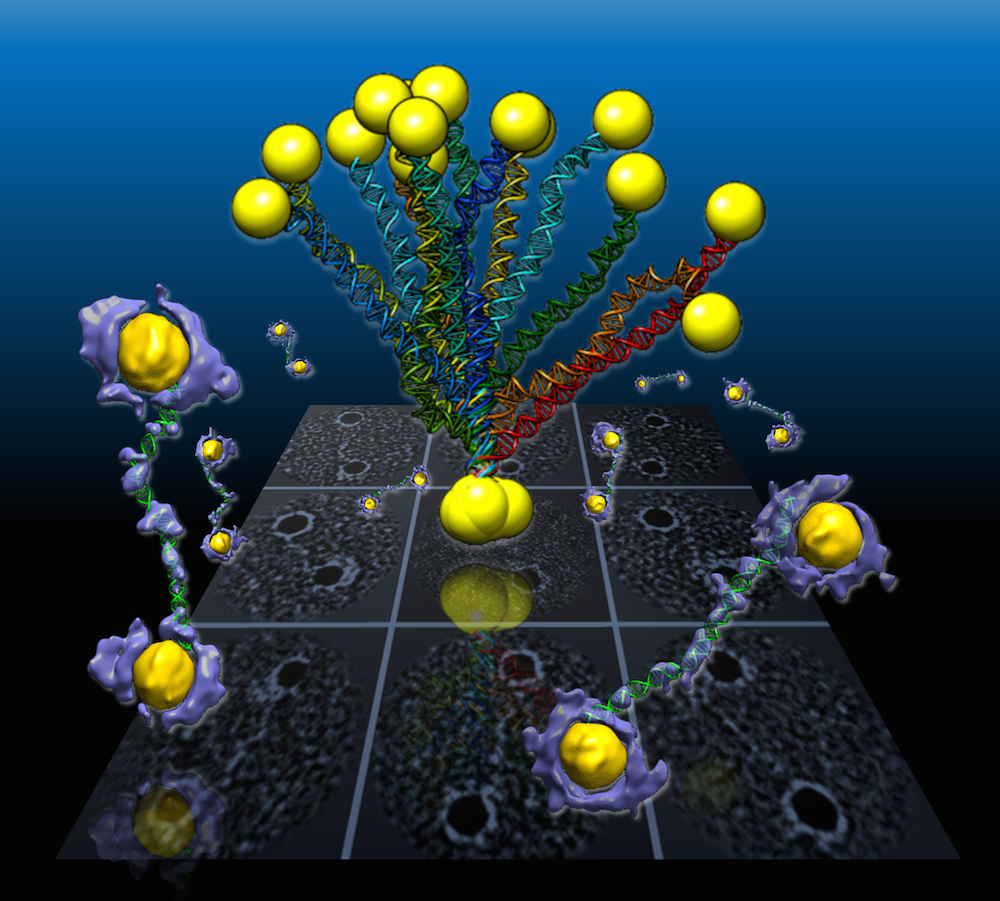
In a Berkeley Lab-led study, flexible double-helix DNA segments (purple, with green DNA models) connected to gold nanoparticles (yellow) are revealed from the 3D density maps reconstructed from individual samples using a Berkeley Lab-developed technique called individual-particle electron tomography (IPET). Projections of the structures are shown in the green background grid. (credit: Berkeley Lab)

An international research team working at the Lawrence Berkeley National Laboratory (Berkeley Lab) has captured the first high-resolution 3D images of double-helix DNA segments attached at either end to gold nanoparticles — which could act as building blocks for molecular computer memory and electronic devices (see World’s smallest electronic diode made from single DNA molecule), nanoscale drug-delivery systems, and as markers for biological research and for imaging disease-relevant proteins.

The researchers connected coiled DNA strands between polygon-shaped gold nanoparticles and then reconstructed 3D images, using a cutting-edge electron microscope technique coupled with a protein-staining process and sophisticated software that provided structural details at the scale of about 2 nanometers.

“We had no idea about what the double-strand DNA would look like between the gold nanoparticles,” said Gang “Gary” Ren, a Berkeley Lab scientist who led the research. “This is the first time for directly visualizing an individual double-strand DNA segment in 3D,” he said.

The results were published in an open-access paper in the March 30 edition of Nature Communications.

The method developed by this team, called individual-particle electron tomography (IPET), had earlier captured the 3-D structure of a single protein that plays a key role in human cholesterol metabolism. By grabbing 2D images of an object from different angles, the technique allows researchers to assemble a 3D image of that object.

The team has also used the technique to uncover the fluctuation of another well-known flexible protein, human immunoglobulin 1, which plays a role in the human immune system.


Berkeley Lab | 3-D Reconstructions of Double strand DNA and Gold Nanoparticle Structures

For this new study of DNA nanostructures, Ren used an electron-beam study technique called cryo-electron microscopy (cryo-EM) to examine frozen DNA-nanogold samples, and used IPET to reconstruct 3-D images from samples stained with heavy metal salts. The team also used molecular simulation tools to test the natural shape variations (“conformations”) in the samples, and compared these simulated shapes with observations.

First visualization of DNA strand dynamics without distorting x-ray crystallography

Ren explained that the naturally flexible dynamics of samples, like a man waving his arms, cannot be fully detailed by any method that uses an average of many observations.

A popular way to view the nanoscale structural details of delicate biological samples is to form them into crystals and zap them with X-rays, but that destroys their natural shape, especially fir the DNA-nanogold samples in this study, which the scientists say are incredibly challenging to crystallize. Other common research techniques may require a collection of thousands of near-identical objects, viewed with an electron microscope, to compile a single, averaged 3-D structure. But an averaged 3D image may not adequately show the natural shape fluctuations of a given object.

The samples in the latest experiment were formed from individual polygon gold nanostructures, measuring about 5 nanometers across, connected to single DNA-segment strands with 84 base pairs. Base pairs are basic chemical building blocks that give DNA its structure. Each individual DNA segment and gold nanoparticle naturally zipped together with a partner to form the double-stranded DNA segment with a gold particle at either end.


Berkeley Lab | These views compare the various shape fluctuations obtained from different samples of the same type of double-helix DNA segment (DNA renderings in green, 3D reconstructions in purple) connected to gold nanoparticles (yellow).

The samples were flash-frozen to preserve their structure for study with cryo-EM imaging. The distance between the two gold nanoparticles in individual samples varied from 20 to 30 nanometers, based on different shapes observed in the DNA segments.

Researchers used a cryo-electron microscope at Berkeley Lab’s Molecular Foundry for this study. They collected a series of tilted images of the stained objects, and reconstructed 14 electron-density maps that detailed the structure of individual samples using the IPET technique.

Sub-nanometer images next

Ren said that the next step will be to work to improve the resolution to the sub-nanometer scale.

“Even in this current state we begin to see 3-D structures at 1- to 2-nanometer resolution,” he said. “Through better instrumentation and improved computational algorithms, it would be promising to push the resolution to that visualizing a single DNA helix within an individual protein.”

In future studies, researchers could attempt to improve the imaging resolution for complex structures that incorporate more DNA segments as a sort of “DNA origami,” Ren said. Researchers hope to build and better characterize nanoscale molecular devices using DNA segments that can, for example, store and deliver drugs to targeted areas in the body.

“DNA is easy to program, synthesize and replicate, so it can be used as a special material to quickly self-assemble into nanostructures and to guide the operation of molecular-scale devices,” he said. “Our current study is just a proof of concept for imaging these kinds of molecular devices’ structures.”

The team included researchers at UC Berkeley, the Kavli Energy NanoSciences Institute at Berkeley Lab and UC Berkeley, and Xi’an Jiaotong University in China. This work was supported by the National Science Foundation, DOE Office of Basic Energy Sciences, National Institutes of Health, the National Natural Science Foundation of China, Xi’an Jiaotong University in China, and the Ministry of Science and Technology in China. View more about Gary Ren’s research group here.


Abstract of Three-dimensional structural dynamics and fluctuations of DNA-nanogold conjugates by individual-particle electron tomography

DNA base pairing has been used for many years to direct the arrangement of inorganic nanocrystals into small groupings and arrays with tailored optical and electrical properties. The control of DNA-mediated assembly depends crucially on a better understanding of three-dimensional structure of DNA-nanocrystal-hybridized building blocks. Existing techniques do not allow for structural determination of these flexible and heterogeneous samples. Here we report cryo-electron microscopy and negative-staining electron tomography approaches to image, and three-dimensionally reconstruct a single DNA-nanogold conjugate, an 84-bp double-stranded DNA with two 5-nm nanogold particles for potential substrates in plasmon-coupling experiments. By individual-particle electron tomography reconstruction, we obtain 14 density maps at ~2-nm resolution. Using these maps as constraints, we derive 14 conformations of dsDNA by molecular dynamics simulations. The conformational variation is consistent with that from liquid solution, suggesting that individual-particle electron tomography could be an expected approach to study DNA-assembling and flexible protein structure and dynamics.

MIT AI Lab 3D-prints first mobile robot made of solids and liquids

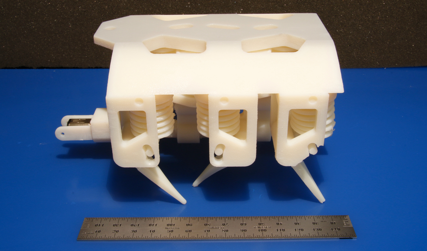
This 3D-printed hexapod robot moves via a single motor that spins a crankshaft that pumps fluid to the robot’s legs. Every component except the motor, battery, and added electronics is printed in a single step, no assembly required. (credit: R. MacCurdy/MIT CSAIL)

Ever want to just push a button and print out a hydraulically-powered robot that can immediately get to work for you?

MIT’s Computer Science and Artificial Intelligence Laboratory (CSAIL) researchers have designed a system that does just that: 3D-prints moving robots with solid and liquid materials in a single step and on a commercially available 3D printer. No assembly required.

“All you have to do is stick in a battery and motor, and you have a robot that can practically walk right out of the printer,” says CSAIL Director Daniela Rus, who oversaw the project and co-wrote the paper.

Printable hydraulics

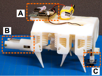
The 3D-printed six-legged demo robot weighs about 1.5 pounds and is less than 6 inches long. The robot uses a tripod gait. A single DC motor spins a central crankshaft that pumps fluid via banks of bellows pumps. Fluid is forced out of the pumps and distributed to each leg actuator by pipes embedded within the robot’s body. Added after printing to enhance control, an onboard microcontroller (A) controls a motor (B) and enables responses to environmental stimuli via a sensor (C), with cellphone control via Bluetooth. (credit: R. MacCurdy/MIT CSAIL)

To demonstrate their “printable hydraulics” concept, the researchers 3D-printed a tiny six-legged robot that can crawl, using 12 hydraulic pumps embedded within its body. They also added an onboard microcontroller, sensor, and remote control via cellphone.

The inkjet printer deposits individual droplets of material that are each 20 to 30 micrometers in diameter. The printer proceeds layer-by-layer from the bottom up. For each layer, the printer deposits different materials in different parts, and then uses high-intensity UV light to solidify all of the materials (except the liquids). Each layer consist of a “photopolymer” (a solid) and “a non-curing material” (a liquid).

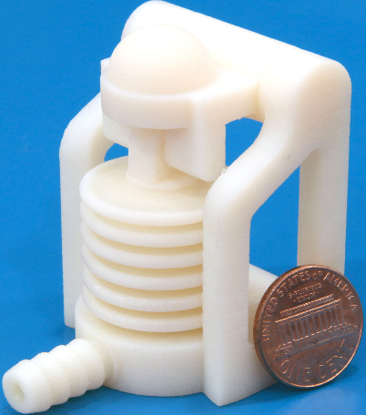
3D-printed bellows produced via inkjet printer in a single print. Co-deposition of liquids and solids allows fine internal channels to be fabricated and pre-filled. The part is ready to use when it is removed from the printer. (credit: R. MacCurdy/MIT CSAIL)

With current methods, liquid printing requires an additional post-printing step such as melting the materials away or having a human manually scrape them clean, making it hard for liquid-based methods to be used in factory-scale manufacturing.

Liquids also often interfere with the droplets that are supposed to solidify. To handle that issue, the team printed dozens of test geometries with different orientations to determine the proper resolutions for printing solids and liquids together.

The team also 3-D printed a silicone-rubber robotic hand with fluid-actuated fingers. They developed this “soft gripper” for the Baxter research robot, designed by former CSAIL director Rodney Brooks as part of his spinoff company Rethink Robotics.

“Printable hydraulics” allows for a customizable design template that can create robots of different sizes, shapes, and functions and is compatible with any multimaterial 3-D inkjet printer.

“The CSAIL team has taken multi-material printing to the next level by printing not just a combination of different polymers or a mixture of metals, but essentially a self-contained working hydraulic system,” says Hod Lipson, a professor of engineering at Columbia University and co-author of Fabricated: The New World of 3-D Printing. “It’s an important step towards the next big phase of 3-D printing — moving from printing passive parts to printing active integrated systems.”

An open-access paper will be included in this summer’s IEEE International Conference on Robotics and Automation (ICRA) proceedings. The research was funded in part by a grant from the National Science Foundation.


MIT CSAIL | Researchers at MIT’s Computer Science and Artificial Intelligence Laboratory have developed the first-ever technique for 3D-printing robots that involves printing solid and liquid materials at the same time.


Abstract of Printable Hydraulics: A Method for Fabricating Robots by 3D Co-Printing Solids and Liquids

This paper introduces a novel technique for fabricating functional robots using 3D printers. Simultaneously depositing photopolymers and a non-curing liquid allows complex, pre-filled fluidic channels to be fabricated. This new printing capability enables complex hydraulically actuated robots and robotic components to be automatically built, with no assembly required. The technique is showcased by printing linear bellows actuators, gear pumps, soft grippers and a hexapod robot, using a commercially-available 3D printer. We detail the steps required to modify the printer and describe the design constraints imposed by this new fabrication approach.