Machine learning rivals human skills in cancer detection

Two announcements yesterday (April 21) suggest that deep learning algorithms rival human skills in detecting cancer from ultrasound images and in identifying cancer in pathology reports.
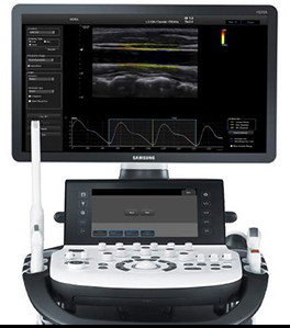
Samsung Medison RS80A ultrasound imaging system (credit: Samsung)

Samsung Medison, a global medical equipment company and an affiliate of Samsung Electronics, has just updated its RS80A ultrasound imaging system with a deep learning algorithm for breast-lesion analysis.

The “S-Detect for Breast” feature uses big data collected from breast-exam cases and recommends whether the selected lesion is benign or malignant. It’s used in in lesion segmentation, characteristic analysis, and assessment processes, providing “more accurate results.”

“We saw a high level of conformity from analyzing and detecting lesion in various cases by using the S-Detect,” said professor Han Boo Kyung, a radiologist at Samsung Medical Center.

“Users can reduce taking unnecessary biopsies and doctors-in-training will likely have more reliable support in accurately detecting malignant and suspicious lesions.”

Deep learning is better than humans in extracting meaning from cancer pathology reports

Meanwhile, researchers from the Regenstrief Institute and Indiana University School of Informatics and Computing at Indiana University-Purdue University Indianapolis say they’ve found that open-source machine learning tools are as good as — or better than — humans in extracting crucial meaning from free-text (unstructured) pathology reports and detecting cancer cases. The computer tools are also faster and less resource-intensive.

(U.S. states require cancer cases to be reported to statewide cancer registries for disease tracking, identification of at-risk populations, and recognition of unusual trends or clusters. This free-text information can be difficult for health officials to interpret, which can further delay health department action, when action is needed.)

“We think that its no longer necessary for humans to spend time reviewing text reports to determine if cancer is present or not,” said study senior author Shaun Grannis*, M.D., M.S., interim director of the Regenstrief Center of Biomedical Informatics.

Awash in oceans of data

“We have come to the point in time that technology can handle this. A human’s time is better spent helping other humans by providing them with better clinical care. Everything — physician practices, health care systems, health information exchanges, insurers, as well as public health departments — are awash in oceans of data. How can we hope to make sense of this deluge of data? Humans can’t do it — but computers can.”

This is especially relevant for underserved nations, where a majority of clinical data is collected in the form of unstructured free text, he said.

The researchers sampled 7,000 free-text pathology reports from over 30 hospitals that participate in the Indiana Health Information Exchange and used open source tools, classification algorithms, and varying feature selection approaches to predict if a report was positive or negative for cancer. The results indicated that a fully automated review yielded results similar or better than those of trained human reviewers, saving both time and money.

Major infrastructure advance

“We found that artificial intelligence was as least as accurate as humans in identifying cancer cases from free-text clinical data. For example the computer ‘learned’ that the word ‘sheet’ or ‘sheets’ signified cancer as ‘sheet’ or ‘sheets of cells’ are used in pathology reports to indicate malignancy.

“This is not an advance in ideas; it’s a major infrastructure advance — we have the technology, we have the data, we have the software from which we saw accurate, rapid review of vast amounts of data without human oversight or supervision.”

The study was published in the April 2016 issue of the Journal of Biomedical Informatics. It was conducted with support from the Centers for Disease Control and Prevention.

Co-authors of the study include researchers at the IU Fairbanks School of Public Health, the IU School of Medicine and the School of Science at IUPUI.

* Grannis, a Regenstrief Institute investigator and an associate professor of family medicine at the IU School of Medicine, is the architect of the Regenstrief syndromic surveillance detector for communicable diseases and led the technical implementation of Indiana’s Public Health Emergency Surveillance System — one of the nation’s largest. Studies over the past decade have shown that this system detects outbreaks of communicable diseases seven to nine days earlier and finds four times as many cases as human reporting while providing more complete data.

Yann Lecun is Director of AI Research, Facebook and a noted deep-learning expert. 

Scientists shoot anticancer drugs deep into tumors

Schematic of a magnetic microbubble used in the study, containing gas core (blue) and shell of magnetic iron-oxide nanoparticles (red) that form a dense shell (center) around drug-containing nanoparticles. When stimulated by ultrasound at resonant frequencies, the microbubbles explode, releasing the nanoparticles, which can travel hundreds of micrometers into tumor tissue to deliver anticancer drugs and can also be imaged on an MRI machine. (credit: Yu Gao et al./NPG Asia Materials)

Scientists at Nanyang Technological University (NTU Singapore) have invented a new way to deliver cancer drugs deep into tumor cells.

They created micro-sized gas bubbles coated with anticancer drug particles embedded in iron oxide nanoparticles and used MRI or other magnetic sources to direct these microbubbles to gather around a specific tumor. Then they used ultrasound to vibrate the microbubbles, providing the energy to direct the drug particles into a targeted kill zone in the tumor. The magnetic nanoparticles also allow for imaging in an MRI machine.

The microbubbles were successfully tested in mice and the study has been published by the Nature Publishing Group in Asia Materials.

Overcoming limitations of chemotherapy

This innovative technique was developed by a multidisciplinary team of scientists led by Asst Prof C. J. Xu from the School of Chemical and Biomedical Engineering and Assoc. Prof Claus-Dieter Ohl from the School of Physical and Mathematical Sciences.

Xu, who is also a researcher at the NTU-Northwestern Institute for Nanomedicine, said their new method may solve some of the most pressing problems faced in chemotherapy used to treat cancer.

The main problem is that current chemotherapy drugs cannot be easily targeted. The drug particles flow in the bloodstream, damaging both healthy and cancerous cells. Typically, these drugs are flushed away quickly in organs such as the lungs and liver, limiting their effectiveness.

The remaining drugs are also unable to penetrate deep into the core of the tumor, leaving some cancer cells alive, which could lead to a resurgence in tumor growth.

Delivering anticancer drugs deep into tumors

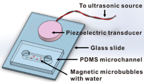
Schematic of the apparatus used to investigate magnetic microbubble oscillation and nanoparticle release (credit: Yu Gao et al./NPG Asia Materials)

The microbubbles are magnetic, so after injecting them into the bloodstream, they can be clustered around the tumor using magnets to ensure that they don’t kill the healthy cells, explains Xu, who has been working on cancer diagnosis and drug delivery systems since 2004.

“More importantly, our invention is the first of its kind that allows drug particles to be directed deep into a tumor in a few milliseconds. They can penetrate a depth of 50 cell layers or more (about 200 micrometers) — twice the width of a human hair. This helps to ensure that the drugs can reach the cancer cells on the surface and also inside the core of the tumor.”

According to Clinical Associate Professor Chia Sing Joo, a Senior Consultant at the Tan Tock Seng Hospital’s Endoscopy Centre and the Urology & Continence Clinic, “For anticancer drugs to achieve their best effectiveness, they need to penetrate into the tumor efficiently in order to reach the cytoplasm of all the cancer cells that are being targeted without affecting the normal cells.

“Currently, this can [only] be achieved by means of a direct injection into the tumor or by administering a large dosage of anticancer drugs, which can be painful, expensive, impractical and might have various side effects. If successful, I envisage [the new drug-delivery system] can be a good alternative treatment in the future, one which is low cost and yet effective for the treatment of cancers involving solid tumors, as it might minimize the side effects of drugs.” Joo is a surgeon experienced in the treatment of prostate, bladder and kidney cancer and a consultant for this study.

According to Ohl, an expert in biophysics who has published previous studies involving drug delivery systems and bubble dynamics, “most prototype drug delivery systems on the market face three main challenges before they can be commercially successful: they have to be non-invasive, patient-friendly, and yet cost-effective. We were able to come up with our solution that addresses these three challenges.”

The 12-person study team included scientists from City University of Hong Kong and Technion – Israel Institute of Technology (Technion). The team plans to use this new drug delivery system in studies on lung and liver cancer using animal models, and eventually clinical studies.

They estimate that it will take another eight to ten years before it reaches human clinical trials.


Abstract of Controlled nanoparticle release from stable magnetic microbubble oscillations

Magnetic microbubbles (MMBs) are microbubbles (MBs) coated with magnetic nanoparticles (NPs). MMBs not only maintain the acoustic properties of MBs, but also serve as an important contrast agent for magnetic resonance imaging. Such dual-modality functionality makes MMBs particularly useful for a wide range of biomedical applications, such as localized drug/gene delivery. This article reports the ability of MMBs to release their particle cargo on demand under stable oscillation. When stimulated by ultrasound at resonant frequencies, MMBs of 450 nm to 200 μm oscillate in volume and surface modes. Above an oscillation threshold, NPs are released from the MMB shell and can travel hundreds of micrometers from the surface of the bubble. The migration of NPs from MMBs can be described with a force balance model. With this technology, we deliver doxorubicin-containing poly(lactic-co-glycolic acid) particles across a physiological barrier bothin vitro and in vivo, with a 18-fold and 5-fold increase in NP delivery to the heart tissue of zebrafish and tumor tissue of mouse, respectively. The penetration of released NPs in tissues is also improved. The ability to remotely control the release of NPs from MMBs suggests opportunities for targeted drug delivery through/into tissues that are not easily diffused through or penetrated.

Ultrathin organic material enhances e-skin displays

Top left: System outline of a blood oxygen level monitor. Top right: Red and green polymer light-emitting diodes (PLEDs) are directed to shine into the finger. Reflected light from inside the finger is caught by an ultraflexible organic photodetector. This reflected light provides a measure of blood oxygen and pulse rate. Bottom: The output of the sensor can be shown on a PLED display. (credit: Someya Laboratory)

University of Tokyo researchers have developed technology to enable creation of electronic skin (e-skin) displays of blood oxygen level, e-skin heart rate sensors for medical, athletic uses, and other applications.

To serve as a demo, they’ve created an ultrathin, ultraflexible, protective layer and created an air-stable, organic light-emitting diode (OLED) display.

For use in electronic devices integrated into the human body, wearable electronics need to be thin and flexible to minimize impact where they attach to the body.

But most devices developed so far have required millimeter-scale-thickness glass or plastic substrates with limited flexibility, and micrometer-scale thin flexible organic devices have not been stable enough to survive in air.

The research group of Professor Takao Someya and Dr. Tomoyuki Yokota at the University of Tokyo’s Graduate School of Engineering has now developed a high-quality protective film less than two micrometers thick that enables the production of ultrathin, ultraflexible, high-performance wearable electronic displays and other devices.

The group developed the protective film by alternating layers of inorganic (Silicon Oxynitrite) and organic (Parylene) material. The protective film prevented passage of oxygen and water vapor in the air, extending device lifetimes from the few hours seen in prior research to several days. In addition, the research group was able to attach transparent indium tin oxide (ITO) electrodes to an ultrathin substrate without damaging it, making the e-skin display possible.

Using the new protective layer and ITO electrodes, the research group created polymer light-emitting diodes (PLEDs) and organic photodetectors (OPDs). These were thin enough to be attached to the skin and flexible enough to distort and crumple in response to body movement.

The PLEDs were just three micrometers thick and more than six times more efficient than previously reported ultrathin PLEDs. This reduced heat generation and power consumption, making them particularly suitable for direct attachment to the body for medical applications such as displays for blood oxygen concentration or pulse rate. The research group also combined red and green PLEDs with a photodetector to demonstrate a blood oxygen sensor.

In addition to medical uses, Someya sees this technology as a kind of ultra mood ring. “The advent of mobile phones has changed the way we communicate. While these communication tools are getting smaller and smaller, they are still discrete devices that we have to carry with us,” he says. “What would the world be like if we had displays that could adhere to our bodies and even show our emotions or level of stress or unease? They might enhance the way we interact with those around us or add a whole new dimension to how we communicate.”


Someya Laboratory | The red seven-segment PLED display in operation on the back of a hand.

Abstract of Ultraflexible organic photonic skin

Thin-film electronics intimately laminated onto the skin imperceptibly equip the human body with electronic components for health-monitoring and information technologies. When electronic devices are worn, the mechanical flexibility and/or stretchability of thin-film devices helps to minimize the stress and discomfort associated with wear because of their conformability and softness. For industrial applications, it is important to fabricate wearable devices using processing methods that maximize throughput and minimize cost. We demonstrate ultraflexible and conformable three-color, highly efficient polymer light-emitting diodes (PLEDs) and organic photodetectors (OPDs) to realize optoelectronic skins (oe-skins) that introduce multiple electronic functionalities such as sensing and displays on the surface of human skin. The total thickness of the devices, including the substrate and encapsulation layer, is only 3 μm, which is one order of magnitude thinner than the epidermal layer of human skin. By integrating green and red PLEDs with OPDs, we fabricate an ultraflexible reflective pulse oximeter. The device unobtrusively measures the oxygen concentration of blood when laminated on a finger. On-skin seven-segment digital displays and color indicators can visualize data directly on the body.

Creating custom drugs on a portable refrigerator-size device

This device built by MIT researchers can be reconfigured to manufacture several different types of pharmaceuticals (credit: courtesy of the researchers)

MIT researchers have developed a compact, portable pharmaceutical manufacturing system that can be reconfigured to produce a variety of drugs on demand — if you have the right chemicals.

The device could be rapidly deployed to produce drugs needed to handle an unexpected disease outbreak, to prevent a drug shortage caused by a manufacturing plant shutdown, or produce small quantities of drugs needed for clinical trials or to treat rare diseases, the researchers say.

Traditional “batch processing” drug manufacturing can take weeks or months. Active pharmaceutical ingredients are synthesized in chemical manufacturing plants and then shipped to other sites to be converted into a form that can be given to patients, such as tablets, drug solutions, or suspensions.

With research funded by DARPA’s Make-It program, the new system prototype can produce four drugs formulated as solutions or suspensions: Benadryl, Lidocaine, Valium, and Prozac. Using this apparatus, the researchers can manufacture about 1,000 doses of a given drug in 24 hours.

The key to the new system: chemical reactions that can take place as the reactants flow through relatively small tubes as opposed to the huge vats in which most pharmaceutical reactions now take place. Traditional batch processing is limited by the difficulty of cooling these vats, but the flow system allows reactions that produce a great deal of heat to be run safely.*

Personalized “orphan drugs”

One of the advantages of this small-scale system is that it could be used to make small amounts of drugs that would be prohibitively expensive to make in a large-scale plant. This would be useful for “orphan drugs” — drugs needed by a small number of patients. “Sometimes it’s very difficult to get those drugs, because economically it makes no sense to have a huge production operation for those,” says Klavs Jensen, the Warren K. Lewis Professor of Chemical Engineering at MIT and a senior author of a paper describing the new system in the March 31 online edition of Science.

The researchers are now working on the second phase of the project, which includes making the system about 40 percent smaller and producing drugs whose chemical syntheses are more complex. They are also working on producing tablets, which are more complicated to manufacture than liquid drugs.

*The chemical reactions required to synthesize each drug take place in the first of two modules. The reactions were designed so that they can take place at temperatures up to 250 degrees Celsius and pressures up to 17 atmospheres. By swapping in different module components, the researchers can easily reconfigure the system to produce different drugs. “Within a few hours we could change from one compound to the other,” Jensen says.

In the second module, the crude drug solution is purified by crystallization, filtered, and dried to remove solvent, then dissolved or suspended in water as the final dosage form. The researchers also incorporated an ultrasound monitoring system that ensures the formulated drug solution is at the correct concentration.


Abstract of On-demand continuous-flow production of pharmaceuticals in a compact, reconfigurable system

Pharmaceutical manufacturing typically uses batch processing at multiple locations. Disadvantages of this approach include long production times and the potential for supply chain disruptions. As a preliminary demonstration of an alternative approach, we report here the continuous-flow synthesis and formulation of active pharmaceutical ingredients in a compact, reconfigurable manufacturing platform. Continuous end-to-end synthesis in the refrigerator-sized [1.0 meter (width) × 0.7 meter (length) × 1.8 meter (height)] system produces sufficient quantities per day to supply hundreds to thousands of oral or topical liquid doses of diphenhydramine hydrochloride, lidocaine hydrochloride, diazepam, and fluoxetine hydrochloride that meet U.S. Pharmacopeia standards. Underlying this flexible plug-and-play approach are substantial enabling advances in continuous-flow synthesis, complex multistep sequence telescoping, reaction engineering equipment, and real-time formulation.

Nanoparticle ‘cluster bombs’ destroy cancer cells

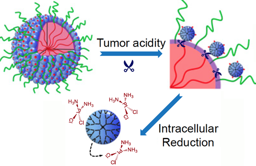
The nanoparticles start out relatively large (100 nm) (large blue circle, upper left) to enable smooth transport into the tumor through leaky blood vessels. Then, in acidic conditions found close to tumors, the particles discharge “bomblets” (right, small blue circles) just 5 nm in size. Once inside tumor cells, a second chemical step activates the platinum-based drug cisplatin (bottom) to attack the cancer directly. (credit: Emory Health Sciences)

Scientists have devised a triple-stage stealth “cluster bomb” system for delivering the anti-cancer chemotherapy drug cisplatin, using nanoparticles designed to break up when they reach a tumor:

  1. The nanoparticles start out relatively large  — 100 nanometers wide — so that they can move through the bloodstream and smoothly transport into the tumor through leaky blood vessels.
  2. As they detect acidic conditions close to tumors, the nanoparticles discharge “bomblets” just 5 nanometers in size to penetrate tumor cells.
  3. Once inside tumor cells, the bomblets release the platinum-based cisplatin, which kills by crosslinking and damaging DNA.

Doctors have used cisplatin to fight several types of cancer for decades, but toxic side effects — to the kidneys, nerves and inner ear — have limited its effectiveness. But in research with three different mouse tumor models*, the researchers have now shown that their nanoparticles can enhance cisplatin drug accumulation in tumor tissues for several types of cancer.

Details of the research — by teams led by professor Jun Wang, PhD, at the University of Science and Technology of China and by professor Shuming Nie, PhD, in the Wallace H. Coulter Department of Biomedical Engineering at Georgia Tech and Emory — were published this week in the journal PNAS.

* When mice bearing human pancreatic tumors were given the same doses of free cisplatin or cisplatin clothed in pH-sensitive nanoparticles, the level of platinum in tumor tissues was seven times higher with the nanoparticles. This suggests the possibility that nanoparticle delivery of a limited dose of cisplatin could restrain the toxic side effects during cancer treatment.

The researchers also showed that the nanoparticles were effective against a cisplatin-resistant lung cancer model and an invasive metastatic breast cancer model in mice. In the lung cancer model, a dose of free cisplatin yielded just 10 percent growth inhibition, while the same dose clothed in nanoparticles yielded 95 percent growth inhibition, the researchers report. In the metastatic breast cancer model, treating mice with cisplatin clothed in nanoparticles prolonged animal survival by weeks; 50 percent of the mice were surviving at 54 days with nanoparticles compared with 37 days for the same dose of free cisplatin.


Abstract of Stimuli-responsive clustered nanoparticles for improved tumor penetration and therapeutic efficacy

A principal goal of cancer nanomedicine is to deliver therapeutics effectively to cancer cells within solid tumors. However, there are a series of biological barriers that impede nanomedicine from reaching target cells. Here, we report a stimuli-responsive clustered nanoparticle to systematically overcome these multiple barriers by sequentially responding to the endogenous attributes of the tumor microenvironment. The smart polymeric clustered nanoparticle (iCluster) has an initial size of ∼100 nm, which is favorable for long blood circulation and high propensity of extravasation through tumor vascular fenestrations. Once iCluster accumulates at tumor sites, the intrinsic tumor extracellular acidity would trigger the discharge of platinum prodrug-conjugated poly(amidoamine) dendrimers (diameter ∼5 nm). Such a structural alteration greatly facilitates tumor penetration and cell internalization of the therapeutics. The internalized dendrimer prodrugs are further reduced intracellularly to release cisplatin to kill cancer cells. The superior in vivo antitumor activities of iCluster are validated in varying intractable tumor models including poorly permeable pancreatic cancer, drug-resistant cancer, and metastatic cancer, demonstrating its versatility and broad applicability.

New type of molecular tag makes MRI 10,000 times more sensitive

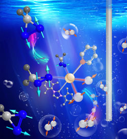
Duke scientists have discovered a new class of inexpensive, long-lived molecular tags that enhance MRI signals by 10,000 times. To activate the tags, the researchers mix them with a newly developed catalyst (center) and a special form of hydrogen (gray), converting them into long-lived magnetic resonance “lightbulbs” that might be used to track disease metabolism in real time. (credit: Thomas Theis, Duke University)

Duke University researchers have discovered a new form of MRI that’s 10,000 times more sensitive and could record actual biochemical reactions, such as those involved in cancer and heart disease, and in real time.


Let’s review how MRI (magnetic resonance imaging) works: MRI takes advantage of a property called spin, which makes the nuclei in hydrogen atoms act like tiny magnets. By generating a strong magnetic field (such as 3 Tesla) and a series of radio-frequency waves, MRI induces these hydrogen magnets in atoms to broadcast their locations. Since most of the hydrogen atoms in the body are bound up in water, the technique is used in clinical settings to create detailed images of soft tissues like organs (such as the brain), blood vessels, and tumors inside the body.


MRI’s ability to track chemical transformations in the body has been limited by the low sensitivity of the technique. That makes it impossible to detect small numbers of molecules (without using unattainably more massive magnetic fields).

So to take MRI a giant step further in sensitivity, the Duke researchers created a new class of molecular “tags” that can track disease metabolism in real time, and can last for more than an hour, using a technique called hyperpolarization.* These tags are biocompatible and inexpensive to produce, allowing for using existing MRI machines.

“This represents a completely new class of molecules that doesn’t look anything at all like what people thought could be made into MRI tags,” said Warren S. Warren, James B. Duke Professor and Chair of Physics at Duke, and senior author on the study. “We envision it could provide a whole new way to use MRI to learn about the biochemistry of disease.”

Sensitive tissue detection without radiation

The new molecular tags open up a new world for medicine and research by making it possible to detect what’s happening in optically opaque tissue instead of requiring expensive positron emission tomography (PET), which uses a radioactive tracer chemical to look at organs in the body and only works for (typically) about 20 minutes, or CT x-rays, according to the researchers.

This research was reported in the March 25 issue of Science Advances. It was supported by the National Science Foundation, the National Institutes of Health, the Department of Defense Congressionally Directed Medical Research Programs Breast Cancer grant, the Pratt School of Engineering Research Innovation Seed Fund, the Burroughs Wellcome Fellowship, and the Donors of the American Chemical Society Petroleum Research Fund.

* For the past decade, researchers have been developing methods to “hyperpolarize” biologically important molecules. “Hyperpolarization gives them 10,000 times more signal than they would normally have if they had just been magnetized in an ordinary magnetic field,” Warren said. But while promising, Warren says these hyperpolarization techniques face two fundamental problems: incredibly expensive equipment — around 3 million dollars for one machine — and most of these molecular “lightbulbs” burn out in a matter of seconds.

“It’s hard to take an image with an agent that is only visible for seconds, and there are a lot of biological processes you could never hope to see,” said Warren. “We wanted to try to figure out what molecules could give extremely long-lived signals so that you could look at slower processes.”

So the researchers synthesized a series of molecules containing diazarines — a chemical structure composed of two nitrogen atoms bound together in a ring. Diazirines were a promising target for screening because their geometry traps hyperpolarization in a “hidden state” where it cannot relax quickly. Using a simple and inexpensive approach to hyperpolarization called SABRE-SHEATH, in which the molecular tags are mixed with a spin-polarized form of hydrogen and a catalyst, the researchers were able to rapidly hyperpolarize one of the diazirine-containing molecules, greatly enhancing its magnetic resonance signals for over an hour.

The scientists believe their SABRE-SHEATH catalyst could be used to hyperpolarize a wide variety of chemical structures at a fraction of the cost of other methods.


Abstract of Direct and cost-efficient hyperpolarization of long-lived nuclear spin states on universal 15N2-diazirine molecular tags

Conventional magnetic resonance (MR) faces serious sensitivity limitations, which can be overcome by hyperpolarization methods, but the most common method (dynamic nuclear polarization) is complex and expensive, and applications are limited by short spin lifetimes (typically seconds) of biologically relevant molecules. We use a recently developed method, SABRE-SHEATH, to directly hyperpolarize 15N2 magnetization and long-lived 15N2 singlet spin order, with signal decay time constants of 5.8 and 23 min, respectively. We find >10,000-fold enhancements generating detectable nuclear MR signals that last for more than an hour. 15N2-diazirines represent a class of particularly promising and versatile molecular tags, and can be incorporated into a wide range of biomolecules without significantly altering molecular function.

A wearable graphene-based biomedical device to monitor and treat diabetes

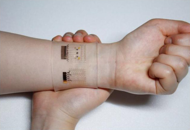
Graphene-based patch for non-invasive blood-sugar diabetes monitoring and painless drug delivery (credit: IBS)

A  wearable graphene-based patch that allows for accurate non-invasive blood-sugar diabetes monitoring and painless drug delivery has been developed by researchers at The Institute for Basic Science (IBS) Center for Nanoparticle Research in South Korea.

The device uses a hybrid of gold-doped graphene and a serpentine-shape gold mesh to measure pH (blood acidity level) and temperature by measuring the amount of glucose in sweat. If abnormally high levels of glucose are detected, an insulin drug (such as Metformin) is released into a patient’s bloodstream via drug-loaded microneedles.*

Wireless smartphone monitoring of glucose levels (credit: IBS)

The current treatments available to diabetics are painful, inconvenient, and costly, requiring regular visits to a doctor, the researchers note. Home testing kits are available to record glucose levels, but for treatment, patients have to inject uncomfortable insulin** shots to regulate glucose levels. The IBS device provides non-invasive, painless, and stress-free monitoring of important markers of diabetes using multifunctional wearable devices, reducing lengthy and expensive cycles of visiting doctors and pharmacies, according to the researchers.

“The device shows dramatic advances over current treatment methods by allowing non-invasive treatments,” according to Center for Nanoparticle Research scientist Kim Dae-Hyeong.

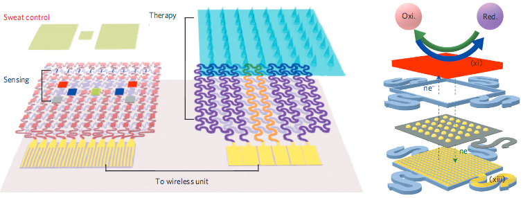
Diabetes monitoring and drug-delivery device. (Left) Schematic of diabetes patch, composed of sweat-control (sweat-uptake layer and waterproof film) and sensing (humidity, glucose, pH and tremor sensors) components; (Middle) therapeutic components (microneedles, heater, and temperature sensor); (Right) graphene-hybrid electrochemical unit: electrochemically active and soft functional materials (red), gold-doped graphene (yellow spheres), and serpentine gold mesh (bottom). (credit: Hyunjae Lee et al./Nature Nanotechnology)

* The researchers tested the therapeutic effects by experimenting on diabetic mice. They applied the device near the abdomen of the  mouse. Microneedles pierced the skin of the mouse and released Metformin, an insulin-regulating drug, into the bloodstream. The group treated with microneedles showed a significant suppression of blood glucose concentrations with respect to control groups. Two healthy human males also participated in tests to demonstrate the sweat-based glucose sensing of the device. Glucose and pH levels of both subjects were recorded; a statistical analysis confirmed a reliable correlation between sweat-glucose data from the diabetes patch and those from commercial glucose tests. 

** Insulin is produced in the pancreas and regulates the use of glucose, maintaining a balance in blood sugar levels. Diabetes causes an imbalance: insufficient amounts of insulin results in high blood glucose levels, known as hyperglycemia. Type 2 diabetes is the most common form of diabetes with no known cure.


Abstract of A graphene-based electrochemical device with thermoresponsive microneedles for diabetes monitoring and therapy

Owing to its high carrier mobility, conductivity, flexibility and optical transparency, graphene is a versatile material in micro- and macroelectronics. However, the low density of electrochemically active defects in graphene synthesized by chemical vapour deposition limits its application in biosensing. Here, we show that graphene doped with gold and combined with a gold mesh has improved electrochemical activity over bare graphene, sufficient to form a wearable patch for sweat-based diabetes monitoring and feedback therapy. The stretchable device features a serpentine bilayer of gold mesh and gold-doped graphene that forms an efficient electrochemical interface for the stable transfer of electrical signals. The patch consists of a heater, temperature, humidity, glucose and pH sensors and polymeric microneedles that can be thermally activated to deliver drugs transcutaneously. We show that the patch can be thermally actuated to deliver Metformin and reduce blood glucose levels in diabetic mice.

Major steps toward a bioengineered heart for transplantation

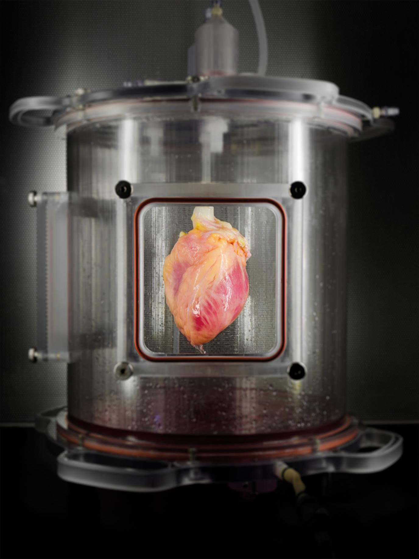
A human whole heart was stripped of cells, leaving behind a “structural scaffold” of connective tissue, which was then partially re-seeded with human heart-like cells grown in the laboratory from skin cells. Once these new heart-like cells were re-seeded into the heart scaffold, the heart was regenerated in this bioreactor, which delivers a nutrient solution and replicates some of the environmental conditions in a living heart. (credit: Bernhard Jank, MD/Ott Lab, Center for Regenerative Medicine, Massachusetts General Hospital)

Massachusetts General Hospital (MGH) researchers have taken early steps towards producing a bioengineered heart for transplantation that would use cells from the patient receiving the heart.

Using a patient’s own cells would help to overcome some of the problems associated with receiving a heart donated by another person, including immune rejection of the donated heart, as well as the long-term side effects of life-long treatment with the immunosuppressive drugs needed to suppress the immune system and reduce the risk of rejection.

To achieve those steps, Jacques Guyette, PhD, of the Massachusetts General Hospital’s Center for Regenerative Medicine, lead author of a paper in Circulation Research, says the research team had to overcome three major technical challenges.

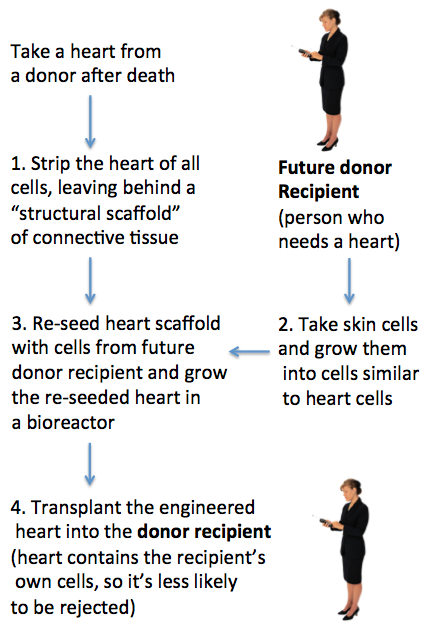
Future methodology for producing bioengineered hearts (credit: Julie Milland)

1. Create a structural scaffold from a human heart

One challenge is producing a structural scaffold able to support new functioning heart cells. To do this, the researchers take a human heart and remove the heart muscle and other cells and components that would stimulate the recipient’s immune system to reject the organ. Once stripped, the structural and connective tissues that give the heart its 3D structure are left behind.

If researchers can re-seed this structural scaffold with viable heart-like cells from the patient who will receive the heart (steps 2 and 3 below), the engineered heart would have the potential to reduce the risk of rejection and the resulting need for long-term immunosuppressive treatment.

Team leader Harald Ott, MD, an assistant professor of surgery at Harvard Medical School, has pioneered a method for stripping the living cells from donor organs and then re-populating the remaining scaffold with new cells.*

2. Grow cells that will function and contract like heart cells

Another challenge is developing a method that will enable researchers to use cells from the potential heart recipient to produce cells that will function like heart cells. These are generated from induced pluripotent stem cells (iPSCs) — cells with the potential to be turned into many different types of cells.

The team generated the heart-like muscle cells from reprogrammed skin cells. Once they had checked the quality of these cells, they grew them in the laboratory for several days and showed that the cells developed into tissue that spontaneously contracted like heart cells.**

3. Re-seed the human heart scaffold and grow it in an automated bioreactor

The final challenge was developing an automated bioreactor system capable of supporting a whole human heart while the re-seeded heart cells take hold.*** In this initial study, the researchers only partially re-seeded the scaffold — many more cells would be needed to totally re-populate a functioning heart scaffold.

After incubating the engineered heart in the bioreactor, the researchers showed that the regenerated tissue behaved like immature cardiac muscle tissue that was able to contract in response to electrical stimulation.

Next steps

“Regenerating a whole heart is most certainly a long-term goal that is several years away,” says Guyette. “Among the next steps that we are pursuing are improving methods to generate even more cardiac cells.”

He says re-seeding a whole heart would take tens of billions of cells, so the team needs to optimize the bioreactor techniques to improve the maturation and function of engineered cardiac tissue. They also need to integrate the electrical function of the regenerated tissue in the bioengineered heart. In the meantime, Guyette says the team is working on engineering a functional myocardial patch that could be used to replace tissue damaged after a heart attack or heart failure.

The study was supported by National Institutes of Health Director’s New Innovator Award and by National Heart Lung and Blood Institute grants.

* Since 2008, Ott’s team has used the approach to generate functional rat kidneys and lungs and has stripped cells from large-animal hearts, lungs and kidneys.

This study used 73 human hearts donated through the New England Organ Bank, unsuitable for transplantation and recovered under research consent. Using a scaled-up version of a process originally developed in rat hearts, the team stripped cells from the hearts from both brain-dead donors and from those who had undergone cardiac death. The consequent cardiac scaffolds showed a high retention of matrix proteins and preserved blood vessels. The structure was free of cardiac cells and other molecules that could induce rejection.

** Instead of using genetic manipulation to generate iPSCs from adult cells, the team used a newer method to reprogram skin cells, which should be both more efficient and less likely to run into regulatory hurdles. They then induced the iPSCs to create cardiac muscle cells or cardiomyocytes, documenting patterns of gene expression that reflected developmental milestones and generating cells in sufficient quantity for possible clinical application. Cardiomyocytes were then reseeded into three-dimensional matrix tissue, first into thin matrix slices and then into 15 mm fibers, which developed into spontaneously contracting tissue after several days in culture.

*** The team delivered about 500 million heart-like cells grown from iPSC into the left ventricular wall of the heart scaffolds. The re-seeded organs were mounted for 14 days in an automated bioreactor system developed by the team. The automated bioreactor system supplied the organ with a circulating nutrient solution and applied environmental conditions to reproduce conditions within a living heart.


Abstract of Bioengineering Human Myocardium on Native Extracellular Matrix

Rationale: More than 25 million individuals have heart failure worldwide, with ≈4000 patients currently awaiting heart transplantation in the United States. Donor organ shortage and allograft rejection remain major limitations with only ≈2500 hearts transplanted each year. As a theoretical alternative to allotransplantation, patient-derived bioartificial myocardium could provide functional support and ultimately impact the treatment of heart failure.

Objective: The objective of this study is to translate previous work to human scale and clinically relevant cells for the bioengineering of functional myocardial tissue based on the combination of human cardiac matrix and human induced pluripotent stem cell–derived cardiomyocytes.

Methods and Results: To provide a clinically relevant tissue scaffold, we translated perfusion-decellularization to human scale and obtained biocompatible human acellular cardiac scaffolds with preserved extracellular matrix composition, architecture, and perfusable coronary vasculature. We then repopulated this native human cardiac matrix with cardiomyocytes derived from nontransgenic human induced pluripotent stem cells and generated tissues of increasing 3-dimensional complexity. We maintained such cardiac tissue constructs in culture for 120 days to demonstrate definitive sarcomeric structure, cell and matrix deformation, contractile force, and electrical conduction. To show that functional myocardial tissue of human scale can be built on this platform, we then partially recellularized human whole-heart scaffolds with human induced pluripotent stem cell–derived cardiomyocytes. Under biomimetic culture, the seeded constructs developed force-generating human myocardial tissue and showed electrical conductivity, left ventricular pressure development, and metabolic function.

New synthesized molecule could reduce brain damage in stroke victims

This graphic depicts a new inhibitor, 6S, locking up an enzyme (red) to block the production of hydrogen sulfide (yellow and white). Hydrogen sulfide concentrations have been shown to climb after the onset of a stroke, leading to brain damage. (credit: Matthew Beio, University of Nebraska-Lincoln)

A new molecule known as 6S has reduced the death of brain tissue from ischemic stroke by up to 66 percent in rats while reducing the accompaning inflammation, researchers at the University of Nebraska-Lincoln and the National University of Singapore reported March 9 in an open-access paper published by the journal ACS Central Science.

The inhibitor molecule works by binding to cystathionine beta-synthase (CBS), an enzyme that normally helps regulate cellular function, but can also trigger production of toxic levels of hydrogen sulfide in the brain. (That buildup initiates brain damage after strokes by disrupting blood flow, which prevents oxygen and glucose from reaching brain tissue, ultimately killing neurons and other cells.)

The researchers modeled the inhibitor on a naturally occurring molecule produced by the CBS enzyme, tailoring the molecule’s structure to improve its performance.* That increased the inhibitor’s binding time from less than a second to hours.

Because the 6S inhibitor has only demonstrated its effects in cell cultures and the brain tissue of rats, the researchers cautioned that it represents just an initial step toward developing a stroke-treating drug for humans.

Research and facilities that contributed to the study were partly funded by the American Heart Association, the National Science Foundation, and the National Institutes of Health.

The World Health Organization has estimated that stroke kills more than 6 million people annually.

* The researchers replaced functional groups of atoms known as amines with hydrazines.


Abstract of “Zipped Synthesis” by Cross-Metathesis Provides a Cystathionine β-Synthase Inhibitor that Attenuates Cellular H2S Levels and Reduces Neuronal Infarction in a Rat Ischemic Stroke Model

The gaseous neuromodulator H2S is associated with neuronal cell death pursuant to cerebral ischemia. As cystathionine β-synthase (CBS) is the primary mediator of H2S biogenesis in the brain, it has emerged as a potential target for the treatment of stroke. Herein, a “zipped” approach by alkene cross-metathesis into CBS inhibitor candidate synthesis is demonstrated. The inhibitors are modeled after the pseudo-C2-symmetric CBS product (l,l)-cystathionine. The “zipped” concept means only half of the inhibitor needs be constructed; the two halves are then fused by olefin cross-metathesis. Inhibitor design is also mechanism-based, exploiting the favorable kinetics associated with hydrazine-imine interchange as opposed to the usual imine–imine interchange. It is demonstrated that the most potent “zipped” inhibitor 6S reduces H2S production in SH-SY5Y cells overexpressing CBS, thereby reducing cell death. Most importantly, CBS inhibitor 6S dramatically reduces infarct volume (1 h post-stroke treatment; ∼70% reduction) in a rat transient middle cerebral artery occlusion model for ischemia.

Are you ready for soft, morphing, crawling robots with glowing skin displays?

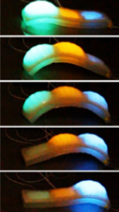
Multi-pixel electroluminescent displays in various states of deformation and illumination (credit: C. Larson et al./Science)

Your future robot or mobile device could have soft, morphable, stretchable “skin” that displays information, according to research by Cornell University engineers. Imagine a health-care robot that displays your blood glucose level and oxygenation, and even your mood — perhaps also your remote physician’s face in 3D.

“When robots become more and more a part of our lives, the ability for them to have an emotional connection with us will be important,” says research team leader Rob Shepherd, an assistant professor of mechanical and aerospace engineering.

Soft robots are currently in use for safe human robot interaction, but they can’t stretch continuously or dynamically display information on their body; and in most cases, can’t sense external and internal stimuli. So the engineers have developed octopus-inspired electroluminescent “skin” that stretches to more than six times its original size, and can also change shape and color.

An undulating gait produced by pressurizing the chambers in sequence along the length of the crawler (credit: C. Larson et al./Science)

The new technology uses a “hyper-elastic light-emitting capacitor” (HLEC), consisting of layers of transparent hydrogel electrodes sandwiching an insulating elastomer (a polymer with viscoelasticity, meaning it has both viscosity and elasticity) sheet.

The elastomer changes luminance and capacitance (the ability to store an electrical charge) when stretched, rolled, and otherwise deformed.

The HLEC skin also endows soft robots with the ability to sense their actuated state and environment and communicate optically — and (for small robots) even crawl.

The engineers created a prototype crawling soft robot, using three of the six-layer HLEC panels bound together. The top four layers made up the illuminated skin and the bottom two served as pneumatic actuators. The chambers were alternately inflated and deflated; the resulting curvature created an undulating, walking motion.

It could also make for a fun pet, we’re guessing.

The team’s research was published in the March 3 online edition of the journal Science. It was supported by a grant from the Army Research Office, a 2015 award from the Air Force Office of Scientific Research, and two grants from the National Science Foundation.

Cornell University | Electroluminescent Skin Demonstration


Abstract of Highly stretchable electroluminescent skin for optical signaling and tactile sensing

Cephalopods such as octopuses have a combination of a stretchable skin and color-tuning organs to control both posture and color for visual communication and disguise. We present an electroluminescent material that is capable of large uniaxial stretching and surface area changes while actively emitting light. Layers of transparent hydrogel electrodes sandwich a ZnS phosphor-doped dielectric elastomer layer, creating thin rubber sheets that change illuminance and capacitance under deformation. Arrays of individually controllable pixels in thin rubber sheets were fabricated using replica molding and were subjected to stretching, folding, and rolling to demonstrate their use as stretchable displays. These sheets were then integrated into the skin of a soft robot, providing it with dynamic coloration and sensory feedback from external and internal stimuli.