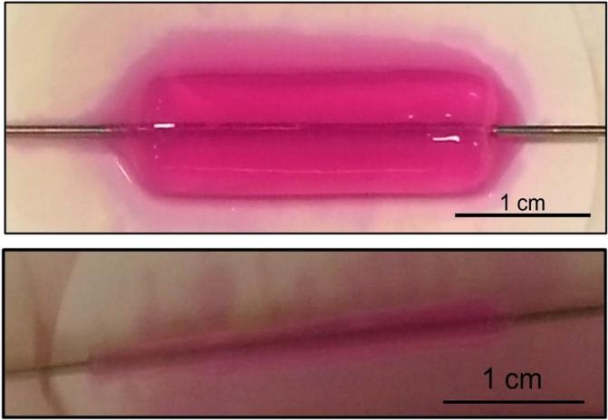
An amputee feels texture in real time: Signals from sensors in an artificial fingertip are converted to neural-like spikes and delivered to nerves in the upper arm. (credit: Ecole polytechnique fédérale de Lausanne)
Amputee Dennis Aabo Sørensen is the first person in the world to recognize texture (smoothness vs. roughness) using an artificial “bionic” fingertip surgically connected to nerves in his upper arm. The experimental system was developed by EPFL (Ecole polytechnique fédérale de Lausanne) and SSSA (Scuola Superiore Sant’Anna).
“The stimulation felt almost like what I would feel with my hand,” says Sørensen. “I felt the texture sensations at the tip of the index finger of my phantom hand.”
Bionic fingertip electronics and plastic gratings with rough and smooth textures (credit: Hillary Sanctuary/EPFL)
As a test, a machine controlled the movement of the fingertip over different pieces of plastic engraved with different patterns, smooth or rough, as sensors generated an electrical signal. This signal was translated into a series of electrical spikes, imitating the language of the nervous system, then delivered to the nerves.
But how does this sensation relate to the feeling of touch from a real finger? The scientists tested that by comparing brain-wave activity of amputees to non-amputees. The brain scans were similar.
“This study provides additional evidence that research in neuroprosthetics can contribute to [understanding] neuronal mechanisms of the human sense of touch,” says Calogero Oddo of the BioRobotics Institute of SSSA. “It will also be translated to other applications, such as artificial touch in robotics for surgery, rescue, and manufacturing.”
The research is described in an open-access paper on the journal e-Life. It was carried out in collaboration with Università di Pisa, IRCCS San Raffaele Pisana, Università Cattolica del Sacro Cuore, and Università Campus Biomedico.
The work was partly supported by EU Grants TIME, NEBIAS and NANOBIOTOUCH; by the ENABLE project, funded by the Wyss Center for Bio and Neuroengineering; by the Swiss National Competence Center in Research in Robotics; and by Italian grants NEMESIS (funded by the Italian Ministry of Health), PRIN/HandBot (funded by the Italian Ministry of Research), and PPR2 (funded by the National Institute for Insurance against Industrial Injuries).
Abstract of Intraneural stimulation elicits discrimination of textural features by artificial fingertip in intact and amputee humans
Restoration of touch after hand amputation is a desirable feature of ideal prostheses. Here, we show that texture discrimination can be artificially provided in human subjects by implementing a neuromorphic real-time mechano-neuro-transduction (MNT), which emulates to some extent the firing dynamics of SA1 cutaneous afferents. The MNT process was used to modulate the temporal pattern of electrical spikes delivered to the human median nerve via percutaneous microstimulation in four intact subjects and via implanted intrafascicular stimulation in one transradial amputee. Both approaches allowed the subjects to reliably discriminate spatial coarseness of surfaces as confirmed also by a hybrid neural model of the median nerve. Moreover, MNT-evoked EEG activity showed physiologically plausible responses that were superimposable in time and topography to the ones elicited by a natural mechanical tactile stimulation. These findings can open up novel opportunities for sensory restoration in the next generation of neuro-prosthetic hands.
The technology to deliver this sophisticated tactile information was developed by Silvestro Micera and his team at together with Calogero Oddo and his team at SSSA.
The muscle layer of the engineered human arteries function well. After just one week, the arteries contain two types of proteins important for muscle contraction — actin (stained red, left) and calponin (stained red, right). These two protein molecules allow the arteries to contract and dilate in response to environmental stimuli. (credit: George Truskey Lab, Duke University)
Duke University researchers have developed a rapid new technique for making small-scale artificial human arteries for use in a system for testing drugs — one that is more accurate and reliable than using animal models. That means promising drugs could be better tested before entering human trials.
The new technique produces the artificial arteries ten times faster than current methods and the arteries are functional.
Arterial blood vessel walls have multiple layers of cells, including the endothelium and media. The endothelium is the innermost lining that interacts with circulating blood. The media is made mostly of smooth muscle cells that help control the flow of blood and blood pressure. These two layers communicate and control how blood vessels react to stimuli such as drugs and exercise. (credit: Wikimedia Commons)
The researchers successfully engineered artificial arteries containing the lining (endothelium) and muscle (media) layers of arteries. They also showed that both layers could communicate and function normally.
“We wanted to focus on arteries because that’s where most of the damage is caused in coronary diseases,” said George Truskey, senior author, Professor of Biomedical Engineering and Dean of the Pratt School of Engineering at Duke University.
How to create an artificial artery
To rapidly construct strong human artificial arteries, cells were embedded in collagen gels (top) and then more than 90 per cent of the water was removed (bottom). These arteries were prepared in only three hours. The mechanical strength of these engineered arteries means researchers no longer need to wait six to eight weeks for the tissues to mature. (credit: George Truskey Lab, Duke University)
Graduate student Cristina Fernandez developed the technique to create arteries; but instead of full-size arteries, they were scaled down to one-tenth the size of a typical human artery. With this smaller diameter, the researchers were able to make a lot of these artificial vessels in a short amount of time and use them in experiments in just a few hours, instead of spending several weeks developing each one.
Despite the smaller size, these engineered arteries behaved normally, with statin drugs blocking inflammation just as they do in patients. The endothelial cells also released chemical signals to relax and constrict the media layer, again just like they do in the normal human body.
“Many of the [previous] techniques for creating artificial tissue also were rather lengthy, which was frustrating,” said Truskey. The previous studies also focused on the media cells rather than the endothelial cells, and “nobody had shown how the two would interact,” he added.
Replacing arteries in patients
“While our arteries are small and intended for testing, they’re just as mechanically strong as those intended to be put inside of the body,” said Truskey. “So the technique could be beneficial to researchers trying to create artificial arteries to replace damaged ones in patients as well.” The arteries could also be used to look at how some select rare genetic diseases affect arteries, he added.
The study was reported online (open access) in Nature Scientific Reports. The work was supported by the National Institutes of Health and the American Heart Association.
Abstract of Human Vascular Microphysiological System for in vitro Drug Screening
In vitro human tissue engineered human blood vessels (TEBV) that exhibit vasoactivity can be used to test human toxicity of pharmaceutical drug candidates prior to pre-clinical animal studies. TEBVs with 400–800 μM diameters were made by embedding human neonatal dermal fibroblasts or human bone marrow-derived mesenchymal stem cells in dense collagen gel. TEBVs were mechanically strong enough to allow endothelialization and perfusion at physiological shear stresses within 3 hours after fabrication. After 1 week of perfusion, TEBVs exhibited endothelial release of nitric oxide, phenylephrine-induced vasoconstriction, and acetylcholine-induced vasodilation, all of which were maintained up to 5 weeks in culture. Vasodilation was blocked with the addition of the nitric oxide synthase inhibitor L-NG-Nitroarginine methyl ester (L-NAME). TEBVs elicited reversible activation to acute inflammatory stimulation by TNF-α which had a transient effect upon acetylcholine-induced relaxation, and exhibited dose-dependent vasodilation in response to caffeine and theophylline. Treatment of TEBVs with 1 μM lovastatin for three days prior to addition of Tumor necrosis factor – α (TNF-α) blocked the injury response and maintained vasodilation. These results indicate the potential to develop a rapidly-producible, endothelialized TEBV for microphysiological systems capable of producing physiological responses to both pharmaceutical and immunological stimuli.
1. Electronic stethoscope records patient’s breathing. Lung sounds are sent to a phone or tablet and analyzed by an app. 3. Medical professionals can listen and see the results in real time from any location to diagnose the patient. (credit: Hiroshima University)
The traditional stethoscope has just been superseded by an electronic stethoscope and an app called Respiratory Sounds Visualizer, which can automatically classify lung sounds into five common diagnostic categories.* The system was developed by three physician researchers at Hiroshima University and Fukushima Medical University in collaboration with Pioneer Corporation.
The respiratory specialist doctors recorded and classified lung sounds of 878 patients, then turned these diagnoses into templates to create a mathematical formula that evaluates the length, frequency, and intensity of lung sounds. The resulting app can recognize the sound patterns consistent with five different respiratory diagnoses.
How the Respiratory Sounds Visualizer app works
Based on an analysis of the characteristics of respiratory sounds, the Respiratory Sounds Visualizer app generates this diagnostic chart. The total area in red represents the overall volume of sound, and the proportion of red around each line from the center to each vertex represents the proportion of the overall sound that each respiratory sound contributes. (credit: Shinichiro Ohshimo et al./Annals of Internal Medicine)
The app analyzes the lung sounds and maps them on a five-sided chart. Each of the five axes represents one of the five types of lung sounds. Doctors and patients can see the likely diagnosis based on the length of the axis covered in red.
A doctor working in less-than-ideal circumstances, such as a noisy emergency room or field hospital, could rely on the computer program to “hear” what they might otherwise miss, and the new system could help student doctors learn.
The results from the computer program are simple to interpret and can be saved and shared electronically. In the future, this convenience may allow patients to track and record their own lung function during chronic conditions, like chronic obstructive pulmonary disease (COPD) or cystic fibrosis.
“We plan to use the electronic stethoscope and Respiratory Sounds Visualizer with our own patients after further improving [the mathematical calculations]. We will also release the computer program as a downloadable application to the public in the near future,” said Shinichiro Ohshimo, MD, PhD, an emergency physician in the Department of Emergency and Critical Care Medicine at Hiroshima University Hospital and one of the researchers involved in developing the technology.
* Despite advances in technology, respiratory physiology still depends primarily on chest auscultation, [which is] subjective and requires sufficient training. In addition, identification of the five respiratory sounds specified by the International Lung Sounds Association is difficult because their frequencies overlap:The frequency of normal respiratory sound is 100 to 1000 Hz, wheeze is 100 to 5000 Hz, rhonchus is 150 Hz, coarse crackle is 350 Hz, and fine crackle is 650 Hz. — Shinichiro Ohshimo et al./Annals of Internal Medicine.
(Left) Normal coronary artery with normal blood flow. Right: a coronary artery narrowed by plaque, limiting the flow of oxygen-rich blood through the artery. (credit: NIH)
Mayo Clinic researchers have demonstrated the first study in which repeated treatments to remove senescent cells (cells that stop dividing due to age or stress) in mice improve age-related vascular conditions — and may possibly reduce cardiovascular disease and death.
The researchers intermittently gave the mice a cocktail of two senolytic drugs (ones that selectively induce cell death): dasatinib (a cancer drug, trade name Sprycel) and quercetin*. The drugs cleared (killed off) senescent cells in naturally aged and atherosclerotic mice. The treatment did not reduce the size of plaques in mice with high cholesterol, but did reduce calcification of existing plaques on the interior of vessel walls.**
The findings appear online (open access) in Aging Cell.
“Our finding that senolytic drugs can reduce cardiovascular calcification is very exciting, since blood vessels with calcified plaques are notoriously difficult to reduce in size, and patients with heart-valve calcification currently do not have any treatment options other than surgery,” says Jordan Miller, Ph.D., Mayo cardiovascular surgery researcher and senior author of the paper.
“While more research is needed, our findings are encouraging that one day removal of senescent cells in humans may be used as a complementary therapy along with traditional management of risk factors to reduce surgery, disability, or death resulting from cardiovascular disease.”
The coauthors include two researchers from Newcastle University. The research was supported by the National Institutes of Health, Mayo Clinic Center for Regenerative Medicine, and the Connor Group and Noaber Foundation. Drs. Kirkland, Tchkonia, Zhu, Pirtskhalava, and Ms. Palmer have a financial interest related to the research.
* Quercetin is found in many fruits, vegetables, leaves and grains. It can be used as an ingredient in supplements, beverages, or foods. — Wikipedia
** Prior studies at Mayo showed chronic removal of the cells from genetically-altered mice can alter or delay many of these conditions, and short-term treatment with drugs that remove senescent cells can improve the function of the endothelial cells that line the blood vessels. This study, however, looked at the structural and functional impacts of cell clearance using a unique combination of drugs on blood vessels over time. Mice were 24 months old when the drugs were administered orally over a three-month period following those initial two years. A separate set of mice with high cholesterol was allowed to develop atherosclerotic plaques for 4 months and were then treated with the drug cocktail for two months. — Mayo Clinic
Abstract of Chronic senolytic treatment alleviates established vasomotor dysfunction in aged or atherosclerotic mice
Rationale: While reports suggest a single dose of senolytics may improve vasomotor function, the structural and functional impact of long-term senolytic treatment is unknown.
Objective: To determine whether long-term senolytic treatment improves vasomotor function, vascular stiffness, and intimal plaque size and composition in aged or hypercholesterolemic mice with established disease.
Methods and Results: Senolytic treatment (intermittent treatment with Dasatinib + Quercetin via oral gavage) resulted in significant reductions in senescent cell markers (TAF+ cells) in the medial layer of aorta from aged and hypercholesterolemic mice, but not in intimal atherosclerotic plaques. While senolytic treatment significantly improved vasomotor function (isolated organ chamber baths) in both groups of mice, this was due to increases in nitric oxide bioavailability in aged mice and increases in sensitivity to NO donors in hypercholesterolemic mice. Genetic clearance of senescent cells in aged normocholesterolemic INK-ATTAC mice phenocopied changes elicited by D+Q. Senolytics tended to reduce aortic calcification (alizarin red) and osteogenic signaling (qRT-PCR, immunohistochemistry) in aged mice, but both were significantly reduced by senolytic treatment in hypercholesterolemic mice. Intimal plaque fibrosis (picrosirius red) was not changed appreciably by chronic senolytic treatment.
Conclusions: This is the first study to demonstrate that chronic clearance of senescent cells improves established vascular phenotypes associated with aging and chronic hypercholesterolemia, and may be a viable therapeutic intervention to reduce morbidity and mortality from cardiovascular diseases.
Just lopped off your ring finger slicing carrots (some time in the future)? No problem. Just speed-read this article while you’re waiting for the dronebulance. …
“Epimorphic regeneration” — growing digits, maybe even limbs, with full 3D structure and functionality — may one day be possible. So say scientists at Tulane University, the University of Washington, and the University of Pittsburgh, writing in a review article just published in Tissue Engineering, Part B, Reviews (open access until March 8).
The process of amphibian epimorphic regeneration may offer hints for humans. After amputation, the wound heals to form an epidermal layer, the underlying tissues undergo matrix remodeling, and cells in the region secrete soluble factors. A heterogeneous cell mass, or blastema, forms from the proliferation and migration of cells from the adjacent tissues. The blastema then gives rise to the various new tissues that are spatially patterned to reconstruct the original limb structure. (credit: Lina M. Quijano et al./Tissue Engineering Part B)
Epimorphic generation occurs in certain animals, such as salamanders and frogs, which are able to regenerate limbs, tails, jaws, and even eye lenses; and in deer antlers and mouse ears.
Turns out there are also rare cases of children and young adults who have had tips of digits regenerated. And there are specific “steps of epimorphic regeneration to promote the partial or complete restoration of a biological digit or limb after amputations,” the scientists believe.
Epimorphic regeneration in the murine (type of mouse) digit is level-specific and provides an opportunity for comparative studies of mammalian epimorphic regeneration. Transection through the P2 element results in the frequent outcome of fibrotic scar tissue formation. Transection through the more distal P3 element instead results in the regeneration of missing tissue. (credit: Lina M. Quijano et al./Tissue Engineering Part B)
Some of those steps are suggested by what’s possible in mice, where the digit tip has been found capable of regrowing multiple structures, including bone, after amputation.
What about humans?
The highly ambitious goal of epimorphic regeneration for humans would require the regrowth of multiple tissues that have been assembled in the proper conformation and patterns to create a fully functional limb, according to the authors.
Epimorphic regeneration has been observed in distal finger tips of children and young adults. Converting such random events into designed clinical outcomes will require altering the default postamputation progression. It may include the transplantation of cells, scaffolds, and/or soluble factors, as well as controlling microenvironmental aspects, such as oxygen concentration, tissue hydration, mechanical, and electrical cues. (credit: Lina M. Quijano et al./Tissue Engineering Part B)
They note that “it may be possible to suture an engineered epithelial layer, much like the present skin grafts, across the injury site. In-depth understanding of the proper soluble factor communication necessary, however, could lead to a more direct approach of delivering growth factors to the region, leveraging drug delivery paradigms that create spatiotemporal gradients.These interventions are intended to mimic the signals that induce a stable cell mass that functions as a blastema [a mass of cells capable of growth and regeneration into organs or body parts].
“Initial studies in mice have already shown the promise of introducing solubilized [extra-cellular matrices, bone morphogenetic proteins,and matrix metalloproteinases, generated by immune cells] in promoting recruitment/mobilization of endogenous cells to proliferate at the transected bone front. Furthermore, the injury response may also be influenced with external bioreactors … that can control parameters, such as hydration, pH, oxygen concentration, and electrical stimulation.”
The research was supported by the National Institutes of Health and the Fulbright Scholars Program.
Abstract of Looking Ahead to Engineering Epimorphic Regeneration of a Human Digit or Limb
Approximately 2 million people have had limb amputations in the United States due to disease or injury, with more than 185,000 new amputations every year. The ability to promote epimorphic regeneration, or the regrowth of a biologically based digit or limb, would radically change the prognosis for amputees. This ambitious goal includes the regrowth of a large number of tissues that need to be properly assembled and patterned to create a fully functional structure. We have yet to even identify, let alone address, all the obstacles along the extended progression that limit epimorphic regeneration in humans. This review aims to present introductory fundamentals in epimorphic regeneration to facilitate design and conduct of research from a tissue engineering and regenerative medicine perspective. We describe the clinical scenario of human digit healing, featuring published reports of regenerative potential. We then broadly delineate the processes of epimorphic regeneration in nonmammalian systems and describe a few mammalian regeneration models. We give particular focus to the murine digit tip, which allows for comparative studies of regeneration-competent and regeneration-incompetent outcomes in the same animal. Finally, we describe a few forward-thinking opportunities for promoting epimorphic regeneration in humans.
(Left): Control rabbit brain, showing neuropil near the CA1 band in the hippocampus. (Right): Vitrified rabbit brain, same location. Synapses, vesicles, and microfilaments are clear. The myelinated axon shows excellent preservation. (credit: Robert L. McIntyre and Gregory M. Fahy/Cryobiology)
The Brain Preservation Foundation (BPF) has announced that a team at 21st Century Medicine led by Robert McIntyre, PhD., has won the Small Mammal Brain Preservation Prize, which carries an award of $26,735.
The Small Mammalian Brain Preservation Prize was awarded after the determination that the protocol developed by McIntyre, termed Aldehyde-Stabilized Cryopreservation, was able to preserve an entire rabbit brain with well-preserved ultrastructure, including cell membranes, synapses, and intracellular structures such as synaptic vesicles (full protocol here).
The judges for the prize were Kenneth Hayworth, PhD., Brain Preservation Foundation President and neuroscientist at the Howard Hughes Medical Institute; and Prof.Sebastian Seung, PhD., Princeton Neuroscience Institute and Computer Science Department.
First preservation of the connectome
“This is a milestone in the development of brain preservation techniques: it is the first time that the preservation of the connectome has been demonstrated in a whole brain (prior to this only small parts of brains have been preserved to this level of detail),” said the BPF announcement.
“Current models of the brain suggest that the connectome contains all the information necessary for personal identity (i.e., memory and personality). This technique is not the same as conventional cryonics (rapidly freezing the brain), which has never demonstrated preservation of the ultrastructure of the brain. Thus the winning of this prize represents a significant advance in the methods available for large scale studies of the connectome and could lead to procedures that preserve a complete human brain.
Kenneth Hayworth (KH) (President of the Brain Preservation Foundation (BPF)) and Michael Shermer (member of BPF advisory board) witnessed (on Sept. 25, 2015) the full Aldehyde Stabilized Cryopreservation surgical procedure performed on this rabbit at the laboratories of 21 Century Medicine under the direction of 21CM lead researcher Robert McIntyre. This included the live rabbit’s carotid arteries being perfused with glutaraldehyde and subsequent perfusion with cryoprotectant agent (CPA). KH witnessed this rabbit brain being put in -135 degrees C storage, removal from storage the following day (verifying that it had vitrified solid), and KH witnessed all subsequent tissue processing steps involved in the evaluation process. (credit: The Brain Preservation Foundation)
“The key breakthrough was the rapid perfusion of a deadly chemical fixative (glutaraldehyde) through the brain’s vascular system, instantly stopping metabolic decay and fixing all proteins in place by covalent crosslinks. This stabilized the tissue and vasculature so that cryoprotectant could be perfused at an optimal temperature and rate. The result was an intact rabbit brain filled with such a high concentration of cryoprotectants that it could be stored as a solid ‘vitrified’ block at a temperature of -135 degrees Celsius.”
Winning the award also required that the procedure be published in a peer reviewed scientific publication. McIntyre satisfied this requirement and published the protocol in an open-access paper in the Journal of Cryobiology.
3D microscope evaluation of the rabbit brain tissue preservation (credit: Brain Preservation Foundation)
The Brain Preservation Foundation plans to continue to promote the idea that brain preservation following legal death, by using scientifically validated techniques, is a reasonable choice for consenting individuals to make. Focus now shifts to the final Large Mammal phase of the contest, which requires an intact pig brain to be preserved with similar fidelity in a manner that could be directly adapted to terminal patients in a hospital setting.
The 21st Century Medicine team has recently submitted to the BPF such a preserved pig brain for official evaluation. Lead researcher Robert McIntyre has started Nectome to further develop this method.
“Of course, [the demonstrated brain preservation procedure] is only useful if you think all the relevant information is preserved in the fixation,” said Anders Sandberg, PhD., of the Future of Humanity Institute/Oxford Martin School. “Protein states and small molecule chemical information may be messed up.”
GPA | Will You Preserve Your Brain?
Background and significance (statement by BPF)
Proponents of cryonics have long sought a technique that could put terminal patients into longterm stasis, the goal being a form of medical time travel in which patients are stabilized against decay with the hope of being biologically revived and cured by future technologies. Despite decades of research, this goal of reversible cryopreservation remains far out of reach — too much damage occurs during the cryopreservation itself.
This has led a new generation of researchers to focus on a more achievable and demonstrable goal–preservation of brain structure only. Specifically preservation of the delicate pattern of synaptic connections (the “connectome”) which neuroscience contends encodes a person’s memory and identity. Instead of biological revival, these new researchers often envision a future “synthetic revival” comprising nanometer-scale scanning of the preserved brain to serve as the basis for mind uploading.
This shift in focus toward “synthetic” revival has completely transformed the cryonics debate, opening up new avenues of research and bringing it squarely within the purview of today’s scientific investigation. Hundreds of neuroscience papers have detailed how memory and personality are encoded structurally in synaptic connections, and recent advances in connectome imaging and brain simulation can be seen as a preview of the synthetic revival technologies to come.
Until now, the crucial unanswered questions were “How well does cryonics preserve the brain’s connectome?” and “Are there alternatives/modifications to cryonics that might preserve the connectome better and in a manner that could be demonstrated today?” The Brain Preservation Prize was put forward in 2010 to spur research that could definitively answer these questions. Now, five years later, these questions have been answered: Traditional cryonics procedures were not able to demonstrate (to the BPF’s satisfaction) preservation of the connectome, but the newly invented “Aldehyde-Stabilized Cryopreservation” technique was.
This result directly answers what has for decades been the main skeptical and scientific criticism against cryonics –that it does not provably preserve the delicate synaptic circuitry of the brain. As such, this research sets the stage for renewed interest within the scientific community, and offers a potential challenge to medical researchers to develop a human surgical procedure based on these successful animal experiments.
Abstract of Aldehyde-stabilized cryopreservation
We describe here a new cryobiological and neurobiological technique, aldehyde-stabilized cryopreservation (ASC), which demonstrates the relevance and utility of advanced cryopreservation science for the neurobiological research community. ASC is a new brain-banking technique designed to facilitate neuroanatomic research such as connectomics research, and has the unique ability to combine stable long term ice-free sample storage with excellent anatomical resolution. To demonstrate the feasibility of ASC, we perfuse-fixed rabbit and pig brains with a glutaraldehyde-based fixative, then slowly perfused increasing concentrations of ethylene glycol over several hours in a manner similar to techniques used for whole organ cryopreservation. Once 65% w/v ethylene glycol was reached, we vitrified brains at −135 °C for indefinite long-term storage. Vitrified brains were rewarmed and the cryoprotectant removed either by perfusion or gradual diffusion from brain slices. We evaluated ASC-processed brains by electron microscopy of multiple regions across the whole brain and by Focused Ion Beam Milling and Scanning Electron Microscopy (FIB-SEM) imaging of selected brain volumes. Preservation was uniformly excellent: processes were easily traceable and synapses were crisp in both species. Aldehyde-stabilized cryopreservation has many advantages over other brain-banking techniques: chemicals are delivered via perfusion, which enables easy scaling to brains of any size; vitrification ensures that the ultrastructure of the brain will not degrade even over very long storage times; and the cryoprotectant can be removed, yielding a perfusable aldehyde-preserved brain which is suitable for a wide variety of brain assays.
Cellular senescence serves as an important anticancer growth-arrest mechanism, but also contributes to aging. This study shows that mitochondria are a candidate target for interventions to reduce the deleterious impact of senescence in age. (credit: Clara Correia‐Melo et al./EMBO Journal)
An international team of scientists led by João Passos at Newcastle University has for the first time shown that mitochondria (the “batteries” of the cells) are major triggers for aging, and eliminating them upon the induction of senescence prevents senescence in the aging mouse liver.
As we grow old, cells in our bodies accumulate different types of damage and have increased inflammation, factors that are thought to contribute to the aging process.
As described Feb. 4 in an open-access paper in the EMBO Journal, the team carried out a series of genetic experiments involving human cells grown in the laboratory and succeeded in eliminating the majority, if not all, the mitochondria from aging cells.
Tricking mitochondria
Components of a typical mitochondrion (credit: Kelvinsong/Creative Commons)
Cells can normally eliminate faulty mitochondria by a process called mitophagy. The scientists were able to “trick” the cells into inducing this process in a grand scale, until all the mitochondria within the cells were physically removed.
To their surprise, they observed that the aging cells, after losing their mitochondria, showed characteristics similar to younger cells — that is, they became rejuvenated. The levels of inflammatory molecules, oxygen free radicals and expression of genes, which are among the makers of cellular aging, dropped to the level that would be expected in younger cells.
“This is a very exciting and surprising discovery,” said Passos. “We already had some clues that mitochondria played a role in the aging of cells, but scientists around the world have struggled to understand exactly how and to what extent these were involved.”
The team, involving other universities in the UK and the U.S., also deciphered a new mechanism by which mitochondria contribute to aging: mitochondrial biogenesis, the complex process by which mitochondria replicate themselves, is a major driver of cellular aging.
This work was funded by the UK Biotechnology and Biological Sciences Research Council.
Abstract of Mitochondria are required for pro-ageing features of the senescent phenotype
Cell senescence is an important tumour suppressor mechanism and driver of ageing. Both functions are dependent on the development of the senescent phenotype, which involves an overproduction of pro‐inflammatory and pro‐oxidant signals. However, the exact mechanisms regulating these phenotypes remain poorly understood. Here, we show the critical role of mitochondria in cellular senescence. In multiple models of senescence, absence of mitochondria reduced a spectrum of senescence effectors and phenotypes while preserving ATP production via enhanced glycolysis. Global transcriptomic analysis by RNA sequencing revealed that a vast number of senescent‐associated changes are dependent on mitochondria, particularly the pro‐inflammatory phenotype. Mechanistically, we show that the ATM, Akt and mTORC1 phosphorylation cascade integrates signals from the DNA damage response (DDR) towards PGC‐1β‐dependent mitochondrial biogenesis, contributing to a ROS‐mediated activation of the DDR and cell cycle arrest. Finally, we demonstrate that the reduction in mitochondrial content in vivo, by either mTORC1 inhibition or PGC‐1β deletion, prevents senescence in the ageing mouse liver. Our results suggest that mitochondria are a candidate target for interventions to reduce the deleterious impact of senescence in ageing tissues.
Aged mice with and without senescent cell clearance (credit: Mayo Clinic)
Researchers at Mayo Clinic have discovered that senescent cells — cells that no longer divide and accumulate with age — shorten lifespan by as much as 35 percent in normal mice.
Removing these aging cells delays tumor formation, preserves tissue and organ function, and extends lifespan without observed adverse effects, the researchers found, writing Feb. 3 in Nature.
“Cellular senescence is a biological mechanism that functions as an ‘emergency brake’ used by damaged cells to stop dividing,” says Jan van Deursen, Ph.D., Chair of Biochemistry and Molecular biology at Mayo Clinic, and senior author of the paper. “While halting cell division of these cells is important for cancer prevention, it has been theorized that once the ‘emergency brake’ has been pulled, these cells are no longer necessary.”
As the immune system becomes less effective, senescent cells build up and damage adjacent cells, causing chronic inflammation, which is closely associated with frailty and age-related diseases.
Mayo Clinic researchers used a compound called AP20187 to remove senescent cells, which delayed tumor formation and reduced age-related deterioration of several organs, extending mediian lifespan of treated mice by 17 to 35 percent. The mice also had a healthier appearance and less inflammation in fat, muscle and kidney tissue.
The research was supported by the National Institutes of Health, the Paul F. Glenn Foundation, the Ellison Medical Foundation, the Noaber Foundation, and the Mayo Clinic Robert and Arlene Kogod Center on Aging.
Van Deursen is a co-inventor of the technology that has been licensed by Mayo Clinic to Unity Biotechnology. Mayo Clinic and Van Deursen have a financial interest in the technology.
Mayo Clinic | Researchers Extend Lifespan by as Much as 35 Percent in Mice
Abstract of Naturally occurring p16Ink4a-positive cells shorten healthy lifespan
Cellular senescence, a stress-induced irreversible growth arrest often characterized by expression of p16Ink4a (encoded by the Ink4a/Arf locus, also known as Cdkn2a) and a distinctive secretory phenotype, prevents the proliferation of preneoplastic cells and has beneficial roles in tissue remodelling during embryogenesis and wound healing. Senescent cells accumulate in various tissues and organs over time, and have been speculated to have a role in ageing. To explore the physiological relevance and consequences of naturally occurring senescent cells, here we use a previously established transgene, INK-ATTAC, to induce apoptosis in p16Ink4a-expressing cells of wild-type mice by injection of AP20187 twice a week starting at one year of age. We show that compared to vehicle alone, AP20187 treatment extended median lifespan in both male and female mice of two distinct genetic backgrounds. The clearance of p16Ink4a-positive cells delayed tumorigenesis and attenuated age-related deterioration of several organs without apparent side effects, including kidney, heart and fat, where clearance preserved the functionality of glomeruli, cardio-protective KATP channels and adipocytes, respectively. Thus, p16Ink4a-positive cells that accumulate during adulthood negatively influence lifespan and promote age-dependent changes in several organs, and their therapeutic removal may be an attractive approach to extend healthy lifespan.
Researchers have developed a way to grow crystals in a spherical shape — a possible future drug-delivery platform. (credit: Drexel University)
A Drexel Universitymaterials scientist has discovered a way to encapsulate medication to deliver it more effectively inside the body.
Until now, crystals have grown in rigid, structured formations (like the snowflake) — with a web of straight lines connecting to making a grid that grows into the crystalline flake.*
Li was able to overcome crystal’s edge-forming tendencies by creating a tiny bubble of oil to encase water molecules. When the surfactant bubble was cooled to the appropriate temperature, the molecules inside began to crystalize. But rather than forming an angular web of connections, the molecules, instead, lined up along the interior of the oil bubble — crystallizing in a hollow, spherical shape.
Liposomes, composite structures made of phospholipids, can carry drugs to target tissues (Kosi Gramatikoff/Wikimedia Commons)
Early tests indicate that Li’s “crystalsome” (named for their similarity to liposomes — tiny bubbles with the same membrane as cells that are being explored for use as biological packages for delivering drug treatments) is a few hundred times stronger than liposomes, making them a sturdier option for medicine encapsulation.
Crystalsome fabriation process: 1) Emulsification; (2) quench to the crystallization temperature; and (3–5) different stages of crystal growth (credit: Wenda Wang et al./Nature Communications)
With funding from the National Science Foundation, Li’s team is now exploring ways to control the shape and strength of the spheres by making them out of various different molecules.
* Crystals form this way because their molecules are predisposed to align themselves in a way that links them via the strongest electrochemical bond available. If molecules are floating freely, as they are in a water vapor for example, they are able to follow this default course to connect with other molecules and, eventually, form a crystal — an ice crystal, or snowflake, in the case of water molecules.
Abstract of Highly robust crystalsome via directed polymer crystallization at curved liquid/liquid interface
Lipids and amphiphilic block copolymers spontaneously self-assemble in water to form a plethora of micelles and vesicles. They are typically fluidic in nature and often mechanically weak for applications such as drug delivery and gene therapeutics. Mechanical properties of polymeric materials could be improved by forming crystalline structures. However, most of the self-assembled micelles and vesicles have curved surfaces and precisely tuning crystallization within a nanoscale curved space is challenging, as the curved geometry is incommensurate with crystals having three-dimensional translational symmetry. Herein, we report using a miniemulsion crystallization method to grow nanosized, polymer single-crystal-like capsules. We coin the name crystalsome to describe this unique structure, because they are formed by polymer lamellar crystals and their structure mimics liposomes and polymersomes. Using poly(L-lactic acid) (PLLA) as the model polymer, we show that curved water/p-xylene interface formed by the miniemulsion process can guide the growth of PLLA single crystals. Crystalsomes with the size ranging from ~148 nm to over 1 μm have been formed. Atomic force microscopy measurement demonstrate a two to three orders of magnitude increase in bending modulus compared with conventional polymersomes. We envisage that this novel structure could shed light on investigating spherical crystallography and drug delivery.
These microscopic images reveal the dramatic effect that a novel mechanotherapy has on muscle regeneration of injured tissue over a period of two weeks: no treatment is pictured (left), vs. direct mechanical stimulation of the muscle (right), resulting in two-and-a-half-fold improvement in muscle regeneration, reduced tissue scarring and fibrosis, and a visible increase in the density of muscle cells. (credit: Wyss Institute at Harvard University)
Engineers and biomedical scientists at the Wyss Institute for Biologically Inspired Engineering at Harvard University and the Harvard School of Engineering and Applied Sciences have developed a promising new approach for repairing severely damaged skeletal muscles: direct mechanical stimulation. It may be appropriate for major injuries commonly caused by motor vehicle accidents, other traumas, or nerve damage, which can lead to extensive scarring, fibrous tissue, and loss of muscle function.
The new method could one day promote skeletal muscle regeneration and replace or enhance drug- and cell-based regenerative treatments. The finding was published on January 25 in the journal Proceedings of the National Academy of Sciences.
Two mechanotherapy methods of generating cyclic mechanical compressions (credit: Christine A. Cezar et al./PNAS)
The team investigated two potential mechanotherapies for a two-week period, using mice with muscle injury and hind limb ischemia:
Ferrogel implant. They implanted a biocompatible magnetized gel called a “biphasic ferrogel,” located in direct contact with the damaged tissue. They then used external magnetic pulses to apply cyclic stimulation to the muscle.
Pressurized cuff. They fitted a soft robotic, non-invasive pressurized cuff over the injured leg and used pulses of air to cyclically massage the hind leg.
Finding: mechanical stimulation of muscle alone can foster regeneration
To their surprise, the researchers discovered that both cyclic mechanical stimulation — provided by either magnetized gel or robotic cuff — resulted in a two-and-a-half-fold improvement in muscle regeneration and reduced tissue scarring over the course of two weeks (compared to no-treatment controls), ultimately leading to an improvement in muscle function.
That suggested that the use of non-invasive pressurized cuffs or devices alone could one day help heal patients suffering from severe muscle injuries — no invasive procedure required. The direct stimulation of muscle tissue increases the transport of oxygen, nutrients, fluids and waste removal from the site of the injury, which are all vital components of muscle health and repair, according to the reseachers.
Replacing drugs and cells
“Until now, most approaches to muscle regeneration have been biologic, relying on the use of drugs or cells,” said Christine Cezar, Ph.D., lead author on the study, who completed her doctoral research at the Wyss Institute and Harvard SEAS. “Our finding that mechanical stimulation alone is enough to enhance muscle repair could open the door to new non-biologic therapies, or even combinatorial therapies that employ both mechanical and biological interventions to treat severely damaged skeletal muscles.”
According to Harvard bioengineer David Mooney, Ph.D., senior author on the new study, one of the most exciting aspects of this research is that its translation to the clinic in the form of a stimulatory device could be relatively rapid as compared to drug or cell therapies. The team plans to explore how the findings can make the jump from the laboratory to the clinic.*
“Chemistry tends to dominate the way we think about medicine, but it has become clear that physical and mechanical factors play very critical roles in regulating biology,” said Mooney, a Wyss Institute Core Faculty member and the Robert P. Pinkas Family Professor of Bioengineering at the Harvard John A. Paulson School of Engineering and Applied Sciences (SEAS). “The results of our new study demonstrate how direct physical and mechanical intervention can impact biological processes and can potentially be exploited to improve clinical outcomes.”
Down the road, the principle of using mechanical stimulation to enhance regeneration or reduce formation of scarring or fibrosis could also be applied to a wide range of medical devices that interface mechanical components with body tissues, the researchers say. Currently, such medical devices are often plagued by the formation of thickened tissue capsules that form at the intersection of machine and man.
DIY muscle treatment?
The finding raises a question: what about testing conventional post-operative external pneumatic compression devices (such as an inflatable cuff or bootie that’s inflated and deflated to apply intermittent pressure) in clinic or at home (with physician oversight)? An improved device could have doctor- or patient-adjustable pressure settings for various levels of injury or various stages of healing. And what about deep massage**, which has the advantage of patient feedback on excessive pain (indicating possible tissue damage)?
* The multi-disciplinary team spanning the Wyss Institute’s Programmable Nanomaterials and Bioinspired Robotics platforms was led by Mooney and also included soft roboticist Conor Walsh, Ph.D., who is a Wyss Core Faculty member, Associate Professor of Mechanical and Biomedical Engineering at Harvard SEAS and Founder of the Harvard Biodesign Lab, and biomechanical engineer Georg Duda, Ph.D., who is a Wyss Associate Core Faculty member, Vice-Director of the Berlin-Brandenburg Center for Regenerative Therapies (BCRT) and the Director of the Julius Wolff Institute for Biomechanics and Musculoskeletal Regeneration at Charité-Universitätsmedizin Berlin.
** In the PNAS paper, the authors note that “a role for massage therapy as a potent immunomodulator after exercise-induced muscle damage in humans has been reported” and that there is some evidence that massage therapy or physical manipulation may promote recovery by increasing blood flow.
Abstract of Biologic-free mechanically induced muscle regeneration
Severe skeletal muscle injuries are common and can lead to extensive fibrosis, scarring, and loss of function. Clinically, no therapeutic intervention exists that allows for a full functional restoration. As a result, both drug and cellular therapies are being widely investigated for treatment of muscle injury. Because muscle is known to respond to mechanical loading, we investigated instead whether a material system capable of massage-like compressions could promote regeneration. Magnetic actuation of biphasic ferrogel scaffolds implanted at the site of muscle injury resulted in uniform cyclic compressions that led to reduced fibrous capsule formation around the implant, as well as reduced fibrosis and inflammation in the injured muscle. In contrast, no significant effect of ferrogel actuation on muscle vascularization or perfusion was found. Strikingly, ferrogel-driven mechanical compressions led to enhanced muscle regeneration and a ∼threefold increase in maximum contractile force of the treated muscle at 2 wk compared with no-treatment controls. Although this study focuses on the repair of severely injured skeletal muscle, magnetically stimulated bioagent-free ferrogels may find broad utility in the field of regenerative medicine.