A molecular light-driven nanosubmarine

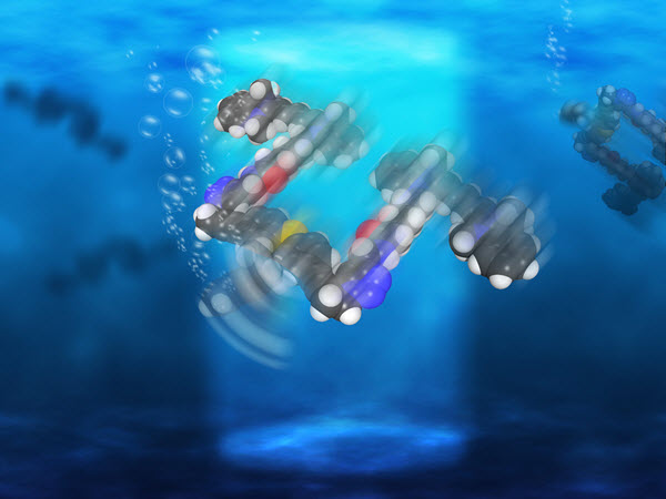
Rice University scientists have created light-driven, single-molecule submersibles that contain just 244 atoms (credit: Loïc Samuel/Rice University)

The Rice University lab of chemist James Tour has created single-molecule, 244-atom submersibles with motors powered by ultraviolet light, as they reported this month in the American Chemical Society journal Nano Letters.

With each full revolution, the motor’s tail-like propeller moves the sub forward 18 nanometers, but with the motors running at more than a million RPM, that translates into almost 1 inch per second — a breakneck pace on the molecular scale, says Tour. “These are the fastest-moving molecules ever seen in solution,” he said.

This chemical schematic shows the design of single-molecule nanosubmersibles created at Rice University. The nanosub’s fluorescent pontoons are blue; the motor is red. (credit: Victor García-López/Rice University)

While they can’t be steered yet, the study proves molecular motors are powerful enough to drive the sub-10-nanometer subs through solutions of moving molecules of about the same size. “This is akin to a person walking across a basketball court with 1,000 people throwing basketballs at him,” Tour said.

In 2006, Tour’s lab introduced the world to nanocars, single-molecule cars with four wheels, axles, and independent suspensions that could be “driven” across a surface (see “Rice scientists attach motor to single-molecule car“).

Tour said many scientists have created microscopic machines with motors over the years, but most have either used or generated toxic chemicals. He said a motor that was conceived in 2009 by a group in the Netherlands proved suitable for Rice’s submersibles, which were produced in a 20-step chemical synthesis.

“These motors are well-known and used for different things,” said lead author and Rice graduate student Victor García-López. “But we were the first ones to propose they can be used to propel nanocars and now submersibles.”

Operate like a bacteria’s flagellum

A nanosubmarine found in nature: this whip-like flagellum powers a bacterium’s swimming (credit: LadyofHats/CC)

The motors, which operate more like a bacteria’s flagellum than a propeller, complete each revolution in four steps. When excited by light, the double bond that holds the rotor to the body becomes a single bond, allowing it to rotate a quarter step. As the motor seeks to return to a lower energy state, it jumps adjacent atoms for another quarter turn. The process repeats as long as the light is on.

For comparison tests, the lab also made submersibles with no motors, slow motors, and motors that paddle back and forth. All versions of the submersibles have pontoons that fluoresce red when excited by a laser, according to the researchers.

Once built, the sub’s performance was independently confirmed by Gufeng Wang at North Carolina State University.

Rice’s researchers hope future nanosubs will be able to carry cargoes for medical and other purposes. “There’s a path forward,” García-López said. “This is the first step, and we’ve proven the concept. Now we need to explore opportunities and potential applications.”

Wang is an assistant professor of analytical chemistry at North Carolina State. Tour is the T.T. and W.F. Chao Chair in Chemistry as well as a professor of computer science and of materials science and nanoengineering.

The National Science Foundation, the National Institutes of Health, the Welch Foundation, and North Carolina State supported the research.


Abstract of Unimolecular Submersible Nanomachines. Synthesis, Actuation, and Monitoring

Unimolecular submersible nanomachines (USNs) bearing light-driven motors and fluorophores are synthesized. NMR experiments demonstrate that the rotation of the motor is not quenched by the fluorophore and that the motor behaves in the same manner as the corresponding motor without attached fluorophores. No photo or thermal decomposition is observed. Through careful design of control molecules with no motor and with a slow motor, we found using single molecule fluorescence correlation spectroscopy that only the molecules with fast rotating speed (MHz range) show an enhancement in diffusion by 26% when the motor is fully activated by UV light. This suggests that the USN molecules give ∼9 nm steps upon each motor actuation. A non-unidirectional rotating motor also results in a smaller, 10%, increase in diffusion. This study gives new insight into the light actuation of motorized molecules in solution.

‘Super natural killer cells’ destroy cancer in lymph nodes to halt metastasis

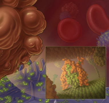
Nanoscale liposomes (orange) containing TRAIL protein (green) attach to the surface of white blood cells (blue), bump into cancer cells (brown), and program them to die (credit: Cornell University)

Cornell biomedical engineers have developed specialized white blood cells they call “super natural killer cells” that seek out cancer cells in lymph nodes with only one purpose: to destroy them, halting the onset of cancer tumor cell metastasis.

“We want to see lymph-node metastasis become a thing of the past,” said Michael R. King, the Daljit S. and Elaine Sarkaria Professor of Biomedical Engineering and senior author of a paper in the journal Biomaterials.

For tumor cells, the lymph nodes are a staging area in the body and play a key role in advancing metastasis throughout the body. In the study with mice, the biomedical engineers killed cancerous tumor cells within days by injecting liposomes (spherical vesicles that can act as carriers) armed with TRAIL (Tumor necrosis factor Related Apoptosis-Inducing Ligand). The liposomes attached to “natural killer” cells — a type of white blood cell — residing in the lymph nodes.

Inducing cancer-cell suicide

King says these natural killer cells in the body became the “super natural killer cells,” which found the cancerous cells and induced apoptosis (cell suicide). The cancer cells self-destruct and disintegrate, preventing the lymphatic spread of cancer any further by “completely eliminating lymph node metastases in mice,” said King.

In cancer progression, there are four stages. At stage I, the tumor is small and has yet to progress to the lymph nodes. In stages II and III, the tumors have grown and likely will have spread to the lymph nodes. At stage IV, the cancer has advanced from the lymph nodes to organs and other parts of the body.

Between 29 and 37 percent of patients with breast, colorectal, and lung cancers are diagnosed with metastases in their tumor-draining lymph nodes — those lymph nodes that lie downstream from the tumor — and those patients are at a higher risk for distant-organ metastases and later-stage cancer diagnoses.

In January 2014, King and his colleagues published research (see “Piggy-backing proteins ride white blood cells to destroy metastasizing cancer“) that demonstrated that by attaching the TRAIL protein to white blood cells, metastasizing cancer cells in the bloodstream were annihilated.

“So, now we [also] have technology to eliminate lymph node metastases,” King said. He said human testing of the TRAIL drug could be done “short of a few years from now.”


Cornell University Media Relations | Cornell scientists develop “killer cells” to destroy cancer in lymph nodes


Abstract of Super natural killer cells that target metastases in the tumor draining lymph nodes

Tumor draining lymph nodes are the first site of metastasis in most types of cancer. The extent of metastasis in the lymph nodes is often used in staging cancer progression. We previously showed that nanoscale TRAIL liposomes conjugated to human natural killer cells enhance their endogenous therapeutic potential in killing cancer cells cultured in engineered lymph node microenvironments. In this work, it is shown that liposomes decorated with apoptosis-inducing ligand TRAIL and an antibody against a mouse natural killer cell marker are carried to the tumor draining inguinal lymph nodes and prevent the lymphatic spread of a subcutaneous tumor in mice. It is shown that targeting natural killer cells with TRAIL liposomes enhances their retention time within the tumor draining lymph nodes to induce apoptosis in cancer cells. It is concluded that this approach can be used to kill cancer cells within the tumor draining lymph nodes to prevent the lymphatic spread of cancer.

Experimental drug targeting Alzheimer’s disease shows anti-aging effects

As mice age, those treated with J147 (right) showed improved physiology, memory and appearance that more closely resembled younger mice (credit: the Salk Institute for Biological Studies)

Salk Institute researchers have found that an experimental drug candidate called called J147, which was aimed at combating Alzheimer’s disease, also has a host of unexpected anti-aging effects in animals.

The team used a mouse model of aging not typically used in Alzheimer’s research. When these mice were treated with J147, they had better memory and cognition, healthier blood vessels in the brain, and other improved physiological features, as detailed Nov. 12 in an open-access paper in the journal Aging.

The researchers used a comprehensive set of assays to measure the expression of all genes in the brain, along with 500 small molecules involved with metabolism in the brains and blood of three groups of the rapidly aging mice. The three groups included one set that was young, one set that was old, and one set that was old but fed J147 as they aged.

The old mice that received J147 performed better on memory and other tests for cognition and also displayed more robust motor movements. The mice treated with J147 also had fewer pathological signs of Alzheimer’s in their brains. Importantly, because of the large amount of data collected on the three groups of mice, it was possible to demonstrate that many aspects of gene expression and metabolism in the old mice fed J147 were very similar to those of young animals. These included markers for increased energy metabolism, reduced brain inflammation, and reduced levels of oxidized fatty acids in the brain.

Another notable effect was that J147 prevented the leakage of blood from the microvessels in the brains of old mice. “Damaged blood vessels are a common feature of aging in general, and in Alzheimer’s, it is frequently much worse,” says Antonio Currais, the lead author and a member of Professor David Schubert’s Cellular Neurobiology Laboratory at Salk.

The team aims to begin human trials of J147 next year.

Alzheimer’s disease is a progressive brain disorder, recently ranked as the third leading cause of death in the United States and affecting more than five million Americans. It is also the most common cause of dementia in older adults, according to the National Institutes of Health. While most drugs developed in the past 20 years target the amyloid plaque deposits in the brain (which are a hallmark of the disease), few have proven effective in the clinic.


Abstract of A comprehensive multiomics approach toward understanding the relationship between aging and dementia

Because age is the greatest risk factor for sporadic Alzheimer’s disease (AD), phenotypic screens based upon old age-associated brain toxicities were used to develop the potent neurotrophic drug J147. Since certain aspects of aging may be primary cause of AD, we hypothesized that J147 would be effective against AD-associated pathology in rapidly aging SAMP8 mice and could be used to identify some of the molecular contributions of aging to AD. An inclusive and integrative multiomics approach was used to investigate protein and gene expression, metabolite levels, and cognition in old and young SAMP8 mice. J147 reduced cognitive deficits in old SAMP8 mice, while restoring multiple molecular markers associated with human AD, vascular pathology, impaired synaptic function, and inflammation to those approaching the young phenotype. The extensive assays used in this study identified a subset of molecular changes associated with aging that may be necessary for the development of AD.

Beyond telomerase: another enzyme discovered critical to maintaining telomere length

ATM inhibition shortens telomeres and ATM activation elongates telomeres. (credit: Stella Suyong Lee et al./Cell Reports)

Johns Hopkins researchers report they have uncovered the role of an another enzyme crucial to telomere length in addition to the enzyme telomerase, discovered in 1984.

The researchers say the new test they used to find the enzyme should speed discovery of other proteins and processes that determine telomere length. Shortened telomeres have been implicated in aging and in diseases as diverse as lung and bone marrow disorders, while overly long telomeres are linked to cancer.

Their results appear in an open-access paper in the Nov. 24 issue of Cell Reports.

“We’ve known for a long time that telomerase doesn’t tell the whole story of why chromosomes’ telomeres are a given length, but with the tools we had, it was difficult to figure out which proteins were responsible for getting telomerase to do its work,” says Carol Greider, Ph.D., the Daniel Nathans Professor and Director of Molecular Biology and Genetics in the Johns Hopkins Institute for Basic Biomedical Sciences. Greider won the 2009 Nobel Prize in Physiology or Medicine for the discovery of telomerase.

Figuring out exactly what’s needed to lengthen telomeres has broad health implications, Greider notes. Telomeres naturally shorten each time DNA is copied in preparation for cell division, so cells need a well-tuned process to keep adding the right number of building blocks back onto telomeres over an organism’s lifetime.

But until now, researchers have been saddled with a limiting and time-consuming test for whether a given protein is involved in maintaining telomere length, a test that first requires blocking a suspected protein’s action in lab-grown cells, then getting the cells to grow and divide for about three months so that detectable differences in telomere length can emerge. In addition to being time consuming, the test could not be used at all for proteins whose loss would kill the cells before the three-month mark.

ATM kinase found needed to lengthen telomeres

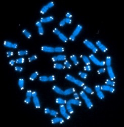
Telomeres glow at the ends of chromosomes (credit: Hesed Padilla-Nash and Thomas Ried/NIH)

For their trial run of the new test, dubbed “addition of de novo initiated telomeres (ADDIT),” Greider’s group examined an enzyme called ATM kinase. “ATM kinase was known to be involved in DNA repair, but there were conflicting reports about whether it had a role in telomere lengthening,” says Greider.

Her team blocked the enzyme in lab-grown mouse cells, and used ADDIT to find that it was indeed needed to lengthen telomeres. They verified the result using the old, three-month-long telomere test, and got the same result.

The team also found that in normal mouse cells, a drug that blocks an enzyme called PARP1 would activate ATM kinase and spur telomere lengthening. This finding offers a proof of principle for drug-based telomere elongation to treat short-telomere diseases, such as bone marrow failure, Greider says — but she cautions that PARP1 inhibitor drug itself doesn’t have the same telomere-elongating effect in human cells as it does in mouse cells.

Greider’s group plans to use ADDIT to find out more about the telomere-lengthening biochemical pathway that ATM kinase is a part of, as well as other pathways that help determine telomere length.

“The potential applications are very exciting,” says graduate student Stella Suyong Lee, who conducted the research, which took nearly five years. “Ultimately ADDIT can help us understand how cells strike a balance between aging and the uncontrolled cell growth of cancer, which is very intriguing.”


Abstract of ATM Kinase Is Required for Telomere Elongation in Mouse and Human Cells

Short telomeres induce a DNA damage response, senescence, and apoptosis, thus maintaining telomere length equilibrium is essential for cell viability. Telomerase addition of telomere repeats is tightly regulated in cells. To probe pathways that regulate telomere addition, we developed the ADDIT assay to measure new telomere addition at a single telomere in vivo. Sequence analysis showed telomerase-specific addition of repeats onto a new telomere occurred in just 48 hr. Using the ADDIT assay, we found that ATM is required for addition of new repeats onto telomeres in mouse cells. Evaluation of bulk telomeres, in both human and mouse cells, showed that blocking ATM inhibited telomere elongation. Finally, the activation of ATM through the inhibition of PARP1 resulted in increased telomere elongation, supporting the central role of the ATM pathway in regulating telomere addition. Understanding this role of ATM may yield new areas for possible therapeutic intervention in telomere-mediated disease.

Multi-layer nanoparticles glow when exposed to invisible near-infrared light

An artist’s rendering shows the layers of a new, onion-like nanoparticle whose specially crafted layers enable it to efficiently convert invisible near-infrared light to higher-energy blue and UV light (credit: Kaiheng Wei)

A new onion-like nanoparticle developed at the State University of New York University at Buffalo could open new frontiers in biomaging, solar-energy harvesting, and light-based security techniques.

The particle’s innovation lies in its layers: a coating of organic dye, a neodymium-containing shell, and a core that incorporates ytterbium and thulium. Together, these strata convert invisible near-infrared light to higher energy blue and UV light with record-high efficiency.

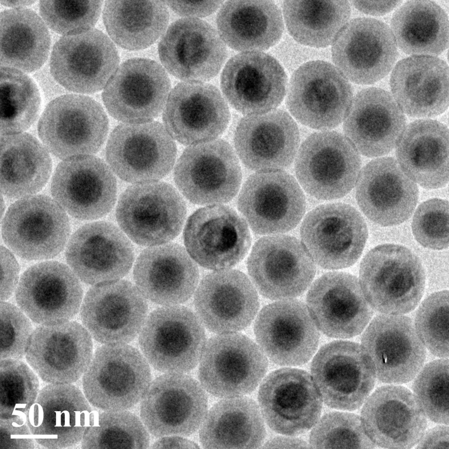
A transmission electron microscopy image of the new nanoparticles, which convert invisible near-infrared light to higher-energy blue and UV light with high efficiency. Each particle is about 50 nanometers in diameter. (credit: Institute for Lasers, Photonics and Biophotonics, University at Buffalo)

Light-emitting nanoparticles placed by a surgeon inside the body could provide high-contrast images of areas of interest. Nanoparticle-infused inks could also be incorporated into currency designs using ink that is invisible to the naked eye but glows blue when hit by a low-energy near-infrared laser pulse — which would very difficult for counterfeiters to reproduce.

The researchers say the nanoparticle is about 100 times more efficient at “upconverting” [increasing the frequency of] light than similar nanoparticles.

Peeling back the layers

Energy-cascaded upconversion (credit: Guanying Chen et al./Nano Letters)

Converting low-energy light to light of higher energies is difficult to do. It involves capturing two or photons from a low-energy light source, and combining their energy to form a single, higher-energy photon. Each of the three layers of this onionesque nanoparticle fulfills a unique function:

  • The outermost layer is a coating of organic dye. This dye is adept at absorbing photons from low-energy near-infrared light sources. It acts as an “antenna” for the nanoparticle, harvesting light and transferring energy inside, Ohulchanskyy says.
  • The next layer is a neodymium-containing shell. This layer acts as a bridge, transferring energy from the dye to the particle’s light-emitting core.*
  • Inside the light-emitting core, ytterbium and thulium ions work in concert. The ytterbium ions draw energy into the core and pass the energy on to the thulium ions, which have special properties that enable them to absorb the energy of three, four or five photons at once, and then emit a single higher-energy photon of blue and UV light.

The research was published online in Nano Letters on Oct. 21. It was led by the Institute for Lasers, Photonics, and Biophotonics at UB, and the Harbin Institute of Technology in China, with contributions from the Royal Institute of Technology in Sweden, Tomsk State University in Russia, and the University of Massachusetts Medical School.

* The neodymium-containing layer is necessary for transferring energy efficiently from dye to core. When molecules or ions in a material absorb a photon, they enter an “excited” state from which they can transfer energy to other molecules or ions. The most efficient transfer occurs between molecules or ions whose excited states require a similar amount of energy to obtain, but the dye and ytterbium ions have excited states with very different energies. So the team added neodymium — whose excited state is in between that of the dye and thulium’s — to act as a bridge between the two, creating a “staircase” for the energy to travel down to reach emitting thulium ions.


Abstract of Energy-Cascaded Upconversion in an Organic Dye-Sensitized Core/Shell Fluoride Nanocrystal

Lanthanide-doped upconversion nanoparticles hold promises for bioimaging, solar cells, and volumetric displays. However, their emission brightness and excitation wavelength range are limited by the weak and narrowband absorption of lanthanide ions. Here, we introduce a concept of multistep cascade energy transfer, from broadly infrared-harvesting organic dyes to sensitizer ions in the shell of an epitaxially designed core/shell inorganic nanostructure, with a sequential nonradiative energy transfer to upconverting ion pairs in the core. We show that this concept, when implemented in a core–shell architecture with suppressed surface-related luminescence quenching, yields multiphoton (three-, four-, and five-photon) upconversion quantum efficiency as high as 19% (upconversion energy conversion efficiency of 9.3%, upconversion quantum yield of 4.8%), which is about ∼100 times higher than typically reported efficiency of upconversion at 800 nm in lanthanide-based nanostructures, along with a broad spectral range (over 150 nm) of infrared excitation and a large absorption cross-section of 1.47 × 10⁻¹⁴ cm2 per single nanoparticle. These features enable unprecedented three-photon upconversion (visible by naked eye as blue light) of an incoherent infrared light excitation with a power density comparable to that of solar irradiation at the Earth surface, having implications for broad applications of these organic–inorganic core/shell nanostructures with energy-cascaded upconversion.

Blood-brain barrier opened non-invasively for the first time in humans, using focused ultrasound

Opening up the blood-brain barrier to deliver drugs (credit: Focused Ultrasound Foundation)

The blood-brain barrier has been non-invasively opened in a human patient for the first time. A team at Sunnybrook Health Sciences Centre in Toronto used focused ultrasound to temporarily open the blood-brain barrier (BBB), allowing for effective delivery of chemotherapy into a patient’s malignant brain tumor.

The team infused the chemotherapy agent doxorubicin, along with tiny gas-filled bubbles, into the bloodstream of a patient with a brain tumor. They then applied focused ultrasound to areas in the tumor and surrounding brain, causing the bubbles to vibrate, loosening the tight junctions of the cells comprising the BBB, and allowing high concentrations of the chemotherapy to enter targeted tissues.

This patient treatment is part of a pilot study of up to 10 patients to establish the feasibility, safety, and preliminary efficacy of focused ultrasound to temporarily open the blood-brain barrier to deliver chemotherapy to brain tumors. The Focused Ultrasound Foundation is currently funding this trial through their Cornelia Flagg Keller Memorial Fund for Brain Research. Based on these two pre-clinical studies, a pilot clinical trial using focused ultrasound to treat Alzheimer’s is being organized.

Dr. Kullervo Hynynen, senior scientist at the Sunnybrook Research Institute, has been performing similar pre-clinical studies for about a decade. In 2012, his team was able to bypass the BBB of a rat model non-invasively (see Bypassing the blood-brain barrier with MRI and ultrasound).

Previous methods where invasive, requiring an operation, such as an implanted mucosal graft in the nose (see A drug-delivery technique to bypass the blood-brain barrier and Researchers bypass the blood-brain barrier, widening treatment options for neurodegenerative and central nervous system disease) or inserting needle electrodes into the diseased tissue and applying multiple bursts of pulsed electric energy (see Blood-brain-barrier disruption with high-frequency pulsed electric fields).

Fighting disease

The researchers suggest that focused ultrasound could also be used to deliver other types of drugs, DNA-loaded nanoparticles, viral vectors, and antibodies to the brain to treat a range of neurological conditions, including various types of brain tumors, Parkinson’s, Alzheimer’s and some psychiatric diseases.

For example, the temporary opening of the blood-brain barrier appears to facilitate the brain’s clearance of a key pathologic protein related to Alzheimer’s and improves cognitive function, the researchers found. And a recent study at the Queensland Brain Institute in Australia demonstrated that opening the blood-brain barrier with focused ultrasound reduced brain plaques and improved memory in a mouse model of Alzheimer’s disease.


Focused Ultrasound Foundation


Abstract of Scanning ultrasound removes amyloid-β and restores memory in an Alzheimer’s disease mouse model

Transgenic mice with increased amyloid-β (Aβ) production show several aspects of Alzheimer’s disease, including Aβ deposition and memory impairment. By repeatedly treating these Aβ-forming mice with scanning ultrasound, Leinenga and Götz now demonstrate that Aβ is removed and memory is restored as revealed by improvement in three memory tasks. These improvements were achieved without the use of any therapeutic agent, and the scanning ultrasound treatment did not induce any apparent damage to the mouse brain. The authors then showed that scanning ultrasound activated resident microglial cells that took up Aβ into their lysosomes. These findings suggest that repeated scanning ultrasound may be a noninvasive method with potential for treating Alzheimer’s disease.

New ‘tricorder’ technology might be able to ‘hear’ tumors

Capacitive micromachined ultrasonic detectors used in the experiments, with a detail view of the front and back of one device (credit: Hao Nan et al./Applied Physics Letters)

Stanford electrical engineers have developed an enhancement of technology intended to safely find buried plastic explosives and spot fast-growing tumors, using a combination of microwaves and ultrasound to develop a detector similar to the legendary Star Trek tricorder.

The work, led by Assistant Professor Amin Arbabian and Research Professor Pierre Khuri-Yakub, grows out of DARPA research designed to detect buried plastic explosives, but the researchers said the technology could also provide a new way to detect early stage cancers.

The new work was spurred by a challenge posed by the Defense Advanced Research Projects Agency (DARPA), which sought a system to detect plastic explosives (improvised explosive devices or IEDs) buried underground, which are currently invisible to metal detectors. The detection device could not touch the surface in question, so as not to trigger an explosion.

The engineers developed a system based on the principle that all materials expand and contract when heated, but not at identical rates. In a potential battlefield application, the microwaves would heat the suspect area, causing the muddy ground to absorb energy and expand, and thus squeeze the plastic. Pulsing the microwaves would then generate a series of ultrasound pressure waves that could be detected and interpreted to disclose the presence of buried plastic explosives.

Touchless ultrasound detection

Sound waves propagate differently in solids than air, with a drastic transmission loss occurring when sound jumps from the solid to air. So the Stanford team accommodated for this loss by building highly sensitive capacitive micromachined ultrasonic transducers (CMUTs) that can specifically discern the weaker ultrasound signals that jumped from the solid, through the air, to the detector.

Solving the technical challenges of detecting ultrasound after it left the ground gave the Stanford researchers the experience to take aim at their ultimate goal: Using the device in medical applications without touching the skin.

Schematic of the non-contact thermoacoustic detection setup. H is the thickness of the surrounding packaging material (set to between 1 and 3 cm of water or Agarose), corresponding to the surrounding flesh-like tissue. T is the thickness of the embedded target (Rexolite, in this case, set to 4mm layers and target area of 4 square cm), corresponding to a tumor. In microwave-induced thermoacoustic imaging, the target absorbs a portion of the microwave electromagnetic energy (from the microwave signal generator) based on the the target tissue’s dielectric properties, producing an ultrasonic wave that is then detected by the airborne capacitive micromachined ultrasonic transducers (CMUT). The corresponding data is then captured for use in reconstructing the target image. (credit: Hao Nan et al./Applied Physics Letters)

Arbabian’s team used brief microwave pulses to heat a flesh-like material that had been implanted with a sample “target.” Holding the device at a standoff distance of 30 cm, the material was heated by a mere thousandth of a degree, well within safety limits. Yet even that slight heating caused the material to expand and contract, which, in turn, created ultrasound waves that the Stanford team was able to detect to disclose the location of the  4 square centimeter embedded target, all without touching the “flesh” — just like the Star Trek tricorder.

Prior medical research showed that tumors grow additional blood vessels to nourish their cancerous growth. Like wet ground, blood vessels absorb heat differently than surrounding tissue, so tumors should show up as ultrasound hotspots.

“We think we could develop instrumentation sufficiently sensitive to disclose the presence of tumors, and perhaps other health anomalies, much earlier than current detection systems, non-intrusively and with a handheld portable device,” Arbabian said.

The researchers believe that their microwave and ultrasound detection system will be practical and widely available within 10 to 15 years. It would be more portable and less expensive than other medical imaging devices such as MRI or CT, and safer than X-rays.

The experiments are detailed in Applied Physics Letters and were presented at the International Ultrasonics Symposium in Taipei, Taiwan.


Stanford University | Stanford Engineers Test Tricorder-Like Detector


Abstract of Non-contact thermoacoustic detection of embedded targets using airborne-capacitive micromachined ultrasonic transducers

A radio frequency (RF)/ultrasound hybrid imaging system using airborne capacitive micromachined ultrasonic transducers (CMUTs) is proposed for the remote detection of embedded objects in highly dispersive media (e.g., water, soil, and tissue). RF excitation provides permittivity contrast, and ultra-sensitive airborne-ultrasound detection measures thermoacoustic-generated acoustic waves that initiate at the boundaries of the embedded target, go through the medium-air interface, and finally reach the transducer. Vented wideband CMUTs interface to 0.18 μm CMOS low-noise amplifiers to provide displacement detectionsensitivity of 1.3 pm at the transducer surface. The carefully designed vented CMUT structure provides a fractional bandwidth of 3.5% utilizing the squeeze-film damping of the air in the cavity.

3D-printed microchannels deliver oxygen, nutrients from artery to tissue implant

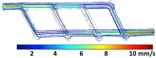
A miniature 3D-printed network of microchannels designed to link up an artery to a tissue implant to ensure blood flow of oxygen and nutrients. Inlet and outlet are ~1 millimeter in diameter; multiple smaller vessels are ~ 600 to 800 microns in diameter. Flow streamlines are color-coded corresponding to flow rate. Flow rate at the inlet is equal to 0.12 mL/min. (credit: Renganaden Sooppan et al./Tissue Engineering Part C: Methods)

Scientists have designed an innovative structure containing an intricate microchannel network of simulated blood vessels that solves one of the biggest challenges in regenerative medicine: How to deliver oxygen and nutrients to all cells in an artificial organ or tissue implant that takes days or weeks to grow in the lab prior to surgery.

The new study was performed by a research team led by Jordan Miller, assistant professor of bioengineering at Rice, and Pavan Atluri, assistant professor of surgery at Penn.

Stayin’ alive, stayin’ alive …

Miller explained that one of the hurdles of engineering large artificial tissues, such as livers or kidneys, is keeping the cells inside them alive. Tissue engineers have typically relied on the body’s own ability to grow blood vessels — for example, by implanting engineered tissue scaffolds inside the body and waiting for blood vessels from nearby tissues to spread via arbolization to the engineered constructs.

But that process can take weeks, and cells deep inside the constructs often starve or die from lack of oxygen before they’re reached by the slow-approaching blood vessels.

“What a surgeon needs in order to do transplant surgery isn’t just a mass of cells; the surgeon needs a vessel inlet and an outlet that can be directly connected to arteries and veins,” he said.

3D-printing pastry-inspired sugar glass to form an intricate microchannel capillary lattice

“We wondered if there were a way to implant a 3-D printed construct where we could connect host arteries directly to the construct and get perfusion [blood flow] immediately. In this study, we are taking the first step toward applying an analogy from transplant surgery to 3-D printed constructs we make in the lab.”

Miller turned to a method inspired by the intricate sugar glass cages crafted by pastry chefs to garnish desserts and that he had pioneered in 2012.

Description of sugar glass printing and initial flow testing. A: Extrusion print head in the process of printing a sugar glass lattice. B: Final sugar lattice prior to casting. The lattice contains a network of filaments supported by a surrounding well. Red line denotes the outer edge of the well that will be filled with PDMS silicone gel during casting. C: Schematic of printed sugar glass network. Drawing on the left denotes sugar filaments after printing, while the figure on the right shows trimmed filaments prior to casting. D: Final cast PDMS gel with microchannel network. (credit: Renganaden Sooppan et al./Tissue Engineering Part C: Methods)

Using an open-source 3-D printer to lay down individual filaments of sugar glass one layer at a time, the researchers printed a lattice of would-be blood vessels. Once the sugar hardened, they placed it in a mold and poured in silicone gel. After the gel cured, Miller’s team dissolved the sugar, leaving behind a network of small channels in the silicone.

“They don’t yet look like the blood vessels found in organs, but they have some of the key features relevant for a transplant surgeon,” Miller said. “We created a construct that has one inlet and one outlet, which are about 1 millimeter in diameter, and these main vessels branch into multiple smaller vessels, which are about 600 to 800 microns.”

Passing the surgeon-oriented test: normal blood flow

Collaborating surgeons at Penn in Atluri’s group then connected the inlet and outlet of the engineered gel to a major (femoral) artery in a small animal model. Using Doppler imaging technology, the team observed and measured blood flow through the construct and found that it withstood physiologic pressures and remained open and unobstructed for up to three hours.

They found that blood flowed normally through test constructs that were surgically connected to native blood vessels.

“This study provides a first step toward developing a transplant model for tissue engineering where the surgeon can directly connect arteries to an engineered tissue,” Miller said. “In the future, we aim to utilize a biodegradable material that also contains live cells next to these perfusable vessels for direct transplantation and monitoring long term.”
The report was published in an open-access paper in the journal Tissue Engineering Part C: Methods.

Abstract of Tissue Engineering Part C: Methods

The field of tissue engineering has advanced the development of increasingly biocompatible materials to mimic the extracellular matrix of vascularized tissue. However, a majority of studies instead rely on a multi-day inosculation between engineered vessels and host vasculature, rather than the direct connection of engineered microvascular networks with host vasculature. We have previously demonstrated that the rapid casting of 3D printed sacrificial carbohydrate glass is an expeditious and reliable method of creating scaffolds with 3D microvessel networks. Here, we describe a new surgical technique to directly connect host femoral arteries to patterned microvessel networks. Vessel networks were connected in vivo in a rat femoral artery graft model. We utilized laser Doppler imaging to monitor hind limb ischemia for several hours after implantation and thus measured the vascular patency of implants that were anastomosed to the femoral artery. This study may provide a method to overcome the challenge of rapid oxygen and nutrient delivery to engineered vascularized tissues implanted in vivo.

A new 3-​​D printing method for creating patient-​​specific medical devices

The 3D magnetic printing process systematically aligns and selectively polymerizes groupings of voxels (volume “pixels”) programmed to have specific reinforcement orientation within each layer of printed material based upon a shifting field. The 3-D printer build plate peels after a layer is complete to print additional layers. (credit: Joshua J. Martin et al./Nature Communications)

Northeastern University engineers have devel­oped a 3-D printing process that uses mag­netic fields to shape com­posite materials (mixes of plas­tics and ceramics) into patient-specific biomedical devices, such as catheters.

The devices are intended to be stronger and lighter than cur­rent models and the cus­tomized design could ensure an appro­priate fit, said Ran­dall Erb, assis­tant pro­fessor in the Depart­ment of Mechan­ical and Indus­trial Engi­neering.

The magnetic field enables the engineers to con­trol how the ceramic fibers are arranged, allowing for con­trol of the mechan­ical prop­er­ties of the mate­rial. That con­trol is crit­ical if you’re crafting devices with com­plex archi­tec­tures, such as cus­tomized minia­ture bio­med­ical devices. Within a single patient-specific device, the cor­ners, the curves, and the holes must all be rein­forced by ceramic fibers arranged in just the right con­fig­u­ra­tion to make the device durable.

This is the strategy taken by many nat­ural com­pos­ites from bones to trees. Fibers of cal­cium phos­phate, the min­eral com­po­nent of bone, are nat­u­rally ori­ented precisely around the holes for blood ves­sels to ensure the bone’s strength and sta­bility to enable, say, your femur to with­stand a daily jog.

Aligning fibers with magnets

The 3D magnetic-printer setup. A digital light processor (DLP) photo-polymerizes resin with UV while a magnetic field is simultaneously applied via electromagnetic solenoids. (credit: Joshua J. Martin et al./Nature Communications)

Erb ini­tially described the role of magnets in the composite-making process in a 2012 paper in the journal Sci­ence. First the researchers “mag­ne­tize” the ceramic fibers by dusting them very lightly with iron oxide, which has been FDA-approved for drug-delivery appli­ca­tions.

They then apply ultra-low mag­netic fields to indi­vidual sec­tions of the com­posite material — the ceramic fibers immersed in liquid plastic — to align the fibers according to the exacting spec­i­fi­ca­tions dic­tated by the product they are printing.

In a video accom­pa­nying the Sci­ence article, you can see the fibers spring to atten­tion when the mag­netic field is turned on. “Mag­netic fields are very easy to apply,” says Erb. “They’re safe, and they pen­e­trate not only our bodies but many other materials.”

Finally, in a process called “stere­olith­o­g­raphy,” they build the product, layer by layer, using a computer-controlled laser beam that hardens the plastic. Each six-by-six inch layer takes a minute to complete.

Using mag­nets, the new printing method aligns each minus­cule fiber in the direc­tion that con­forms pre­cisely to the geom­etry of the item being printed.

“If you can print a catheter whose geom­etry is spe­cific to the indi­vidual patient, you can insert it up to a cer­tain crit­ical spot, you can avoid punc­turing veins, and you can expe­dite delivery of the contents.”

The engineers’ open-access paper on the new tech­nology appears in the Oct. 23 issue of Nature Com­mu­ni­ca­tions.

Custom-designing neonatal catheters

Erb has received a $225,000 Small Busi­ness Tech­nology Transfer grant from the National Institutes of Health to develop neonatal catheters with a local com­pany. “Another of our goals is to use cal­cium phos­phate fibers and bio­com­pat­ible plas­tics to design sur­gical implants.”

Neonatal preemie with catheters (credit: March of Dimes Foundation)

The new technology is especially valuable for prema­ture babies (“preemies”) in neonatal care units, some weighing just a bit over a pound, with plastic tubes snaking through their nose or mouth, or dis­ap­pearing into veins or other parts of the body. Those tubes, or “catheters,” are how the babies get the nec­es­sary oxygen, nutri­ents, fluid, and med­ica­tions to stay alive.

The problem is, today’s catheters only come in stan­dard sizes and shapes, which means they cannot accom­mo­date the needs of all pre­ma­ture babies. “With neonatal care, each baby is a dif­ferent size, each baby has a dif­ferent set of prob­lems,” says Erb.

Worldwide, “15 million babies are born too soon every year” and of those, “1 million children die each year due to complications of preterm birth,” according to a report by the World Health Organization. This data was cited in the “March of Dimes Premature Birth Report Card,” issued today (Nov. 5) by March of Dimes. “Babies who survive an early birth often face serious and lifelong health problems, including breathing problems, jaundice, vision loss, cerebral palsy, and intellectual delays,” the March of Dimes report noted.

The report provides rates and grades for major cities or counties in each U.S. state and Puerto Rico. It also provides preterm birth rates by race and ethnicity. The U.S. preterm birth rate ranks among the worst of high-resource countries, the March of Dimes says.


Abstract of Designing bioinspired composite reinforcement architectures via 3D magnetic printing

Discontinuous fibre composites represent a class of materials that are strong, lightweight and have remarkable fracture toughness. These advantages partially explain the abundance and variety of discontinuous fibre composites that have evolved in the natural world. Many natural structures out-perform the conventional synthetic counterparts due, in part, to the more elaborate reinforcement architectures that occur in natural composites. Here we present an additive manufacturing approach that combines real-time colloidal assembly with existing additive manufacturing technologies to create highly programmable discontinuous fibre composites. This technology, termed as ‘3D magnetic printing’, has enabled us to recreate complex bioinspired reinforcement architectures that deliver enhanced material performance compared with monolithic structures. Further, we demonstrate that we can now design and evolve elaborate reinforcement architectures that are not found in nature, demonstrating a high level of possible customization in discontinuous fibre composites with arbitrary geometries.

Just one junk-food snack triggers signals of metabolic syndrome

(credit: iStock)

Just one high-calorie milkshake was enough to make metabolic syndrome worse for some people. And overindulgence in just a single meal or snack (especially junk food) is enough to trigger the beginnings of metabolic syndrome, which is associated with the risk of developing cardiovascular disease and diabetes (obesity around the waist and trunk is the main sign).

That finding by researchers at the Microbiology and Systems Biology Group of the Netherlands Organisation for Applied Scientific Research (TNO) was reported in the online edition of the Nov. 2015 issue of The FASEB Journal.

For some people, “acute effects of diet are mostly small, but may have large consequences in the long run,” said TNO researcher Suzan Wopereis, Ph.D., senior author of the report.

The researchers gave male volunteers in two groups a high-fat milkshake consisting of 53% whipping cream, 3% sugar, and 44% water (1.6 g protein, 16 g fat, and 3.2 g carbohydrates).

The first group included 10 healthy male volunteers. They were also given a snack diet consisting of an additional 1300 kcal per day, in the form of sweets and savory products such as candy bars, tarts, peanuts, and crisps for four weeks.

The second group included nine volunteers with metabolic syndrome and who had a combination of two or more risk factors for heart disease, such as unhealthy cholesterol levels, high blood pressure, high blood sugar, high blood lipids, and abdominal fat.

Test results: not good

Both groups had blood samples taken, before and after the snacks. In these blood samples, the researchers measured 61 biomarkers, such as cholesterol and blood sugar.

For the subjects with metabolic syndrome, the blood tests showed that biochemical processes related to sugar metabolism, fat metabolism, and inflammation were abnormal.

For the 10 healthy male volunteers, the blood tests showed that signaling molecules such as hormones regulating the control of sugar and fat metabolism and inflammation were changed, resembling the very subtle start of negative health effects similar to those found at the start of metabolic disease.

“Eating junk food is one of those situations where our brains say ‘yes’ and our bodies say ‘no,’” said Gerald Weissmann, M.D., Editor-in-Chief of The FASEB Journal. “Unfortunately for us, this report shows that we need to use our brains and listen to our bodies. Even one unhealthy snack has negative consequences that extend far beyond any pleasure it brings.”


Abstract of Quantifying phenotypic flexibility as the response to a high-fat challenge test in different states of metabolic health

Metabolism maintains homeostasis at chronic hypercaloric conditions, activating postprandial response mechanisms, which come at the cost of adaptation processes such as energy storage, eventually with negative health consequences. This study quantified the metabolic adaptation capacity by studying challenge response curves. After a high-fat challenge, the 8 h response curves of 61 biomarkers related to adipose tissue mass and function, systemic stress, metabolic flexibility, vascular health, and glucose metabolism was compared between 3 metabolic health stages: 10 healthy men, before and after 4 wk of high-fat, high-calorie diet (1300 kcal/d extra), and 9 men with metabolic syndrome (MetS). The MetS subjects had increased fasting concentrations of biomarkers representing the 3 core processes, glucose, TG, and inflammation control, and the challenge response curves of most biomarkers were altered. After the 4 wk hypercaloric dietary intervention, these 3 processes were not changed, as compared with the preintervention state in the healthy subjects, whereas the challenge response curves of almost all endocrine, metabolic, and inflammatory processes regulating these core processes were altered, demonstrating major molecular physiologic efforts to maintain homeostasis. This study thus demonstrates that change in challenge response is a more sensitive biomarker of metabolic resilience than are changes in fasting concentrations.—Kardinaal, A. F. M., van Erk, M. J., Dutman, A. E., Stroeve, J. H. M., van de Steeg, E., Bijlsma, S., Kooistra, T., van Ommen, B., Wopereis, S. Quantifying phenotypic flexibility as the response to a high-fat challenge test in different states of metabolic health.