Single-agent phototherapy system diagnoses and kills cancer cells

A new single-agent phototherapy system combines silicon naphthalocyanine (which is toxic to cancer) and PEG-PCL (biodegradable carrier) for diagnosis and treatment of cancer (credit: Oregon State University)

Researchers at Oregon State University have announced a new single-agent phototherapy (light-based) approach to combating cancer, using a single chemical compound (SiNc-PNP), for both diagnosis and treatment.

The compound makes cancer cells glow when exposed to near-infrared light so a surgeon can identify the cancer. The compound includes a copolymer called PEG-PCL as the biodegradable carrier. The carrier causes the silicon naphthalocyanine to accumulate selectively in cancer cells and reach a maximum level in the cells after about one day. At that point, doctors would do surgery, and then use phototherapy treatment to kill the remaining cancer cells. The compounds are naturally and completely excreted from the body.

In tests completed with laboratory animals, tumors were completely eradicated without side effects, and did not return.

The findings were presented Thursday (Nov. 29) at the annual meeting of the American Association of Pharmaceutical Scientists in Orlando, Florida, and were also recently published in Chemistry of Materials, a publication of the American Chemical Society.

An alternative to surgery, radiation, and chemotherapy

The researchers believe that phototherapy may become a new and promising addition to the three primary ways that most cancer is treated today: surgery, radiation, and/or chemotherapy. Phototherapy may have special value with cancers that have formed resistance to chemotherapeutic drugs, or present other problems that can’t be managed with existing therapies, the researchers suggest.

Their research so far has studied ovarian cancers in laboratory animals, but the treatment may also be useful for other solid tumors, they suggest. There were no apparent side effects on animals tested.

“A single-agent based system is simple and very good at targeting only cancer tumors and should significantly improve outcomes,” said Oleh Taratula, an assistant professor in the Oregon State University/Oregon Health & Science University College of Pharmacy. “It’s small, nontoxic, and highly efficient.”

In continued research with the OSU College of Veterinary Medicine, the treatment will eventually move on to human clinical trials.


Abstract of Naphthalocyanine-Based Biodegradable Polymeric Nanoparticles for Image-Guided Combinatorial Phototherapy

Image-guided phototherapy is extensively considered as a promising therapy for cancer treatment. To enhance translational potential of this modality, we developed a single agent-based biocompatible nanoplatform that provides both real time near-infrared (NIR) fluorescence imaging and combinatorial phototherapy with dual photothermal and photodynamic therapeutic mechanisms. The developed theranostic nanoplatform consists of two building blocks: (1) silicon naphthalocyanine (SiNc) as a NIR fluorescence imaging and phototherapeutic agent and (2) a copolymer, poly(ethylene glycol)-block-poly(ε-caprolactone) (PEG–PCL) as the biodegradable SiNc carrier. Our simple, highly reproducible, and robust approach results in preparation of spherical, monodisperse SiNc-loaded PEG–PCL polymeric nanoparticles (SiNc-PNP) with a hydrodynamic size of 37.66 ± 0.26 nm (polydispersity index = 0.06) and surface charge of −2.76 ± 1.83 mV. The SiNc-loaded nanoparticles exhibit a strong NIR light absorption with an extinction coefficient of 2.8 × 10⁵ M–1 cm–1 and efficiently convert the absorbed energy into fluorescence emission (ΦF = 11.8%), heat (ΔT ∼ 25 °C), and reactive oxygen species. Moreover, the SiNc-PNP are characterized by superior photostability under extensive photoirradiation and structure integrity during storage at room temperature over a period of 30 days. Following intravenous injection, the SiNc-PNP accumulated selectively in tumors and provided high lesion-to-normal tissue contrast for sensitive fluorescence detection. Finally, adriamycin-resistant tumors treated with a single intravenous dose of SiNc-PNP (1.5 mg/kg) combined with 10 min of a 785 nm light irradiation (1.3 W/cm2) were completely eradicated from the mice without cancer recurrence or side effects. The reported characteristics make the developed SiNc-PNP a promising platform for future clinical application.

Long-term aerobic exercise prevents age-related brain deterioration

Schematic illustration of age-related changes in the neurovascular unit that are prevented by exercise. In the aged cortex of sedentary mice, neurovascular dysfunction is evident by decreased numbers of pericytes (surrounding capillaries, pink), decline in basement membrane (BM) coverage (blue), increased transcytosis (a process that transports macromolecules across cells, allowing pathogens to invade) on endothelial cells (green), reduced expression of AQP4 in astrocytes, down-regulation of Apoe (an essential protein, light purple), decrease in synaptic proteins such as synaptophysin (SYN, green), and increased proinflammatory IBA1+ microglia/monocytes (indicating age-related neuroinflammation, yellow). These age-related changes were successfully prevented (horizontal T line, “Exercise”)  by 6 months of voluntary running during aging. (credit: Ileana Soto et al./PLOS Biology

A study of the brains of mice shows that structural deterioration associated with old age can be prevented by long-term aerobic exercise starting in mid-life, according to the authors of an open-access paper in the journal PLOS Biology yesterday (October 29).

Old age is the major risk factor for Alzheimer’s disease, like many other diseases, as the authors at The Jackson Laboratory in Bar Harbor, Maine, note. Age-related cognitive deficits are due partly to changes in neuronal function, but also correlate with deficiencies in the blood supply to the brain and with low-level inflammation.

“Collectively, our data suggests that normal aging causes significant dysfunction to the cortical neurovascular unit, including basement membrane reduction and pericyte (cells that wrap around blood capillaries) loss. These changes correlate strongly with an increase in microglia/monocytes in the aged cortex,” said Ileana Soto, lead author on the study.*

Benefits of aerobic exercise

However, the researchers found that if they let the mice run freely, the structural changes that make the blood-brain barrier leaky and result in inflammation of brain tissues in old mice can be mitigated. That suggests that there are also beneficial effects of exercise on dementia in humans.**

Further work will be required to establish the mechanism(s): what is the role of the complement-producing microglia/macrophages, how does Apoe decline contribute to age-related neurovascular decline, does the leaky blood-brain barrier allow the passage of damaging factors from the circulation into the brain?

This work was funded in part by The Jackson Laboratory Nathan Shock Center, the Fraternal Order of the Eagle, the Jane B Cook Foundation and NIH.

* The authors investigated the changes in the brains of normal young and aged laboratory mice by comparing by their gene expression profiles using a technique called RNA sequencing, and by comparing their structures at high-resolution by using fluorescence microscopy and electron microscopy. The gene expression analysis indicated age-related changes in the expression of genes relevant to vascular function (including focal adhesion, vascular smooth muscle and ECM-receptor interactions), and inflammation (especially related to the complement system, which clears foreign particles) in the brain cortex.

These changes were accompanied by a decline in the function of astrocytes (key support cells in the brain) and loss of pericytes (the contractile cells that surround small capillaries and venules and maintain the blood-brain barrier). There were also effects on the basement membrane, which forms an integral part of the blood-brain barrier, as well as an increase in the density and functional activation of the immune cells known as microglia/monocytes, which scavenge the brain for infectious agents and damaged cells.

** To investigate the impact of long-term physical exercise on the brain changes seen in the aging mice, the researchers provided the animals with a running wheel from 12 months old (equivalent to middle aged in humans) and assessed their brains at 18 months (equivalent to ~60yrs old in humans, when the risk of Alzheimer’s disease is greatly increased). Young and old mice alike ran about two miles per night, and this physical activity improved the ability and motivation of the old mice to engage in the typical spontaneous behaviors that seem to be affected by aging.

This exercise significantly reduced age-related pericyte loss in the brain cortex and improved other indicators of dysfunction of the vascular system and blood-brain barrier. Exercise also decreased the numbers of microglia/monocytes expressing a crucial initiating component of the complement pathway that others have shown previously to play are role in age-related cognitive decline. Interestingly, these beneficial effects of exercise were not seen in mice deficient in a gene called Apoe, variants of which are a major genetic risk factor for Alzheimer’s disease. The authors also report that Apoe expression in the brain cortex declines in aged mice and this decline can also be prevented by exercise.


Abstract of APOE Stabilization by Exercise Prevents Aging Neurovascular Dysfunction and Complement Induction

Aging is the major risk factor for neurodegenerative diseases such as Alzheimer’s disease, but little is known about the processes that lead to age-related decline of brain structures and function. Here we use RNA-seq in combination with high resolution histological analyses to show that aging leads to a significant deterioration of neurovascular structures including basement membrane reduction, pericyte loss, and astrocyte dysfunction. Neurovascular decline was sufficient to cause vascular leakage and correlated strongly with an increase in neuroinflammation including up-regulation of complement component C1QA in microglia/monocytes. Importantly, long-term aerobic exercise from midlife to old age prevented this age-related neurovascular decline, reduced C1QA+ microglia/monocytes, and increased synaptic plasticity and overall behavioral capabilities of aged mice. Concomitant with age-related neurovascular decline and complement activation, astrocytic Apoe dramatically decreased in aged mice, a decrease that was prevented by exercise. Given the role of APOE in maintaining the neurovascular unit and as an anti-inflammatory molecule, this suggests a possible link between astrocytic Apoe, age-related neurovascular dysfunction and microglia/monocyte activation. To test this, Apoe-deficient mice were exercised from midlife to old age and in contrast to wild-type (Apoe-sufficient) mice, exercise had little to no effect on age-related neurovascular decline or microglia/monocyte activation in the absence of APOE. Collectively, our data shows that neurovascular structures decline with age, a process that we propose to be intimately linked to complement activation in microglia/monocytes. Exercise prevents these changes, but not in the absence of APOE, opening up new avenues for understanding the complex interactions between neurovascular and neuroinflammatory responses in aging and neurodegenerative diseases such as Alzheimer’s disease.

How to 3-D print a heart

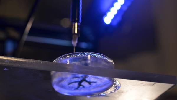
Coronary artery structure being 3-D bioprinted (credit: Carnegie Mellon University College of Engineering)

Carnegie Mellon scientists are creating cutting-edge technology that could one day solve the shortage of heart transplants, which are currently needed to repair damaged organs.

“We’ve been able to take MRI images of coronary arteries and 3-D images of embryonic hearts and 3-D bioprint them with unprecedented resolution and quality out of very soft materials like collagens, alginates and fibrins,” said Adam Feinberg, an associate professor of Materials Science and Engineering and Biomedical Engineering at Carnegie Mellon University.

Feinberg leads the Regenerative Biomaterials and Therapeutics Group, and the group’s study was published in an open-access paper today (Oct. 23) in the journal Science Advances.


College of Engineering, Carnegie Mellon University | Adam Feinberg Demonstrates 3-D Bioprinting Process

“The challenge with soft materials is that they collapse under their own weight when 3-D printed in air,” explained Feinberg. “So we developed a method of printing these soft materials inside a support bath material. Essentially, we print one gel inside of another gel, which allows us to accurately position the soft material as it’s being printed, layer-by-layer.”

A FRESH idea

A schematic of the FRESH process showing the hydrogel (green) — representing an artery — being added to the gelatin slurry support bath (yellow). The 3D object is built layer by layer and, when completed, is released by heating to 37°C and melting the gelatin. (credit: Thomas J. Hinton et al./Science Advances)

With this new FRESH (Freeform Reversible Embedding of Suspended Hydrogels) technique, after printing, the support gel can be easily melted away and removed by heating to body temperature, which does not damage the delicate biological molecules or living cells that were bioprinted.

(Left) A model of a section of a human right coronary arterial tree created from a 3D MRI image is processed at full scale into machine code for FRESH printing. (Right) An example of the arterial tree printed in alginate (black) and embedded in the gelatin slurry support bath. Scale bar: 10 mm. (credit: Thomas J. Hinton et al./Science Advances)

As a next step, the group is working toward incorporating real heart cells into these 3-D printed tissue structures, providing a scaffold to help form contractile muscle.

Accessible bioprinters

Most 3-D bioprinters cost more than $100,000 and/or require specialized expertise to operate, limiting wider-spread adoption. Feinberg’s group, however, has been able to implement their technique on a range of consumer-level 3-D printers, which cost less than $1,000 and use open-source hardware and software.

“Not only is the cost low, but by using open-source software, we have access to fine-tune the print parameters, optimize what we’re doing, and maximize the quality of what we’re printing,” Feinberg said.

More than 4,000 Americans are currently on the waiting list to receive a heart transplant. With failing hearts, these patients have no other options; heart tissue, unlike other parts of the body, is unable to heal itself once it is damaged.


Abstract of Three-dimensional printing of complex biological structures by freeform reversible embedding of suspended hydrogels

We demonstrate the additive manufacturing of complex three-dimensional (3D) biological structures using soft protein and polysaccharide hydrogels that are challenging or impossible to create using traditional fabrication approaches. These structures are built by embedding the printed hydrogel within a secondary hydrogel that serves as a temporary, thermoreversible, and biocompatible support. This process, termed freeform reversible embedding of suspended hydrogels, enables 3D printing of hydrated materials with an elastic modulus <500 kPa including alginate, collagen, and fibrin. Computer-aided design models of 3D optical, computed tomography, and magnetic resonance imaging data were 3D printed at a resolution of ~200 μm and at low cost by leveraging open-source hardware and software tools. Proof-of-concept structures based on femurs, branched coronary arteries, trabeculated embryonic hearts, and human brains were mechanically robust and recreated complex 3D internal and external anatomical architectures.

Custom 3-D printed ear models help surgeons carve new ears

Children with under-formed or missing ears can undergo surgeries to fashion a new ear from rib cartilage, as shown in the above photo. But aspiring surgeons lack lifelike practice models. (credit: University of Washington)

A University of Washington (UW) otolaryngology resident and a bioengineering student have used 3-D printing to create a low-cost pediatric rib cartilage model that more closely resembles the feel of real cartilage, which is used in an operation called auricular reconstruction (ear replacement).

The innovation could make it possible for aspiring surgeons to become proficient in the sought-after but challenging procedure. And because the UW models are printed from a CT scan, they mimic an individual’s specific unique anatomy. That offers the opportunity for even an experienced surgeon to practice a particular tricky surgery ahead of time on a patient-specific rib model.

As part of the study, three experienced surgeons practiced carving, bending, and suturing the UW team’s silicone models, which were produced from a 3-D printed mold modeled from a CT scan of an 8-year-old patient. They compared their firmness, feel, and suturing quality to real rib cartilage, and to a more expensive material made out of dental impression material. They preferred the 3-D printed versions.

The UW team used a 3-D printer to create a negative mold of a patient’s ribs from a CT scan. Surgeons take pieces of those ribs and “carve” them into a new ear. (credit: University of Washington)

Co-author Sharon Newman, who graduated from the UW with a bioengineering degree in June, teamed up with lead author Angelique Berens, a UW School of Medicine otolaryngologist, while they both worked in the UW BioRobotics Lab under electrical engineering professor Blake Hannaford.

Newman figured out how to upload and process a CT scan through a series of free, open-source modeling and imaging programs, and ultimately use a 3-D printer to print a negative mold of a patient’s ribs.

Newman had previously tested different combinations of silicone, corn starch, mineral oil and glycerin to replicate human tissue that the lab’s surgical robot could manipulate. She poured them into the molds and let them cure to see which mixture most closely resembled rib cartilage.

The team’s next steps are to get the models into the hands of surgeons and surgeons-in-training, and hopefully to demonstrate that more lifelike practice models can elevate their skills and abilities.

“With one 3-D printed mold, you can make a billion of these models for next to nothing,” said Berens. “What this research shows is that we can move forward with one of these models and start using it.”

Long waiting list

Kathleen Sie, a UW Medicine professor of otolaryngology – head and neck surgery and director of the Childhood Communication Center at Seattle Children’s, said the lack of adequate training models makes it difficult for surgeons to become comfortable performing the delicate technical procedure.

There’s typically a six- to 12-month waiting list for children to have the procedure done at Seattle Children’s, she said.

“It’s a surgery that more people could do, but this is often the single biggest roadblock,” Sie said. “They’re hesitant to start because they’ve never carved an ear before.”

Their study results were presented at the American Academy of Otolaryngology — Head and Neck Surgery conference in Dallas.

How to control heartbeats more precisely, using light

Using computer-generated light patterns, researchers were able to control the direction of spiraling electrical waves in heart cells. (credit: Eana Park)

Researchers from Oxford and Stony Brook universities has found a way to precisely control the electrical waves that regulate the rhythm of our heartbeat — using light. Their results are published in the journal Nature Photonics.

Cardiac cells in the heart and neurons in the brain communicate by electrical signals, and these messages of communication travel fast from cell to cell as “excitation waves.”

For heart patients there are currently two options to keep these waves in check: electrical devices (pacemakers or defibrillators) or drugs (e.g., beta blockers). However, these methods are relatively crude: they can stop or start waves but cannot provide fine control over the wave speed and direction.

Gil Bub, from Oxford University explained: ‘When there is scar tissue in the heart or fibrosis, this can cause part of the wave to slow down. That can cause re-entrant waves which spiral back around the tissue, causing the heart to beat much too quickly, which can be fatal. If we can control these spirals, we could prevent that.

The optogenetics solution

The solution the researchers found was optogenetics, which uses genetic modification to alter cells so that they can be activated by light. Until now, it has mainly been used to activate individual cells or to trigger excitation waves in tissue, especially in neuroscience research. “We wanted to use it to very precisely control the activity of millions of cells,” said Bub.

A light-activated protein called channelrhodopsin was delivered to heart cells using gene therapy techniques so that they could be controlled by light. Then, using a computer-controlled light projector, the team was able to control the speed of the cardiac waves, their direction and even the orientation of spirals in real time — something that never been shown for waves in a living system before.

In the short term, the ability to provide fine control means that researchers are able to carry out experiments at a level of detail previously only available using computer models. They can now compare those models to experiments with real cells, potentially improving our understanding of how the heart works. The research can also be applied to the physics of such waves in other processes. In the long run, it might be possible to develop precise treatments for heart conditions.

“Precise control of the direction, speed and shape of such excitation waves would mean unprecedented direct control of organ-level function, in the heart or brain, without having to focus on manipulating each cell individually,” said Stony Brook University scientist Emilia Entcheva.

The team stresses that there are significant hurdles before this could offer new treatments; a key issue is being able to alter the heart to be light-sensitized and being able to get the light to desired locations. However, as gene therapy moves into the clinic and with miniaturization of optical devices, use of this all-optical technology may become possible.

In the meantime, the research enables scientists to look into the physics behind many biological processes, including those in our own brains and hearts.

University of Oxford | Controlling heart tissue with light


Abstract of Optical control of excitation waves in cardiac tissue

In nature, macroscopic excitation waves are found in a diverse range of settings including chemical reactions, metal rust, yeast, amoeba and the heart and brain. In the case of living biological tissue, the spatiotemporal patterns formed by these excitation waves are different in healthy and diseased states. Current electrical and pharmacological methods for wave modulation lack the spatiotemporal precision needed to control these patterns. Optical methods have the potential to overcome these limitations, but to date have only been demonstrated in simple systems, such as the Belousov–Zhabotinsky chemical reaction. Here, we combine dye-free optical imaging with optogenetic actuation to achieve dynamic control of cardiac excitation waves. Illumination with patterned light is demonstrated to optically control the direction, speed and spiral chirality of such waves in cardiac tissue. This all-optical approach offers a new experimental platform for the study and control of pattern formation in complex biological excitable systems.

Carbon nanotubes found in cells from airways of asthmatic children in Paris

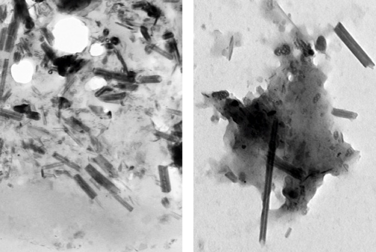
Carbon nanotubes (rods) and nanoparticles (black clumps) found inside a lung cell vacuole (left) are similar to those found in vehicle exhaust in tailpipes of cars in Paris (right) (credit: Fathi Moussa/Paris-Saclay University)

Carbon nanotubes (CNTs) have been found in cells extracted from the airways of Parisian children under routine treatment for asthma, according to a report in the journal EBioMedicine (open access) by scientists in France and at Rice University.

The cells were taken from 69 randomly selected asthma patients aged 2 to 17 who underwent routine fiber-optic bronchoscopies as part of their treatment. The researchers analyzed particulate matter found in the alveolar macrophage cells (also known as dust cells), which help stop foreign materials like particles and bacteria from entering the lungs.

The study partially answers the question of what makes up the black material inside alveolar macrophages, the original focus of the study. The researchers found single-walled and multiwalled carbon nanotubes and amorphous carbon among the cells.

The nanotube aggregates in the cells ranged in size from 10 to 60 nanometers in diameter and up to several hundred nanometers in length, small enough that optical microscopes would not have been able to identify them in samples from former patients. The new study used more sophisticated tools, including high-resolution transmission electron microscopy, X-ray spectroscopy, Raman spectroscopy, and near-infrared fluorescence microscopy to definitively identify them in the cells and in the environmental samples.

“The concentrations of nanotubes are so low in these samples that it’s hard to believe they would cause asthma, but you never know,” said Rice chemist Lon Wilson, a corresponding author of the paper. “What surprised me the most was that carbon nanotubes were the major component of the carbonaceous pollution we found in the samples.”

The study notes but does not make definitive conclusions about the controversial proposition that carbon nanotube fibers may act like asbestos, a proven carcinogen. But the authors did note that “long carbon nanotubes and large aggregates of short ones can induce a granulomatous (inflammation) reaction.”

The researchers also suggested previous studies that link the carbon content of airway macrophages and the decline of lung function should be reconsidered in light of the new findings. The researchers also suggested that the large surface areas of nanotubes and their ability to adhere to substances may make them effective carriers for other pollutants.

Carbon nanotubes from forest fires and cars?

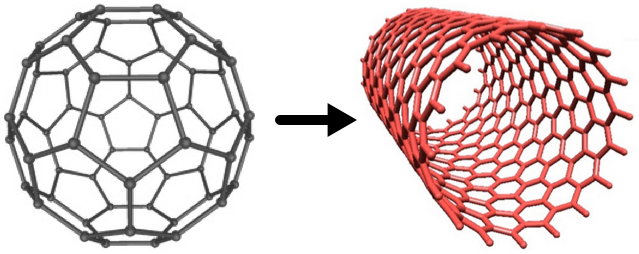
Fullerenes (left) can be converted to carbon nanotubes (right) with a catalytic process, according to Rice chemists (credits: Soroush83/CC and Matías Soto/Rice University)

However, similar nanotubes have been found in samples from the exhaust pipes of Paris vehicles, in dust gathered from various places around the city, in spider webs in India, and even in ice cores, the paper notes.

“We know that carbon nanoparticles are found in nature,” Wilson said, noting that round fullerene (C60) molecules are commonly produced by volcanoes, forest fires, and other combustion of carbon materials. “All you need is a little catalysis to make carbon nanotubes instead of fullerenes.”

A car’s catalytic converter, which turns toxic carbon monoxide into safer emissions, bears at least a passing resemblance to the Rice-invented high-pressure carbon monoxide, or HiPco, process to make carbon nanotubes, he said. “So it is not a big surprise, when you think about it,” Wilson said.

“Based on our discovery of CNTs in tailpipes, we propose that the catalytic converters of the automobiles are manufacturing carbon nanotubes, Wilson told KurzweilAI. “However, we have not actually proven that.”

We are all carbon-nanotube bearers now

For ethical reasons, no cells from healthy patients were analyzed, but because nanotubes were found in all of the samples, the study led the researchers to conclude that carbon nanotubes are likely to be found in everybody.

“It’s kind of ironic. In our laboratory, working with carbon nanotubes, we wear facemasks to prevent exactly what we’re seeing in these samples, yet everyone walking around out there in the world probably has at least a small concentration of carbon nanotubes in their lungs,” he said.

The study followed one released by Rice and Baylor College of Medicine earlier this month with the similar goal of analyzing the black substance found in the lungs of smokers who died of emphysema. That study found carbon black nanoparticles that were the product of the incomplete combustion of such organic material as tobacco.

Co-authors are from Paris-Saclay University, the Paediatric Pulmonology and Allergy Center and the Department of Anatomo-Pathology of the Groupe hospitalier La Roche-Guyon, and Paris Diderot University. The Welch Foundation partially supported the research.


Abstract of Anthropogenic Carbon Nanotubes Found in the Airways of Parisian Children

Compelling evidence shows that fine particulate matters (PM) from air pollution penetrate lower airways and are associated with adverse health effects even within concentrations below those recommended by the WHO. A paper reported a dose-dependent link between carbon content in alveolar macrophages (assessed only by optical microscopy) and the decline in lung function. However, to the best of our knowledge, PM had never been accurately characterized inside human lung cells and the most responsible components of the particulate mix are still unknown. On another hand carbon nanotubes (CNTs) from natural and anthropogenic sources might be an important component of PM in both indoor and outdoor air.

We used high-resolution transmission electron microscopy and energy dispersive X-ray spectroscopy to characterize PM present in broncho-alveolar lavage-fluids (n = 64) and inside lung cells (n = 5 patients) of asthmatic children. We show that inhaled PM mostly consist of CNTs. These CNTs are present in all examined samples and they are similar to those we found in dusts and vehicle exhausts collected in Paris, as well as to those previously characterized in ambient air in the USA, in spider webs in India, and in ice core. These results strongly suggest that humans are routinely exposed to CNTs.

Surgeons reroute nerves to restore hand, arm movement to quadriplegic patients

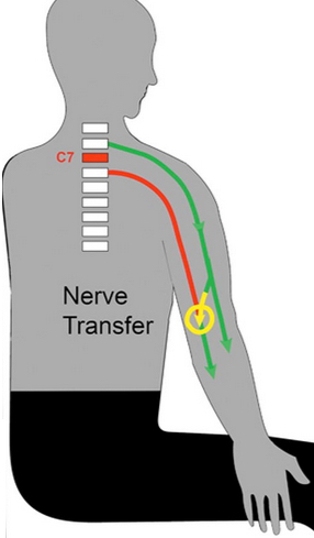
A nerve transfer bypasses the zone of a spinal cord injury (C7). Functional nerves (green) that are under volitional control are rerouted (yellow) to nerves (red) that come off below the spinal cord injury. (credit: Washington University in St. Louis)

A pioneering surgical technique has restored some hand and arm movement to nine patients immobilized by spinal cord injuries in the neck, reports a new study at Washington University School of Medicine in St. Louis.

Bypassing the spinal cord, the surgeons rerouted healthy nerves sitting above the injury site, usually in the shoulders or elbows, to paralyzed nerves in the hand or arm. Once a connection was established, patients underwent extensive physical therapy to train the brain to recognize the new nerve signals, a process that takes about 6–18 months.

The technique targets patients with injuries at the C6 or C7 vertebra, the lowest bones in the neck. It typically does not help patients who have lost all arm function due to higher injuries in vertebrae C1 through C5.

“Physically, nerve-transfer surgery provides incremental improvements in hand and arm function. However, psychologically, these small steps are huge for a patient’s quality of life,” said the study’s lead author, Ida K. Fox, MD, assistant professor of plastic and reconstructive surgery.

One of the most humbling effects of spine damage is the inability to manage bladder or bowel functions. “People with spinal cord injuries cannot control those functions because their brains can’t talk to the nerves in the lower body,” said Fox, who performs surgeries at Barnes-Jewish Hospital.

The study is published in an open-access paper in the October issue of the American Society of Plastic Surgeons’ journal, Plastic and Reconstructive Surgery.

Ultimately, medical professionals hope to discover a way to restore full movement to the estimated 250,000 people in the U.S. living with spinal cord injuries. More than half of such injuries involve the neck. However, until a cure is found, progress in regaining basic independence in routine tasks is important.


Abstract of Nerve transfers to restore upper extremity function in cervical spinal cord injury: Update and preliminary outcomes

Background: Cervical spinal cord injury can result in profound loss of upper extremity function. Recent interest in the use of nerve transfers to restore volitional control is an exciting development in the care of these complex patients. In this article, the authors review preliminary results of nerve transfers in spinal cord injury.

Methods: Review of the literature and the authors’ cases series of 13 operations in nine spinal cord injury nerve transfer recipients was performed. Representative cases were reviewed to explore critical concepts and preliminary outcomes.

Results: The nerve transfers used expendable donors (e.g., teres minor, deltoid, supinator, and brachialis) innervated above the level of the spinal cord injury to restore volitional control of missing function such as elbow extension, wrist extension, and/or hand function (posterior interosseous nerve or anterior interosseous nerve/finger flexors reinnervated). Results from the literature and the authors’ patients (after a mean postsurgical follow-up of 12 months) indicate gains in function as assessed by both manual muscle testing and patients’ self-reported outcomes measures.

Conclusions: Nerve transfers can provide an alternative and consistent means of reestablishing volitional control of upper extremity function in people with cervical level spinal cord injury. Early outcomes provide evidence of substantial improvements in self-reported function despite relatively subtle objective gains in isolated muscle strength. Further work to investigate the optimal timing and combination of nerve transfer operations, the combination of these with traditional treatments (tendon transfer and functional electrical stimulation), and measurement of outcomes is imperative for determining the precise role of these operations.

Smaller silver nanoparticles more likely to be absorbed by aquatic life, UCLA study finds

Researchers studied zebrafish because they have some genetic similarities to humans and their embryos and larvae are transparent, which makes them easier to observe (credit: Tunde Akinloye/CNSI)

A study led by UCLA scientists has found that smaller silver nanoparticles entered fish’s bodies more deeply and persisted longer than larger silver nanoparticles or fluid silver nitrate.

More than 2,000 consumer products today contain nanoparticles — particles so small that they are measured in billionths of a meter. Manufacturers use nanoparticles to help sunscreen work better against the sun’s rays and to make athletic apparel better at wicking moisture away from the body, among many other purposes.

Of those products, 462 contain nanoparticles made from silver, which are used for their ability to kill bacteria. But that benefit might be coming at a cost to the environment. In many cases, simply using the products as-intended causes silver nanoparticles to wind up in rivers and other bodies of water, where they can be ingested by fish and interact with other marine life.

The new study by the University of California Center for Environmental Implications of Nanotechnology, published online in the journal ACS Nano, was intend to begin addressing the question: to what extent do organisms retain those particles and what effects might they have?

Absorption of silver nanoparticles by fish

According to Andre Nel, director of UCLA’s Center for Environmental Implications of Nanotechnology (CEIN) and associate director of the California NanoSystems Institute at UCLA, it is not yet known whether silver nanoparticles are harmful, but the research team wanted to first identify whether they were even being absorbed by fish.

Deposits of 20-nanometer silver nanoparticles in zebrafish gill filaments (outlined in red) (credit: Olivia J. Osborne et al./ACS Nano)

In the study, researchers placed zebrafish in water that contained fluid silver nitrate and two sizes of silver nanoparticles — some measuring 20 nanometers in diameter and others 110 nanometers. The researchers found that the two sizes of particles affected the fish very differently.

The researchers used zebrafish in the study because they have some genetic similarities to humans, and their embryos and larvae are transparent (which makes them easier to observe). In addition, they tend to absorb chemicals and other substances from water.

The team focused its research on the fish’s gills and intestines because they are the organs most susceptible to silver exposure.

The gills showed a significantly higher silver content for the 20-nanometer than the 110-nanometer particles, while the values were more similar in the intestines; both sizes of the silver particles were retained in the intestines even after the fish spent seven days in clean water.

The experiment was one of the most comprehensive in vivo studies to date on silver nanoparticles, as well as the first to compare silver nanoparticle toxicity by extent of organ penetration and duration with different-sized particles, and the first to demonstrate a mechanism for the differences.

Osborne said the results seem to indicate that smaller particles penetrated deeper into the fishes’ organs and stayed there longer because they dissolve faster than the larger particles and are more readily absorbed by the fish.

Nel said the team’s next step is to determine whether silver particles are potentially harmful. “Our research will continue in earnest to determine what the long-term effects of this exposure can be,” he said.

The research was supported by the National Science Foundation and the Environmental Protection Agency.


Abstract of Organ-Specific and Size-Dependent Ag Nanoparticle Toxicity in Gills and Intestines of Adult Zebrafish

We studied adult zebrafish to determine whether the size of 20 and 110 nm citrate-coated silver nanoparticles (AgC NPs) differentially impact the gills and intestines, known target organs for Ag toxicity in fish. Following exposure for 4 h, 4 days, or 4 days plus a 7 day depuration period, we obtained different toxicokinetic profiles for different particle sizes, as determined by Ag content of the tissues. Ionic AgNO3 served as a positive control. The gills showed a significantly higher Ag content for the 20 nm particles at 4 h and 4 days than the 110 nm particles, while the values were more similar in the intestines. Both particle types were retained in the intestines even after depuration. These toxicokinetics were accompanied by striking size-dependent differences in the ultrastructural features and histopathology in the target organs in response to the particulates. Ag staining of the gills and intestines confirmed prominent Ag deposition in the basolateral membranes for the 20 nm but not for the 110 nm particles. Furthermore, it was possible to link the site of tissue deposition to disruption of the Na+/K+ ion channel, which is also localized to the basolateral membrane. This was confirmed by a reduction in ATPase activity and immunohistochemical detection of the α subunit of this channel in both target organs, with the 20 nm particles causing significantly higher inhibition and disruption than the larger size particles or AgNO3. These results demonstrate the importance of particle size in determining the hazardous impact of AgNPs in the gills and intestines of adult zebrafish.

Sleep may strengthen long-term memories in the immune system

A model of memory formation in the central nervous system (upper section) and the immune system (lower section) (credit: Westermann et al./Trends in Neurosciences 2015)

Deep (slow-wave*) sleep, which helps retain memories in the brain, may also strengthen immunological memories of encountered pathogens, German and Dutch neuroscientists propose in an Opinion article published September 29 in Trends in Neurosciences.

The immune system “remembers” an encounter with a bacteria or virus by collecting fragments from the microbe to create memory T cells, which last for months or years and help the body recognize a previous infection and quickly respond. These memory T cells appear to abstract “gist information” about the pathogens, allowing memory T cells to detect new pathogens that are similar, but not identical, to previously encountered bacteria or viruses.

Studies in humans have shown that long-term increases in memory T cells are associated with deep slow-wave sleep on the nights after vaccination. Taken together, the findings support the view that slow-wave sleep contributes to the formation of long-term memories of abstract, generalized information, which leads to adaptive behavioral and immunological responses.

How lack of sleep puts your body at risk

The obvious implication is that sleep deprivation could put your body at risk. “If we didn’t sleep, then the immune system might focus on the wrong parts of the pathogen,” says senior author Jan Born of the University of Tuebingen.

“For example, many viruses can easily mutate some parts of their proteins to escape from immune responses. If too few antigen-recognizing cells [the cells that present the fragments to T cells] are available, then they might all be needed to fight off the pathogen. In addition to this, there is evidence that the hormones released during sleep benefit the crosstalk between antigen-presenting and antigen-recognizing cells, and some of these important hormones could be lacking without sleep.”

Born says that future research should examine what information is selected during sleep for storage in long-term memory, and how this selection is achieved. This research could have important clinical implications.

“In order to design effective vaccines against HIV, malaria, and tuberculosis, which are based on immunological memory, the correct memory model must be available,” Born says. “It is our hope that by comparing the concepts of neuronal and immunological memory, a model of immunological memory can be developed which integrates the available experimental data and serves as a helpful basis for vaccine development.”

* Slow wave sleep (SWS) is the constructive phase of sleep for recuperation of the mind-body system in which it rebuilds itself after each day. Substances that have been ingested into the body while an organism is awake are synthesized into complex proteins of living tissue. Growth hormones are also secreted to facilitate the healing of muscles as well as repairing damage to any tissues. Lastly, glial cells within the brain are restored with sugars to provide energy for the brain. Longer periods of SWS occur in the first part of the night, primarily in the first two sleep cycles (roughly three hours). — Wikipedia


Abstract of System Consolidation during Sleep — A Common Principle Underlying Psychological and Immunological Memory Formation

Sleep benefits the consolidation of psychological memory, and there are hints that sleep likewise supports immunological memory formation. Comparing psychological and immunological domains, we make the case for active system consolidation that is similarly established in both domains and partly conveyed by the same sleep-associated processes. In the psychological domain, neuronal reactivation of declarative memory during slow-wave sleep (SWS) promotes the redistribution of representations initially stored in hippocampal circuitry to extra-hippocampal circuitry for long-term storage. In the immunological domain, SWS seems to favor the redistribution of antigenic memories initially held by antigen-presenting cells, to persisting T cells serving as a long-term store. Because storage capacities are limited in both systems, system consolidation presumably reduces information by abstracting ‘gist’ for long-term storage.

A fast cell sorter shrinks to cell phone size

An artist’s conception of an acoustic cell sorter is the cover image on the current issue of Lab on a Chip (credit: Huang Group/Penn State)

Penn State researchers have developed a new lab-on-a-chip cell sorting device based on acoustic waves that is capable of the kind of high sorting throughput necessary to compete with commercial fluorescence activated cell sorters, described in the cover story in the current issue of the British journal Lab on a Chip.

Commercial fluorescence activated cell sorters have been highly successful in the past 40 years at rapidly and accurately aiding medical diagnosis and biological studies, but they are bulky and too expensive ($200,000 -$1,000,000) for many labs or doctors’ offices.

“The current benchtop cell sorters are too expensive, too unsafe, and too high-maintenance. More importantly, they have very low biocompatibility. The cell-sorting process can reduce cell viability and functions by 30–99 percent for many fragile or sensitive cells such as neurons, stem cells, liver cells and sperm cells,” said Tony Jun Huang, Penn State professor of engineering science and mechanics and the paper’s corresponding author. “We are developing an acoustic cell sorter that has the potential to address all these problems.”

High-speed sorting

Schematic of the standing surface acoustic waves (SSAWs)-based sorter excited by focused interdigital transducers (FIDTs) (credit: Liqiang Ren et al./Lab on a Chip)

Microfluidic cell sorters are a promising new tool for single cell sequencing, rare cell isolation, and drug screening. However, many of them operate at only a few hundred cells per second, far too slow to compete with commercial devices that operate on the order of tens of thousands of operations per second. The Penn State system can sort about 3,000 cells per second, with the potential to sort more than 13,000 cells per second.

The speed is generated by using focused transducers to create standing surface acoustic waves (SSAWs). When the waves are not focused, the acoustic field spreads out, slowing the sorting process. The narrow field allows the sorting to take place at high speed while gently manipulating individual cells.

“Our high-throughput acoustic cell sorter is expected to maintain cell integrity by preserving not only high viability, but also other cellular features such as gene expression, post translational modification, and cell function,” said Huang.

“The acoustic power intensity and frequency used in our device are in a similar range as those used in ultrasonic imaging, which has proven to be extremely safe for health monitoring, even during various stages of pregnancy. With the gentle nature of low-power acoustic waves, I believe that our device has the best chance in preserving cell integrity, even for fragile, sensitive cells. Such an ability is important for numerous applications such as animal reproduction, cell immunotherapy and biological research.”

Because the device is built on a lab-on-a chip system, it is both compact and inexpensive — about the size and cost of a cell phone in its current configuration. With the addition of optics, the device would still be only as large as a book.

The acoustic cell sorter was fabricated in Penn State’s Nanofabrication Laboratory using standard lithography techniques and co-developed with Ascent Bio-Nano Technologies and the National Heart, Lung, and Blood Institute, a part of the National Institutes of Health.

In future work, the researchers plan to integrate their acoustic cell-sorting unit with an optical cell-detecting unit, with the goal of increasing throughput to 10,000 events per second.


Abstract of A high-throughput acoustic cell sorter

Acoustic-based fluorescence activated cell sorters (FACS) have drawn increased attention in recent years due to their versatility, high biocompatibility, high controllability, and simple design. However, the sorting throughput for existing acoustic cell sorters is far from optimum for practical applications. Here we report a high-throughput cell sorting method based on standing surface acoustic waves (SSAWs). We utilized a pair of focused interdigital transducers (FIDTs) to generate SSAW with high resolution and high energy efficiency. As a result, the sorting throughput is improved significantly from conventional acoustic-based cell sorting methods. We demonstrated the successful sorting of 10 μm polystyrene particles with a minimum actuation time of 72 μs, which translates to a potential sorting rate of more than 13800 events per second. Without using a cell-detection unit, we were able to demonstrate an actual sorting throughput of 3300 events per second. Our sorting method can be conveniently integrated with upstream detection units, and it represents an important development towards a functional acoustic-based FACS system.