A biomimetic dental prosthesis

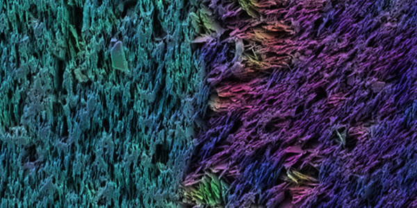
Cross section of an artificial tooth under an electron microscope (pseudocolor). Ceramic platelets are oriented vertically in the enamel and horizontally in the dentin. (credit: Hortense Le Ferrand/ETH Zürich)

A new procedure that can mimic the complex fine structure of biological composite materials, such as teeth or seashells, has been developed by ETH Zurich researchers. It could allow for creating synthetic materials that are as hard and tough as their natural counterparts.

The secret of these hard natural biomaterials is in their unique fine structure: they are composed of different layers in which numerous micro-platelets are joined together, aligned in identical orientation in each layer.

Although methods exist that allow material scientists to imitate nacre (mother of pearl), it was a challenge to create a material that imitates the entire seashell, with comparable properties and structural complexity, according to the researchers, led by André Studart, Professor of Complex Materials.

The new procedure mimics the natural model almost perfectly. It recreates the multiple layers of micro-platelets with identical orientation in each layer in a single complex piece by using a “magnetically assisted slip casting” (MASC) procedure.

How to create a tooth

Here’s how the procedure works:

  1. Create a plaster cast to serve as a mold.
  2. Pour in a suspension containing magnetized ceramic platelets, such as aluminum oxide platelets. The pores of the plaster mold slowly absorb the liquid from the suspension, which causes the material to solidify and to harden from the outside in.
  3. Create an ordered layer-like structure by applying a magnetic field during the casting process, changing its orientation at regular intervals. As long as the material remains liquid, the ceramic platelets align to the magnetic field. In the solidified material, the platelets retain their orientation.

This continuous process can produce multiple layers with differing material properties in a single object and are almost perfect imitations of their natural models, such as nacre or tooth enamel, says Florian Bouville, a post-doc and co-lead author of the study, which is published in the journal Nature Materials. “Our technique is similar to 3-D printing, but 10 times faster and much more cost-effective.”

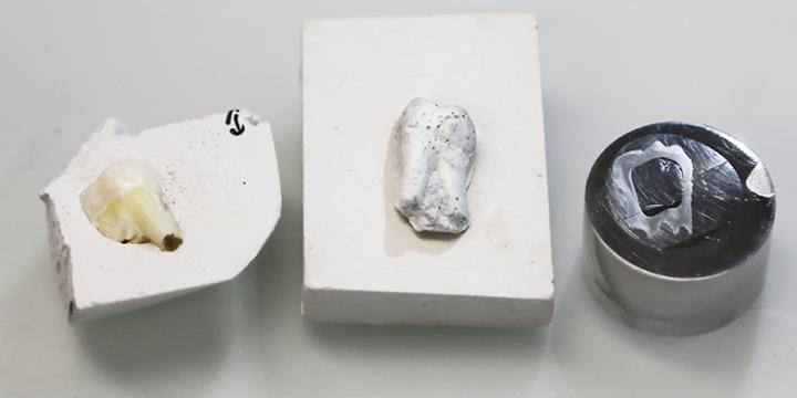
The left structure is showing the natural tooth in its gypsum mold. The middle structure is the artificial tooth (sintered but not yet polymer infiltrated). The model on the right has been sintered and polymer infiltrated. It is embedded in a “puck” to enable polishing and coated with platinum to prevent charging in the electron microscope. (credit: Tobias Niebel/ETH Zurich)

To demonstrate the process, Studart’s research group produced an artificial tooth with a microstructure that mimics that of a real tooth. The surface of the artificial tooth is as hard and structurally complex as real tooth enamel, while the layer beneath is as tough as the dentine of the natural model.

They began by creating a plaster cast of a human wisdom tooth. They then filled this mold with a suspension containing aluminum oxide platelets and glass nanoparticles as mortar. Using a magnet, they aligned the platelets perpendicular to the surface of the object. Once the first layer was dry, the scientists poured a second suspension without glass particles into the same mold. The aluminium oxide platelets in the second layer were aligned horizontally to the surface of the tooth using the magnet.

This double-layered structure was then sintered (“fired” in a kiln) at 1,600 degrees C to densify and harden the material. Finally, the researchers filled the pores that remained after the sintering with a synthetic monomer used in dentistry, which subsequently polymerized (formed into a complex material).

Artificial teeth that behave just like the real thing

“The profile of hardness and toughness obtained from the artificial tooth corresponds exactly with that of a natural tooth,” says Studart.

The current study is an initial proof-of-concept, which shows that the natural fine structure of a tooth can be reproduced in the laboratory, he says. “The appearance of the material has to be significantly improved before it can be used for dental prostheses.

He noted that the base substances and the orientation of the platelets can be combined as required, “which rapidly and easily makes a wide range of different material types with varying properties feasible.” For example, copper platelets could be used in place of aluminum oxide platelets, allowing for use in electronics.

One part of the MASC process, the magnetization and orientation of the ceramic platelets, has been patented.


Abstract of Magnetically assisted slip casting of bioinspired heterogeneous composites

Natural composites are often heterogeneous to fulfil functional demands. Manufacturing analogous materials remains difficult, however, owing to the lack of adequate and easily accessible processing tools. Here, we report an additive manufacturing platform able to fabricate complex-shaped parts exhibiting bioinspired heterogeneous microstructures with locally tunable texture, composition and properties, as well as unprecedentedly high volume fractions of inorganic phase (up to 100%). The technology combines an aqueous-based slip-casting process with magnetically directed particle assembly to create programmed microstructural designs using anisotropic stiff platelets in a ceramic, metal or polymer functional matrix. Using quantitative tools to control the casting kinetics and the temporal pattern of the applied magnetic fields, we demonstrate that this approach is robust and can be exploited to design and fabricate heterogeneous composites with thus far inaccessible microstructures. Proof-of-concept examples include bulk composites with periodic patterns of microreinforcement orientation, and tooth-like bilayer parts with intricate shapes exhibiting site-specific composition and texture.

A new class of anti-obesity compounds with potential anti-diabetic properties

Prevalence of Self-Reported Obesity Among U.S. Adults by State and Territory, BRFSS, 2014 (credit: Behavorial Risk Factor Surveillance System/CDC)

A molecule known as MnTBAP* has rapidly reversed obesity in mice and could be effective for humans in the future, according to researchers from Skidmore College and the Perelman School of Medicine at the University of Pennsylvania.

“In the span of a month, mice with pre-existing obesity lost 20 percent of their body weight and about 50 percent of their fat mass,” said Thomas H. Reynolds, PhD., an associate professor of Health and Exercise Sciences at Skidmore College. The weight loss is explained partly by a decrease in food consumption, but other mechanisms are also at play, according to the study published Wednesday (Sept. 23, 2015) in an open-access paper in PLOS One.

The authors report that MnTBAP also has beneficial effects on type 2 diabetes by improving insulin action in muscle and fat. Insulin is the hormone that allows tissues to take up glucose. “In type 2 diabetics, insulin action is impaired, causing the pancreas to go into overdrive in an attempt to maintain normal blood glucose levels.

“Over time, the pancreas becomes exhausted and can’t keep up, leading to rising blood glucose levels and the development of diabetes,” Jonathan Brestoff, the study’s first author, said in an exclusive interview with KurzweilAI. Brestoff is now at the Perelman School of Medicine at the University of Pennsylvania.

He said this molecule represents “a new class of anti-obesity compounds with potential anti-diabetic properties.” He has co-founded the biotech company Symmetry Therapeutics, Inc. to translate the team’s science into clinical applications to treat obesity via an anti-obesity compound called SYM401.

Symmetry is taking an unusual approach with its drug development by crowdfunding part of its research on the platform IndieGoGo. Brestoff said their efforts have gained international attention for attempting to add transparency to an otherwise secretive industry, and have attracted donations and private investors. Their campaign ends October 6, 2015.

On Monday Sept. 21, the CDC published new statistics on obesity in the United States indicating that it remains one of the biggest public health problems facing this country.

* Also known as Manganese [III] 5,10,15,20-tetrakis benzoic acid porphyrin.


Abstract of Manganese [III] Tetrakis [5,10,15,20]-Benzoic Acid Porphyrin Reduces Adiposity and Improves Insulin Action in Mice with Pre-Existing Obesity

The superoxide dismutase mimetic manganese [III] tetrakis [5,10,15,20]-benzoic acid porphyrin (MnTBAP) is a potent antioxidant compound that has been shown to limit weight gain during short-term high fat feeding without preventing insulin resistance. However, whether MnTBAP has therapeutic potential to treat pre-existing obesity and insulin resistance remains unknown. To investigate this, mice were treated with MnTBAP or vehicle during the last five weeks of a 24-week high fat diet (HFD) regimen. MnTBAP treatment significantly decreased body weight and reduced white adipose tissue (WAT) mass in mice fed a HFD and a low fat diet (LFD). The reduction in adiposity was associated with decreased caloric intake without significantly altering energy expenditure, indicating that MnTBAP decreases adiposity in part by modulating energy balance. MnTBAP treatment also improved insulin action in HFD-fed mice, a physiologic response that was associated with increased protein kinase B (PKB) phosphorylation and expression in muscle and WAT. Since MnTBAP is a metalloporphyrin molecule, we hypothesized that its ability to promote weight loss and improve insulin sensitivity was regulated by heme oxygenase-1 (HO-1), in a similar fashion as cobalt protoporphyrins. Despite MnTBAP treatment increasing HO-1 expression, administration of the potent HO-1 inhibitor tin mesoporphyrin (SnMP) did not block the ability of MnTBAP to alter caloric intake, adiposity, or insulin action, suggesting that MnTBAP influences these metabolic processes independent of HO-1. These data demonstrate that MnTBAP can ameliorate pre-existing obesity and improve insulin action by reducing caloric intake and increasing PKB phosphorylation and expression.

Smart robot accelerates cancer treatment research by finding optimal treatment combinations

Iterative search for anti-cancer drug combinations. The procedure starts by generating an initial generation (population) of drug combinations randomly or guided by biological prior knowledge and assumptions. In each iteration the aim is to propose a new generation of drug combinations based on the results obtained so far. The procedure iterates through a number of generations until a stop criterion for a predefined fitness function is satisfied. (credit: M. Kashif et al./Scientific Reports)

A new smart research system developed at Uppsala University accelerates research on cancer treatments by finding optimal treatment drug combinations. It was developed by a research group led by Mats Gustafsson, Professor of Medical Bioinformatics.

The “lab robot” system plans and conducts experiments with many substances, and draws its own conclusions from the results. The idea is to gradually refine combinations of substances so that they kill cancer cells without harming healthy cells.

Instead of just combining a couple of substances at a time, the new lab robot can handle about a dozen drugs simultaneously. The future aim is to handle many more, preferably hundreds.

There are a few such laboratories in the world with this type of lab robot, but researchers “have only used the systems to look for combinations that kill the cancer cells, not taking the side effects into account,” says Gustafsson.

The next step: make the robot system more automated and smarter. The scientists also want to build more knowledge into the guiding algorithm of the robot, such as prior knowledge about drug targets and disease pathways.

For patients with the same cancer type returning multiple times, sometimes the cancer cells develop resistance against the pharmacotherapy used. The new robot systems may also become important in the efforts to find new drug compounds that make these resistant cells sensitive again.

The research is described in an open-access article published Tuesday (Sept. 22, 2015) in Scientific Reports.


Abstract of In vitro discovery of promising anti-cancer drug combinations using iterative maximisation of a therapeutic index

In vitro-based search for promising anti-cancer drug combinations may provide important leads to improved cancer therapies. Currently there are no integrated computational-experimental methods specifically designed to search for combinations, maximizing a predefined therapeutic index (TI) defined in terms of appropriate model systems. Here, such a pipeline is presented allowing the search for optimal combinations among an arbitrary number of drugs while also taking experimental variability into account. The TI optimized is the cytotoxicity difference (in vitro) between a target model and an adverse side effect model. Focusing on colorectal carcinoma (CRC), the pipeline provided several combinations that are effective in six different CRC models with limited cytotoxicity in normal cell models. Herein we describe the identification of the combination (Trichostatin A, Afungin, 17-AAG) and present results from subsequent characterisations, including efficacy in primary cultures of tumour cells from CRC patients. We hypothesize that its effect derives from potentiation of the proteotoxic action of 17-AAG by Trichostatin A and Afungin. The discovered drug combinations against CRC are significant findings themselves and also indicate that the proposed strategy has great potential for suggesting drug combination treatments suitable for other cancer types as well as for other complex diseases.

3D-printed silicone guide with chemical cues helps regenerate complex nerves after injury

3-D scans of a nerve from different angles are used to create a custom regeneration guide for complex nerves (credit: University of Minnesota)

A national team of researchers used a combination of 3-D imaging and 3-D printing techniques to create a custom silicone guide implanted with biochemical cues to help nerve regeneration after an injury.

Nerve regeneration is a complex process, which is why regrowth of nerves after injury or disease is very rare and often permanent, according to the Mayo Clinic.

As a test, the researchers used a 3-D scanner to reverse-engineer the structure of a rat’s sciatic nerve. They then used a specialized, custom-built 3-D printer to print a regeneration guide containing 3D-printed chemical cues to promote both motor and sensory nerve regeneration within the same structure. The guide was then implanted into the rat by surgically grafting it to the cut ends of the nerve. Within about 10 to 12 weeks, the rat’s ability to walk again was improved.

A 3D-printed complex nerve-regeneration pathway implanted in a rat helped to improve walking in 10 to 12 weeks after implantation (credit: University of Minnesota)

“Someday we hope that we could have a 3D scanner and printer right at the hospital to create custom nerve guides right on site to restore nerve function,” said University of Minnesota mechanical engineering professor Michael McAlpine, the study’s lead researcher.

Conventional nerve guidance channels are typically fabricated around cylindrical substrates, so the resulting guidance devices are limited to linear structures. This is is the first time a study has shown the creation of a custom guide for regrowth of a complex nerve like the Y-shaped sciatic nerve, which has both sensory and motor branches.

“The exciting next step would be to implant these guides in humans rather than rats,” McAlpine said. For cases where a patient’s nerve is unavailable for scanning, McAlpine said there could someday be a “library” of scanned nerves from other people or cadavers that hospitals could use to create closely matched 3D-printed guides for patients.

The study by researchers from the University of Minnesota, Virginia Tech, University of Maryland, Princeton University, and Johns Hopkins University was published Thursday (Sept. 17) in the journal Advanced Functional Materials.


UMN College of Science and Engineering | 3D printing of a nerve regeneration guide [no audio]


Abstract of 3D Printed Anatomical Nerve Regeneration Pathways

A 3D printing methodology for the design, optimization, and fabrication of a custom nerve repair technology for the regeneration of complex peripheral nerve injuries containing bifurcating sensory and motor nerve pathways is introduced. The custom scaffolds are deterministically fabricated via a microextrusion printing principle using 3D models, which are reverse engineered from patient anatomies by 3D scanning. The bifurcating pathways are augmented with 3D printed biomimetic physical cues (microgrooves) and path-specific biochemical cues (spatially controlled multicomponent gradients). In vitro studies reveal that 3D printed physical and biochemical cues provide axonal guidance and chemotractant/chemokinetic functionality. In vivo studies examining the regeneration of bifurcated injuries across a 10 mm complex nerve gap in rats showed that the 3D printed scaffolds achieved successful regeneration of complex nerve injuries, resulting in enhanced functional return of the regenerated nerve. This approach suggests the potential of 3D printing toward advancing tissue regeneration in terms of: (1) the customization of scaffold geometries to match inherent tissue anatomies; (2) the integration of biomanufacturing approaches with computational modeling for design, analysis, and optimization; and (3) the enhancement of device properties with spatially controlled physical and biochemical functionalities, all enabled by the same 3D printing process.

Low vitamin D associated with faster decline in cognitive function

(Credit: iStock)

A research team has found that Vitamin D insufficiency was associated with faster decline in cognitive functions among a group of ethnically diverse older adults, according to an open-access paper published in JAMA Neurology.*

According to the researchers — Joshua W. Miller, Ph.D., of Rutgers University, New Brunswick, N.J., and coauthors from the University of California, Davis — Vitamin D may influence all organ systems, not just calcium absorption and bone health.

Both the vitamin D receptor and the enzyme that converts 25-hydroxyvitamin D (25-OHD) to the active form of the vitamin are expressed in all human organs, including the brain. So research has increasingly examined the association between vitamin D status and a variety of health outcomes, including dementia and age-associated cognitive decline.

JAMA |Vitamin D and Cognitive Decline in Multiethnic Cohort of Older Adults

The authors report:

  • The average 25-OHD level among participants was 19.2 ng/mL, with 26.2 percent of participants being vitamin D deficient and 35.1 percent vitamin D insufficient.
  • Average 25-OHD levels were lower for African American and Hispanic participants compared with their white counterparts (17.9, 17.2 and 21.7 ng/mL, respectively).
  • Average 25-OHD levels were lower in the dementia group compared with mild cognitive impairment and cognitively normal groups (16.2, 20.0 and 19.7 ng/mL, respectively.
  • During an average follow-up of 4.8 years, rates of decline in episodic memory and executive function among vitamin D deficient and vitamin D insufficient participants were greater than those with adequate vitamin D status after adjusting for a variety of patient factors.
  • Vitamin D status was not significantly associated with decline in semantic memory or visuospatial ability.

The authors note limitations to their study including that they did not directly measure dairy intake, sun exposure or exercise, each of which can influence vitamin D levels.

“Our data support the common occurrence of VitD [vitamin D] insufficiency among older individuals. In addition, these data show that African American and Hispanic individuals are more likely to have VitD insufficiency or deficiency.

“Independent of race or ethnicity, baseline cognitive ability, and a host of other risk factors, VitD insufficiency was associated with significantly faster declines in both episodic memory and executive function performance, which may correspond to elevated risk for incident AD [Alzheimer disease] dementia.

“Given that VitD insufficiency is medically correctable, well-designed clinical trials that emphasize enrollment of individuals of nonwhite race/ethnicity with hypovitaminosis D could be useful for testing the effect of VitD replacement on dementia prevention,” the study concludes.

* The researchers examined baseline vitamin D status and change in subdomains of cognitive function as measured on assessment scales in an ethnically diverse group of 382 older adults.

Serum (blood) 25-OHD was measured and vitamin D status was defined as follows: deficient was less than 12 ng/mL; insufficient was 12 to less than 20 ng/mL; adequate was 20 to less than 50 ng/mL; and high was 50 ng/mL or higher.

Study participants were an average age of 75.5 years and nearly 62 percent were female, while 41.4 percent of the group was white, 29.6 percent were African American and 25.1 percent were Hispanic. At study enrollment, 17.5 percent of the participants had dementia, 32.7 percent had mild cognitive impairment and 49.5 percent were cognitively normal.


Abstract of Vitamin D Status and Rates of Cognitive Decline in a Multiethnic Cohort of Older Adults

Importance:  Vitamin D (VitD) deficiency is associated with brain structural abnormalities, cognitive decline, and incident dementia.

Objective:  To assess associations between VitD status and trajectories of change in subdomains of cognitive function in a cohort of ethnically diverse older adults.

Design, Setting, and Participants:  Longitudinal multiethnic cohort study of 382 participants in an outpatient clinic enrolled between February 2002 and August 2010 with baseline assessment and yearly follow-up visits. Serum 25-hydroxyvitamin D (25-OHD) was measured, with VitD status defined as the following: deficient, less than 12 ng/mL (to convert to nanomoles per liter, multiply by 2.496); insufficient, 12 to less than 20 ng/mL; adequate, 20 to less than 50 ng/mL; or high, 50 ng/mL or higher. Subdomains of cognitive function were assessed using the Spanish and English Neuropsychological Assessment Scales. Associations were evaluated between 25-OHD levels (as continuous and categorical [deficient, insufficient, or adequate]) and trajectories of cognitive decline.

Main Outcomes and Measures:  Serum 25-OHD levels, cognitive function, and associations between 25-OHD levels and trajectories of cognitive decline.

Results:  Participants (N = 382 at baseline) had a mean (SD) age of 75.5 (7.0) years; 61.8% were women; and 41.4% were white, 29.6% African American, 25.1% Hispanic, and 3.9% other race/ethnicity. Diagnosis at enrollment included 17.5% with dementia, 32.7% with mild cognitive impairment, and 49.5% cognitively normal. The mean (SD) 25-OHD level was 19.2 (11.7) ng/mL, with 26.2% of participants being VitD deficient and 35.1% insufficient. The mean (SD) 25-OHD levels were significantly lower for African American and Hispanic participants compared with white participants (17.9 [15.8] and 17.2 [8.4] vs 21.7 [10.0] ng/mL, respectively; P < .001 for both). The mean (SD) 25-OHD levels were similarly lower in the dementia group compared with the mild cognitive impairment and cognitively normal groups (16.2 [9.4] vs 20.0 [10.3] and 19.7 [13.1] ng/mL, respectively; P = .006). The mean (SD) follow-up was 4.8 (2.5) years. Rates of decline in episodic memory and executive function among VitD-deficient (episodic memory: β = −0.04 [SE = 0.02], P = .049; executive function: β = −0.05 [SE = 0.02], P = .01) and VitD-insufficient (episodic memory: β = −0.06 [SE = 0.02], P < .001; executive function: β = −0.04 [SE = 0.02], P = .008) participants were greater than those with adequate status after controlling for age, sex, education, ethnicity, body mass index, season of blood draw, vascular risk, and apolipoprotein E4 genotype. Vitamin D status was not significantly associated with decline in semantic memory or visuospatial ability. Exclusion of participants with dementia did not substantially affect the associations between VitD status and rates of cognitive decline.

Conclusions and Relevance:  Low VitD status was associated with accelerated decline in cognitive function domains in ethnically diverse older adults, including African American and Hispanic individuals who exhibited a high prevalence of VitD insufficiency or deficiency. It remains to be determined whether VitD supplementation slows cognitive decline.

Vitamin D3 sources and dosage (update Sept. 17, 2015)

The U.S. National Institutes of Health explains that Vitamin D can be obtained from “sun exposure, food, and supplements,” notes food sources, and makes recommended dietary allowances (RDA). Some sources, such as “The Real RDA for Vitamin D Is 10 Times Higher Than Currently Recommended,” recommend higher RDA.

Controlling brain cells with ultrasound

For the first time, sound waves are used to control brain cells. Salk scientists developed the new technique, dubbed sonogenetics, to selectively and noninvasively turn on groups of neurons in worms that could be a boon to science and medicine. (credit: Salk Institute)

Salk scientists have developed a new method, dubbed sonogenetics, to selectively activate brain, heart, muscle and other cells using ultrasonic waves (the same type of waves used in medical sonograms).

This new method may have advantages over the similar light-based approach known as optogenetics, particularly for human therapeutics. It is described today (Sept. 15, 2015) in the journal Nature Communications.

Experiment setup. (Left) Schematic of the computer-controlled imaging and ultrasound exposure system. (Right) Top-down view of petri dish with agar (food) plate with animals corralled into a small area by a copper barrier. (credit: Stuart Ibsen et al./Nature Communications)

Sreekanth Chalasani, an assistant professor in Salk’s Molecular Neurobiology Laboratory and senior author of the study, and his colleagues activated neurons in the nematode C. elegans that don’t usually react to ultrasound.

They found that microbubbles of gas outside of the worm were able to amplify the low-intensity ultrasound waves. “The microbubbles grow and shrink in [sync] with the ultrasound pressure waves,” Ibsen says. “These oscillations can then propagate noninvasively into the worm.”

They also found that when the ultrasound hits gas bubbles, they cause a membrane ion channel, TRP-4, to open up and activate the cell. Armed with that knowledge, the team tried adding the TRP-4 channel to neurons that don’t normally have it.

Possible human uses next

Worm exhibits reversals and omega bends upon ultrasound stimulus (single 10 ms pulse at 2.25 MHz with peak negative pressure of 0.9 MPa) in the presence of micro bubbles (credit: Stuart Ibsen et al./Nature Communications)

So far, sonogenetics has only been applied to C. elegans neurons. But TRP-4 could be added to any calcium-sensitive cell type in any organism including humans, Chalasani says. Here’s how it would work:

  1. The microbubbles are injected into the bloodstream, and are distributed throughout the body —- an approach already used in some human imaging techniques.
  2. Ultrasound could then noninvasively reach any tissue of interest, including the brain, be amplified by the microbubbles, and activate the cells of interest through TRP-4. (Many cells in the human body, he points out, can respond to the influxes of calcium caused by TRP-4.)

The researchers have already begun testing the approach in mice.

Both optogenetics and sonogenetics approaches, Chalasani says, hold promise in basic research by letting scientists study the effect of cell activation. And they also may be useful in therapeutics through the activation of cells affected by disease.

However, for either technique to be used in humans, researchers first need to develop safe ways to deliver the light or ultrasound-sensitive channels to target cells.

The work and the researchers involved were supported by a Salk Institute Pioneer Fund Postdoctoral Fellowship, a Salk Institute Innovation Grant, the Rita Allen Foundation, the W.M. Keck Foundation and the National Institutes of Health. A University of California, San Diego researcher was also involved in the research.


Abstract of Sonogenetics is a non-invasive approach to activating neurons in Caenorhabditis elegans

A major challenge in neuroscience is to reliably activate individual neurons, particularly those in  deeper brain regions. Current optogenetic approaches require invasive surgical procedures to  deliver light of specific wavelengths to target cells in order to activate or silence them. Here, we demonstrate the use of low-pressure ultrasound as a non-invasive trigger to activate specific ultrasonically-sensitized neurons in the nematode, Caenorhabditis elegans. We first show that wild-type animals are insensitive to low pressure ultrasound and require gas-filled microbubbles to transduce the ultrasound wave. We find that neuron-specific misexpression of TRP-4, the pore-forming subunit of a mechanotransduction channel, sensitizes neurons to ultrasound stimulus resulting in motor outputs. Furthermore, we use this approach to manipulate the  function of sensory neurons and interneurons and identify a role for the PVD sensory neurons in modifying locomotory behaviors. We suggest this method can be broadly applied to manipulate cellular functions in vivo.

Cocoa flavanols lower blood pressure and increase blood-vessel function in healthy people

Two recently published studies in the journals Age and the British Journal of Nutrition (BJN) demonstrate that consuming cocoa flavanols improves cardiovascular function and lessens the burden on the heart that comes with the aging and stiffening of arteries, while reducing the risk of developing cardiovascular disease (CVD)

As we age, our blood vessels become less flexible and less able to expand to let blood flow and circulate normally, and the risk of hypertension also increases. Arterial stiffness and blood vessel dysfunction are linked with cardiovascular disease — the number one cause of deaths worldwide.

Cocoa flavanols increase blood vessel flexibility and lower blood pressure

Cocoa pods (credit: Wikimedia Commons)

Cocoa flavanols are plant-derived bioactives from the cacao bean. Dietary intake of flavanols has been shown to have a beneficial effect on cardiovascular health, but the compounds are often destroyed during normal food processing.

Earlier studies have demonstrated that cocoa flavanol intake improves the elasticity of blood vessels and lowers blood pressure.

But, for the most part, these investigations have focused on high-risk individuals like smokers and people that have already been diagnosed with conditions like hypertension and coronary heart disease.

These two studies are the first to look at the different effects dietary cocoa flavanols can have on the blood vessels of healthy, low-risk individuals with no signs or symptoms of cardiovascular disease.

In the study* published in Age, they found that vasodilation was significantly improved in both age groups that consumed flavanols over the course of the study (by 33% in the younger age group and 32% in the older age group over the control intervention).

In the older age group, a statistically and clinically significant decrease in systolic blood pressure of 4 mmHg over control was also seen.

Improving cardiovascular health and lowering the risk of CVD

In the second study**, published in BJN, the researchers extended their investigations to a larger group (100) of healthy middle-aged men and women (35–60 years) with low risk of CVD.

“We found that intake of flavanols significantly improves several of the hallmarks of cardiovascular health,” says Professor Kelm. In particular, the researchers found that consuming flavanols for four weeks significantly increased flow-mediated vasodilation by 21%.

Increased flow-mediated vasodilation is a sign of improved endothelial function and has been shown by some studies to be associated with decreased risk of developing CVD. In addition, taking flavanols decreased blood pressure (systolic by 4.4 mmHg, diastolic by 3.9 mmHg), and improved the blood cholesterol profile by decreasing total cholesterol (by 0.2 mmol/L), decreasing LDL cholesterol (by 0.17 mmol/L), and increasing HDL cholesterol (by 0.1 mmol/L).

Data source: United Nations Dept. of Economic and Social Affairs Population Division (2014). World Population Ageing 2013 (credit: MARS Cocoa Health Science)

The researchers also calculated the Framingham Risk Score — a widely used model to estimate the 10-year cardiovascular risk of an individual — and found that flavanol intake reduced the risk of CVD. “Our results indicate that dietary flavanol intake reduces the 10-year risk of being diagnosed with CVD by 22% and the 10-year risk of suffering a heart attack by 31%,” says Professor Kelm.

The combined results of these studies demonstrate that flavanols are effective at mitigating age-related changes in blood vessels, and could thereby reduce the risk of CVD in healthy individuals.

The application of 10-year Framingham Risk Scores should be interpreted with caution as the duration of the BJN study was weeks not years and the number of participants was around 100, not reaching the scale of the Framingham studies.

Other longer-term studies, such as the 5-year COcoa Supplement and Multivitamin Outcomes Study (COSMOS) of 18,000 men and women, are now underway to investigate the health potential of flavanols on a much larger scale.

Poor diet and high blood pressure now number one risk factors for death

A related huge international study of global causes of death has revealed high blood pressure is the number one individual risk factor associated with Australian deaths, contributing to 28,500 deaths a year.

Smoking and high body mass index are number two and three respectively, while drug use is among the fastest growing risk factors for poor health in Australia, up 53 per cent between 1990 and 2013.

However, deaths from high cholesterol have decreased by 25 per cent, and deaths from diets low in fruit and vegetables have decreased by 10 per cent.

The finding is from a new analysis of global cause-of-death data by the University of Melbourne and the University of Washington published in The Lancet last week.

Researchers looked at 79 risk factors for death in 188 countries between 1990 and 2013.

The risk factors examined in the study contributed to almost 31 million deaths worldwide in 2013, up from 25 million deaths in 1990. The top risks associated with the deaths of both men and women in Australia are high blood pressure, smoking, high body mass index, and high fasting plasma glucose.

Top risk factors for the rest of the world include:

  • In much of the Middle East and Latin America, high body mass index is the number-one risk associated with health loss.
  • In South and Southeast Asia, household air pollution is a leading risk, and India also grapples with high risks of unsafe water and childhood under-nutrition.
  • Alcohol is the number-two risk in Russia.
  • Smoking is the number-one risk in many high-income countries, including the United Kingdom.
  • The most marked differences are found in sub-Saharan Africa, which, unlike other regions, is dominated by a combination of childhood malnutrition, unsafe water and lack of sanitation, unsafe sex, and alcohol use.
  • Wasting (low weight) accounts for one in five deaths of children under five-years-old, highlighting the importance of child malnutrition as a risk factor.
  • Unsafe sex took a huge toll on global health, contributing to 82 per cent of HIV/AIDS deaths and 94 per cent of HIV/AIDS deaths among 15- to 19-year-olds in 2013. This has a greater impact on South Africa than any other country, 38 per cent of South African deaths were attributed to unsafe sex. The global burden of unsafe sex grew from 1990 and peaked in 2005.

The study included several risk factors in its analysis for the first time: wasting (low weight for a person’s height), stunting (low height for a person’s age), unsafe sex, HIV, no hand-washing with soap, intimate partner violence.

In Australia, increases in deaths due to high body mass index and diabetes-related illnesses have been increased 35 per cent and 47 per cent respectively. Australians are also grappling with poor kidney function and low physical activity, both of which are not among the top-10 global risk factors.

The leading risk factors associated with poor health in Australia in 2013 were high body mass index, smoking, and high blood pressure. While these were also in the top-five risk factors in 1990, smoking has decreased slightly, by 4 per cent.

Senior author on the study, University of Melbourne Professor Alan Lopez, said many of these risk factors for Australian deaths are preventable with lifestyle changes.

“While our study shows that public policy in Australia has been effective in reducing the health impacts of high cholesterol and insufficient fruit and vegetables in our diet, progress against some large, avoidable risks has been less impressive,” Prof Lopez said.

“Smoking, high blood pressure and obesity are still prevalent among adult Australians and remain a large cause of disease burden. We can, and ought, to be more conscientious in reducing these exposures among all Australians, not only those considered at high risk.”

The study was conducted by an international consortium of researchers working on the Global Burden of Disease project and led by the Institute for Health Metrics and Evaluation (IHME) at the University of Washington.

* Two groups of 22 young (<35 years of age) and 20 older (50-80 years of age) healthy men consumed either a flavanol-containing drink, or a flavanol-free control drink, twice a day for two weeks. The researchers then measured the effect of flavanols on hallmarks of cardiovascular aging, such as arterial stiffness (as measured by pulse wave velocity), blood pressure and flow-mediated vasodilation (the extent to which blood vessels dilate in response to nitric oxide).

** The participants were randomly and blindly assigned into groups that consumed either a flavanol-containing drink or a flavanol-free control drink, twice a day for four weeks. The researchers also measured cholesterol levels in the study groups, in addition to vasodilation, arterial stiffness and blood pressure.


Abstract of Cocoa flavanol intake improves endothelial function and Framingham Risk Score in healthy men and women: a randomised, controlled, double-masked trial: the Flaviola Health Study

Cocoa flavanol (CF) intake improves endothelial function in patients with cardiovascular risk factors and disease. We investigated the effects of CF on surrogate markers of cardiovascular health in low risk, healthy, middle-aged individuals without history, signs or symptoms of CVD. In a 1-month, open-label, one-armed pilot study, bi-daily ingestion of 450 mg of CF led to a time-dependent increase in endothelial function (measured as flow-mediated vasodilation (FMD)) that plateaued after 2 weeks. Subsequently, in a randomised, controlled, double-masked, parallel-group dietary intervention trial (Clinicaltrials.gov: NCT01799005), 100 healthy, middle-aged (35–60 years) men and women consumed either the CF-containing drink (450 mg) or a nutrient-matched CF-free control bi-daily for 1 month. The primary end point was FMD. Secondary end points included plasma lipids and blood pressure, thus enabling the calculation of Framingham Risk Scores and pulse wave velocity. At 1 month, CF increased FMD over control by 1·2 % (95 % CI 1·0, 1·4 %). CF decreased systolic and diastolic blood pressure by 4·4 mmHg (95 % CI 7·9, 0·9 mmHg) and 3·9 mmHg (95 % CI 6·7, 0·9 mmHg), pulse wave velocity by 0·4 m/s (95 % CI 0·8, 0·04 m/s), total cholesterol by 0·20 mmol/l (95 % CI 0·39, 0·01 mmol/l) and LDL-cholesterol by 0·17 mmol/l (95 % CI 0·32, 0·02 mmol/l), whereas HDL-cholesterol increased by 0·10 mmol/l (95 % CI 0·04, 0·17 mmol/l). By applying the Framingham Risk Score, CF predicted a significant lowering of 10-year risk for CHD, myocardial infarction, CVD, death from CHD and CVD. In healthy individuals, regular CF intake improved accredited cardiovascular surrogates of cardiovascular risk, demonstrating that dietary flavanols have the potential to maintain cardiovascular health even in low-risk subjects.


Abstract of Global, regional, and national comparative risk assessment of 79 behavioural, environmental and occupational, and metabolic risks or clusters of risks in 188 countries, 1990–2013: a systematic analysis for the Global Burden of Disease Study 2013

Background: The Global Burden of Disease, Injuries, and Risk Factor study 2013 (GBD 2013) is the first of a series of annual updates of the GBD. Risk factor quantification, particularly of modifiable risk factors, can help to identify emerging threats to population health and opportunities for prevention. The GBD 2013 provides a timely opportunity to update the comparative risk assessment with new data for exposure, relative risks, and evidence on the appropriate counterfactual risk distribution.

Methods: Attributable deaths, years of life lost, years lived with disability, and disability-adjusted life-years (DALYs) have been estimated for 79 risks or clusters of risks using the GBD 2010 methods. Risk–outcome pairs meeting explicit evidence criteria were assessed for 188 countries for the period 1990–2013 by age and sex using three inputs: risk exposure, relative risks, and the theoretical minimum risk exposure level (TMREL). Risks are organised into a hierarchy with blocks of behavioural, environmental and occupational, and metabolic risks at the first level of the hierarchy. The next level in the hierarchy includes nine clusters of related risks and two individual risks, with more detail provided at levels 3 and 4 of the hierarchy. Compared with GBD 2010, six new risk factors have been added: handwashing practices, occupational exposure to trichloroethylene, childhood wasting, childhood stunting, unsafe sex, and low glomerular filtration rate. For most risks, data for exposure were synthesised with a Bayesian meta-regression method, DisMod-MR 2.0, or spatial-temporal Gaussian process regression. Relative risks were based on meta-regressions of published cohort and intervention studies. Attributable burden for clusters of risks and all risks combined took into account evidence on the mediation of some risks such as high body-mass index (BMI) through other risks such as high systolic blood pressure and high cholesterol.

Findings: All risks combined account for 57·2% (95% uncertainty interval [UI] 55·8–58·5) of deaths and 41·6% (40·1–43·0) of DALYs. Risks quantified account for 87·9% (86·5–89·3) of cardiovascular disease DALYs, ranging to a low of 0% for neonatal disorders and neglected tropical diseases and malaria. In terms of global DALYs in 2013, six risks or clusters of risks each caused more than 5% of DALYs: dietary risks accounting for 11·3 million deaths and 241·4 million DALYs, high systolic blood pressure for 10·4 million deaths and 208·1 million DALYs, child and maternal malnutrition for 1·7 million deaths and 176·9 million DALYs, tobacco smoke for 6·1 million deaths and 143·5 million DALYs, air pollution for 5·5 million deaths and 141·5 million DALYs, and high BMI for 4·4 million deaths and 134·0 million DALYs. Risk factor patterns vary across regions and countries and with time. In sub-Saharan Africa, the leading risk factors are child and maternal malnutrition, unsafe sex, and unsafe water, sanitation, and handwashing. In women, in nearly all countries in the Americas, north Africa, and the Middle East, and in many other high-income countries, high BMI is the leading risk factor, with high systolic blood pressure as the leading risk in most of Central and Eastern Europe and south and east Asia. For men, high systolic blood pressure or tobacco use are the leading risks in nearly all high-income countries, in north Africa and the Middle East, Europe, and Asia. For men and women, unsafe sex is the leading risk in a corridor from Kenya to South Africa.

Interpretation: Behavioural, environmental and occupational, and metabolic risks can explain half of global mortality and more than one-third of global DALYs providing many opportunities for prevention. Of the larger risks, the attributable burden of high BMI has increased in the past 23 years. In view of the prominence of behavioural risk factors, behavioural and social science research on interventions for these risks should be strengthened. Many prevention and primary care policy options are available now to act on key risks.

Funding: Bill & Melinda Gates Foundation.
‘Lab-on-a-Chip’ microfluidics technology may cut costs of lab tests for diseases and disorders

The Rutgers lab-on-a chip is three inches long and an inch wide — the size of a glass microscope slide (credit: Mehdi Ghodbane)

Rutgers engineers have developed a breakthrough microfluidics device that can significantly reduce the cost of sophisticated lab tests while using 90 percent less sample fluid than needed in conventional tests.

It uses miniaturized channels and valves to replace “benchtop” assays — tests that require large samples of blood or other fluids and expensive chemicals that lab technicians manually mix in trays of tubes or plastic plates with cup-like depressions.

The device also requires only one-tenth of the chemicals used in a conventional multiplex immunoassay, which can cost as much as $1500, and it automates much of the skilled labor involved in performing tests, according to Mehdi Ghodbane, who earned his doctorate in biomedical engineering at Rutgers and now works in biopharmaceutical research and development at GlaxoSmithKline.

Ghodbane and six Rutgers researchers recently published their results in the Royal Society of Chemistry’s journal, Lab on a Chip.

A schematic diagram of the Rutgers lab-on-a-chip (credit: Mehdi Ghodbane)

“The results are as sensitive and accurate as the standard benchtop assay,’’ said Martin Yarmush, the Paul and Mary Monroe Chair and Distinguished Professor of biomedical engineering at Rutgers and Ghodbane’s adviser.

Until now, animal research on central nervous system disorders, such as spinal cord injury and Parkinson’s disease, has been limited because researchers could not extract sufficient cerebrospinal fluid to perform conventional assays. “With our technology, researchers will be able to perform large-scale controlled studies with comparable accuracy to conventional assays,” Yarmush said.

The discovery could also lead to more comprehensive research on autoimmune joint diseases such as rheumatoid arthritis through animal studies. As with spinal fluid, the amount of joint fluid, or synovial fluid, researchers are able to collect from lab animals is minuscule.

The Rutgers team has combined several capabilities for the first time in the device they’ve dubbed “ELISA-on-a-chip” (for enzyme-linked immunosorbent assay). A single device analyzes 32 samples at once and can measure widely varying concentrations of as many as six proteins in a sample.

While there has been a lot of research in lab-on-a-chip technology covered on KurzweilAI, the new device is unique in using commercially available reagents, which allows the analytes of interest to be easily changed and can measure 6 proteins in 32 samples simultaneously using only 4.2 microliters of sample volume, the researchers note in the paper.

The researchers are exploring the commercial potential of their technology. The research was partially funded by the National Institute of Health grants, the National Institute of Health Rutgers Biotechnology Training Program, the National Science Foundation Integrated Science and Engineering of Stem Cells Program, The New Jersey Commission on Brain Injury Research, and Corning Inc.


Abstract of Development and validation of a microfluidic immunoassay capable of multiplexing parallel samples in microliter volumes

Immunoassays are widely utilized due to their ability to quantify a vast assortment of biomolecules relevant to biological research and clinical diagnostics. Recently, immunoassay capabilities have been improved by the development of multiplex assays that simultaneously measure multiple analytes in a single sample. However, these assays are hindered by high costs of reagents and relatively large sample requirements. For example, in vitro screening systems currently dedicate individual wells to each time point of interest and this limitation is amplified in screening studies when the investigation of many experimental conditions is necessary; resulting in large volumes for analysis, a correspondingly high cost and a limited temporal experimental design. Microfluidics based immunoassays have been developed in order to overcome these drawbacks. Together, previous studies have demonstrated on-chip assays with either a large dynamic range, high performance sensitivity, and/or the ability to process samples in parallel on a single chip. In this report, we develop a multiplex immunoassay possessing all of these parallel characteristics using commercially available reagents, which allows the analytes of interest to be easily changed. The device presented can measure 6 proteins in 32 samples simultaneously using only 4.2 μL of sample volume. High quality standard curves are generated for all 6 analytes included in the analysis, and spiked samples are quantified throughout the working range of the assay. In addition, we demonstrate a strong correlation (R2 = 0.8999) between in vitro supernatant measurements using our device and those obtained from a bench-top multiplex immunoassay. Finally, we describe cytokine secretion in an in vitro inflammatory hippocampus culture system, establishing proof-of-concept of the ability to use this platform as an in vitro screening tool. The low-volume, multiplexing abilities of the microdevice described in this report could be broadly applied to numerous situations where sample volumes and costs are limiting.

Cancer patient receives 3D-printed ribs in world-first surgery

(Credit: Anatomics)

A Spanish cancer patient has received a 3D-printed titanium sternum and rib cage.

Suffering from a chest wall sarcoma (a type of cancerous tumor that grows, in this instance, around the rib cage), the 54 year old man needed his sternum and a portion of his rib cage replaced. This part of the chest is notoriously tricky to recreate with prosthetics, due to the complex geometry and design required for each patient.

Thoracic surgeons typically use flat and plate implants for the chest. However, these can come loose over time and increase the risk of complications. The patient’s surgical team at the Salamanca University Hospital thought a fully customized 3D-printed implant could replicate the intricate structures of the sternum and ribs, providing a safer option for the patient.

So they turned to Melbourne-based medical device company Anatomics, which designed and manufactured the implant using the Melbourne CSIRO 3D printing facility.

Using high resolution CT (computed tomography) data, the Anatomics team was able to create a 3-D reconstruction of the chest wall and tumor, allowing the surgeons to plan and accurately define resection margins. Twelve days after the surgery, the patient was discharged and has recovered well.


CSIRO | Cancer patient receives 3D printed ribs in world first surgery

First known magnetic wormhole created

(Left) 3-D diagram of the magnetic wormhole shows how the magnetic field lines (in red) leaving a magnet on the right side of the sphere pass through the wormhole. (Right) As seen by a magnet, the magnetic field seems to disappear on the right side of the sphere only to reappear on the left in the form of a magnetic monopole. (credit: Jordi Prat-Camps/Universitat Autònoma de Barcelona)

A wormhole* that can connect two regions of space magnetically has been created in the laboratory and experimentally demonstrated by physicists at Universitat Autònoma de Barcelona in Spain.

This is not a wormhole in space, as in the movie Interstellar. It’s a special design that transfers a magnetic field from one location in space to another in such a way that the process is magnetically undetectable and invisible (only visible by light). This is explained in the diagrams above.

The researchers used metamaterials and metasurfaces to build the tunnel experimentally. The magnetic field from a source, such as a magnet or an electromagnet, appears at the other end of the wormhole as an isolated magnetic monopole (a magnet with only one pole, whether north or south, which does not exist in nature).

That causes the magnetic field to appear to magically travel from one location to another through a dimension that appears to (or actually?) lies outside the conventional three dimensions.

How to build a wormhole

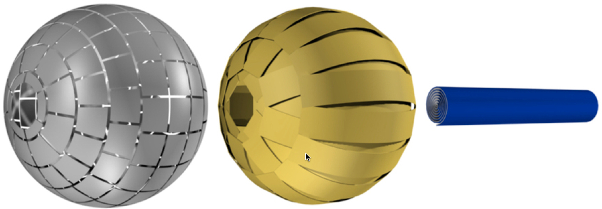
The magnetic wormhole device is composed of (from left to right) an outer spherical ferromagnetic metasurface, an inner spherical superconducting layer, and inside that, an inner spirally wound ferromagnetic sheet (credit: (credit: Jordi Prat-Camps/Universitat Autònoma de Barcelona)

To create the wormhole used in this experiment, the researchers designed a sphere made of three layers: an external layer with a ferromagnetic (as in a standard iron magnet) surface, a second inner layer made of superconducting material, and a ferromagnetic sheet rolled into a cylinder that crosses the sphere from one end to the other and conducts the magnetic field.

The magnetic wormhole is analogous to a gravitational wormhole — it “changes the topology of space, as if the inner region has been magnetically erased from space,” explains Àlvar Sánchez, the lead researcher.

Practical applications

The study was published in an open-access paper in Scientific Reports, but the researchers first published the fundamental concept in a paper in Physical Review Letters, where they described it as a “magnetic hose.”

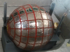
Experimental magnetic wormhole (credit: Jordi Prat-Camps/Universitat Autònoma de Barcelona)

The researchers built a magnetic “hose” capable of channeling a magnetic field from a source to a distance more than 10 centimeters away. (However, that version was detectable magnetically.)

As Steven Anlage, a professor of Physics at the University of Maryland, explains in this open-access article in the journal Physics, channeling magnetic fields could improve the spatial resolution of magnetic-field images, for example.

“Another application would be magnetic resonance imaging, in which the patient and superconducting magnet required to generate the magnetic field could be physically separated, and the intense magnetic field could be applied locally to the patient” (instead of having to enter an MRI machine).

Or it could allow MRI images of different parts of the body to be obtained simultaneously.

There may be other possible uses. Any ideas? Please comment below.

* Wormholes are cosmic tunnels that can connect two distant regions of the universe, popularized by science fiction like Stargate, Star Trek or, more recently, Interstellar. Using present-day technology, it would be impossible to create a gravitational wormhole because the field would have to be manipulated with huge amounts of gravitational energy, which no one yet knows how to generate. In electromagnetism, however, advances in metamaterials and invisibility have allowed researchers to put forward several designs to achieve this.


Abstract of A Magnetic Wormhole

Wormholes are fascinating cosmological objects that can connect two distant regions of the universe. Because of their intriguing nature, constructing a wormhole in a lab seems a formidable task. A theoretical proposal by Greenleaf et al. presented a strategy to build a wormhole for electromagnetic waves. Based on metamaterials, it could allow electromagnetic wave propagation between two points in space through an invisible tunnel. However, an actual realization has not been possible until now. Here we construct and experimentally demonstrate a magnetostatic wormhole. Using magnetic metamaterials and metasurfaces, our wormhole transfers the magnetic field from one point in space to another through a path that is magnetically undetectable. We experimentally show that the magnetic field from a source at one end of the wormhole appears at the other end as an isolated magnetic monopolar field, creating the illusion of a magnetic field propagating through a tunnel outside the 3D space. Practical applications of the results can be envisaged, including medical techniques based on magnetism.