Building and transplanting a bioengineered forelimb

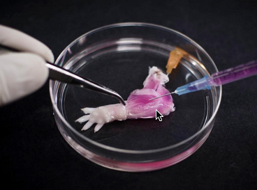
A suspension of muscle progenitor cells is injected into the cell-free matrix of a decellularized rat limb, which provides shape and structure onto which regenerated tissue can grow (credit: Bernhard Jank, MD, Ott Laboratory, Massachusetts General Hospital Center for Regenerative Medicine)

A team of Massachusetts General Hospital (MGH) investigators has made the first steps towards developing bioartificial replacement limbs suitable for transplantation.

In a Biomaterials journal report, the researchers describe using an experimental approach previously used to build bioartificial organs to engineer rat forelimbs with functioning vascular and muscle tissue. They also provided evidence that the same approach could be applied to the limbs of primates.

“The composite nature of our limbs makes building a functional biological replacement particularly challenging,” explains Harald Ott, MD, of the MGH Department of Surgery and the Center for Regenerative Medicine and assistant professor of Surgery at Harvard Medical School, senior author of the paper.

The progenitor cells needed to regenerate all of the tissues that make up a limb could be provided by the potential recipient. The problem is that limbs contain muscles, bone, cartilage, blood vessels, tendons, ligaments and nerves — each of which has to be rebuilt and requires a specific supporting structure (“matrix”), a step that has been a missing, he explained.

“We have shown that we can maintain the matrix of all of these tissues in their natural relationships to each other, that we can culture the entire construct over prolonged periods of time, and that we can repopulate the vascular system and musculature.”

Engineering a bioartificial limb
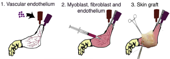
Procedure for composite tissue engineering. (1) Vascular endothelial cells are instilled into the vascular system of acellular composite tissue grafts. (2) Myoblasts, fibroblasts and endothelial cells are injected into the muscle compartment on day 2 of whole organ culture. (3) Full-thickness skin grafts are transplanted onto engineered constructs on day 10 of in vitro culture. (credit: B.J. Jank et al. / Biomaterials)

The current study uses technology Ott discovered as a research fellow at the University of Minnesota, in which living cells are stripped from a donor organ with a detergent solution and the remaining matrix is then repopulated with progenitor cells appropriate to the specific organ.

His team and others at MGH and elsewhere have used this decellularization technique to regenerate kidneyslivershearts, and lungs from animal models, but this is the first reported use to engineer the more complex tissues of a bioartificial limb.

The same decellularization process used in the whole-organ studies — perfusing a detergent solution through the vascular system — was used to strip all cellular materials from forelimbs removed from deceased rats in a way that preserved the primary vasculature and nerve matrix.

After thorough removal of cellular debris — a process that took a week — what remained was the cell-free matrix that provides structure to all of a limb’s composite tissues.  At the same time, populations of muscle and vascular cells were being grown in culture.

Bioreactor for growing a forelimb
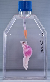
After vascular and muscle progenitors have been introduced into a decellularized rat limb, it is suspended in a bioreactor, which provides a nutrient solution and electrical stimulation to support and promote the growth of new tissues. (Bernhard Jank, MD, Ott Laboratory, Massachusetts General Hospital Center for Regenerative Medicine)

The research team then cultured the forelimb matrix in a bioreactor, within which vascular cells were injected into the limb’s main artery to regenerate veins and arteries.  Muscle progenitors were injected directly into the matrix sheaths that define the position of each muscle.

After five days in culture, electrical stimulation was applied to the potential limb graft to further promote muscle formation, and after two weeks, the grafts were removed from the bioreactor.

Analysis of the bioartificial limbs confirmed the presence of vascular cells along blood vessel walls and muscle cells aligned into appropriate fibers throughout the muscle matrix.

Functional testing of the isolated limbs showed that electrical stimulation of muscle fibers caused them to contract with a strength 80 percent of what would be seen in newborn animals.

The vascular systems of bioengineered forelimbs transplanted into recipient animals quickly filled with blood, which continued to circulate, and electrical stimulation of muscles within transplanted grafts flexed the wrists and digital joints of the animals’ paws.

The research team also successfully decellularized baboon forearms to confirm the feasibility of using this approach on the scale that would be required for human patients.

Replicating with human cells

Ott notes that, while regrowing nerves within a limb graft and reintegrating them into a recipient’s nervous system is one of the next challenges that needs to be faced, the experience of patients who have received hand transplants is promising.

“In clinical limb transplantation, nerves do grow back into the graft,  enabling both motion and sensation, and we have learned that this process is largely guided by the nerve matrix within the graft. We hope in future work to show that the same will apply to bioartificial grafts.

“Additional next steps will be replicating our success in muscle regeneration with human cells and expanding that to other tissue types, such as bone, cartilage and connective tissue.”

The study was supported by a New Innovator Award from the National Institutes of Health.

The authors note that more than 1.5 million individuals in the U.S. have lost a limb. Over the past two decades, a number of patients have received donor hand transplants, which also expose recipients to the risks of life-long immunosuppressive therapy.


Ott Laboratory | Rat Tissue Decellularization


Abstract of Engineered composite tissue as a bioartificial limb graft

The loss of an extremity is a disastrous injury with tremendous impact on a patient’s life. Current mechanical prostheses are technically highly sophisticated, but only partially replace physiologic function and aesthetic appearance. As a biologic alternative, approximately 70 patients have undergone allogeneic hand transplantation to date worldwide. While outcomes are favorable, risks and side effects of transplantation and long-term immunosuppression pose a significant ethical dilemma. An autologous, bio-artificial graft based on native extracellular matrix and patient derived cells could be produced on demand and would not require immunosuppression after transplantation. To create such a graft, we decellularized rat and primate forearms by detergent perfusion and yielded acellular scaffolds with preserved composite architecture. We then repopulated muscle and vasculature with cells of appropriate phenotypes, and matured the composite tissue in a perfusion bioreactor under electrical stimulation in vitro. After confirmation of composite tissue formation, we transplanted the resulting bio-composite grafts to confirm perfusion in vivo.

First multi-organ transplant that includes skull and scalp

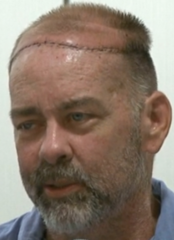
James Boyson (credit: CNN)

James Boysen, a 55-year-old software developer from Austin, Texas has become the first patient to receive a scalp and skull transplant while receiving kidney and pancreas transplants.

More than 50 health care professionals from Houston Methodist Hospital and The University of Texas MD Anderson Cancer Center assisted with or supported the double surgery over a period of more than 24 hours.

“This was a very complex surgery because we had to transplant the tissues utilizing microsurgery,” said Michael Klebuc, M.D., the surgeon who led the Houston Methodist Hospital Plastic Surgery Team.

“Imagine connecting blood vessels 1/16 of an inch under a microscope with tiny stitches about half the diameter of a human hair being done with tools that one would use to make a fine Swiss watch.”

In 2006, Boysen was diagnosed with leiomyosarcoma, a rare cancer of the smooth muscle, on his scalp. Successfully treated with chemotherapy and radiation, he was left with a large, deep wound on his head that included the scalp and the full thickness of his skull down to his brain.

In addition to the wound, which would require a major reconstructive undertaking, Boysen’s kidney and pancreas, which were first transplanted in 1992, were failing. Diagnosed with diabetes at age 5, Boysen’s declining condition over the years prompted the original double-organ transplant.

CNN video


Medical ‘millirobots’ could replace invasive surgery

Cross-section: three-component Gauss gun before (top) and after (bottom) firing (credit: Aaron T. Becker et al./Proceedings of the IEEE)

University of Houston researchers have developed a concept for MRI-powered millimeter-size “millirobots” that could one day perform unprecedented minimally invasive medical treatments.

This technology could be used to treat hydrocephalus, for example. Current treatments require drilling through the skull to implant pressure-relieving shunts, said Aaron T. Becker, assistant professor of electrical and computer engineering at the University of Houston.

But MRI scanners alone don’t produce enough force to pierce tissues (or insert needles). So the researchers drew upon the principle of the “Gauss gun.”


K&J Magnetics | Gauss Gun Demonstrations

Here’s how the a Gauss gun works: a single steel ball rolls down a chamber, setting off a chain reaction when it smashes into the next ball, etc., until the last ball flies forward, moving much more quickly the initial ball.

Based on that concept, the researchers imagine a medical robot with a barrel self-assembled from three small high-impact 3D-printed plastic components, with slender titanium rod spacers separating two steel balls.

Millirobot components (credit: Aaron T. Becker et al./Proceedings of the IEEE)

Aaron T. Becker, assistant professor of electrical and computer engineering at the University of Houston, said the potential technology could be used to treat hydrocephalus and other conditions, allowing surgeons to avoid current treatments that require cutting through the skull to implant pressure-relieving shunts.

Becker was first author of a paper presented at ICRA, the conference of the IEEE Robotics and Automation Society, nominated for best conference paper and best medical robotics paper.

“Hydrocephalus, among other conditions, is a candidate for correction by our millirobots because the ventricles are fluid-filled and connect to the spinal canal,” Becker said. “Our noninvasive approach would eventually require simply a hypodermic needle or lumbar puncture to introduce the components into the spinal canal, and the components could be steered out of the body afterwards.”

Future work will focus on exploring clinical context, miniaturizing the device, and optimizing material selection.


Abstract of Toward Tissue Penetration by MRI-powered Millirobots Using a Self-Assembled Gauss Gun

MRI-based navigation and propulsion of millirobots is a new and promising approach for minimally invasive therapies. The strong central field inside the scanner, however, precludes torque-based control. Consequently, prior propulsion techniques have been limited to gradient-based pulling through fluid-filled body lumens. This paper introduces a technique for generating large impulsive forces that can be used to penetrate tissue. The approach is based on navigating multiple robots to a desired location and using self-assembly to trigger the conversion of magnetic potential energy into sufficient kinetic energy to achieve penetration. The approach is illustrated through analytical modeling and experiments in a clinical MRI scanner.

A chip implanted under the skin allows for precise, real-time medical monitoring

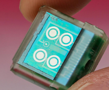
Under-the-skin chip (credit: EPFL)

A tiny (one-centimeter-square) biosensor chip developed at EPFL is designed to be implanted under your skin to continuously monitor concentrations of pH, temperature, and metabolism-related molecules like glucose, lactate and cholesterol, as well as some drugs.

The chip would replace blood work, which may take  hours — or even days — for analysis and is a limited snapshot of conditions at the moment the blood is drawn.

Developer Sandro Carrara unveiled the chip Tuesday (May 26) at the International Symposium on Circuits and Systems (ISCAS) in Lisbon.

The electrochemical sensors work with or without enzymes, which means the device can react to a wide range of compounds, and it can do so for several days or even weeks.

Wireless power and monitoring

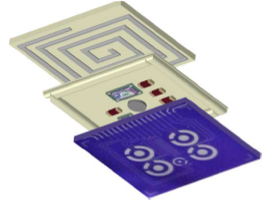
Implantable biosensor chip with three layers: a passive sensing platform (bottom), integrated circuits (middle) to analyze electrochemical measurements and generate a Bluetooth signal, and a coil (top) for through-the-skin data transmission and power via an external battery (credit: Camilla Baj-Rossi et al./IEEE Transactions on Biomedical Circuits and Systems)

The biochip contains three main components: a circuit with six sensors, a control unit that analyzes incoming signals, and a Bluetooth module for sending the results immediately to a mobile phone.

It also has an induction coil that wirelessly draws power from an external battery attached to the skin by a patch.

To ensure biocompatibility, an epoxy-enhanced polyurethane membrane was used to cover the device.

The chip was successfully tested in vivo on mice at the Institute for Research in Biomedicine (IRB) in Bellinzona, where researchers were able to constantly monitor glucose and paracetamol levels without a wire tracker getting in the way of the animals’ daily activities.

The results were promising, so clinical tests on humans could take place in three to five years — especially since the procedure is minimally invasive, the researchers say.

“Knowing the precise and real-time effect of drugs on the metabolism is one of the keys to the type of personalised, precision medicine that we are striving for,” said Carrara.

Fly-catching robot speeds biomedical research

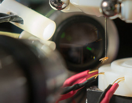
A fruit fly hangs unharmed at the end of the robot’s suction tube. The robot uses machine vision to inspect and analyze the captured fly. (credit: Stanforf Bio-X)

Stanford Bio-X scientists have created a robot that speeds and extends biomedical research with a common laboratory organism — fruit flies (Drosophila).

The robot can visually inspect awake flies and carry out behavioral experiments that were impossible with anesthetized flies. The work is described today (May 25) in the journal Nature Methods.

“Robotic technology offers a new prospect for automated experiments and enables fly researchers to do several things they couldn’t do previously,” said research team leader Mark Schnitzer, an associate professor of biology and of applied physics.

“For example, it can do studies with large numbers of flies inspected in very precise ways.” The group did one study of 1,000 flies in 10 hours, a task that would have taken much longer for even a highly skilled human.

Zap, you’re part of an experiment

When the robot’s fly-snatching apparatus is ready to grab a fly, it flashes a brief infrared blast of light that is invisible to the fly. The light reflects off its thorax, indicating the precise location of each fly and allowing the robot to recognize each individual fly by its reflection pattern. Then, a tiny, narrow suction tube strikes one of the illuminated thoraxes, painlessly sucking onto the fly and lifting it up.

Once the fly is attached, the robot uses machine vision to analyze the fly’s physical attributes, sort the flies by male and female, and even carry out a microdissection to reveal the fly’s minuscule brain. In one experiment, the robot’s machine vision was able to differentiate between two strains of flies so similar they are indistinguishable to the human eye.

Speeding disease research

All this is good news to the legion of graduate students who still spend hours a day looking at flies under a microscope as part of work that continues to uncover mechanisms in human aging, cancer, diabetes and a range of other diseases.

Although flies and humans have obvious differences, in many cases our cells and organs behave in similar ways and it is easier to study those processes in flies than in humans. The earliest information about how radiation causes gene mutations came from fruit flies, as did an understanding of our daily sleep/waking rhythms. And many of the molecules that are now famous for their roles in regulating how cells communicate were originally discovered by scientists hunched over microscopes staring at the unmoving bodies of anesthetized flies.

Now, that list of fruit fly contributions can be expended to include behavioral studies, previously impossible because the humans carrying out the analysis can neither see fly behaviors clearly nor distinguish between individuals.

In their paper, Schnitzer and his team had the robot pick up a fly and carry it to a trackball. Once there, they exposed the fly to different smells and could record how the fly behaved — racing along the trackball to get closer or attempting to turn away.

The work was funded by the W.M. Keck Foundation, the Stanford Bio-X program, an NIH Director’s Pioneer Award, and the Stanford-NIBIB Training Program in Biomedical Imaging Instrumentation.

Converting blood stem cells to sensory neural cells to predict and treat pain

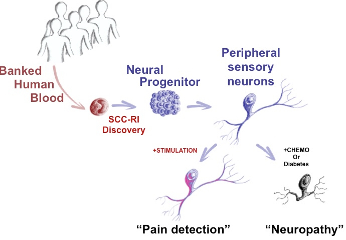
McMaster University scientists have discovered how to make adult sensory neurons from a patient’s blood sample to measure pain (credit: McMaster University

Stem-cell scientists at McMaster University have developed a way to directly convert adult human blood cells to sensory neurons, providing the first objective measure of how patients may feel things like pain, temperature, and pressure, the researchers reveal in an open-access paper in the journal Cell Reports.

Currently, scientists and physicians have a limited understanding of the complex issue of pain and how to treat it. “The problem is that unlike blood, a skin sample or even a tissue biopsy, you can’t take a piece of a patient’s neural system,” said Mick Bhatia, director of the McMaster Stem Cell and Cancer Research Institute and research team leader. “It runs like complex wiring throughout the body and portions cannot be sampled for study.

“Now we can take easy to obtain blood samples, and make the main cell types of neurological systems in a dish that is specialized for each patient,” said Bhatia. “We can actually take a patient’s blood sample, as routinely performed in a doctor’s office, and with it we can produce one million sensory neurons, [which] make up the peripheral nerves. We can also make central nervous system cells.”

Testing pain drugs

The new technology has “broad and immediate applications,” said Bhatia: It allows researchers to understand disease and improve treatments by asking questions such as: Why is it that certain people feel pain versus numbness? Is this something genetic? Can the neuropathy that diabetic patients experience be mimicked in a dish?

It also paves the way for the discovery of new pain drugs that don’t just numb the perception of pain. Bhatia said non-specific opioids used for decades are still being used today. “If I was a patient and I was feeling pain or experiencing neuropathy, the prized pain drug for me would target the peripheral nervous system neurons, but do nothing to the central nervous system, thus avoiding addictive drug side effects,” said Bhatia.

“Until now, no one’s had the ability and required technology to actually test different drugs to find something that targets the peripheral nervous system, and not the central nervous system, in a patient-specific, or personalized manner.”

A patient time machine 

Bhatia’s team also successfully tested their process with cryopreserved (frozen) blood. Since blood samples are taken and frozen with many clinical trials, this give them “almost a bit of a time machine” to run tests on neurons created from blood samples of patients taken in past clinical trials, where responses and outcomes have already been recorded.

In the future, the process may have prognostic (predictive diagnostic) potential, explained Bhatia: one might be able to look at a patient with Type 2 Diabetes and predict whether they will experience neuropathy, by running tests in the lab using their own neural cells derived from their blood sample.

“This bench-to-bedside research is very exciting and will have a major impact on the management of neurological diseases, particularly neuropathic pain,” said Akbar Panju, medical director of the Michael G. DeGroote Institute for Pain Research and Care, a clinician and professor of medicine.

“This research will help us understand the response of cells to different drugs and different stimulation responses, and allow us to provide individualized or personalized medical therapy for patients suffering with neuropathic pain.”

This research was supported by the Canadian Institutes of Health Research, Ontario Institute of Regenerative Medicine, Marta and Owen Boris Foundation, J.P. Bickell Foundation, the Ontario Brain Institute, and Brain Canada.

Pain insensitivity

In related news, an international team of researchers co-led by the University of Cambridge reported Monday in the journal Nature Genetics that they have identified a gene, PRDM12, that is essential to the production of pain-sensing neurons in humans. Rare individuals — around one in a million people in the UK — are born unable to feel pain, in a condition known as congenital insensitivity to pain (CIP). These people accumulate numerous self-inflicted injuries, often leading to reduced lifespan.

The researchers are hopeful that this new gene could be an excellent candidate for drug development.


Abstract of Single Transcription Factor Conversion of Human Blood Fate to NPCs with CNS and PNS Developmental Capacity

The clinical applicability of direct cell fate conversion depends on obtaining tissue from patients that is easy to harvest, store, and manipulate for reprogramming. Here, we generate induced neural progenitor cells (iNPCs) from neonatal and adult peripheral blood using single-factor OCT4 reprogramming. Unlike fibroblasts that share molecular hallmarks of neural crest, OCT4 reprogramming of blood was facilitated by SMAD+GSK-3 inhibition to overcome restrictions on neural fate conversion. Blood-derived (BD) iNPCs differentiate in vivo and respond to guided differentiation in vitro, producing glia (astrocytes and oligodendrocytes) and multiple neuronal subtypes, including dopaminergic (CNS related) and nociceptive neurons (peripheral nervous system [PNS]). Furthermore, nociceptive neurons phenocopy chemotherapy-induced neurotoxicity in a system suitable for high-throughput drug screening. Our findings provide an easily accessible approach for generating human NPCs that harbor extensive developmental potential, enabling the study of clinically relevant neural diseases directly from patient cohorts.

Nature inspires first artificial molecular pump

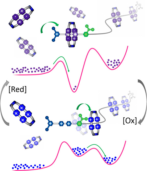
A blueprint for an artificial molecular pump that acts to organize rings in a high-energy state on a polymethylene chain. The flashing energy ratchet mechanism shows how redox chemistry can be used to prime the pump with rings (top) under reducing conditions and then have it pump the rings (bottom) under oxidative conditions into a high-energy state. Hypothetical distributions of the components of the pump on a simplified potential energy surface diagram are indicated by purple dots in the reduced state and blue dots in the oxidized state. A solid green arrow indicates a surmountable energy barrier. (credit: Chuyang Cheng et al./Nature Nanotechnology)

Northwestern University scientists have developed the first entirely artificial molecular pump, in which molecules pump other molecules. The pump might one day be used to power other molecular machines, such as artificial muscles.

The new machine mimics the pumping mechanism of proteins that move small molecules around living cells to metabolize and store energy from food. The artificial pump draws power from chemical reactions, driving molecules step-by-step from a low-energy state to a high-energy state — far away from equilibrium.

While nature has had billions of years to perfect its complex molecular machinery, modern science is now beginning to scratch the surface of what might be possible in tomorrow’s world.

Imitating how nature transfers energy

“Our molecular pump is radical chemistry — an ingenious way of transferring energy from molecule to molecule, the way nature does,” said Sir Fraser Stoddart, the senior author of the study. Stoddart is the Board of Trustees Professor of Chemistry in Northwestern’s Weinberg College of Arts and Sciences.

“All living organisms, including humans, must continuously transport and redistribute molecules around their cells, using vital carrier proteins,” he said. “We are trying to recreate the actions of these proteins using relatively simple small molecules we make in the laboratory.”

“In some respects, we are asking the molecules to behave in a way that they would not do normally,” Cheng said. “It is much like trying to push two magnets together. The ring-shaped molecules we work with repel one another under normal circumstances. The artificial pump is able to syphon off some of the energy that changes hands during a chemical reaction and uses it to push the rings together.”

The tiny molecular machine threads the rings around a nanoscopic chain — a sort of axle — and squeezes the rings together, with only a few nanometers separating them. At present, the artificial molecular pump is able to force only two rings together, but the researchers believe it won’t be long before they can extend its operation to tens of rings and store more energy.

Compared to nature’s system, the artificial pump is very simple, but it is a start, the researchers say. They have designed a novel system, using kinetic barriers, that allows molecules to flow “uphill” energetically.

Powering artificial muscles

“This is non-equilibrium chemistry, moving molecules far away from their minimum energy state, which is essential to life,” said Paul R. McGonigal, an author of the study. “Conducting non-equilibrium chemistry in this way, with simple artificial molecules, is one of the major challenges for science in the 21st century.”

Ultimately, they intend to use the energy stored in their pump to power artificial muscles and other molecular machines. The researchers also hope their design will inspire other chemists working in non-equilibrium chemistry.

“This is completely unlike the process of designing the machinery we are used to seeing in everyday life,” Stoddart said. “In a way, one must learn to see things from the molecules’ point of view, considering forces such as random thermal motion that one would never consider when building an agricultural water pump or any other mechanical device.”

The National Science Foundation supported the research, published May 18 in the journal Nature Nanotechnology.


Northwestern University | Artificial Molecular Pump Animation

Animation shows the steps of the pumping mechanism, which operates in response to redox cycling, with simplified illustrations of the corresponding energy profiles. The dumbbell and the ring repel each other initially, then reduction favors complexation both thermodynamically and kinetically. Oxidation restores the repulsion between the components and causes the ring to be trapped around the dumbbell during thermal relaxation. When another reduction step is performed, attraction of a second ring from the bulk solution is kinetically favored. After oxidation and thermal relaxation, the second ring falls into the same kinetic trap as the first, resulting in the mutually repulsive rings being held in close proximity to one another.


Abstract of An artificial molecular pump

Carrier proteins consume fuel in order to pump ions or molecules across cell membranes, creating concentration gradients. Their control over diffusion pathways, effected entirely through noncovalent bonding interactions, has inspired chemists to devise artificial systems that mimic their function. Here, we report a wholly artificial compound that acts on small molecules to create a gradient in their local concentration. It does so by using redox energy and precisely organized noncovalent bonding interactions to pump positively charged rings from solution and ensnare them around an oligomethylene chain, as part of a kinetically trapped entanglement. A redox-active viologen unit at the heart of a dumbbell-shaped molecular pump plays a dual role, first attracting and then repelling the rings during redox cycling, thereby enacting a flashing energy ratchet mechanism with a minimalistic design. Our artificial molecular pump performs work repetitively for two cycles of operation and drives rings away from equilibrium toward a higher local concentration.

How to print stronger, bigger, conductive 3-D graphene structures for tissue engineering

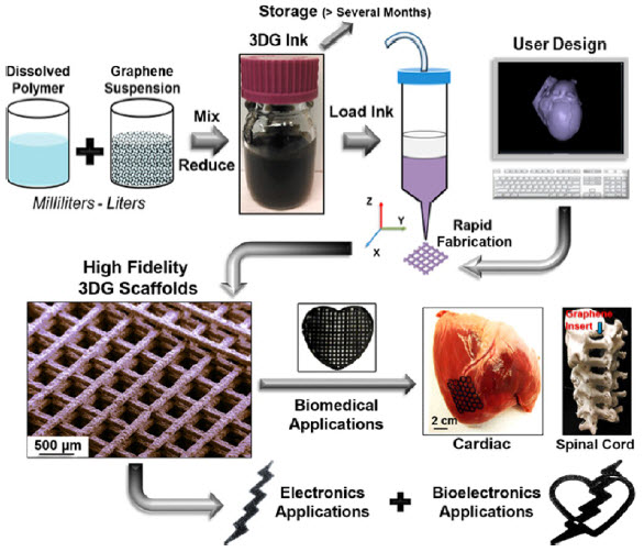
3D graphene inks are produced by simple combination and mixing an elastomer solution with graphene powder in a graded solvent, followed by volume reduction and thickening, a process that can be scaled up to many liters at once (credit: Adam E. Jakus et al./ACS Nano)

Northwestern University researchers have developed a way to print large, robust 3-D structures with graphene-based ink.

The new method could allow for using graphene-printed scaffolds for regenerative medicine and other medical and electronic  applications.

“People have tried to print graphene before,” said Ramille Shah, assistant professor of materials science and engineering at the McCormick School of Engineering and of surgery in the Feinberg School of Medicine.  “But it’s been a mostly polymer composite with graphene making up less than 20 percent of the volume.”

Adding higher volumes of graphene flakes to the mix in these ink systems typically results in printed structures too brittle and fragile to manipulate. At 60–70 percent graphene, the new ink preserves the material’s unique properties, including its electrical conductivity. And it’s flexible and robust enough to print robust macroscopic structures.

The secret: graphene nanoflakes are mixed with a biocompatible elastomer and fast-evaporating solvents.

“After the ink is extruded, one of the solvents in the system evaporates right away, causing the structure to solidify nearly instantly,” Shah explained. “The presence of the other solvents and the interaction with the specific polymer binder chosen also has a significant contribution to its resulting flexibility and properties. Because it holds its shape, we are able to build larger, well-defined objects.”

Could allow neurons to grow and communicate

Shah said her team populated one of the scaffolds with stem cells to surprising results. Not only did the cells survive; they divided, proliferated, and morphed into neuron-like cells.

The printed graphene structure is also flexible and strong enough to be easily sutured to existing tissues, so it could be used for biodegradable sensors and medical implants. Shah said the biocompatible elastomer and graphene’s electrical conductivity most likely contributed to the scaffold’s biological success.

“Cells conduct electricity inherently — especially neurons,” Shah said. “So if they’re on a substrate that can help conduct that signal, they’re able to communicate over wider distances.”

Supported by a Google Gift and a McCormick Research Catalyst Award, the research is described in the paper published in the April 2015 issue of ACS Nano.


Abstract of Three-Dimensional Printing of High-Content Graphene Scaffolds for Electronic and Biomedical Applications

The exceptional properties of graphene enable applications in electronics, optoelectronics, energy storage, and structural composites. Here we demonstrate a 3D printable graphene (3DG) composite consisting of majority graphene and minority polylactide-co-glycolide, a biocompatible elastomer, 3D-printed from a liquid ink. This ink can be utilized under ambient conditions via extrusion-based 3D printing to create graphene structures with features as small as 100 μm composed of as few as two layers (<300 μm thick object) or many hundreds of layers (>10 cm thick object). The resulting 3DG material is mechanically robust and flexible while retaining electrical conductivities greater than 800 S/m, an order of magnitude increase over previously reported 3D-printed carbon materials. In vitro experiments in simple growth medium, in the absence of neurogenic stimuli, reveal that 3DG supports human mesenchymal stem cell (hMSC) adhesion, viability, proliferation, and neurogenic differentiation with significant upregulation of glial and neuronal genes. This coincides with hMSCs adopting highly elongated morphologies with features similar to axons and presynaptic terminals. In vivo experiments indicate that 3DG has promising biocompatibility over the course of at least 30 days. Surgical tests using a human cadaver nerve model also illustrate that 3DG has exceptional handling characteristics and can be intraoperatively manipulated and applied to fine surgical procedures. With this unique set of properties, combined with ease of fabrication, 3DG could be applied toward the design and fabrication of a wide range of functional electronic, biological, and bioelectronic medical and nonmedical devices.

New evidence that electrical stimulation accelerates wound healing

An untreated wound (left) after 10 days is larger than an electrical-stimulation-treated wound (right) (credit: The University of Manchester)

The most detailed study to date of skin wound healing, conducted by University of Manchester scientists with 40 volunteers, has provided new evidence that electrical stimulation accelerates wound healing.

In the new research, half-centimeter harmless wounds were created on each upper arm of the volunteers.  One wound was left to heal normally, while the other was treated with electrical pulses* over a period of two weeks.  The pulses stimulated angiogenesis — the process by which new blood vessels form — increasing blood flow to the damaged area and resulting in wounds healing significantly faster.

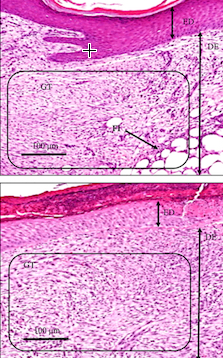
Normal-healing tissues (top) vs. electrical-stimulation-treated healing tissues (bottom). Electrical-stimulation sample showed reorganization and accelerated granulation tissue stage development. ED is epidermis, DE is dermis, GT is granulation tissue, FT is fat/adipose tissue. (credit: Sara Ud-Din et al./PLoS ONE)

“The aim of this study was to further evaluate the role of electrical stimulation (ES) in affecting angiogenesis during the acute phase of cutaneous wound healing over multiple time points to identify if the enhanced effect occurred earlier than day 14,” the researchers note in a paper published in open-access PLoS ONE.

“This research has shown the effectiveness of electrical stimulation in wound healing,” said research leader Ardeshir Bayat of the University’s Institute of Inflammation and Repair. “We believe this technology has the potential to be applied to any situation where faster wound healing is particularly desirable, such as human or veterinary surgical wounds, accidents, military trauma, and sports injuries.”

Based on the findings, the researchers plan to work with Oxford BioElectronics Ltd. on a five-year project to develop and evaluate devices and dressings that use these experimental techniques to stimulate the body’s nervous system to generate nerve impulses directed to the site of skin repair.

How electrical stimulation enhances wound healing

The researchers explain in the paper that “ES in its various forms has been shown to enhance wound healing by promoting the migration of keratinocytes and macrophages, enhancing angiogenesis, stimulating fibroblasts, and influencing protein synthesis throughout the inflammatory, proliferative, and remodeling phases of healing.”

The researchers previously “investigated the in vitro effect of different types of ES on the expression of collagen in skin fibroblasts. Importantly, we highlighted the role of a novel waveform termed degenerate wave (DW is a degenerating sine wave, which deteriorates over time) and demonstrated its beneficial effects compared to other known waveforms such as direct and alternating currents.”

Skin wounds that are slow to heal are a clinical challenge to physicians all over the world. Every year, the NHS in the U.K. alone spends £1 billion on treating chronic wounds such as lower limb venous and diabetic ulcers. (Wounds become chronic when they fail to heal and remain open for longer than six weeks.)

* According to the researchers writing in the PLoS ONE paper, the electrical stimulation device used was the Fenzian system (Fenzian Ltd, Hungerford, UK), a transcutaneous low intensity device that  detects changes in skin impedance and adjusts the outgoing microcurrent electrical biofeedback impulses (20–80V, 6-millisecond “degenerate wave” pulses at 0.004 milliamps, with a frequency default of 60Hz).


Abstract of Angiogenesis Is Induced and Wound Size Is Reduced by Electrical Stimulation in an Acute Wound Healing Model in Human Skin

Angiogenesis is critical for wound healing. Insufficient angiogenesis can result in impaired wound healing and chronic wound formation. Electrical stimulation (ES) has been shown to enhance angiogenesis. We previously showed that ES enhanced angiogenesis in acute wounds at one time point (day 14). The aim of this study was to further evaluate the role of ES in affecting angiogenesis during the acute phase of cutaneous wound healing over multiple time points. We compared the angiogenic response to wounding in 40 healthy volunteers (divided into two groups and randomised), treated with ES (post-ES) and compared them to secondary intention wound healing (control). Biopsy time points monitored were days 0, 3, 7, 10, 14. Objective non-invasive measures and H&E analysis were performed in addition to immunohistochemistry (IHC) and Western blotting (WB). Wound volume was significantly reduced on D7, 10 and 14 post-ES (p = 0.003, p = 0.002, p<0.001 respectively), surface area was reduced on days 10 (p = 0.001) and 14 (p<0.001) and wound diameter reduced on days 10 (p = 0.009) and 14 (p = 0.002). Blood flow increased significantly post-ES on D10 (p = 0.002) and 14 (p = 0.001). Angiogenic markers were up-regulated following ES application; protein analysis by IHC showed an increase (p<0.05) in VEGF-A expression by ES treatment on days 7, 10 and 14 (39%, 27% and 35% respectively) and PLGF expression on days 3 and 7 (40% on both days), compared to normal healing. Similarly, WB demonstrated an increase (p<0.05) in PLGF on days 7 and 14 (51% and 35% respectively). WB studies showed a significant increase of 30% (p>0.05) on day 14 in VEGF-A expression post-ES compared to controls. Furthermore, organisation of granulation tissue was improved on day 14 post-ES. This randomised controlled trial has shown that ES enhanced wound healing by reduced wound dimensions and increased VEGF-A and PLGF expression in acute cutaneous wounds, which further substantiates the role of ES in up-regulating angiogenesis as observed over multiple time points. This therapeutic approach may have potential application for clinical management of delayed and chronic wounds.