Are you a cyborg?

Bioprinting a brain

Cryogenic 3D-printing soft hydrogels. Top: the bioprinting process. Bottom: SEM image of general microstructure (scale bar: 100 µm). (credit: Z. Tan/Scientific Reports)

A new bioprinting technique combines cryogenics (freezing) and 3D printing to create geometrical structures that are as soft (and complex) as the most delicate body tissues — mimicking the mechanical properties of organs such as the brain and lungs.

The idea: “Seed” porous scaffolds that can act as a template for tissue regeneration (from neuronal cells, for example), where damaged tissues are encouraged to regrow — allowing the body to heal without tissue rejection or other problems. Using “pluripotent” stem cells that can change into different types of cells is also a possibility.

Smoothy. Solid carbon dioxide (dry ice) in an isopropanol bath is used to rapidly cool hydrogel ink (a rapid liquid-to-solid phase change) as it’s extruded, yogurt-smoothy-style. Once thawed, the gel is as soft as body tissues, but doesn’t collapse under its own weight — a previous problem.

Current structures produced with this technique are “organoids” a few centimeters in size. But the researchers hope to create replicas of actual body parts with complex geometrical structures — even whole organs. That could allow scientists to carry out experiments not possible on live subjects, or for use in medical training, replacing animal bodies for surgical training and simulations. Then on to mechanobiology and tissue engineering.

Source: Imperial College London, Scientific Reports (open-access).

How to generate electricity with your body

Bending a finger generates electricity in this prototype device. (credit: Guofeng Song et al./Nano Energy)

A new triboelectric nanogenerator (TENG) design, using a gold tab attached to your skin, will convert mechanical energy into electrical energy for future wearables and self-powered electronics. Just bend your finger or take a step.

Triboelectric charging occurs when certain materials become electrically charged after coming into contact with a different material. In this new design by University of Buffalo and Chinese scientists, when a stretched layer of gold is released, it crumples, creating what looks like a miniature mountain range. An applied force leads to friction between the gold layers and an interior PDMS layer, causing electrons to flow between the gold layers.

More power to you. Previous TENG designs have been difficult to manufacture (requiring complex lithography) or too expensive. The new 1.5-centimeters-long prototype generates a maximum of 124 volts but at only 10 microamps. It has a power density of 0.22 millwatts per square centimeter. The team plans larger pieces of gold to deliver more electricity and a portable battery.

Source: Nano Energy. Support: U.S. National Science Foundation, the National Basic Research Program of China, National Natural Science Foundation of China, Beijing Science and Technology Projects, Key Research Projects of the Frontier Science of the Chinese Academy of Sciences ,and National Key Research and Development Plan.

This artificial electrical eel may power your implants

How the eel’s electrical organs generate electricity by moving sodium (Na) and potassium (K) ions across a selective membrane. (credit: Caitlin Monney)

Taking it a giant (and a bit scary) step further, an artificial electric organ, inspired by the electric eel, could one day power your implanted implantable sensors, prosthetic devices, medication dispensers, augmented-reality contact lenses, and countless other gadgets. Unlike typical toxic batteries that need to be recharged, these systems are soft, flexible, transparent, and potentially biocompatible.

Doubles as a defibrillator? The system mimicks eels’ electrical organs, which use thousands of alternating compartments with excess potassium or sodium ions, separated by selective membranes. To create a jolt of electricity (600 volts at 1 ampere), an eel’s membranes allow the ions to flow together. The researchers built a similar system, but using sodium and chloride ions dissolved in a water-based hydrogel. It generates more than 100 volts, but at safe low current — just enough to power a small medical device like a pacemaker.

The researchers say the technology could also lead to using naturally occurring processes inside the body to generate electricity, a truly radical step.

Source: Nature, University of Fribourg, University of Michigan, University of California-San Diego. Funding: Air Force Office of Scientific Research, National Institutes of Health.

E-skin for Terminator wannabes

A section of “e-skin” (credit: Jianliang Xiao / University of Colorado Boulder)

A new type of thin, self-healing, translucent “electronic skin” (“e-skin,” which mimicks the properties of natural skin) has applications ranging from robotics and prosthetic development to better biomedical devices and human-computer interfaces.

Ready for a Terminator-style robot baby nurse? What makes this e-skin different and interesting is its embedded sensors, which can measure pressure, temperature, humidity and air flow. That makes it sensitive enough to let a robot take care of a baby, the University of Colorado mechanical engineers and chemists assure us. The skin is also rapidly self-healing (by reheating), as in The Terminator, using a mix of three commercially available compounds in ethanol.

The secret ingredient: A novel network polymer known as polyimine, which is fully recyclable at room temperature. Laced with silver nanoparticles, it can provide better mechanical strength, chemical stability and electrical conductivity. It’s also malleable, so by applying moderate heat and pressure, it can be easily conformed to complex, curved surfaces like human arms and robotic hands.

Source: University of Colorado, Science Advances (open-access). Funded in part by the National Science Foundation.

Altered Carbon
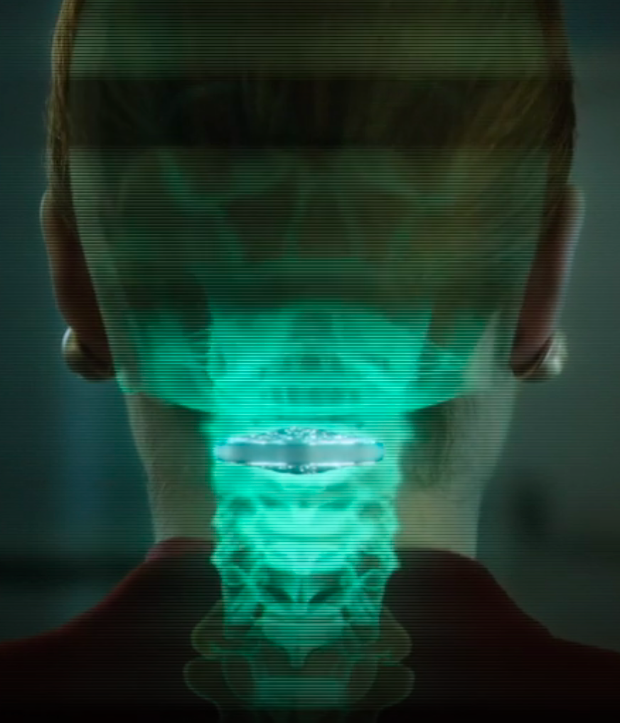
Vertebral cortical stack (credit: Netflix)

Altered Carbon takes place in the 25th century, when humankind has spread throughout the galaxy. After 250 years in cryonic suspension, a prisoner returns to life in a new body with one chance to win his freedom: by solving a mind-bending murder.

Resleeve your stack. Human consciousness can be digitized and downloaded into different bodies. A person’s memories have been encapsulated into “cortical stack” storage devices surgically inserted into the vertebrae at the back of the neck. Disposable physical bodies called “sleeves” can accept any stack.

But only the wealthy can acquire replacement bodies on a continual basis. The long-lived are called Meths, as in the Biblical figure Methuselah. The uber rich are also able to keep copies of their minds in remote storage, which they back up regularly, ensuring that even if their stack is destroyed, the stack can be resleeved (except for periods of time not backed up — as in the hack-murder).

Source: Netflix. Premiered on February 2, 2018. Based on the 2002 novel of the same title by Richard K. Morgan.


Remote-controlled DNA nanorobots could lead to the first nanorobotic production factory

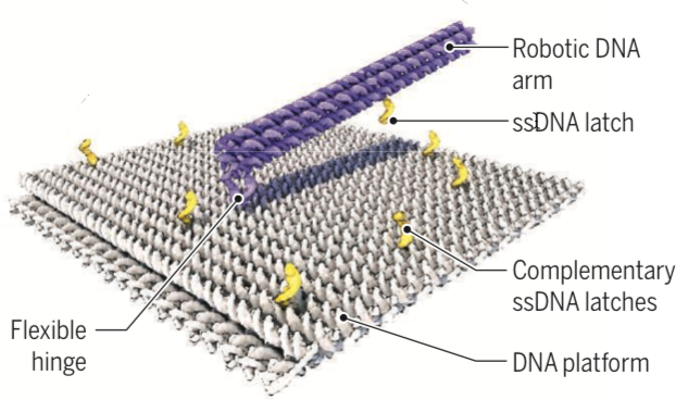
German researchers created a 55-nm-by-55-nm DNA-based molecular platform with a 25-nm-long robotic arm that can be actuated with externally applied electrical fields, under computer control. (credit: Enzo Kopperger et al./Science)

By powering a self-assembling DNA nanorobotic arm with electric fields, German scientists have achieved precise nanoscale movement that is at least five orders of magnitude (hundreds of thousands times) faster than previously reported DNA-driven robotic systems, they suggest today (Jan. 19) in the journal Science.

DNA origami has emerged as a powerful tool to build precise structures. But now, “Kopperger et al. make an impressive stride in this direction by creating a dynamic DNA origami structure that they can directly control from the macroscale with easily tunable electric fields—similar to a remote-controlled robot,” notes Björn Högberg of Karolinska Institutet in a related Perspective in Science, (p. 279).

The nanorobotic arm resembles the gearshift lever of a car. Controlled by an electric field (comparable to the car driver), short, single-stranded DNA serves as “latches” (yellow) to momentarily grab and lock the 25-nanometer-long arm into predefined “gear” positions. (credit: Enzo Kopperger et al./Science)

The new biohybrid nanorobotic systems could even act as a molecular mechanical memory (a sort of nanoscale version of the Babbage Analytical Engine), he notes. “With the capability to form long filaments with multiple DNA robot arms, the systems could also serve as a platform for new inventions in digital memory, nanoscale cargo transfer, and 3D printing of molecules.”

“The robot-arm system may be scaled up and integrated into larger hybrid systems by a combination of lithographic and self-assembly techniques,” according to the researchers. “Electrically clocked synthesis of molecules with a large number of robot arms in parallel could then be the first step toward the realization of a genuine nanorobotic production factory.”


Taking a different approach to a nanofactory, this “Productive Nanosystems: from Molecules to Superproducts” film — a collaborative project of animator and engineer John Burch and pioneer nanotechnologist K. Eric Drexler in 2005 — demonstrated key steps in a hypothetical process that converts simple molecules into a billion-CPU laptop computer. More here.


Abstract of A self-assembled nanoscale robotic arm controlled by electric fields

The use of dynamic, self-assembled DNA nanostructures in the context of nanorobotics requires fast and reliable actuation mechanisms. We therefore created a 55-nanometer–by–55-nanometer DNA-based molecular platform with an integrated robotic arm of length 25 nanometers, which can be extended to more than 400 nanometers and actuated with externally applied electrical fields. Precise, computer-controlled switching of the arm between arbitrary positions on the platform can be achieved within milliseconds, as demonstrated with single-pair Förster resonance energy transfer experiments and fluorescence microscopy. The arm can be used for electrically driven transport of molecules or nanoparticles over tens of nanometers, which is useful for the control of photonic and plasmonic processes. Application of piconewton forces by the robot arm is demonstrated in force-induced DNA duplex melting experiments.

Brainwave ‘mirroring’ neurotechnology improves post-traumatic stress symptoms

Patient receiving a real-time reflection of her frontal-lobe brainwave activity as a stream of audio tones through earbuds. (credit: Brain State Technologies)

You are relaxing comfortably, eyes closed, with non-invasive sensors attached to your scalp that are picking up signals from various areas of your brain. The signals are converted by a computer to audio tones that you can hear on earbuds. Over several sessions, the different frequencies (pitches) of the tones associated with the two hemispheres of the brain create a mirror for your brainwave activity, helping your brain reset itself to reduce traumatic stress.

In a study conducted at Wake Forest School of Medicine, 20 sessions of noninvasive brainwave “mirroring” neurotechnology called HIRREM* (high-resolution, relational, resonance-based electroencephalic mirroring) significantly reduced symptoms of post-traumatic stress resulting from service as a military member or vet.


Example of tones (credit: Brain State Technologies)

“We observed reductions in post-traumatic symptoms**, including insomnia, depressive mood, and anxiety, that were durable through six months after the use of HIRREM, but additional research is needed to confirm these initial findings,” said the study’s principal investigator, Charles H. Tegeler, M.D., professor of neurology at Wake Forest School of Medicine, a part of Wake Forest Baptist.

About 500 patients have participated in HIRREM clinical trials at Wake Forest School of Medicine and other locations, according to Brain State Technologies Founder and CEO Lee Gerdes.


Brain State Technologies | HIRREM process, showing a technologist applying Brain State Technologies’ proprietary HIRREM process with a military veteran client.

HIRREM is intended for medical research. A consumer version of the core underlying brainwave mirroring process is available as “Brainwave Optimization” from Brain State Technologies in Scottsdale, Arizona. The company also offers a wearable device for ongoing brain support, BRAINtellect B2v2.


How HIRREM neurotechnology works

(credit: Brain State Technologies)

HIRREM is a neurotechnology that dynamically measures brain electrical activity. It uses two or more EEG (electroencephalogram, or brain-wave detection) scalp sensors to pick up signals from both sides of the brain. Computer software algorithms then convert dominant brain frequencies in real time into audible tones with varying pitch and timing, which can be heard on earbuds.

In effect, the brain is listening to itself. It the process, it makes self-adjustments towards improved balance (between brain temporal lobe activity in the two hemispheres — sympathetic (right) and parasympathetic (left) — of the brain), resulting in reduced hyper-arousal. No conscious cognitive activity is required. Signals from other areas of the brain can also be studied.

The net effect is to reset stress response patterns that have been wired by repetitive traumatic events (physical or non-physical).***

“Thus, if the stimulus is acoustic response to brain function (often called neurofeedback (NFB), then the response is made based on a threshold of the NFB provider. Since the brain moves three to five times faster than the thoughtful response of the client, the brain’s activity is way beyond any kind of activity which the client can mitigate. The NFB hypothesis is that the operant conditioning can be learned by the brain so it changes itself.

“In a HIRREM placebo-controlled insomnia study, HIRREM showed statistically significant improvement in sleep function over the placebo. Additionally, HIRREM demonstrated that biomarkers for the test were also statistically significant over the placebo. Posters for this study were presented at the International Sleep Conference and at the Dept of Defense Research meeting on sleep. A full length manuscript of the study is in process with hopes to be published Q1 2018).”


The study was published (open access) in the Dec. 22 online edition of the journal Military Medical Research with co-authors at Brain State Technologies. It was supported through the Joint Capability Technology Demonstration Program within the Office of the Under Secretary of Defense and by a grant from The Susanne Marcus Collins Foundation, Inc. to the Department of Neurology at Wake Forest Baptist.

The researchers acknowledge limitations of the study, including the small number of participants and the absence of a control group. It was also an open-label project, meaning that both researchers and participants knew what treatment was being administered.

* HIRREM is a registered trademark of Brain State Technologies based in Scottsdale, Arizona, and has been licensed to Wake Forest University for collaborative research since 2011.  In this single-site study, 18 service members or recent veterans, who experienced symptoms over one to 25 years, received an average of 19½ HIRREM sessions over 12 days. Symptom data were collected before and after the study sessions, and follow-up online interviews were conducted at one-, three- and six-month intervals. In addition, heart rate and blood pressure readings were recorded after the first and second visits to analyze downstream autonomic balance with heart rate variability and baroreflex sensitivity. HIRREM has been used experimentally with more than 500 patients at Wake Forest School of Medicine.

** According to the U.S. Department of Veterans Affairs, approximately 31 percent of Vietnam veterans, 10 percent of Gulf War (Desert Storm) veterans and 11 percent of veterans of the war in Afghanistan experience PTSD. Symptoms can include insomnia, poor concentration, sadness, re-experiencing traumatic events, irritability or hyper-alertness, and diminished autonomic cardiovascular regulation.

*** The effect is based on the “bihemispheric autonomic model” (BHAM ), “which proposes that trauma-related sympathetic hyperarousal may be an expression of maladaptive right temporal lobe activity, whereas the avoidant and dissociative features of the traumatic stress response may be indicators of a parasympathetic “freeze” response that is significantly driven by the left temporal lobe. An implication [is that brain-based] intervention may facilitate the reduction of symptom clusters associated with autonomic disturbances through the mitigation of maladaptive asymmetries.” — Catherine L. Tegeler et al./Military Medical Research.

Update Jan. 10, 2017: What about a control group?

“Our study had an open label design, without a control group,” Tegeler explained to KurzweilAI in an email, in response to reader questions.

“We agree that a randomized design is scientifically a more powerful approach, and one we would have preferred.  The reality was that for this cohort of participants, mostly drawn from the special operations community, constraints due to limitation on allowable time away from duties, training cycle pressures, therapeutic expectations, and available funding, prevented consideration of a controlled design.

“Other studies have used a placebo-controlled design utilizing acoustic stimulation linked to brainwaves, as compared to acoustic stimulation not linked to brainwaves. Manuscripts are being prepared to report those results. Finally, our current studies are all focused on evaluation of the effects and benefits of HIRREM alone, for a variety of symptoms or conditions.  That said, in the future there may be opportunities to seek funding for projects that might combine, or follow up after HIRREM, with other strategies such as meditation, improved nutrition, or exercise.”

“Biofeedback/neurofeedback is an open-loop system indicating that the feedback from the brain or other biological function is provided back to the client as the function being analyzed triggers a stimulus,” Gerdes added.


Abstract of Successful use of closed-loop allostatic neurotechnology for post-traumatic stress symptoms in military personnel: self-reported and autonomic improvements

Background: Military-related post-traumatic stress (PTS) is associated with numerous symptom clusters and diminished autonomic cardiovascular regulation. High-resolution, relational, resonance-based, electroencephalic mirroring (HIRREM®) is a noninvasive, closed-loop, allostatic, acoustic stimulation neurotechnology that produces real-time translation of dominant brain frequencies into audible tones of variable pitch and timing to support the auto-calibration of neural oscillations. We report clinical, autonomic, and functional effects after the use of HIRREM® for symptoms of military-related PTS.

Methods: Eighteen service members or recent veterans (15 active-duty, 3 veterans, most from special operations, 1 female), with a mean age of 40.9 (SD = 6.9) years and symptoms of PTS lasting from 1 to 25 years, undertook 19.5 (SD = 1.1) sessions over 12 days. Inventories for symptoms of PTS (Posttraumatic Stress Disorder Checklist – Military version, PCL-M), insomnia (Insomnia Severity Index, ISI), depression (Center for Epidemiologic Studies Depression Scale, CES-D), and anxiety (Generalized Anxiety Disorder 7-item scale, GAD-7) were collected before (Visit 1, V1), immediately after (Visit 2, V2), and at 1 month (Visit 3, V3), 3 (Visit 4, V4), and 6 (Visit 5, V5) months after intervention completion. Other measures only taken at V1 and V2 included blood pressure and heart rate recordings to analyze heart rate variability (HRV) and baroreflex sensitivity (BRS), functional performance (reaction and grip strength) testing, blood and saliva for biomarkers of stress and inflammation, and blood for epigenetic testing. Paired t-tests, Wilcoxon signed-rank tests, and a repeated-measures ANOVA were performed.

Results: Clinically relevant, significant reductions in all symptom scores were observed at V2, with durability through V5. There were significant improvements in multiple measures of HRV and BRS [Standard deviation of the normal beat to normal beat interval (SDNN), root mean square of the successive differences (rMSSD), high frequency (HF), low frequency (LF), and total power, HF alpha, sequence all, and systolic, diastolic and mean arterial pressure] as well as reaction testing. Trends were seen for improved grip strength and a reduction in C-Reactive Protein (CRP), Angiotensin II to Angiotensin 1–7 ratio and Interleukin-10, with no change in DNA n-methylation. There were no dropouts or adverse events reported.

Conclusions: Service members or veterans showed reductions in symptomatology of PTS, insomnia, depressive mood, and anxiety that were durable through 6 months after the use of a closed-loop allostatic neurotechnology for the auto-calibration of neural oscillations. This study is the first to report increased HRV or BRS after the use of an intervention for service members or veterans with PTS. Ongoing investigations are strongly warranted.

A new low-cost, simple way to measure medical vital signs with radio waves

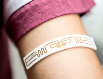
A radio-frequency identification (RFID) tag, used to monitor vital signs, can go into your pocket or be woven into a shirt (credit: Cornell)

Replacing devices based on 19th-century technology* and still in use, Cornell University engineers have developed a simple method for gathering blood pressure, heart rate, and breath rate from multiple patients simultaneously. It uses low-power radio-frequency signals and low-cost microchip radio-frequency identification (RFID) “tags” — similar to the ubiquitous anti-theft tags used in department stores.

The RFID tags measure internal body motion, such as a heart as it beats or blood as it pulses under skin. Powered remotely by electromagnetic energy supplied by a central reader, the tags use a new concept called “near-field coherent sensing.” Mechanical motions (heartbeat, etc.) in the body modulate (modify) radio waves that are bounced off the body and internal organs by passive (no battery required) RFID tags.

The modulated signals detected by the tag then bounce back to an electronic reader, located elsewhere in the room, that gathers the data. Each tag has a unique identification code that it transmits with its signal, allowing up to 200 people to be monitored simultaneously.

Electromagnetic simulations of monitoring vital signs via radio transmission, showing heartbeat sensing (left) and pulse sensing (right) (credit: Xiaonan Hui and Edwin C. Kan/Nature Electronics)

“If this is an emergency room, everybody that comes in can wear these tags or can simply put tags in their front pockets, and everybody’s vital signs can be monitored at the same time. I’ll know exactly which person each of the vital signs belongs to,” said Edwin Kan, a Cornell professor of electrical and computer engineering.

The signal is as accurate as an electrocardiogram or a blood-pressure cuff, according to Kan, who believes the technology could also be used to measure bowel movement, eye movement, and many other internal mechanical motions produced by the body.

The researchers envision embedding the RFID chips in clothing to monitor health in real time, with little or no effort required by the user. They have also developed a method for embroidering the tags directly onto clothing using fibers coated with nanoparticles. A cellphone could read (and display) your vital signs and also transmit them for remote medical monitoring.

The system is detailed in the open-access paper “Monitoring Vital Signs Over Multiplexed Radio by Near-Field Coherent Sensing,” published online Nov. 27 in the journal Nature Electronics. “Current approaches to monitoring vital signs are based on body electrodes, optical absorption, pressure or strain gauges, stethoscope, and ultrasound or radiofrequency (RF) backscattering, each of which suffers particular drawbacks during application,” the paper notes.

* The sphygmomanometer was invented by Samuel Siegfried Karl Ritter von Basch in 1881. Devices based on its basic pressure principle are still in use. (credit: Wellcome Trustees)


Abstract of Monitoring vital signs over multiplexed radio by near-field coherent sensing

Monitoring the heart rate, blood pressure, respiration rate and breath effort of a patient is critical to managing their care, but current approaches are limited in terms of sensing capabilities and sampling rates. The measurement process can also be uncomfortable due to the need for direct skin contact, which can disrupt the circadian rhythm and restrict the motion of the patient. Here we show that the external and internal mechanical motion of a person can be directly modulated onto multiplexed radiofrequency signals integrated with unique digital identification using near-field coherent sensing. The approach, which does not require direct skin contact, offers two possible implementations: passive and active radiofrequency identification tags. To minimize deployment and maintenance cost, passive tags can be integrated into garments at the chest and wrist areas, where the two multiplexed far-field backscattering waveforms are collected at the reader to retrieve the heart rate, blood pressure, respiration rate and breath effort. To maximize reading range and immunity to multipath interference caused by indoor occupant motion, active tags could be placed in the front pocket and in the wrist cuff to measure the antenna reflection due to near-field coherent sensing and then the vital signals sampled and transmitted entirely in digital format. Our system is capable of monitoring multiple people simultaneously and could lead to the cost-effective automation of vital sign monitoring in care facilities.

3D-printing biocompatible living bacteria

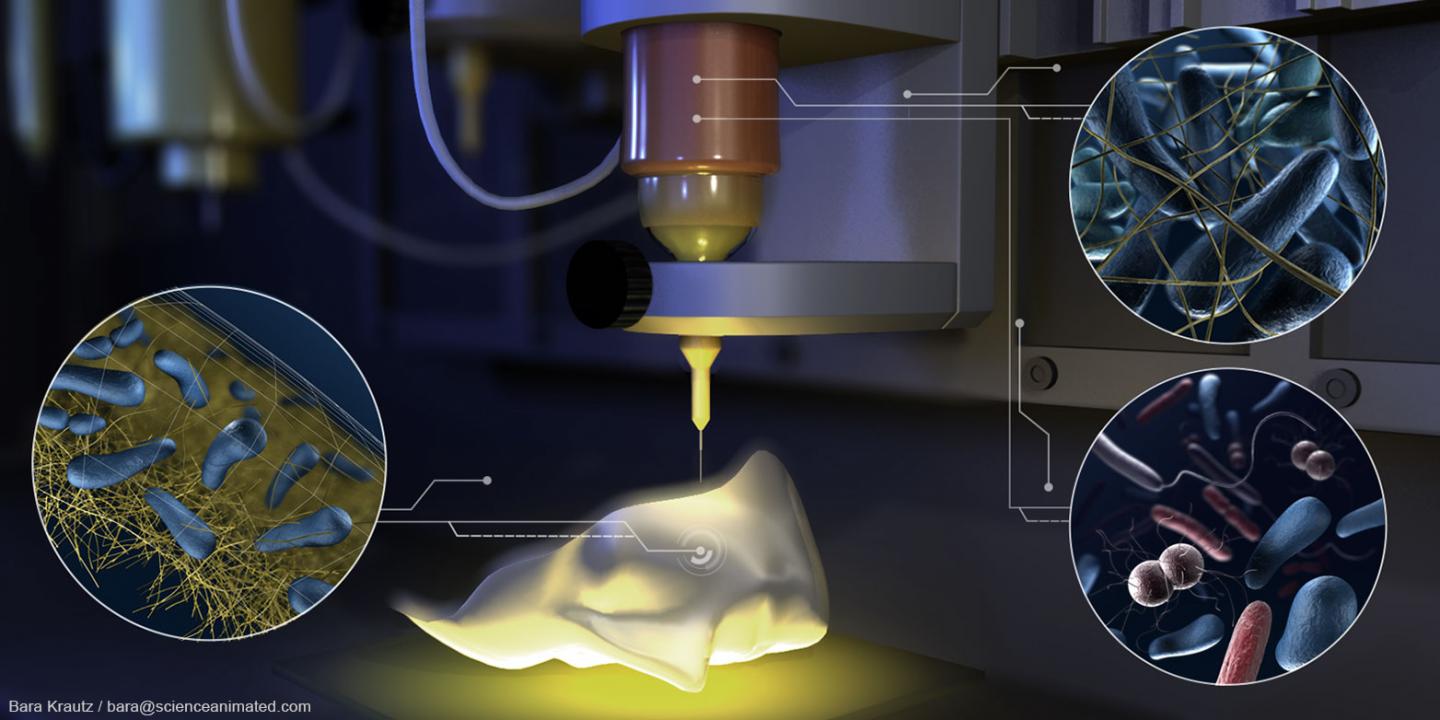
3D-printing with an ink containing living bacteria (credit: Bara Krautz/bara@scienceanimated.com)

Researchers at ETH Zurich university have developed a technique for 3D-printing biocompatible living bacteria for the first time — making it possible to produce produce high-purity cellulose for biomedical applications and nanofilters that can break down toxic substances (in drinking water, for example) or for use in disastrous oil spills, for example.

The technique, called “Flink” (“functional living ink”) allows for printing mini biochemical factories with properties that vary based on which species of bacteria are used. Up to four different inks containing different species of bacteria at different concentrations can be printed in a single pass.

Schematics of the Flink 3D bacteria-printing process for creating two types of functional living materials. (Left and center) Bacteria are embedded in a biocompatible hydrogel (which provides the supporting structure). (Right) The inclusion of P. putida* or A. xylinum* bacteria in the ink yields 3D-printed materials capable of degrading environmental pollutants (top) or forming bacterial cellulose in situ for biomedical applications (bottom), respectively. (credit: Manuel Schaffner et al./Science Advances)

The technique was described Dec. 1, 2017 in the open-access journal Science Advances.

(Left) A. xylinum bacteria were used in printing a cellulose nanofibril network (scanning electron microscope image), which was deposited (Right) on a doll face, forming a cellulose-reinforced hydrogel that, after removal of all biological residues, could serve as a skin transplant. (credit: Manuel Schaffner et al./Science Advances)

“The in situ formation of reinforcing cellulose fibers within the hydrogel is particularly attractive for regions under mechanical tension, such as the elbow and knee, or when administered as a pouch onto organs to prevent fibrosis after surgical implants and transplantations,” the researchers note in the paper. “Cellulose films grown in complex geometries precisely match the topography of the site of interest, preventing the formation of wrinkles and entrapments of contaminants that could impair the healing process. We envision that long-term medical applications will benefit from the presented multimaterial 3D printing process by locally deploying bacteria where needed.”

 * Pseudomonas putida breaks down the toxic chemical phenol, which is produced on a grand scale in the chemical industry; Acetobacter xylinum secretes high-purity nanocellulose, which relieves pain, retains moisture and is stable, opening up potential applications in the treatment of burns.


Abstract of 3D printing of bacteria into functional complex materials

Despite recent advances to control the spatial composition and dynamic functionalities of bacteria embedded in materials, bacterial localization into complex three-dimensional (3D) geometries remains a major challenge. We demonstrate a 3D printing approach to create bacteria-derived functional materials by combining the natural diverse metabolism of bacteria with the shape design freedom of additive manufacturing. To achieve this, we embedded bacteria in a biocompatible and functionalized 3D printing ink and printed two types of “living materials” capable of degrading pollutants and of producing medically relevant bacterial cellulose. With this versatile bacteria-printing platform, complex materials displaying spatially specific compositions, geometry, and properties not accessed by standard technologies can be assembled from bottom up for new biotechnological and biomedical applications.

Using microrobots to diagnose and treat illness in remote areas of the body

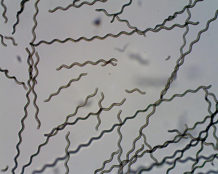
Spirulina algae coated with magnetic particles to form a microrobot. Devices such as these could be developed to diagnose and treat illness in hard-to-reach parts of the body. (credit: Yan et al./Science Robotics)

Imagine a swarm of remote-controlled microrobots, a few micrometers in length (blood-vessel-sized), unleashed into your body to swim through your intestinal track or blood vessels, for example. Goal: to diagnose illness and treat it in hard-to-reach areas of the body.

An international team of researchers, led by the Chinese University of Hong Kong, is now experimenting with this idea (starting with rats) — using microscopic Spirulina algae coated with biocompatible magnetic nanoparticles to form the microswimmers.

Schematic of dip-coating S. platensis algae in a suspension of magnetite nanoparticles and growing microrobots. The time taken for the robots to function and biodegrade within the body could be tailored by adjusting the thickness of the coating. (credit: Xiaohui Yan et al./Science Robotics)

There are two methods being studied: (1) track the microswimmers in tissue close to the skin’s surface by imaging the algae’s natural luminescence; and (2) track them in hard-to-reach deeper tissue by coating with magnetite (Fe3O4) to make them detectable with magnetic resonance imaging (MRI). The devices could also sense chemical changes linked to the onset of illness.

In lab tests, during degradation, the microswimmers were able to release potent compounds from the algae core that selectively attacked cancer cells while leaving healthy cells unharmed. Further research could show whether this might have potential as a treatment, the researchers say.

The study, published in an open-access paper in Science Robotics, was carried out in collaboration with the Universities of Edinburgh and Manchester and was supported by the Research Grants Council of Hong Kong.


Abstract of Multifunctional biohybrid magnetite microrobots for imaging-guided therapy

Magnetic microrobots and nanorobots can be remotely controlled to propel in complex biological fluids with high precision by using magnetic fields. Their potential for controlled navigation in hard-to-reach cavities of the human body makes them promising miniaturized robotic tools to diagnose and treat diseases in a minimally invasive manner. However, critical issues, such as motion tracking, biocompatibility, biodegradation, and diagnostic/therapeutic effects, need to be resolved to allow preclinical in vivo development and clinical trials. We report biohybrid magnetic robots endowed with multifunctional capabilities by integrating desired structural and functional attributes from a biological matrix and an engineered coating. Helical microswimmers were fabricated from Spirulinamicroalgae via a facile dip-coating process in magnetite (Fe3O4) suspensions, superparamagnetic, and equipped with robust navigation capability in various biofluids. The innate properties of the microalgae allowed in vivo fluorescence imaging and remote diagnostic sensing without the need for any surface modification. Furthermore, in vivo magnetic resonance imaging tracked a swarm of microswimmers inside rodent stomachs, a deep organ where fluorescence-based imaging ceased to work because of its penetration limitation. Meanwhile, the microswimmers were able to degrade and exhibited selective cytotoxicity to cancer cell lines, subject to the thickness of the Fe3O4 coating, which could be tailored via the dip-coating process. The biohybrid microrobots reported herein represent a microrobotic platform that could be further developed for in vivo imaging–guided therapy and a proof of concept for the engineering of multifunctional microrobotic and nanorobotic devices.

3D ‘body-on-a-chip’ project aims to accelerate drug testing, reduce costs

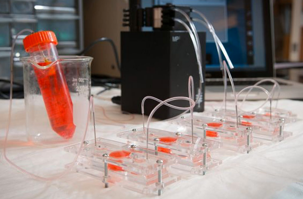
Scientists created miniature models (“organoids”) of heart, liver, and lung  in dishes and combined them into an integrated “body-on-a-chip” system fed with nutrient-rich fluid, mimicking blood. (credit: Wake Forest Baptist Medical Center)

A team of scientists at Wake Forest Institute for Regenerative Medicine and nine other institutions has engineered miniature 3D human hearts, lungs, and livers to achieve more realistic testing of how the human body responds to new drugs.

The “body-on-a-chip” project, funded by the Defense Threat Reduction Agency, aims to help reduce the estimated $2 billion cost and 90 percent failure rate that pharmaceutical companies face when developing new medications. The research is described in an open-access paper in Scientific Reports, published by Nature.

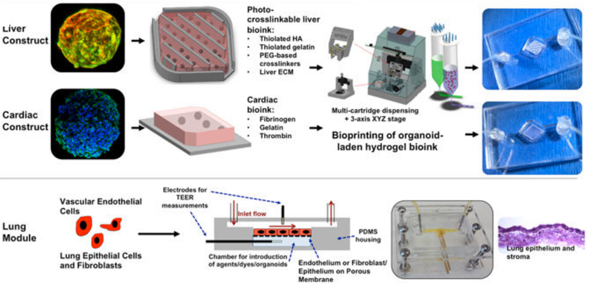
Using the same expertise they’ve employed to build new organs for patients, the researchers connected together micro-sized 3D liver, heart, and lung organs-on-a chip (or “organoids”) on a single platform to monitor their function. They selected heart and liver for the system because toxicity to these organs is a major reason for drug candidate failures and drug recalls. And lungs were selected because they’re the point of entry for toxic particles and for aerosol drugs such as asthma inhalers.

The integrated three-tissue organ-on-a-chip platform combines liver, heart, and lung organoids. (Top) Liver and cardiac modules are created by bioprinting spherical organoids using customized bioinks, resulting in 3D hydrogel constructs (upper left) that are placed into the microreactor devices. (Bottom) Lung modules are formed by creating layers of cells over porous membranes within microfluidic devices. TEER (trans-endothelial [or epithelial] electrical resistance sensors allow for monitoring tissue barrier function integrity over time. The three organoids are placed in a sealed, monitored system with a real-time camera. A nutrient-filled liquid that circulates through the system keeps the organoids alive and is used to introduce potential drug therapies into the system. (credit: Aleksander Skardal et al./Scientific Reports)

Why current drug testing fails

Drug compounds are currently screened in the lab using human cells and then tested in animals. But these methods don’t adequately replicate how drugs affect human organs. “If you screen a drug in livers only, for example, you’re never going to see a potential side effect to other organs,” said Aleks Skardal, Ph.D., assistant professor at Wake Forest Institute for Regenerative Medicine and lead author of the paper.

In many cases during testing of new drug candidates — and sometimes even after the drugs have been approved for use — drugs also have unexpected toxic effects in tissues not directly targeted by the drugs themselves, he explained. “By using a multi-tissue organ-on-a-chip system, you can hopefully identify toxic side effects early in the drug development process, which could save lives as well as millions of dollars.”

“There is an urgent need for improved systems to accurately predict the effects of drugs, chemicals and biological agents on the human body,” said Anthony Atala, M.D., director of the institute and senior researcher on the multi-institution study. “The data show a significant toxic response to the drug as well as mitigation by the treatment, accurately reflecting the responses seen in human patients.”

Advanced drug screening, personalized medicine

The scientists conducted multiple scenarios to ensure that the body-on-a-chip system mimics a multi-organ response.

For example, they introduced a drug used to treat cancer into the system. Known to cause scarring of the lungs, the drug also unexpectedly affected the system’s heart. (A control experiment using only the heart failed to show a response.) The scientists theorize that the drug caused inflammatory proteins from the lung to be circulated throughout the system. As a result, the heart increased beats and then later stopped altogether, indicating a toxic side effect.

“This was completely unexpected, but it’s the type of side effect that can be discovered with this system in the drug development pipeline,” Skardal noted.

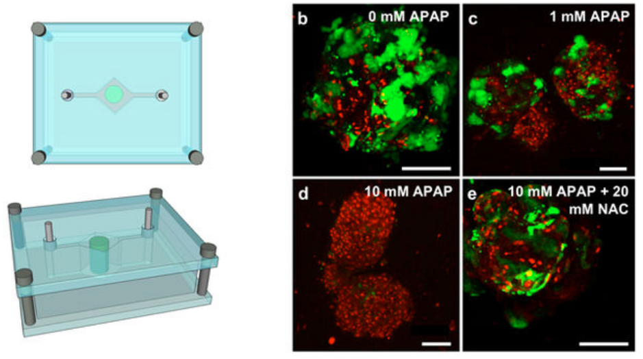
Test of “liver on a chip” response to two drugs to demonstrate clinical relevance. Liver construct toxicity response was assessed following exposure to acetaminophen (APAP) and the clinically-used APAP countermeasure N-acetyl-L-cysteine (NAC). Liver constructs in the fluidic system (left) were treated with no drug (b), 1 mM APAP (c), and 10 mM APAP (d) — showing progressive loss of function and cell death, compared to 10 mM APAP +20 mM NAC (e), which mitigated those negative effects. The data shows both a significant cytotoxic (cell-damage) response to APAP as well as its mitigation by NAC treatment — accurately reflecting the clinical responses seen in human patients. (credit: Aleksander Skardal et al./Scientific Reports)

The scientists are now working to increase the speed of the system for large scale screening and add additional organs.

“Eventually, we expect to demonstrate the utility of a body-on-a-chip system containing many of the key functional organs in the human body,” said Atala. “This system has the potential for advanced drug screening and also to be used in personalized medicine — to help predict an individual patient’s response to treatment.”

Several patent applications comprising the technology described in the paper have been filed.

The international collaboration included researchers at Wake Forest Institute for Regenerative Medicine at the Wake Forest School of Medicine, Harvard-MIT Division of Health Sciences and Technology, Wyss Institute for Biologically Inspired Engineering at Harvard University, Biomaterials Innovation Research Center at Harvard Medical School, Bloomberg School of Public Health at Johns Hopkins University, Virginia Tech-Wake Forest School of Biomedical Engineering and Sciences, Brigham and Women’s Hospital, University of Konstanz, Konkuk University (Seoul), and King Abdulaziz University.


Abstract of Multi-tissue interactions in an integrated three-tissue organ-on-a-chip platform

Many drugs have progressed through preclinical and clinical trials and have been available – for years in some cases – before being recalled by the FDA for unanticipated toxicity in humans. One reason for such poor translation from drug candidate to successful use is a lack of model systems that accurately recapitulate normal tissue function of human organs and their response to drug compounds. Moreover, tissues in the body do not exist in isolation, but reside in a highly integrated and dynamically interactive environment, in which actions in one tissue can affect other downstream tissues. Few engineered model systems, including the growing variety of organoid and organ-on-a-chip platforms, have so far reflected the interactive nature of the human body. To address this challenge, we have developed an assortment of bioengineered tissue organoids and tissue constructs that are integrated in a closed circulatory perfusion system, facilitating inter-organ responses. We describe a three-tissue organ-on-a-chip system, comprised of liver, heart, and lung, and highlight examples of inter-organ responses to drug administration. We observe drug responses that depend on inter-tissue interaction, illustrating the value of multiple tissue integration for in vitro study of both the efficacy of and side effects associated with candidate drugs.

A battery-free origami robot powered and controlled by external magnetic fields

Wirelessly powered and controlled magnetic folding robot arm can grasp and bend (credit: Wyss Institute at Harvard University)

Harvard University researchers have created a battery-free, folding robot “arm” with multiple “joints,” gripper “hand,” and actuator “muscles” — all powered and controlled wirelessly by an external resonant magnetic field.

The design is inspired by the traditional Japanese art of origami (used to transform a simple sheet of paper into complex, three-dimensional shapes through a specific pattern of folds, creases, and crimps). The prototype device is capable of complex, repeatable movements at millimeter to centimeter scales.

The research, by scientists at the Wyss Institute for Biologically Inspired Engineering and the John A. Paulson School of Engineering and Applied Sciences (SEAS), is reported in Science Robotics.

How it works

Design of small-scale-structure prototype of wirelessly controlled robotic arm (credit: Mustafa Boyvat et al./Science Robotics)

The researchers designed a 0.8-gram prototype small-scale-structure* prototype robotic “arm” capable of bending and opening or closing a gripper around an object. The “arm” is constructed with a special origami-like pattern that uses hinges (“joints”) to permit it to bend. There is also a “hand” (gripper — left panel in above image) that opens or closes.

To power the device, an external coil with its own power source (see video below) is used to generate a low-frequency magnetic field that induces an electrical current in three magnetic coils. The current heats the spiral-wire shape-memory-alloy actuator wires (coiled wire shown in inset above). That causes the actuator wires (“muscles”) to contract, making the attached nearby “joints” bend, and folding the robot body.

Mechanism of the origami gripper (for small-scale prototype design). (Left) The coil SMA actuator pushes the center link connected to both fingers and the gripper opens fingers, enabled by dynamic folding at the joints (left). The plate spring, which is a passive compression spring, pulls the link back as the gripper closes the fingers, again by rotations at folding joints (center). (Right) A photo of the gripper showing the SMA actuator wire attached at the center link. (credit: Mustafa Boyvat et al./Science Robotics)

By changing the resonant frequency of the external electromagnetic field, the two longer actuator wires (coiled wires shown in above illustration) are instead heated and stretched, opening the gripper (“hand”).

In both cases, when the external field-induced current stops, the actuators relax, springing back to their “memory” positions and causing the robot body to straighten out or the gripper’s outer triangles to close.

Minimally invasive medicine and surgery applications

As an example of a practical future application, instead of having an uncomfortable endoscope put down their throat to assist a doctor with surgery, a patient could just swallow a micro-robot that could move around and perform simple tasks, like holding tissue or filming, powered by a coil outside their body.

Using a much larger source coil — on the order of yards in diameter — could enable wireless, battery-free communication between multiple “smart” objects in a room or building.

“Medical devices today are commonly limited by the size of the batteries that power them, whereas these remotely powered origami robots can break through that size barrier and potentially offer entirely new, minimally invasive approaches for medicine and surgery in the future,” says Wyss Founding Director Donald Ingber, who is also the Judah Folkman Professor of Vascular Biology at Harvard Medical School and the Vascular Biology Program at Boston Children’s Hospital, as well as a Professor of Bioengineering at Harvard’s School of Engineering and Applied Sciences.

This work was supported by the National Science Foundation, the U.S. Army Research Laboratory, and the Swiss National Science Foundation.

* A large-scale-structure prototype version has minor differences, including 12-cm folding lines vs. 1.7-cm folding lines in the smaller version.

Wyss Institute | Battery-Free Folding Robots


Abstract of Addressable wireless actuation for multijoint folding robots and devices

“Printing” robots and other complex devices through a process of origami-like folding is an emerging and promising manufacturing method due to the inherent simplicity and low cost of folding-based assembly. Folding is used in this class of device to create both complex static structures and flexure-based compliant mechanisms. Dependency on batteries to power these folds with no external wires is a hurdle to giving small-scale folding robots and devices functionality. We demonstrate a battery-free wireless folding method for dynamic multijoint structures, achieving addressable folding motions—both individual and collective folding—using only basic passive electronic components on the device. The method is based on electromagnetic power transmission and resonance selectivity for actuation of resistive shape memory alloy actuators without the need for physical connection or line of sight. We demonstrate the utility of this approach using two folded devices at different sizes using different circuit approaches.

New system allows near-zero-power sensors to communicate data over long distances

This low-cost, flexible epidermal medical-data patch prototype successfully transmitted information at up to 37500 bits per second across a 3,300-square-feet atrium. (credit: Dennis Wise/University of Washington)

University of Washington (UW) researchers have developed a low-cost, long-range data-communication system that could make it possible for medical sensors or billions of low-cost “internet of things” objects to connect via radio signals at long distances (up to 2.8 kilometers) and with 1000 times lower required power (9.25 microwatts in an experiment) compared to existing technologies.

“People have been talking about embedding connectivity into everyday objects … for years, but the problem is the cost and power consumption to achieve this,” said Vamsi Talla, chief technology officer of Jeeva Wireless, which plans to market the system within six months. “This is the first wireless system that can inject connectivity into any device with very minimal cost.”

The new system uses “backscatter,” which uses energy from ambient transmissions (from WiFi, for example) to power a passive sensor that encodes and scatter-reflects the signal. (This article explains how ambient backscatter, developed by UW, works.) Backscatter systems, used with RFID chips, are very low cost, but are limited in distance.

So the researchers combined backscatter with a “chirp spread spectrum” technique, used in LoRa (long-range) wireless data-communication systems.

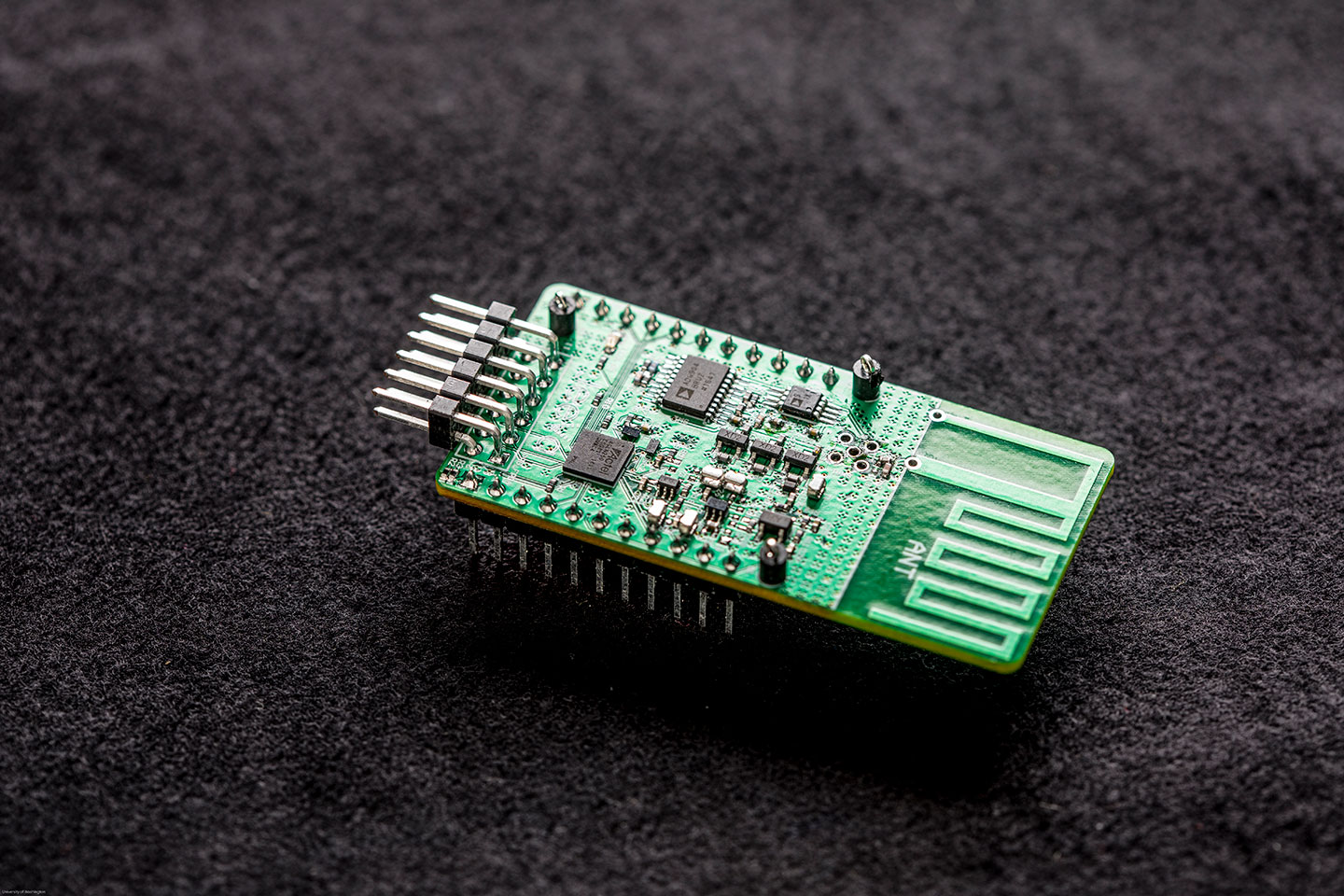
This tiny off-the-shelf spread-spectrum receiver enables extremely-low-power cheap sensors to communicate over long distances. (credit: Dennis Wise/University of Washington)

This new system has three components: a power source (which can be WiFi or other ambient transmission sources, or cheap flexible printed batteries, with an expected bulk cost of 10 to 20 cents each) for a radio signal; cheap sensors (less than 10 cents at scale) that modulate (encode) information (contained in scattered reflections of the signal), and an inexpensive, off-the-shelf spread-spectrum receiver, located as far away as 2.8 kilometers, that decodes the sensor information.

Applications could include, for example, medical monitoring devices that wirelessly transmit information about a heart patient’s condition to doctors; sensor arrays that monitor pollution, noise, or traffic in “smart” cities; and farmers looking to measure soil temperature or moisture, who could affordably blanket an entire field to determine how to efficiently plant seeds or water.

The research team built a contact lens prototype and a flexible epidermal patch that attaches to human skin, which successfully used long-range backscatter to transmit information across a 3300-square-foot building.

The research, which was partially funded by the National Science Foundation, is detailed in an open-access paper presented Sept. 13, 2017 at UbiComp 2017. More information: longrange@cs.washington.edu.


UW (University of Washington) | UW team shatters long-range communication barrier for devices that consume almost no power


Abstract of LoRa Backscatter: Enabling The Vision of Ubiquitous Connectivity

The vision of embedding connectivity into billions of everyday objects runs into the reality of existing communication technologies — there is no existing wireless technology that can provide reliable and long-range communication at tens of microwatts of power as well as cost less than a dime. While backscatter is low-power and low-cost, it is known to be limited to short ranges. This paper overturns this conventional wisdom about backscatter and presents the first wide-area backscatter system. Our design can successfully backscatter from any location between an RF source and receiver, separated by 475 m, while being compatible with commodity LoRa hardware. Further, when our backscatter device is co-located with the RF source, the receiver can be as far as 2.8 km away. We deploy our system in a 4,800 ft2 (446 m2) house spread across three floors, a 13,024 ft2 (1210 m2) office area covering 41 rooms, as well as a one-acre (4046 m2) vegetable farm and show that we can achieve reliable coverage, using only a single RF source and receiver. We also build a contact lens prototype as well as a flexible epidermal patch device attached to the human skin. We show that these devices can reliably backscatter data across a 3,328 ft2 (309 m2) room. Finally, we present a design sketch of a LoRa backscatter IC that shows that it costs less than a dime at scale and consumes only 9.25 &mgr;W of power, which is more than 1000x lower power than LoRa radio chipsets.

Walking DNA nanorobot could deliver a drug to a precise location in your body

DNA nanorobot cargo carrier (artist’s impression) (credit: Ella Maru Studio)

Caltech scientists have developed a “cargo sorting” DNA nanorobot programmed to autonomously “walk” around a surface, pick up certain molecules, and drop them off in designated locations.

The research is described in a paper in the Friday, September 15, 2017 issue of Science.

The major advance in this study is “their methodology for designing simple DNA devices that work in parallel to solve nontrivial tasks,” notes Duke University computer scientist John H. Reif in an article in the same issue of Science.

Such tasks could include synthesizing a drug in a molecular factory or delivering a drug only when a specific signal is present in bloodstreams, say the researchers. “So far, the development of DNA robots has been limited to simple functions,” the researchers note.

Walking nanobots that work in parallel

Conceptual illustration of two DNA nanorobots collectively performing a cargo-sorting task on a DNA origami surface: transporting fluorescent molecules with different colors from initially random locations to ordered destinations. (credit: Demin Liu)

The DNA nanorobot, intended as a proof of concept, has a “leg” with two “feet” for walking, and an “arm” and “hand” for picking up cargo. It also has a segment that can recognize a specific drop-off point and signal to the hand to release its cargo. Each of these building blocks are made of just a few nucleotides (molecules that form DNA) within a single strand of DNA.*

As the robot encounters cargo molecules tethered to pegs, it grabs them with its “hand” components and carries them around (with a 6-nm step size) until it detects the signal of the drop-off point.

Multiple DNA nanorobots independently execute three operations in parallel: [1] cargo pickup, [2] random movement to adjacent stepping stones, and [3] cargo drop-off at ordered locations. (credit: C. Bickel/Science)

In experiments, the nanorobots successfully sorted six randomly scattered molecules into their correct places in 24 hours. The process is slow, but adding more robots to the surface shortened the time it took to complete the task. The very simple robot design utilizes very little chemical energy, according to the researchers.**

“The same system design can be generalized to work with dozens of types of cargos at any arbitrary initial location on the surface,” says lead author Anupama Thubagere. “One could also have multiple robots performing diverse sorting tasks in parallel,” [programmed] like macroscopic robots.”

Future applications

“We don’t develop DNA robots for any specific applications. Our lab focuses on discovering the engineering principles that enable the development of general-purpose DNA robots,” explains Lulu Qian, assistant professor of bioengineering.

“However, it is my hope that other researchers could use these principles for exciting applications, such as synthesizing a therapeutic chemical from its constituent parts in an artificial molecular factory, or sorting molecular components in trash for recycling. Just like electromechanical robots are sent off to faraway places, like Mars, we would like to send molecular robots to minuscule places where humans can’t go, such as the bloodstream.”

Funding was provided by Caltech Summer Undergraduate Research Fellowships, the National Science Foundation, and the Burroughs Wellcome Fund.

* The key to designing DNA machines is the fact that DNA has unique chemical and physical properties that are known and programmable. A single strand of DNA is made up of four different molecules called nucleotides—abbreviated A, G, C, and T—and arranged in a string called a sequence. These nucleotides bond in specific pairs: A with T, and G with C. When a single strand encounters a “reverse complementary strand” — for example, CGATT meets AATCG —the two strands zip together in the classic double-helix shape.

** Using these chemical and physical principles, researchers can also design “playgrounds,” such as molecular pegboards, to test them on, according to the researchers. In the current work, the DNA robot moves around on a 58-nanometer-by-58-nanometer pegboard on which the pegs are made of single strands of DNA complementary to the robot’s leg and foot. The robot binds to a peg with its leg and one of its feet — the other foot floats freely. When random molecular fluctuations cause this free foot to encounter a nearby peg, it pulls the robot to the new peg and its other foot is freed. This process continues with the robot moving in a random direction at each step.


Abstract of A cargo-sorting DNA robot

Two critical challenges in the design and synthesis of molecular robots are modularity and
algorithm simplicity.We demonstrate three modular building blocks for a DNA robot that
performs cargo sorting at themolecular level. A simple algorithm encoding recognition between
cargos and their destinations allows for a simple robot design: a single-stranded DNA with
one leg and two foot domains for walking, and one arm and one hand domain for picking up and
dropping off cargos.The robot explores a two-dimensional testing ground on the surface of
DNA origami, picks up multiple cargos of two types that are initially at unordered locations, and
delivers them to specified destinations until all molecules are sorted into two distinct piles.
The robot is designed to perform a random walk without any energy supply. Exploiting this
feature, a single robot can repeatedly sort multiple cargos. Localization on DNA origami allows
for distinct cargo-sorting tasks to take place simultaneously in one test tube or for multiple
robots to collectively perform the same task.