Miniature MRI simulator chip could help diagnose and treat diseases in the body at sub-millimeter precision

Illustration of an ATOMS microchip localized within the gastrointestinal tract (not to scale; a prototype measures just 0.7 cubic millimeters). The microchip contains a magnetic field sensor, integrated antennas, a wireless powering device, and a circuit that adjusts its radio frequency signal based on the magnetic field strength and wirelessly relays the chip’s precise location. (credit: Ella Marushchenko/Caltech)

Caltech researchers have developed a “Fantastic Voyage” style prototype microchip that could one day be used in “smart pills” to diagnose and treat diseases when inserted into the human body.

Called ATOMS (addressable transmitters operated as magnetic spins), the microchips could one day monitor a patient’s gastrointestinal tract, blood, or brain, measuring factors that indicate a patient’s health — such as pH, temperature, pressure, and sugar concentrations — with sub-millimeter localization and relay that information to doctors. Or the devices could even be instructed to release drugs at precise locations.

An open access paper describing the new device appears in the September issue of the journal Nature Biomedical Engineering. The lead author is Manuel Monge, who now works at Elon Musk’s new Neuralink company.

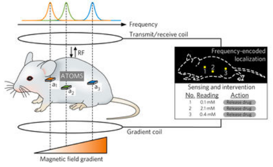
The ATOMS microchips, proven to work in tests with mice, mimic the way nuclear spins in atoms in the body resonate to magnetic fields in a magnetic resonance imaging (MRI) machine and can be precisely identified and localized within the body. Similarly, the ATOMS devices resonate at different frequencies depending on where they are in a magnetic field. (credit: Manuel Monge et al./ Nature Biomedical Engineering)


Abstract of Localization of Microscale Devices In Vivo using Addressable Transmitters Operated as Magnetic Spins

The function of miniature wireless medical devices, such as capsule endoscopes, biosensors and drug-delivery systems, depends critically on their location inside the body. However, existing electromagnetic, acoustic and imaging-based methods for localizing and communicating with such devices suffer from limitations arising from physical tissue properties or from the performance of the imaging modality. Here, we embody the principles of nuclear magnetic resonance in a silicon integrated-circuit approach for microscale device localization. Analogous to the behaviour of nuclear spins, the engineered miniaturized radio frequency transmitters encode their location in space by shifting their output frequency in proportion to the local magnetic field; applied field gradients thus allow each device to be located precisely from its signal’s frequency. The devices are integrated in circuits smaller than 0.7 mm3 and manufactured through a standard complementary-metal-oxide-semiconductor process, and are capable of sub-millimetre localization in vitro and in vivo. The technology is inherently robust to tissue properties, scalable to multiple devices, and suitable for the development of microscale devices to monitor and treat disease.

These fast, low-cost medical technologies will replace ultrasound and X-rays for specific uses

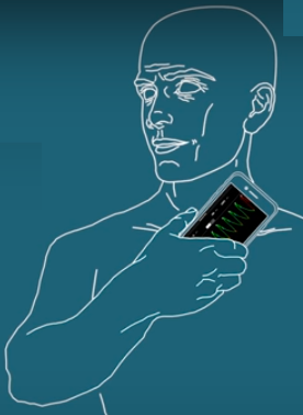
Smartphone instant heart diagnosis (credit: Caltech)

A radical software invention by three Caltech engineers promises to allow your smartphone camera* to provide detailed information about a critical measure of your heart’s health: the “left ventricular ejection fraction” (LVEF) — the amount of blood in the heart that is pumped out to the blood system with each beat. This figure is used by physicians as a base for diagnostic and therapeutic decisions.

You’ll simply hold your phone up to your neck for a minute or two.

In an experiment, the technique was found to be as accurate as a 45-minute echocardiography scan, which currently requires a trained technician operating an expensive ultrasound machine.

The smartphone technique measures how much the carotid artery displaces the skin of the neck as blood pumps through it. In a normal heart, the LVEF measure ranges from 50 to 70 percent. When the heart is weaker, less of the total amount of blood in the heart is pumped out with each beat, and the LVEF value is lower.

Carotid arterial waveform captured using an unmodified iPhone 5S camera by placing the iPhone camera over the carotid pulse (credit: Niema M. Pahlevan et al./Critical Card Medicine)

To test the app, clinical trials were conducted with 72 volunteers between the ages of 20 and 92 at an outpatient magnetic resonance imaging (MRI) facility. MRI is the gold standard in measuring LVEF but is seldom used clinically due to its high cost and limited availability. The measurements made by smartphone had a margin of error of ±19.1 percent compared with those done in an MRI. By way of comparison, the margin of error for echocardiography is around ±20.0 percent.

“This has the potential to revolutionize how doctors and patients can screen for and monitor heart disease, both in the U.S. and the developing world,” says Caltech’s Mory Gharib, the Hans W. Liepmann Professor of Aeronautics and Bioinspired Engineering and senior author of a paper on the study in the July issue of the Journal of Critical Care Medicine.

The researchers have founded a start-up named Avicena, LLC that has licensed this technology and will market the app. They also plan to use this approach to diagnose heart-valve diseases, like aortic stenosis and coronary artery blockage.

* For the study, the team used an iPhone 5, but they say any smartphone with a camera will work.

Seeing through the body

University of Edinburgh and Heriot-Watt University researchers have used a near-infrared camera to see through the chest to track the location of a fiber-optic endomicroscope (a long flexible tube with a light on the end) — replacing X-rays.

A “time-of-flight” camera detects light emitted from an endoscope in sheep lungs. Left: light emitted from the tip of the endoscope, revealing its precise location in the lungs. Right: an image using a conventional camera, with light scattered through the structures of the lung. (credit: Proteus)

Near-infrared light can readily pass through the body, but much of it scatters or bounces off tissues and organs rather than traveling straight through — making it nearly impossible to get a clear picture of where an object is in the body. So this camera uses a “time-of-flight” system: It calculates the distance to the endomicroscope light based on the time it takes individual photons to arrive directly (ignoring scattered photons, which take longer). That’s similar to how this camera can see an object around a corner.

The technology is so sensitive it can detect the miniscule amount of light that passes through 20 centimeters (about 8 inches) of the body’s tissue.

The research is described in an open-access paper in the journal Biomedical Optics Express.


Abstract of Noninvasive iPhone Measurement of Left Ventricular Ejection Fraction Using Intrinsic Frequency Methodology

Objective: The study is based on previously reported mathematical analysis of arterial waveform that extracts hidden oscillations in the waveform that we called intrinsic frequencies. The goal of this clinical study was to compare the accuracy of left ventricular ejection fraction derived from intrinsic frequencies noninvasively versus left ventricular ejection fraction obtained with cardiac MRI, the most accurate method for left ventricular ejection fraction measurement.

Design: After informed consent, in one visit, subjects underwent cardiac MRI examination and noninvasive capture of a carotid waveform using an iPhone camera (The waveform is captured using a custom app that constructs the waveform from skin displacement images during the cardiac cycle.). The waveform was analyzed using intrinsic frequency algorithm.

Setting: Outpatient MRI facility.

Subjects: Adults able to undergo MRI were referred by local physicians or self-referred in response to local advertisement and included patients with heart failure with reduced ejection fraction diagnosed by a cardiologist.

Interventions: Standard cardiac MRI sequences were used, with periodic breath holding for image stabilization. To minimize motion artifact, the iPhone camera was held in a cradle over the carotid artery during iPhone measurements.

Measurements and Main Results: Regardless of neck morphology, carotid waveforms were captured in all subjects, within seconds to minutes. Seventy-two patients were studied, ranging in age from 20 to 92 years old. The main endpoint of analysis was left ventricular ejection fraction; overall, the correlation between ejection fraction–iPhone and ejection fraction–MRI was 0.74 (r = 0.74; p < 0.0001; ejection fraction–MRI = 0.93 × [ejection fraction–iPhone] + 1.9).

Conclusions: Analysis of carotid waveforms using intrinsic frequency methods can be used to document left ventricular ejection fraction with accuracy comparable with that of MRI. The measurements require no training to perform or interpret, no calibration, and can be repeated at the bedside to generate almost continuous analysis of left ventricular ejection fraction without arterial cannulation.


Abstract of Ballistic and snake photon imaging for locating optical endomicroscopy fibres

We demonstrate determination of the location of the distal-end of a fibre-optic device deep in tissue through the imaging of ballistic and snake photons using a time resolved single-photon detector array. The fibre was imaged with centimetre resolution, within clinically relevant settings and models. This technique can overcome the limitations imposed by tissue scattering in optically determining the in vivo location of fibre-optic medical instruments.

Flexible ‘electronic skin’ patch provides wearable health monitoring anywhere on the body

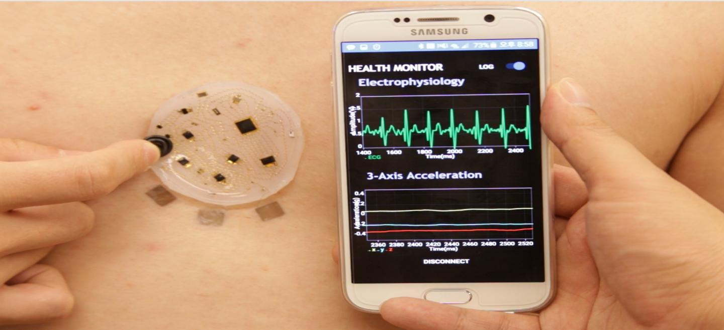
New soft electronic stick-on patch collects, analyzes, and diagnoses biosignals and sends data wirelessly to a mobile app. (credit: DGIST)

A radical new electronic skin monitor developed by Korean and U.S. scientists tracks heart rate, respiration, muscle movement, acceleration, and electrical activity in the heart, muscles, eyes, and brain and wirelessly transmits it to a smartphone, allowing for continuous health monitoring.

KurzweilAI has covered a number of biomedical skin-monitoring devices. This new design is noteworthy because the soft, flexible self-adhesive patch (a soft silicone material about four centimeters or 1.5 inches in diameter) can be instantly stuck just about anywhere on the body as needed — no battery required (it’s powered wirelessly).

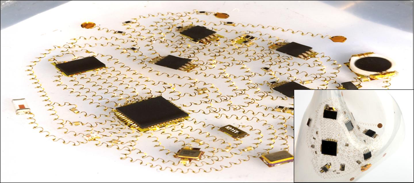
Optical image of the three-dimensional network of helical coils as electrical interconnects for soft electronics. (credit: DGIST)

The patch is designed more like a mattress or creeping vine than a conventional electronic device. It contains about 50 components connected by a network of 250 tiny flexible wire coils embedded in protective silicone. Unlike flat sensors, the tiny helical wire coils, made of gold, chromium and phosphate, are firmly connected to the base only at one end and can stretch and contract like a spring without breaking.

Helical coils serve as 3D electrical interconnects for soft electronics. (credit: DGIST)

The researchers say the microsystem could also be used in soft robotics, virtual reality, and autonomous navigation.

The microsystem was developed by an international team led by Kyung-In Jang, a professor of robotics engineering at South Korea’s Daegu Gyeongbuk Institute of Science and Technology, and John A. Rogers, the director of Northwestern University’s Center for Bio-Integrated Electronics. The research is described in the open-access journal Nature Communications.

“We have several human subject studies ongoing with our medical school at Northwestern — mostly with a focus on health status monitoring in infants,” Rogers told KurzweilAI.


Abstract of Self-assembled three dimensional network designs for soft electronics

Low modulus, compliant systems of sensors, circuits and radios designed to intimately interface with the soft tissues of the human body are of growing interest, due to their emerging applications in continuous, clinical-quality health monitors and advanced, bioelectronic therapeutics. Although recent research establishes various materials and mechanics concepts for such technologies, all existing approaches involve simple, two-dimensional (2D) layouts in the constituent micro-components and interconnects. Here we introduce concepts in three-dimensional (3D) architectures that bypass important engineering constraints and performance limitations set by traditional, 2D designs. Specifically, open-mesh, 3D interconnect networks of helical microcoils formed by deterministic compressive buckling establish the basis for systems that can offer exceptional low modulus, elastic mechanics, in compact geometries, with active components and sophisticated levels of functionality. Coupled mechanical and electrical design approaches enable layout optimization, assembly processes and encapsulation schemes to yield 3D configurations that satisfy requirements in demanding, complex systems, such as wireless, skin-compatible electronic sensors.

Drinking coffee associated with lower risk of death from all causes, study finds

(credit: iStock)

People who drink around three cups of coffee a day may live longer than non-coffee drinkers, a landmark study has found.

The findings — published in the journal Annals of Internal Medicine — come from the largest study of its kind, in which scientists analyzed data from more than half a million people across 10 European countries to explore the effect of coffee consumption on risk of mortality.

Researchers from the International Agency for Research on Cancer (IARC) and Imperial College London found that higher levels of coffee consumption were associated with a reduced risk of death from all causes, particularly from circulatory diseases and diseases related to the digestive tract.

“We found that higher coffee consumption was associated with a lower risk of death from any cause, and specifically for circulatory diseases, and digestive diseases,” said lead author Marc Gunter of the IARC and formerly at Imperial’s School of Public Health. “Importantly, these results were similar across all of the 10 European countries, with variable coffee drinking habits and customs. Our study also offers important insights into the possible mechanisms for the beneficial health effects of coffee.”

Healthier livers, better glucose control

Using data from the EPIC study (European Prospective Investigation into Cancer and Nutrition), the group analysed data from 521,330 people from over the age of 35 from 10 EU countries, including the UK, France, Denmark and Italy. People’s diets were assessed using questionnaires and interviews, with the highest level of coffee consumption (by volume) reported in Denmark (900 mL per day) and lowest in Italy (approximately 92 mL per day). Those who drank more coffee were also more likely to be younger, to be smokers, drinkers, eat more meat and less fruit and vegetables.

After 16 years of follow up, almost 42,000 people in the study had died from a range of conditions including cancer, circulatory diseases, heart failure and stroke. Following careful statistical adjustments for lifestyle factors such as diet and smoking, the researchers found that the group with the highest consumption of coffee had a lower risk for all causes of death, compared to those who did not drink coffee.

They found that decaffeinated coffee had a similar effect.

In a subset of 14,000 people, they also analyzed metabolic biomarkers, and found that coffee drinkers may have healthier livers overall and better glucose control than non-coffee drinkers.

According to the group, more research is needed to find out which of the compounds in coffee may be giving a protective effect or potentially benefiting health.* Other avenues of research to explore could include intervention studies, looking at the effect of coffee drinking on health outcomes.

However, Gunter noted that “due to the limitations of observational research, we are not at the stage of recommending people to drink more or less coffee. That said, our results suggest that moderate coffee drinking is not detrimental to your health, and that incorporating coffee into your diet could have health benefits.”

The study was funded by the European Commission Directorate General for Health and Consumers and the IARC.

* Coffee contains a number of compounds that can interact with the body, including caffeine, diterpenes and antioxidants, and the ratios of these compounds can be affected by the variety of methods used to prepare coffee.


Abstract of Coffee Drinking and Mortality in 10 European Countries: A Multinational Cohort Study

Background: The relationship between coffee consumption and mortality in diverse European populations with variable coffee preparation methods is unclear.

Objective: To examine whether coffee consumption is associated with all-cause and cause-specific mortality.

Design: Prospective cohort study.

Setting: 10 European countries.

Participants: 521 330 persons enrolled in EPIC (European Prospective Investigation into Cancer and Nutrition).

Measurements: Hazard ratios (HRs) and 95% CIs estimated using multivariable Cox proportional hazards models. The association of coffee consumption with serum biomarkers of liver function, inflammation, and metabolic health was evaluated in the EPIC Biomarkers subcohort (n = 14 800).

Results: During a mean follow-up of 16.4 years, 41 693 deaths occurred. Compared with nonconsumers, participants in the highest quartile of coffee consumption had statistically significantly lower all-cause mortality (men: HR, 0.88 [95% CI, 0.82 to 0.95]; P for trend < 0.001; women: HR, 0.93 [CI, 0.87 to 0.98]; P for trend = 0.009). Inverse associations were also observed for digestive disease mortality for men (HR, 0.41 [CI, 0.32 to 0.54]; P for trend < 0.001) and women (HR, 0.60 [CI, 0.46 to 0.78]; P for trend < 0.001). Among women, there was a statistically significant inverse association of coffee drinking with circulatory disease mortality (HR, 0.78 [CI, 0.68 to 0.90]; P for trend < 0.001) and cerebrovascular disease mortality (HR, 0.70 [CI, 0.55 to 0.90]; P for trend = 0.002) and a positive association with ovarian cancer mortality (HR, 1.31 [CI, 1.07 to 1.61]; P for trend = 0.015). In the EPIC Biomarkers subcohort, higher coffee consumption was associated with lower serum alkaline phosphatase; alanine aminotransferase; aspartate aminotransferase; γ-glutamyltransferase; and, in women, C-reactive protein, lipoprotein(a), and glycated hemoglobin levels.

Limitations: Reverse causality may have biased the findings; however, results did not differ after exclusion of participants who died within 8 years of baseline. Coffee-drinking habits were assessed only once.

Conclusion:

Coffee drinking was associated with reduced risk for death from various causes. This relationship did not vary by country.

Primary Funding Source:

European Commission Directorate-General for Health and Consumers and International Agency for Research on Cancer.


Abstract of Association of Coffee Consumption With Total and Cause-Specific Mortality Among Nonwhite Populations

Background: Coffee consumption has been associated with reduced risk for death in prospective cohort studies; however, data in nonwhites are sparse.

Objective: To examine the association of coffee consumption with risk for total and cause-specific death.

Design: The MEC (Multiethnic Cohort), a prospective population-based cohort study established between 1993 and 1996.

Setting: Hawaii and Los Angeles, California.

Participants: 185 855 African Americans, Native Hawaiians, Japanese Americans, Latinos, and whites aged 45 to 75 years at recruitment.

Measurements: Outcomes were total and cause-specific mortality between 1993 and 2012. Coffee intake was assessed at baseline by means of a validated food-frequency questionnaire.

Results: 58 397 participants died during 3 195 484 person-years of follow-up (average follow-up, 16.2 years). Compared with drinking no coffee, coffee consumption was associated with lower total mortality after adjustment for smoking and other potential confounders (1 cup per day: hazard ratio [HR], 0.88 [95% CI, 0.85 to 0.91]; 2 to 3 cups per day: HR, 0.82 [CI, 0.79 to 0.86]; ≥4 cups per day: HR, 0.82 [CI, 0.78 to 0.87]; Pfor trend < 0.001). Trends were similar between caffeinated and decaffeinated coffee. Significant inverse associations were observed in 4 ethnic groups; the association in Native Hawaiians did not reach statistical significance. Inverse associations were also seen in never-smokers, younger participants (<55 years), and those who had not previously reported a chronic disease. Among examined end points, inverse associations were observed for deaths due to heart disease, cancer, respiratory disease, stroke, diabetes, and kidney disease.

Limitation: Unmeasured confounding and measurement error, although sensitivity analysis suggested that neither was likely to affect results.

Conclusion: Higher consumption of coffee was associated with lower risk for death in African Americans, Japanese Americans, Latinos, and whites.

Primary Funding Source: National Cancer Institute.

Meditation, yoga, and tai chi can reverse damaging effects of stress, new study suggests

Gentle exercise like tai chi can reduce the risk of inflammation-related diseases like cancer and accelerated aging. (credit: iStock)

Mind-body interventions such as meditation, yoga*, and tai chi can reverse the molecular reactions in our DNA that cause ill-health and depression, according to a study by scientists at the universities of Coventry and Radboud.

When a person is exposed to a stressful event, their sympathetic nervous system (responsible for the “fight-or-flight” response) is triggered, which increases production of a molecule called nuclear factor kappa B (NF-kB). That molecule then activates genes to produce proteins called cytokines that cause inflammation at the cellular level, affecting the body, brain, and immune system.

That’s useful as a short-lived fight-or-flight reaction. However, if persistent, it leads to a higher risk of cancer, accelerated aging, and psychiatric disorders like depression.

But in a paper published June 16, 2017 in the open-access journal Frontiers in Immunology, the researchers reveal findings of 18 studies (featuring 846 participants over 11 years) indicating that people who practice mind-body interventions exhibit the opposite effect. They showed a decrease in production of NF-kB and cytokines — reducing the pro-inflammatory gene expression pattern and the risk of inflammation-related diseases and conditions.

David Gorski, MD, PhD, has published a critique of this study here. (Lead author Ivana Burić has replied in the comments below.)

Lowering risks from sitting

Brisk walks can offset health hazards of sitting (credit: iStock)

In addition to stress effects, increased sitting is known to be associated with an increased risk of cardiovascular disease, diabetes, and death from all causes.

But regular two-minute brisk walks every 30 minutes (in addition to daily 30-minute walks) significantly reduce levels of triglyceride (lipid, or fatty acid) levels that lead to clogged arteries, researchers from New Zealand’s University of Otago report in a paper published June 19, 2017 in the Journal of Clinical Lipidology.**

The lipid levels were measured in response to a meal consumed around 24 hours after starting the activity. High levels of triglycerides are linked to hardening of the arteries and other cardiovascular conditions.

They previously found that brisk walks for two minutes every 30 minutes also lower blood glucose and insulin levels.

OK, it’s time for that two-minute brisk walk. … So, you’re still sitting there, aren’t you? :)

* However, yoga causes musculoskeletal pain in more than 10 per cent of practitioners per year, according to recent research at the University of Sydney published in the Journal of Bodywork and Movement Therapies. “We also found that yoga can exacerbate existing pain, with 21 per cent of existing injuries made worse by doing yoga, particularly pre-existing musculoskeletal pain in the upper limbs,” said lead researcher Associate Professor Evangelos Pappas from the University’s Faculty of Health Sciences.

“In terms of severity, more than one-third of cases of pain caused by yoga were serious enough to prevent yoga participation and lasted more than 3 months.” The study found that most “new” yoga pain was in the upper extremities (shoulder, elbow, wrist, hand), possibly due to downward dog and similar postures that put weight on the upper limbs. However, 74 per cent of participants in the study reported that existing pain was actually improved by yoga, highlighting the complex relationship between musculoskeletal pain and yoga practice.

** Scientists at the University of Utah School of Medicine previously came to a similar conclusion in a 2015 paper published in the Clinical Journal of the American Society of Nephrology (CJASN).

They used observational data from the National Health and Nutrition Examination Survey (NHANES) to examine whether longer durations of low-intensity activities (e.g., standing) vs. light-intensity activities (e.g., casual walking, light gardening, cleaning) extend the lifespan of people who are sedentary for more than half of their waking hours.

They found that adding two minutes of low-intensity activities every hour (plus 2.5 hours of moderate exercise each week, which strengthens the heart, muscles, and bones) was associated with a 33 percent lower risk of dying. “It was fascinating to see the results because the current national focus is on moderate or vigorous activity,” says lead author Srinivasan Beddhu, M.D., professor of internal medicine. “To see that light activity had an association with lower mortality is intriguing.”

UPDATE July 5, 2017 — Added mention of a critique to the Coventry–Radboud study.


Common antioxidant could slow symptoms of aging in human skin

These cross-section images show three-dimensional human skin models made of living skin cells. Untreated model skin (left panel) shows a thinner dermis layer (black arrow) compared with model skin treated with the antioxidant methylene blue (right panel). A new study suggests that methylene blue could slow or reverse dermal thinning (a sign of aging) and a number of other symptoms of aging in human skin. (credit: Zheng-Mei Xiong/University of Maryland)

University of Maryland (UMD) researchers have found evidence that a common, inexpensive, and safe antioxidant chemical called methylene blue could slow the aging of human skin, based on tests in cultured human skin cells and simulated skin tissue.

“The effects we are seeing are not temporary. Methylene blue appears to make fundamental, long-term changes to skin cells,” said Kan Cao, senior author on the study and an associate professor of cell biology and molecular genetics at UMD.

The researchers tested methylene blue for four weeks in skin cells from healthy middle-aged donors, as well as those diagnosed with progeria — a rare genetic disease that mimics the normal aging process at an accelerated rate. The researchers also tested three other known antioxidants: N-Acetyl-L-Cysteine (NAC), MitoQ and MitoTEMPO (mTEM).

In these experiments, methylene blue outperformed the other three antioxidants, improving several age-related symptoms in cells from both healthy donors and progeria patients. The skin cells (fibroblasts, the cells that produce the structural protein collagen) experienced a decrease in damaging molecules known as reactive oxygen species (ROS), a reduced rate of cell death, and an increase in the rate of cell division throughout the four-week treatment.

Improvements in skin cells from older donors (>80 years old)

Next, Cao and her colleagues tested methylene blue in fibroblasts from older donors (>80 years old), again for a period of four weeks. At the end of the treatment, the cells from older donors had experienced a range of improvements, including decreased expression of two genes commonly used as indicators of cellular aging: senescence-associated beta-galactosidase and p16.

Schematic illustrations of top (left panel) and side (right panel) views of the engineered 3D skin tissue cultured on a microporous membrane insert, used for experiments and skin-irritation tests (credit: Zheng-Mei Xiong et al./Scientific Reports)

The researchers then used simulated human skin to perform several more experiments. This simulated skin — a three-dimensional model made of living skin cells — includes all the major layers and structures of skin tissue, with the exception of hair follicles and sweat glands. The model skin could also be used in skin irritation tests required by the Food and Drug Administration for the approval of new cosmetic products, Cao said.

“This system allowed us to test a range of aging symptoms that we can’t replicate in cultured cells alone,” Cao said. “Most surprisingly, we saw that model skin treated with methylene blue retained more water and increased in thickness—both of which are features typical of younger skin.”

Formulating cosmetics

The researchers also used the model skin to test the safety of cosmetic creams with methylene blue added. The results suggest that methylene blue causes little to no irritation, even at high concentrations. Encouraged by these results, Cao and colleagues hope to develop safe and effective ways for consumers to benefit from the properties of methylene blue.

“We have already begun formulating cosmetics that contain methylene blue. Now we are looking to translate this into marketable products,” Cao said. “Perhaps down the road we can customize the system with bioprinting, such that we might be able to use a patient’s own cells to provide a tailor-made testing platform specific to their needs.”

The study was published online in the Nature journal Scientific Reports on May 30, 2017.

This research was supported by the Maryland Innovation Initiative.


Abstract of Anti-Aging Potentials of Methylene Blue for Human Skin Longevity

Oxidative stress is the major cause of skin aging that includes wrinkles, pigmentation, and weakened wound healing ability. Application of antioxidants in skin care is well accepted as an effective approach to delay the skin aging process. Methylene blue (MB), a traditional mitochondrial-targeting antioxidant, showed a potent ROS scavenging efficacy in cultured human skin fibroblasts derived from healthy donors and from patients with progeria, a genetic premature aging disease. In comparison with other widely used general and mitochondrial-targeting antioxidants, we found that MB was more effective in stimulating skin fibroblast proliferation and delaying cellular senescence. The skin irritation test, performed on an in vitro reconstructed 3D human skin model, indicated that MB was safe for long-term use, and did not cause irritation even at high concentrations. Application of MB to this 3D skin model further demonstrated that MB improved skin viability, promoted wound healing and increased skin hydration and dermis thickness. Gene expression analysis showed that MB treatment altered the expression of a subset of extracellular matrix proteins in the skin, including upregulation of elastin and collagen 2A1, two essential components for healthy skin. Altogether, our study suggests that MB has a great potential for skin care.

3D-printed ‘bionic skin’ could give robots and prosthetics the sense of touch

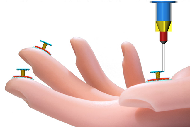
Schematic of a new kind of 3D printer that can print touch sensors directly on a model hand. (credit: Shuang-Zhuang Guo and Michael McAlpine/Advanced Materials )

Engineering researchers at the University of Minnesota have developed a process for 3D-printing stretchable, flexible, and sensitive electronic sensory devices that could give robots or prosthetic hands — or even real skin — the ability to mechanically sense their environment.

One major use would be to give surgeons the ability to feel during minimally invasive surgeries instead of using cameras, or to increase the sensitivity of surgical robots. The process could also make it easier for robots to walk and interact with their environment.

Printing electronics directly on human skin could be used for pulse monitoring, energy harvesting (of movements), detection of finger motions (on a keyboard or other devices), or chemical sensing (for example, by soldiers in the field to detect dangerous chemicals or explosives). Or imagine a future computer mouse built into your fingertip, with haptic touch on any surface.

“While we haven’t printed on human skin yet, we were able to print on the curved surface of a model hand using our technique,” said Michael McAlpine, a University of Minnesota mechanical engineering associate professor and lead researcher on the study.* “We also interfaced a printed device with the skin and were surprised that the device was so sensitive that it could detect your pulse in real time.”

The researchers also visualize use in “bionic organs.”

A unique skin-compatible 3D-printing process

(left) Schematic of the tactile sensor. (center) Top view. (right) Optical image showing the conformally printed 3D tactile sensor on a fingertip. Scale bar = 4 mm. (credit: Shuang-Zhuang Guo et al./Advanced Materials)

McAlpine and his team made the sensing fabric with a one-of-a kind 3D printer they built in the lab. The multifunctional printer has four nozzles to print the various specialized “inks” that make up the layers of the device — a base layer of silicone**, top and bottom electrodes made of a silver-based piezoresistive conducting ink, a coil-shaped pressure sensor, and a supporting layer that holds the top layer in place while it sets (later washed away in the final manufacturing process).

Surprisingly, all of the layers of “inks” used in the flexible sensors can set at room temperature. Conventional 3D printing using liquid plastic is too hot and too rigid to use on the skin. The sensors can stretch up to three times their original size.

The researchers say the next step is to move toward semiconductor inks and printing on a real surface. “The manufacturing is built right into the process, so it is ready to go now,” McAlpine said.

The research was published online in the journal Advanced Materials. It was funded by the National Institute of Biomedical Imaging and Bioengineering of the National Institutes of Health.

* McAlpine integrated electronics and novel 3D-printed nanomaterials to create a “bionic ear” in 2013.

** The silicone rubber has a low modulus of elasticity of 150 kPa, similar to that of skin, and lower hardness (Shore A 10) than that of human skin, according to the Advanced Materials paper.


College of Science and Engineering, UMN | 3D Printed Stretchable Tactile Sensors


Abstract of 3D Printed Stretchable Tactile Sensors

The development of methods for the 3D printing of multifunctional devices could impact areas ranging from wearable electronics and energy harvesting devices to smart prosthetics and human–machine interfaces. Recently, the development of stretchable electronic devices has accelerated, concomitant with advances in functional materials and fabrication processes. In particular, novel strategies have been developed to enable the intimate biointegration of wearable electronic devices with human skin in ways that bypass the mechanical and thermal restrictions of traditional microfabrication technologies. Here, a multimaterial, multiscale, and multifunctional 3D printing approach is employed to fabricate 3D tactile sensors under ambient conditions conformally onto freeform surfaces. The customized sensor is demonstrated with the capabilities of detecting and differentiating human movements, including pulse monitoring and finger motions. The custom 3D printing of functional materials and devices opens new routes for the biointegration of various sensors in wearable electronics systems, and toward advanced bionic skin applications.

New 3D printing method may allow for fast, low-cost, more-flexible medical implants for millions


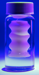
UF Soft Matter | Silicone is 3D-printed into the micro-organogel support material. The printing nozzle follows a predefined trajectory, depositing liquid silicone in its wake. The liquid silicone is supported by the micro-organogel material during this printing process.

University of Florida (UF) researchers have developed a method for 3D printing soft-silicone medical implants that are stronger, quicker, less expensive, more flexible, and more comfortable than the implants currently available. That should be good news for the millions of people every year who need medical devices implanted.

Model 3D-printed silicone trachea implant (credit: University of Florida)

Currently, such devices — such as ports for draining bodily fluids (cerebral spinal fluid in hydrocephalus, for example), implantable bands, balloons, soft catheters, slings and meshes — are mass produced and made through molding processes. To create customized parts for individual patients with molding would be very expensive and could take days or weeks for each job.

The 3D printing method cuts that time to hours, potentially saving lives.

The ability to easily replace silicone implants at low cost is especially important for children, where “implants may need to be replaced frequently as they grow up,” Thomas E. Angelini, an associate professor of mechanical engineering  of the UF Department of Mechanical and Aerospace Engineering, explained to KurzweilAI. Angelini is senior author of a paper published May 10, 2017 in the open-access journal Science Advances.

The research could also pave the way for new therapeutic devices that encapsulate and control the release of drugs or small molecules for guiding tissue regeneration or assisting diseased organs, such as the pancreas or prostate, according to lead author Christopher O’Bryan, a UF mechanical and aerospace engineering doctoral student.


UF Soft Matter | Water is pumped from one reservoir to another using a 3D-printed silicone valve. The silicone valve contains two encapsulated ball valves that allow water to be pumped through the valve by squeezing the lower chamber. The silicone valve demonstrates the ability of the UF 3D-printing method to create multiple encapsulated components in a single part — something that cannot be done with a traditional 3D-printing approach.


Abstract of Self-assembled micro-organogels for 3D printing silicone structures

The widespread prevalence of commercial products made from microgels illustrates the immense practical value of harnessing the jamming transition; there are countless ways to use soft, solid materials that fluidize and become solid again with small variations in applied stress. The traditional routes of microgel synthesis produce materials that predominantly swell in aqueous solvents or, less often, in aggressive organic solvents, constraining ways that these exceptionally useful materials can be used. For example, aqueous microgels have been used as the foundation of three-dimensional (3D) bioprinting applications, yet the incompatibility of available microgels with nonpolar liquids, such as oils, limits their use in 3D printing with oil-based materials, such as silicone. We present a method to make micro-organogels swollen in mineral oil, using block copolymer self-assembly. The rheological properties of this micro-organogel material can be tuned, leveraging the jamming transition to facilitate its use in 3D printing of silicone structures. We find that the minimum printed feature size can be controlled by the yield stress of the micro-organogel medium, enabling the fabrication of numerous complex silicone structures, including branched perfusable networks and functional fluid pumps.

A ‘smart contact lens’ for diabetes and glaucoma diagnosis

Smart contact lens on mannequin eye (credit: UNIST)

Korean researchers have designed a “smart contact lens” that may one day allow patients with diabetes and glaucoma to self-monitor blood glucose levels and internal eye pressure.*

The study was conducted by researchers at Ulsan National Institute of Science and Technology (UNIST) and Kyungpook National University School of Medicine, both of South Korea.

Most previously reported contact lens sensors can only monitor a single analyte (such as glucose) at a time, and generally obstruct the field of vision of the subject.

The design is based on transparent, stretchable sensors that are deposited on commercially available soft-contact lenses.

Electrodes based on a hybrid graphene-silver nanowire material can measure glucose in tears. Internal eye pressure changes are measured by a sandwich structure whose electronic characteristics are modified by pressure.

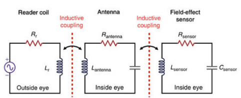
Inductive coupling — batteries not required

Both of these readings are transmitted wirelessly using “inductive coupling” (similar to remote charging of batteries), so no connected power source, such as a battery, is required. This design also allows for 24-hour real-time monitoring by patients.

The researchers conducted in-vivo and in-vitro performance tests using a live rabbit and bovine eyeball.

The team expects that the research could also lead to developing biosensors capable of detecting and treating various other human diseases, or used as a component in other biomedical devices.

The study results were published in the March issue of the journal Nature Communications. The study was supported by the 2017 CooperVision Science and Technology (S&T) Awards Program.

* Diabetes is the most common cause of high blood sugar levels. Intraocular pressure is the largest risk factor for glaucoma, a leading cause of human blindness.


How the smart contact lens works

Schematic of the top portion of the wearable contact-lens sensor. Left: antenna. Insert: Glucose sensor, based on a field-effect transistor (FET), which consists of a graphene channel and graphene/silver nanowire for source/drain. Not shown: chromium/gold interconnect, epoxy layer, and lens (below). (credit: UNIST)

Real-time glucose sensing with graphene/silver hybrid nanostructures. For selective and sensitive detection of glucose, glucose oxidase (GOD) catalyzes oxidation of glucose to gluconic acid and reduction of water to hydrogen peroxide, which produces oxygen, protons and electrons. The concentration of charge carriers in the FET channel, and thus the drain current, increases at higher concentration of glucose. (credit: UNIST)

The FET sensor (right) is modeled as an electrical RLC resonant circuit, comprised of the resistance (R) of the graphene channel, the inductance (L) of the antenna coil made of the graphene-AgNW hybrid, and the capacitance (C) of graphene-AgNW hybrid S/D electrodes. Wireless operation is achieved by mutually coupling the sensor antenna (center) with an external reader antenna (left) at a resonant frequency of 4.1 GHz. (credit: UNIST)

Schematic of intraocular pressure monitoring. A layer of silicone elastomer  was placed between the two inductive spirals made of graphene-AgNW hybrid electrodes in a sandwich structure. The contact lens sensor responds to raised intraocular pressure (ocular hypertension), which increases the corneal radius of curvature, which in turn increases both the capacitance by thinning the dielectric and the inductance by bi-axial lateral expansion of the spiral coils. As a result, ocular hypertension shifts the reflection spectra of the spiral antenna to a lower frequency. (credit: UNIST)


Abstract of Wearable smart sensor systems integrated on soft contact lenses for wireless ocular diagnostics

Wearable contact lenses which can monitor physiological parameters have attracted substantial interests due to the capability of direct detection of biomarkers contained in body fluids. However, previously reported contact lens sensors can only monitor a single analyte at a time. Furthermore, such ocular contact lenses generally obstruct the field of vision of the subject. Here, we developed a multifunctional contact lens sensor that alleviates some of these limitations since it was developed on an actual ocular contact lens. It was also designed to monitor glucose within tears, as well as intraocular pressure using the resistance and capacitance of the electronic device. Furthermore, in-vivo and in-vitro tests using a live rabbit and bovine eyeball demonstrated its reliable operation. Our developed contact lens sensor can measure the glucose level in tear fluid and intraocular pressure simultaneously but yet independently based on different electrical responses.

New nuclear magnetic resonance technique offers ‘molecular window’ for live disease diagnosis

New nuclear magnetic resonance (NMR) system for molecular diagnosis (credit: University of Toronto Scarborough)

University of Toronto Scarborough researchers have developed a new “molecular window” technology based on nuclear magnetic resonance (NMR) that can look inside a living system to get a high-resolution profile of which specific molecules are present, and extract a full metabolic profile.

“Getting a sense of which molecules are in a tissue sample is important if you want to know if it’s cancerous, or if you want to know if certain environmental contaminants are harming cells inside the body,” says Professor Andre Simpson, who led research in developing the new technique.*

An NMR spectrometer generates a powerful magnetic field that causes atomic nuclei to absorb and re-emit energy in distinct patterns, revealing a unique molecular signature — in this example: the chemical ethanol. (credit: adapted from the Bruker BioSpin “How NMR Works” video at www.theresonance.com/nmr-know-how)

Simpson says there’s great medical potential for this new technique, since it can be adapted to work on existing magnetic resonance imaging (MRI) systems found in hospitals. “It could have implications for disease diagnosis and a deeper understanding of how important biological processes work,” by targeting specific biomarker molecules that are unique to specific diseased tissue.

The new approach could detect these signatures without resorting to surgery and could determine, for example, whether a growth is cancerous or benign directly from the MRI alone.

The technique could also provide highly detailed information on how the brain works, revealing the actual chemicals involved in a particular response. “It could mark an important step in unraveling the biochemistry of the brain,” says Simpson.

Overcoming magnetic distortion

Until now, traditional NMR techniques haven’t been able to provide high-resolution profiles of living organisms because of magnetic distortions from the tissue itself.  Simpson and his team were able to overcome this problem by creating tiny communication channels based on “long-range dipole interactions” between molecules.

The next step for the research is to test it on human tissue samples, says Simpson. Since the technique detects all cellular metabolites (substances such as glucose) equally, there’s also potential for non-targeted discovery.

“Since you can see metabolites in a sample that you weren’t able to see before, you can now identify molecules that may indicate there’s a problem,” he explains. “You can then determine whether you need further testing or surgery. So the potential for this technique is truly exciting.”

The research results are published in the journal Angewandte Chemie.

* Simpson has been working on perfecting the technique for more than three years with colleagues at Bruker BioSpin, a scientific instruments company that specializes in developing NMR technology. The technique, called “in-phase intermolecular single quantum coherence” (IP-iSQC), is based on some unexpected scientific concepts that were discovered in 1995, which at the time were described as impossible and “crazed” by many researchers. The technique developed by Simpson and his team builds upon these early discoveries. The work was supported by Mark Krembil of the Krembil Foundation and the Natural Sciences Engineering Research Council of Canada (NSERC).


Abstract of In-Phase Ultra High-Resolution In Vivo NMR

Although current NMR techniques allow organisms to be studied in vivo, magnetic susceptibility distortions, which arise from inhomogeneous distributions of chemical moieties, prevent the acquisition of high-resolution NMR spectra. Intermolecular single quantum coherence (iSQC) is a technique that breaks the sample’s spatial isotropy to form long range dipolar couplings, which can be exploited to extract chemical shift information free of perturbations. While this approach holds vast potential, present practical limitations include radiation damping, relaxation losses, and non-phase sensitive data. Herein, these drawbacks are addressed, and a new technique termed in-phase iSQC (IP-iSQC) is introduced. When applied to a living system, high-resolution NMR spectra, nearly identical to a buffer extract, are obtained. The ability to look inside an organism and extract a high-resolution metabolic profile is profound and should find applications in fields in which metabolism or in vivo processes are of interest.