Travelers to Mars risk leukemia cancer, weakened immune function from radiation, NASA-funded study finds

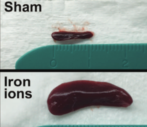
The spleen from a mouse exposed to a mission-relevant dose (20 cGy, 1 GeV/n) of iron ions (bottom) was ~ 30 times the normal volume compared with the spleen from a control mouse (top). (credit: C Rodman et al./Leukemia)

Radiation encountered in deep space travel may increase the risk of leukemia cancer in humans traveling to Mars, NASA-funded researchers at the Wake Forest Institute for Regenerative Medicine and colleagues have found, using mice transplanted with human stem cells.

“Our results are troubling because they show radiation exposure could potentially increase the risk of leukemia,” said Christopher Porada, Ph.D., associate professor of regenerative medicine and senior researcher on the project.

Radiation exposure is believed to be one of the most dangerous aspects of traveling to Mars, according to NASA. The average distance to Mars is 140 million miles, and a round trip could take three years.

The goal of the study, published in the journal Leukemia, was to assess the direct effects of simulated solar energetic particles (SEP) and galactic cosmic ray (GCR) radiation on human hematopoietic stem cells (HSCs). These stem cells comprise less than 0.1% of the bone marrow of adults, but produce the many types of blood cells that circulate through the body and work to transport oxygen, fight infection, and eliminate any malignant cells that arise.

For the study, human HSCs from healthy donors of typical astronaut age (30–55 years) were exposed to Mars mission-relevant doses of protons and iron ions — the same types of radiation that astronauts would be exposed to in deep space, followed by laboratory and animal studies to define the impact of the exposure.

“Radiation exposure at these levels was highly deleterious to HSC function, reducing their ability to produce almost all types of blood cells, often by 60–80 percent,” said Porada. “This could translate into a severely weakened immune system and anemia during prolonged missions in deep space.”

The radiation also caused mutations in genes involved in the hematopoietic process and dramatically reduced the ability of HSCs to give rise to mature blood cells.

Previous studies had already demonstrated that exposure to high doses of radiation, such as from X-rays, can have harmful (even life-threatening) effects on the body’s ability to make blood cells, and can significantly increase the likelihood of cancers, especially leukemias. However, the current study was the first to show a damaging effect of lower, mission-relevant doses of space radiation.

Mice develop T-cell acute lymphoblastic leukemia, weakened immune function

The next step was to assess how the cells would function in the human body. For that purpose, mice were transplanted with GCR-irradiated human HSCs, essentially “humanizing” the animals. The mice developed what appeared to be T-cell acute lymphoblastic leukemia — the first demonstration that exposure to space radiation may increase the risk of leukemia in humans.

“Our results show radiation exposure could potentially increase the risk of leukemia in two ways,” said Porada. “We found that genetic damage to HSCs directly led to leukemia. Secondly, radiation also altered the ability of HSCs to generate T and B cells, types of white blood cells involved in fighting foreign ‘invaders’ like infections or tumor cells. This may reduce the ability of the astronaut’s immune system to eliminate malignant cells that arise as a result of radiation-induced mutations.”

Porada said the findings are particularly troubling given previous work showing that conditions of weightlessness/microgravity present during spaceflight can also cause marked alterations in astronaut’s immune function, even after short duration missions in low-earth orbit, where they are largely protected from cosmic radiation.

Taken together, the results indicate that the combined exposure to microgravity and SEP/GCR radiation that would occur during extended deep space missions, such as to Mars, could potentially exacerbate the risk of immune-dysfunction and cancer,

NASA’s Human Research Program is also exploring conditions of microgravity, isolation and confinement, hostile and closed environments, and distance from Earth. The ultimate goal of the research is to make space missions as safe as possible.

Researchers at Wake Forest Baptist Medical Center, Brookhaven National Laboratory, and the University of California Davis Comprehensive Cancer Center were also involved in the study.


Abstract of In vitro and in vivo assessment of direct effects of simulated solar and galactic cosmic radiation on human hematopoietic stem/progenitor cells

Future deep space missions to Mars and near-Earth asteroids will expose astronauts to chronic solar energetic particles (SEP) and galactic cosmic ray (GCR) radiation, and likely one or more solar particle events (SPEs). Given the inherent radiosensitivity of hematopoietic cells and short latency period of leukemias, space radiation-induced hematopoietic damage poses a particular threat to astronauts on extended missions. We show that exposing human hematopoietic stem/progenitor cells (HSC) to extended mission-relevant doses of accelerated high-energy protons and iron ions leads to the following: (1) introduces mutations that are frequently located within genes involved in hematopoiesis and are distinct from those induced by γ-radiation; (2) markedly reduces in vitro colony formation; (3) markedly alters engraftment and lineage commitment in vivo; and (4) leads to the development, in vivo, of what appears to be T-ALL. Sequential exposure to protons and iron ions (as typically occurs in deep space) proved far more deleterious to HSC genome integrity and function than either particle species alone. Our results represent a critical step for more accurately estimating risks to the human hematopoietic system from space radiation, identifying and better defining molecular mechanisms by which space radiation impairs hematopoiesis and induces leukemogenesis, as well as for developing appropriately targeted countermeasures.

Scientists reverse aging in mice by repairing damaged DNA

A research team led by Harvard Medical School professor of genetics David Sinclair, PhD, has made a discovery that could lead to a revolutionary new drug that allows cells to repair DNA damaged by aging, cancer, and radiation.

In a paper published in the journal Science on Friday (March 24), the scientists identified a critical step in the molecular process related to DNA damage.

The researchers found that a compound known as NAD (nicotinamide adenine dinucleotide), which is naturally present in every cell of our body, has a key role as a regulator in protein-to-protein interactions that control DNA repair. In an experiment, they found that treating mice with a NAD+ precursor called NMN (nicotinamide mononucleotide) improved their cells’ ability to repair DNA damage.

“The cells of the old mice were indistinguishable from the young mice, after just one week of treatment,” said senior author Sinclair.

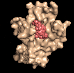
Disarming a rogue agent: When the NAD molecule (red) binds to the DBC1 protein (beige), it prevents DBC1 from attaching to and incapacitating a protein (PARP1) that is critical for DNA repair. (credit: David Sinclair)

Human trials of NMN therapy will begin within the next few months to “see if these results translate to people,” he said. A safe and effective anti-aging drug is “perhaps only three to five years away from being on the market if the trials go well.”

What it means for astronauts, childhood cancer survivors, and the rest of us

The researchers say that in addition to reversing aging, the DNA-repair research has attracted the attention of NASA. The treatment could help deal with radiation damage to astronauts in its Mars mission, which could cause muscle weakness, memory loss, and other symptoms (see “Mars-bound astronauts face brain damage from galactic cosmic ray exposure, says NASA-funded study“), and more seriously, leukemia cancer and weakened immune function (see “Travelers to Mars risk leukemia cancer, weakend immune function from radiation, NASA-funded study finds“).

The treatment could also help travelers aboard aircraft flying across the poles. A 2011 NASA study showed that passengers on polar flights receive about 12 percent of the annual radiation limit recommended by the International Committee on Radiological Protection.

The other group that could benefit from this work is survivors of childhood cancers, who are likely to suffer a chronic illness by age 45, leading to accelerated aging, including cardiovascular disease, Type 2 diabetes, Alzheimer’s disease, and cancers unrelated to the original cancer, the researchers noted.

For the past four years, Sinclair’s team has been working with spinoff MetroBiotech on developing NMN as a drug. Sinclair previously made a link between the anti-aging enzyme SIRT1 and resveratrol. “While resveratrol activates SIRT1 alone, NAD boosters [like NMN] activate all seven sirtuins, SIRT1-7, and should have an even greater impact on health and longevity,” he says.

Sinclair is also a professor at the University of New South Wales School of Medicine in Sydney, Australia.


Abstract of A conserved NAD+ binding pocket that regulates protein-protein interactions during aging

DNA repair is essential for life, yet its efficiency declines with age for reasons that are unclear. Numerous proteins possess Nudix homology domains (NHDs) that have no known function. We show that NHDs are NAD+ (oxidized form of nicotinamide adenine dinucleotide) binding domains that regulate protein-protein interactions. The binding of NAD+ to the NHD domain of DBC1 (deleted in breast cancer 1) prevents it from inhibiting PARP1 [poly(adenosine diphosphate–ribose) polymerase], a critical DNA repair protein. As mice age and NAD+ concentrations decline, DBC1 is increasingly bound to PARP1, causing DNA damage to accumulate, a process rapidly reversed by restoring the abundance of NAD+. Thus, NAD+ directly regulates protein-protein interactions, the modulation of which may protect against cancer, radiation, and aging.

Mayo Clinic discovers high-intensity aerobic training can reverse aging

Mayo Clinic study finds high-intensity aerobic exercise may reverse aging (credit: Flickr user Global Panorama via Creative Commons license)

A Mayo Clinic study says the best training for adults is high-intensity aerobic exercise, which they believe can reverse some cellular aspects of aging.

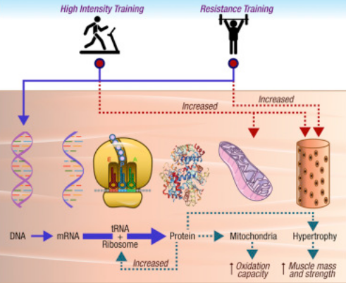
Mayo researchers compared 12 weeks of high-intensity interval training (workouts in which you alternate periods of high-intensity exercise with low-intensity recovery periods), resistance training, and combined training. While all three enhanced insulin sensitivity and lean mass, only high-intensity interval training and combined training improved aerobic capacity and skeletal muscle mitochondrial respiration. (Decline in mitochondrial content and function are common in older adults.)

High-intensity intervals also improved muscle protein content, which enhanced energetic functions and also caused muscle enlargement, especially in older adults. The researchers said exercise training significantly enhanced the cellular machinery responsible for making new proteins. That contributes to protein synthesis, thus reversing a major adverse effect of aging.

12 weeks exercise training in younger and older people (credit: Mayo Clinic)

“We encourage everyone to exercise regularly, but the take-home message for aging adults is that supervised high-intensity training is probably best, because, both metabolically and at the molecular level, it confers the most benefits,” says K. Sreekumaran Nair, M.D., Ph.D., a Mayo Clinic endocrinologist and senior researcher on the study.

He says the high-intensity training reversed some manifestations of aging in the body’s protein function, but noted that increasing muscle strength requires resistance training a couple of days a week.

Other findings

In the study, researchers tracked metabolic and molecular changes in a group of young and older adults over 12 weeks, gathering data 72 hours after individuals in randomized groups completed each type of exercise. General findings showed:

  • Cardio respiratory health, muscle mass, and insulin sensitivity improved with all training.
  • Mitochondrial cellular function declined with age but improved with training.
  • Increase in muscle strength occurred only modestly with high-intensity interval training, but occurred with resistance training alone or when added to the aerobic training.
  • Exercise improves skeletal muscle gene expression independent of age.
  • Exercise substantially enhanced the ribosomal proteins responsible for synthesizing new proteins, which is mainly responsible for enhanced mitochondrial function.
  • Training has no significant effect on skeletal muscle DNA epigenetic changes but promotes skeletal muscle protein expression with maximum effect in older adults.

The research findings appear in Cell Metabolism. The research was supported by the National Institutes of Health, Mayo Clinic, the Robert and Arlene Kogod Center on Aging, and the Murdock-Dole Professorship.


Abstract of Enhanced Protein Translation Underlies Improved Metabolic and Physical Adaptations to Different Exercise Training Modes in Young and Old Humans

The molecular transducers of benefits from different exercise modalities remain incompletely defined. Here we report that 12 weeks of high-intensity aerobic interval (HIIT), resistance (RT), and combined exercise training enhanced insulin sensitivity and lean mass, but only HIIT and combined training improved aerobic capacity and skeletal muscle mitochondrial respiration. HIIT revealed a more robust increase in gene transcripts than other exercise modalities, particularly in older adults, although little overlap with corresponding individual protein abundance was noted. HIIT reversed many age-related differences in the proteome, particularly of mitochondrial proteins in concert with increased mitochondrial protein synthesis. Both RT and HIIT enhanced proteins involved in translational machinery irrespective of age. Only small changes of methylation of DNA promoter regions were observed. We provide evidence for predominant exercise regulation at the translational level, enhancing translational capacity and proteome abundance to explain phenotypic gains in muscle mitochondrial function and hypertrophy in all ages.

Whole-body vibration may be as effective as regular exercise

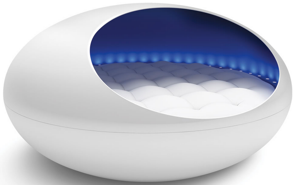
Hate treadmills? No prob. The Tranquility Pod uses “pleasant sound, gentle vibration, and soothing light to transport the body, mind, and spirit to a tranquil state of relaxation” — and maybe lose weight (and $30,000). (credit: Hammacher Schlemmer)

If you’re overweight and find it challenging to exercise regularly, now there’s good news: A less strenuous form of exercise known as whole-body vibration (WBV) can mimic the muscle and bone health benefits of regular exercise — at least in mice — according to a new study published in the Endocrine Society’s journal Endocrinology.

Lack of exercise is contributing to the obesity and diabetes epidemics, according to the researchers. These disorders can also increase the risk of bone fractures. Physical activity can help to decrease this risk and reduce the negative metabolic effects of these conditions.

But the alternative, WBV, can be experienced while sitting, standing, or even lying down on a machine with a vibrating platform. When the machine vibrates, it transmits energy to your body, and your muscles contract and relax multiple times during each second.

“Our study is the first to show that whole-body vibration may be just as effective as exercise at combating some of the negative consequences of obesity and diabetes,” said the study’s first author, Meghan E. McGee-Lawrence, Ph.D., of Augusta University in Georgia. “While WBV did not fully address the defects in bone mass of the obese mice in our study, it did increase global bone formation, suggesting longer-term treatments could hold promise for preventing bone loss as well.”

Just as effective as a treadmill

Glucose and insulin tolerance testing revealed that the genetically obese and diabetic mice showed similar metabolic benefits from both WBV and exercising on a treadmill. Obese mice gained less weight after exercise or WBV than obese mice in the sedentary group, although they remained heavier than normal mice. Exercise and WBV also enhanced muscle mass and insulin sensitivity in the genetically obese mice.

The findings suggest that WBV may be a useful supplemental therapy to combat metabolic dysfunction in individuals with morbid obesity. “These results are encouraging,” McGee-Lawrence said. “However, because our study was conducted in mice, this idea needs to be rigorously tested in humans to see if the results would be applicable to people.”

The authors included researchers at the National Institute of Health’s National Institute of Aging (NIA). Funding was provided by the American Diabetes Association, the National Institutes of Health’s National Institute of Diabetes and Digestive Kidney Diseases, and the National Institute on Aging.

Know a cheaper alternative to the Tranquility Pod? Sound off below!

* To conduct the study, researchers examined two groups of 5-week-old male mice. One group consisted of normal mice, while the other group was genetically unresponsive to the hormone leptin, which promotes feelings of fullness after eating. Mice from each group were assigned to sedentary, WBV or treadmill exercise conditions.

After a week-long period to grow used to the exercise equipment, the groups of mice began a 12-week exercise program. The mice in the WBV group underwent 20 minutes of WBV at a frequency of 32 Hz with 0.5g acceleration each day. Mice in the treadmill group walked for 45 minutes daily at a slight incline. For comparison, the third group did not exercise. Mice were weighed weekly during the study.


Abstract of Whole-body vibration mimics the metabolic effects of exercise in male leptin receptor deficient mice

Whole-body vibration has gained attention as a potential exercise mimetic, but direct comparisons with the metabolic effects of exercise are scarce. To determine whether whole-body vibration recapitulates the metabolic and osteogenic effects of physical activity, we exposed male wildtype (Wt) and leptin receptor deficient (db/db) mice to daily treadmill exercise or whole-body vibration for three months. Body weights were analyzed and compared with Wt and db/db mice that remained sedentary. Glucose and insulin tolerance testing revealed comparable attenuation of hyperglycemia and insulin resistance in db/db mice following treadmill exercise or whole-body vibration. Both interventions reduced body weight in db/db mice and normalized muscle fiber diameter. Treadmill exercise and whole-body vibration also attenuated adipocyte hypertrophy in visceral adipose tissue and reduced hepatic lipid content in db/db mice. Although the effects of leptin receptor deficiency on cortical bone structure were not eliminated by either intervention, exercise and whole-body vibration increased circulating levels of osteocalcin in db/db mice. In the context of increased serum osteocalcin, the modest effects of TE and WBV on bone geometry, mineralization, and biomechanics may reflect subtle increases in osteoblast activity in multiple areas of the skeleton. Taken together, these observations indicate that whole-body vibration recapitulates the effects of exercise on metabolism in type 2 diabetes.

First nanoengineered retinal implant could help the blind regain functional vision

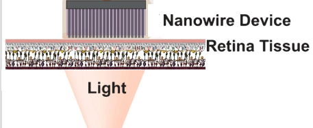
Activated by incident light, photosensitive silicon nanowires 1 micrometer in diameter stimulate residual undamaged retinal cells to induce visual sensations. (credit (image adapted): Sohmyung Ha et al./ J. Neural Eng)

A team of engineers at the University of California San Diego and La Jolla-based startup Nanovision Biosciences Inc. have developed the first nanoengineered retinal prosthesis — a step closer to restoring the ability of neurons in the retina to respond to light.

The technology could help tens of millions of people worldwide suffering from neurodegenerative diseases that affect eyesight, including macular degeneration, retinitis pigmentosa, and loss of vision due to diabetes.

Despite advances in the development of retinal prostheses over the past two decades, the performance of devices currently on the market to help the blind regain functional vision is still severely limited — well under the acuity threshold of 20/200 that defines legal blindness.

The new prosthesis relies on two new technologies: implanted arrays of photosensitive nanowires and a wireless power/data system.

Implanted arrays of silicon nanowires

The new prosthesis uses arrays of nanowires that simultaneously sense light and electrically stimulate the retina. The nanowires provide higher resolution than anything achieved by other devices — closer to the dense spacing of photoreceptors in the human retina, according to the researchers.*

Comparison of retina and electrode geometries between an existing retinal prosthesis and new nanoengineered prosthesis design. (left) Planar platinum electrodes (gray) of the FDA-approved Argus II retinal prosthesis (a 60-element array with 200 micrometer electrode diameter). (center) Retinal photoreceptor cells: rods (yellow) and cones (green). (right) Fabricated silicon nanowires (1 micrometer in diameter) at the same spatial magnification as photoreceptor cells. (credit: Science Photo Library and Sohmyung Ha et al./ J. Neural Eng.)

Existing retinal prostheses require a vision sensor (such as a camera) outside of the eye to capture a visual scene and then transform it into signals to sequentially stimulate retinal neurons (in a matrix). Instead, the silicon nanowires mimic the retina’s light-sensing cones and rods to directly stimulate retinal cells. The nanowires are bundled into a grid of electrodes, directly activated by light.

This direct, local translation of incident light into electrical stimulation makes for a much simpler — and scalable — architecture for a prosthesis, according to the researchers.

Wireless power and telemetry system

For the new device, power is delivered wirelessly, from outside the body to the implant, through an inductive powering telemetry system. Data to the nanowires is sent over the same wireless link at record speed and energy efficiency. The telemetry system is capable of transmitting both power and data over a single pair of inductive coils, one emitting from outside the body, and another on the receiving side in the eye.**

Three of the researchers have co-founded La Jolla-based Nanovision Biosciences, a partner in this study, to further develop and translate the technology into clinical use, with the goal of restoring functional vision in patients with severe retinal degeneration. Animal tests with the device are in progress, with clinical trials following.***

The research was described in a recent issue of the Journal of Neural Engineering. It was funded by Nanovision Biosciences, Qualcomm Inc., and the Institute of Engineering in Medicine and the Clinical and Translational Research Institute at UC San Diego.

* For visual acuity of 20/20,  an electrode pixel size of 5 μm (micrometers) is required; 20/200 visual acuity requires 50 μm. The minimum number of electrodes required for pattern recognition or reading text is estimated to be about 600. The new nanoengineered silicon nanowire electrodes are 1 μm in diameter, and for the experiment, 2500 silicon nanowires were used.

** The device is highly energy efficient because it minimizes energy losses in wireless power and data transmission and in the stimulation process, recycling electrostatic energy circulating within the inductive resonant tank, and between capacitance on the electrodes and the resonant tank. Up to 90 percent of the energy transmitted is actually delivered and used for stimulation, which means less RF wireless power emitting radiation in the transmission, and less heating of the surrounding tissue from dissipated power.

These are primary cortical neurons cultured on the surface of an array of optoelectronic nanowires. Here a neuron is pulling the nanowires, indicating the the cell is doing well on this material. (credit: UC San Diego)

*** For proof-of-concept, the researchers inserted the wirelessly powered nanowire array beneath a transgenic rat retina with rhodopsin P23H knock-in retinal degeneration. The degenerated retina interfaced in vitro with a microelectrode array for recording extracellular neural action potentials (electrical “spikes” from neural activity).


Abstract of Towards high-resolution retinal prostheses with direct optical addressing and inductive telemetry

Objective. Despite considerable advances in retinal prostheses over the last two decades, the resolution of restored vision has remained severely limited, well below the 20/200 acuity threshold of blindness. Towards drastic improvements in spatial resolution, we present a scalable architecture for retinal prostheses in which each stimulation electrode is directly activated by incident light and powered by a common voltage pulse transferred over a single wireless inductive link. Approach. The hybrid optical addressability and electronic powering scheme provides separate spatial and temporal control over stimulation, and further provides optoelectronic gain for substantially lower light intensity thresholds than other optically addressed retinal prostheses using passive microphotodiode arrays. The architecture permits the use of high-density electrode arrays with ultra-high photosensitive silicon nanowires, obviating the need for excessive wiring and high-throughput data telemetry. Instead, the single inductive link drives the entire array of electrodes through two wires and provides external control over waveform parameters for common voltage stimulation. Main results. A complete system comprising inductive telemetry link, stimulation pulse demodulator, charge-balancing series capacitor, and nanowire-based electrode device is integrated and validated ex vivo on rat retina tissue. Significance. Measurements demonstrate control over retinal neural activity both by light and electrical bias, validating the feasibility of the proposed architecture and its system components as an important first step towards a high-resolution optically addressed retinal prosthesis.

A biocompatible stretchable material for brain implants and ‘electronic skin’

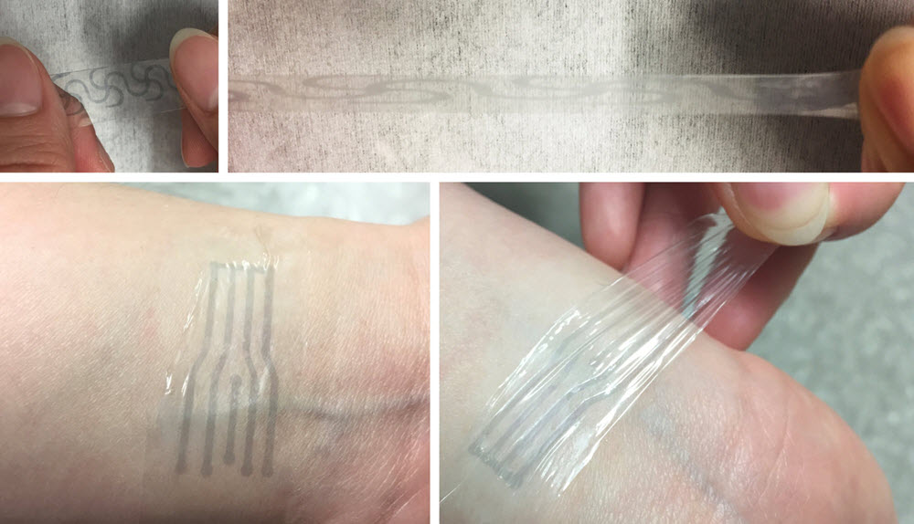
A printed electrode pattern of a new polymer being stretched to several times of its original length (top), and a transparent, highly stretchy “electronic skin” patch (bottom) from the same material, forming an intimate interface with the human skin to potentially measure various biomarkers (credit: Bao Lab)

Stanford chemical engineers have developed a soft, flexible plastic electrode that stretches like rubber but carries electricity like wires — ideal for brain interfaces and other implantable electronics, they report in an open-access March 10 paper in Science Advances.

Developed by Zhenan Bao, a professor of chemical engineering, and his team, the material is still a laboratory prototype, but the team hopes to develop it as part of their long-term focus on creating flexible materials that interface with the human body.

Flexible interface

“One thing about the human brain that a lot of people don’t know is that it changes volume throughout the day,” says postdoctoral research fellow Yue Wang, the first author on the paper. “It swells and de-swells.” The current generation of electronic implants can’t stretch and contract with the brain, making it complicated to maintain a good connection.

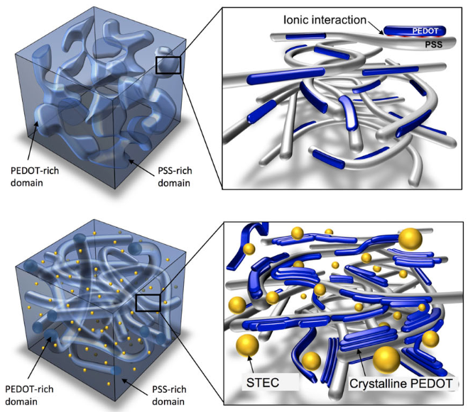
Illustration showing incorporation of ionic liquid-assisted stretchability and electrical conductivity (STEC) enhancers to convert conventional PEDOT:PSS film (top) to stretchable film (bottom). (credit: Wang et al., Sci. Adv.)

To create this flexible electrode, the researchers began with a plastic (PEDOT:PSS) with high electrical conductivity and biocompatibility (could be safely brought into contact with the human body), but was brittle. So they added a “STEC” (stretchability and electrical conductivity) molecule similar to the kind of additives used to thicken soups in industrial kitchens.

This additive transformed the plastic’s chunky and brittle molecular structure into a fishnet pattern with holes in the strands to allow the material to stretch and deform. The resulting plastic remained very conductive even when stretched 800 percent its original length.

Scientists at SLAC National Accelerator Laboratory, UCLA, the Materials Science Institute of Barcelona, and Samsung Advanced Institute of Technology were also involved in the research, which was funded by Samsung Electronics and the Air Force Office of Science Research.


Stanford University School of Engineering | Stretchable electrodes pave way for flexible electronics


Abstract of A highly stretchable, transparent, and conductive polymer

Previous breakthroughs in stretchable electronics stem from strain engineering and nanocomposite approaches. Routes toward intrinsically stretchablemolecularmaterials remain scarce but, if successful,will enable simpler fabrication processes, such as direct printing and coating, mechanically robust devices, and more intimate contact with objects. We report a highly stretchable conducting polymer, realized with a range of enhancers that serve dual functions to changemorphology andas conductivity-enhancingdopants inpoly(3,4-ethylenedioxythiophene):poly(styrenesulfonate) (PEDOT:PSS). The polymer films exhibit conductivities comparable to the best reported values for PEDOT:PSS, with higher than 3100 S/cm under 0% strain and higher than 4100 S/cm under 100% strain—among the highest for reported stretchable conductors. It is highly durable under cyclic loading,with the conductivitymaintained at 3600 S/cm even after 1000 cycles to 100% strain. The conductivity remained above 100 S/cm under 600% strain, with a fracture strain as high as 800%, which is superior to even the best silver nanowire– or carbon nanotube–based stretchable conductor films. The combination of excellent electrical andmechanical properties allowed it to serve as interconnects for field-effect transistor arrays with a device density that is five times higher than typical lithographically patterned wavy interconnects.

Groundbreaking technology rewarms large-scale animal tissues preserved at low temperatures

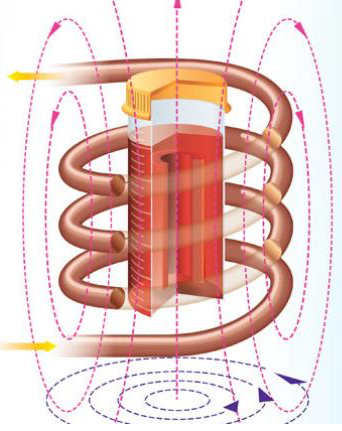
Inductive radio-frequency heating of magnetic nanoparticles embedded in tissue (red material in container) preserved at very low temperatures restored the tissue without damage (credit: Navid Manuchehrabadi et al./Science Translational Medicine)

A research team led by the University of Minnesota has discovered a way to rewarm large-scale animal heart valves and blood vessels preserved at very low (cryogenic) temperatures without damaging the tissue. The discovery could one day lead to saving millions of human lives by creating cryogenic tissue and organ banks of organs and tissues for transplantation.

The research was published March 1 in an open-access paper in Science Translational Medicine.

Long-term preservation methods like vitrification cool biological samples to an ice-free glassy state, using very low temperatures between -160 and -196 degrees Celsius, but tissues larger than 1 milliliter (0.03 fluid ounce) often suffer major damage during the rewarming process, making them unusable for tissues.

In the new research, the researchers were able to restore 50 milliliters (1.7 fluid ounces) of tissue with warming at more than 130°C/minute without damage.

Radiofrequency inductive heating of iron nanoparticles

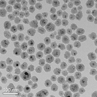
To achieve that, they developed a revolutionary new method using silica-coated iron-oxide nanoparticles dispersed throughout a cryoprotectant solution around the tissue. The nanoparticles act as tiny heaters around the tissue when they are activated using noninvasive radiofrequency inductive energy, rapidly and uniformly warming the tissue.

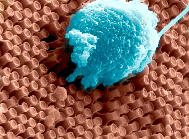
This transmission electron microscopy (TEM) image shows the iron oxide nanoparticles (coated in mesoporous silica) that are used in the tissue warming process. (credit: Haynes research group/University of Minnesota)

The results showed that none of the tissues displayed signs of harm — unlike control samples using vitrification and rewarmed slowly over ice or using convection warming. The researchers were also able to successfully wash away the iron oxide nanoparticles from the sample following the warming.

“This is the first time that anyone has been able to scale up to a larger biological system and demonstrate successful, fast, and uniform warming of hundreds of degrees Celsius per minute of preserved tissue without damaging the tissue,” said University of Minnesota mechanical engineering and biomedical engineering professor John Bischof, the senior author of the study.

Organs next

Bischof said there is a strong possibility they could scale up to even larger systems, like organs. The researchers plan to start with rodent organs (such as rat and rabbit) and then scale up to pig organs and then, hopefully, human organs. The technology might also be applied beyond cryogenics, including delivering lethal pulses of heat to cancer cells.

The researchers’ goal is to eliminate transplant waiting lists. Currently, hearts and lungs donated for transplantation must be discarded because these tissues cannot be kept on ice for longer than a matter of hours, according to the researchers.*

It will be interesting to see if the technology can one day be extended to cryonics.

The research was funded by the National Science Foundation (NSF), National Institutes of Health (NIH), U.S. Army Medical Research and Materiel Command, Minnesota Futures Grant from the University of Minnesota, and the University of Minnesota Carl and Janet Kuhrmeyer Chair in Mechanical Engineering. Researchers at Carnegie Mellon University, Clemson University and Tissue Testing Technologies LLC were also involved in the study.

* “A major limitation of transplantation is the ischemic injury that tissue and organs sustain during the time between recovery from the donor and implantation in the recipient. The maximum tolerable organ preservation for transplantation by hypothermic storage is typically 4 hours for heart and lungs; 8 to 12 hours for liver, intestine, and pancreas; and up to 36 hours for kidney transplants. In many cases, such limits actually prevent viable tissue or organs from reaching recipients. For instance, more than 60% of donor hearts and lungs are not used or transplanted partly because their maximum hypothermic preservation times have been exceeded. Further, if only half of these discarded organs were transplanted, then it has been estimated that wait lists for these organs could be extinguished within 2 to 3 years.” — Navid Manuchehrabadi et al./Science Translational Medicine


Abstract of Improved tissue cryopreservation using inductive heating of magnetic nanoparticles

Vitrification, a kinetic process of liquid solidification into glass, poses many potential benefits for tissue cryopreservation including indefinite storage, banking, and facilitation of tissue matching for transplantation. To date, however, successful rewarming of tissues vitrified in VS55, a cryoprotectant solution, can only be achieved by convective warming of small volumes on the order of 1 ml. Successful rewarming requires both uniform and fast rates to reduce thermal mechanical stress and cracks, and to prevent rewarming phase crystallization. We present a scalable nanowarming technology for 1- to 80-ml samples using radiofrequency-excited mesoporous silica–coated iron oxide nanoparticles in VS55. Advanced imaging including sweep imaging with Fourier transform and microcomputed tomography was used to verify loading and unloading of VS55 and nanoparticles and successful vitrification of porcine arteries. Nanowarming was then used to demonstrate uniform and rapid rewarming at >130°C/min in both physical (1 to 80 ml) and biological systems including human dermal fibroblast cells, porcine arteries and porcine aortic heart valve leaflet tissues (1 to 50 ml). Nanowarming yielded viability that matched control and/or exceeded gold standard convective warming in 1- to 50-ml systems, and improved viability compared to slow-warmed (crystallized) samples. Last, biomechanical testing displayed no significant biomechanical property changes in blood vessel length or elastic modulus after nanowarming compared to untreated fresh control porcine arteries. In aggregate, these results demonstrate new physical and biological evidence that nanowarming can improve the outcome of vitrified cryogenic storage of tissues in larger sample volumes.

Why you should eat 10 portions of fruit or vegetables a day

image credit | iStock

Eating 800 grams a day (about ten portions*) of fruit or vegetables could reduce your chance of heart attack, stroke, cancer, and early death, scientists from Imperial College London conclude from a meta-analysis of 95 studies on fruit and vegetable intake.

The study, published in an open-access paper in the International Journal of Epidemiology, included 2 million people worldwide and assessed up to 43,000 cases of heart disease, 47,000 cases of stroke, 81,000 cases of cardiovascular disease, 112,000 cancer cases and 94,000 deaths.

About 7.8 million premature deaths worldwide could be potentially prevented yearly if people followed this protocol, the researchers say.

Compared to not eating any fruits and vegetables, a daily intake of 200 grams (two and a half portions) was associated with a 16% reduced risk of heart disease, an 18% reduced risk of stroke, a 13% reduced risk of cardiovascular disease, a 4% reduced risk in cancer risk, and a 15% reduction in the risk of premature death.

However, a higher intake of fruits and vegetables of 800 grams a day was associated with 24% reduced risk of heart disease, a 33% reduced risk of stroke, a 28% reduced risk of cardiovascular disease, a 13% reduced risk of total cancer** and a 31% reduction in dying prematurely.***

The current UK guidelines suggest you eat at least five portions or 400 grams per day, but fewer than one in three UK adults are thought to even meet this target. The U.S. Health and Human Services/USDA guidelines use a different metric: “The recommended amount of vegetables in the Healthy U.S.-Style Eating Pattern at the 2,000-calorie level is 2½ cup-equivalents of vegetables per day and 2 cup-equivalents of fruit per day.


Foods that are best at disease prevention, according to the study

To prevent heart disease, stroke, cardiovascular disease, and early death: apples, pears, citrus fruits, salads, and green leafy vegetables such as spinach, lettuce and chicory, and cruciferous vegetables such as broccoli, cabbage and cauliflower.

To reduce cancer risk: green vegetables, such as spinach or green beans, yellow vegetables, such as peppers and carrots, and cruciferous vegetables.


Reasons for health benefits

So why do fruit and vegetables have such profound health benefits? According to Dagfinn Aune, PhD, lead author of the research, from the School of Public Health at Imperial: “Fruit and vegetables have been shown to reduce cholesterol levels, blood pressure, and to boost the health of our blood vessels and immune system. This may be due to the complex network of nutrients they hold. For instance they contain many antioxidants, which may reduce DNA damage, and lead to a reduction in cancer risk.”

He also noted that compounds called glucosinolates in cruciferous vegetables, such as broccoli, activate enzymes that may help prevent cancer. And fruit and vegetables may also have a beneficial effect on the naturally-occurring bacteria in our gut.

image credit | iStock

Most beneficial compounds can’t be easily replicated in a pill, he said: “Most likely it is the whole package of beneficial nutrients you obtain by eating fruits and vegetables that is crucial is health.

“This is why it is important to eat whole plant foods to get the benefit, instead of taking antioxidant or vitamin supplements, which have not been shown to reduce disease risk.”

In the paper, the researchers qualify these statements, noting that they assume the observed associations are causal (there could be other causes of improved health). The team, however, took into account some other factors, such as a person’s weight, smoking, physical activity levels, and overall diet.

“We need further research into the effects of specific types of fruits and vegetables and preparation methods of fruit and vegetables,” Aune suggested. “We also need more research on the relationship between fruit and vegetable intake with causes of death other than cancer and cardiovascular disease. However, it is clear from this work that a high intake of fruit and vegetables hold tremendous health benefits, and we should try to increase their intake in our diet.”

This project was funded by Olav og Gerd Meidel Raagholt’s Stiftelse for Medisinsk Forskning, the Liaison Committee between the Central Norway Regional Health Authority (RHA) and the Norwegian University of Science and Technology (NTNU), and the Imperial College National Institute of Health Research (NIHR) Biomedical Research Centre (BRC).

* A portion (80 grams) of fruit equals approximately one small banana, apple, pear or large mandarin; three heaped tablespoons of cooked vegetables such as spinach, peas, broccoli or cauliflower count as one portion.

** For cancer, no further reductions in risk were observed above 600 grams per day.

*** The team was not able to investigate intakes greater than 800 g a day. The team also did not find significant differences between raw and cooked vegetables in relation to early death, and they noted that that other specific fruits and vegetables as well as preparation methods may also play a role.


image credit | iStock

Abstract of Fruit and vegetable intake and the risk of cardiovascular disease, total cancer and all-cause mortality–a systematic review and dose-response meta-analysis of prospective studies

Background: Questions remain about the strength and shape of the dose-response relationship between fruit and vegetable intake and risk of cardiovascular disease, cancer and mortality, and the effects of specific types of fruit and vegetables. We conducted a systematic review and meta-analysis to clarify these associations.

Methods: PubMed and Embase were searched up to 29 September 2016. Prospective studies of fruit and vegetable intake and cardiovascular disease, total cancer and all-cause mortality were included. Summary relative risks (RRs) were calculated using a random effects model, and the mortality burden globally was estimated; 95 studies (142 publications) were included.

Results: For fruits and vegetables combined, the summary RR per 200 g/day was 0.92 [95% confidence interval (CI): 0.90–0.94, I2 = 0%, n = 15] for coronary heart disease, 0.84 (95% CI: 0.76–0.92, I2 = 73%, n = 10) for stroke, 0.92 (95% CI: 0.90–0.95, I2 = 31%, n = 13) for cardiovascular disease, 0.97 (95% CI: 0.95–0.99, I2 = 49%, n = 12) for total cancer and 0.90 (95% CI: 0.87–0.93, I2 = 83%, n = 15) for all-cause mortality. Similar associations were observed for fruits and vegetables separately. Reductions in risk were observed up to 800 g/day for all outcomes except cancer (600 g/day). Inverse associations were observed between the intake of apples and pears, citrus fruits, green leafy vegetables, cruciferous vegetables, and salads and cardiovascular disease and all-cause mortality, and between the intake of green-yellow vegetables and cruciferous vegetables and total cancer risk. An estimated 5.6 and 7.8 million premature deaths worldwide in 2013 may be attributable to a fruit and vegetable intake below 500 and 800 g/day, respectively, if the observed associations are causal.

Conclusions: Fruit and vegetable intakes were associated with reduced risk of cardiovascular disease, cancer and all-cause mortality. These results support public health recommendations to increase fruit and vegetable intake for the prevention of cardiovascular disease, cancer, and premature mortality.

How to build your own bio-bot

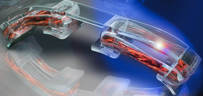
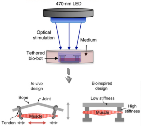
Bio-bot design inspired by the muscle-tendon-bone complex found in the human body, with 3D-printed flexible skeleton. Optical stimulation of the muscle tissue (orange), which is genetically engineered to contract in response to blue light, makes the bio-bot walk across a surface in the direction of the light. (credit: Ritu Raman et al./Nature Protocols)

For the past several years, researchers at the University of Illinois at Urbana-Champaign have reverse-engineered native biological tissues and organs — creating tiny walking “bio-bots” powered by muscle cells and controlled with electrical and optical pulses.

Now, in an open-access cover paper in Nature Protocols, the researchers are sharing a protocol with engineering details for their current generation of millimeter-scale soft robotic bio-bots*.

Using 3D-printed skeletons, these devices would be coupled to tissue-engineered skeletal muscle actuators to drive locomotion across 2D surfaces, and could one day be used for studies of muscle development and disease, high-throughput drug testing, and dynamic implants, among other applications.

In a new design, the researchers worked with MIT optogenetics experts to genetically engineer a light-responsive skeletal muscle cell line that could be stimulated to contract by pulses of blue light. (credit: Ritu Raman et al./Nature Protocols)

The future of bio-bots

The researchers envision future generations of bio-bots as biological building blocks that lead to the machines of the future. The bio-bots would integrate multiple cell and tissue types, including neuronal networks for sensing and processing, and vascular networks for delivery of nutrients and other biochemical factors. They might also have some of the higher-order properties of biological materials, such as self-organization and self-healing.

“These next iterations of biohybrid machines could, for example, be designed to sense chemical toxins, locomote toward them, and neutralize them through cell-secreted factors. Such a functionality could have broad relevance in medical diagnostics and targeted therapeutics in vivo, or even be extended to environmental use as a method of cleaning pathogens from public water supplies,” the research note in the paper.

“This protocol is essentially intended to be a one-stop reference for any scientist around the world who wants to replicate the results we showed in our PNAS 2016 and PNAS 2014 papers, and give them a framework for building their own bio-bots for a variety of applications,” said Bioengineering Professor Rashid Bashir**, who heads the bio-bots research group.

Bashir’s group has been a pioneer in designing and building bio-bots, less than a centimeter in size, made of flexible 3D printed hydrogels and living cells. In 2012, the group demonstrated bio-bots that could “walk” on their own, powered by beating heart cells from rats. In 2014, they switched to muscle cells controlled with electrical pulses, giving researchers unprecedented command over their function.

* Not to be confused with swimming biobots and rescue biobots using remotely controlled cockroaches.

** Bashir is also Grainger Distinguished Chair in Engineering and head of the Department of Bioengineering. Work on the bio-bots was conducted at the Micro + Nanotechnology Lab at Illinois.


NewsAtIllinois | Light illuminates the way for bio-bots


Abstract of A modular approach to the design, fabrication, and characterization of muscle-powered biological machines

Biological machines consisting of cells and biomaterials have the potential to dynamically sense, process, respond, and adapt to environmental signals in real time. As a first step toward the realization of such machines, which will require biological actuators that can generate force and perform mechanical work, we have developed a method of manufacturing modular skeletal muscle actuators that can generate up to 1.7 mN (3.2 kPa) of passive tension force and 300 μN (0.56 kPa) of active tension force in response to external stimulation. Such millimeter-scale biological actuators can be coupled to a wide variety of 3D-printed skeletons to power complex output behaviors such as controllable locomotion. This article provides a comprehensive protocol for forward engineering of biological actuators and 3D-printed skeletons for any design application. 3D printing of the injection molds and skeletons requires 3 h, seeding the muscle actuators takes 2 h, and differentiating the muscle takes 7 d.