Soft robotic sleeve developed to aid failing hearts

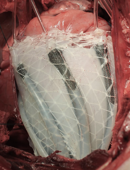
A soft robotic sleeve placed around the heart in a pig model of acute heart failure. The actuators embedded in the sleeve support heart function by mimicking the outer heart muscles that induce the heart to beat. (credit: Harvard SEAS)

An international team of scientists has developed a soft robotic sleeve that can be implanted on the external surface of the heart to restore blood circulation in pigs (and possibly humans in the future) whose hearts have stopped beating.

The device is a silicone-based system with two layers of actuators: one that squeezes circumferentially and one that squeezes diagonally, both designed to mimic the movement of healthy hearts when they beat.

Heart failure affects 41 million people worldwide. The concept of an artificial pump that aids cardiac function is not new and has been employed clinically with ventricular assist devices (VADs). “I’ve been implanting VADs in patients for a long time,” says Frank Pigula, MD, a former professor at Harvard and Boston Children’s Hospital who is co-corresponding author of an open-access paper published Jan. 18 in Science Translational Medicine. Pigula is now chief of pediatric cardiac surgery at Norton Children’s Hospital in Louisville, Kentucky.

The current-generation VADs systems directly expose a patient’s blood to artificial materials such as tubing and rotors. When blood touches a device’s components, it has a tendency to clot, which could lead to heart attacks, pulmonary embolism, strokes, or other complications, Pigula explained. To prevent clots from forming, it’s necessary to use blood thinning and anticoagulatory medications such as heparin and warfarin, which are complication-prone.

How to make a soft robotic sleeve for a human heart

That’s where the soft robotic sleeve developed by Pigula’s team comes in.
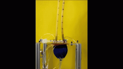
In vitro demonstration of the pumping of the soft robotic sleeve (credit: Ellen Roche/Harvard University)

The robotic sleeve can be implanted on the external surface of the heart, preventing circulating blood from ever coming in contact with the device’s components, possibly eliminating or decreasing the need for anticoagulatory drugs.

In designing the soft robotic sleeve, the researchers were inspired by the structure and movement of the heart itself. The two actuators are formed in thin layers of silicone that are layered on each other in roughly the same orientation as the muscle fibers in the heart. The sleeve is about half a millimeter thick, which is about the width of 5–10 human hairs.

To adhere the sleeve to pig hearts, the researchers used an FDA-approved adherent on the apex, or tip, of the heart. However, the researchers found this caused severe inflammation at the point of adherence, which could interfere with the ability to implant the sleeve for long periods of time. So the researchers employed a gel to adhere the sleeve to the heart, a technique that lessened the inflammation.

In vivo demonstration of cardiac assist in a porcine model of acute heart failure (credit: Ellen Rouche/Harvard SEAS)

To test the sleeves, the scientists implanted them on the hearts of pigs and induced acute heart failure, resulting in a 50–60% drop in cardiac output. Turning on the sleeve restored 97% of the original cardiac output.

“The soft robotic actuators are essentially artificial muscles,” says Nikolay Vasilyev, MD, a staff scientist in cardiac surgery research at Boston Children’s Hospital and co-author on the recent study. “In this sense, the robotic sleeve mimics both ventricles of the heart.”

The soft robotic heart sleeve also contains sophisticated sensing abilities that measure pressure at specific points on the heart’s surface.

“This work represents an exciting proof-of-concept result for this soft robot, demonstrating that it can safely interact with soft tissue and lead to improvements in cardiac function,” said Conor Walsh, senior author of the paper and the John L. Loeb Associate Professor of Engineering and Applied Sciences at SEAS and Core Faculty Member at Harvard’s Wyss Institute. “We envision many other future applications where such devices can deliver mechanotherapy both inside and outside of the body.”

However, “the human body is remarkably good at detecting foreign materials and mounting immune responses to them, so it will be tricky business to find a biologically inert material that will not, over the long run, scar the tissues it’s physically associated with,” according to a physician who was not involved in the research. “This is less critical for non-vital organs like soft-tissue silicone implants, but a thin layer of scar tissue around the heart could have serious implications for the stiffness, structural integrity, and function of native heart tissue.”

The research was a collaboration between Harvard’s SEAS and Wyss Institute, Boston Children’s Hospital, National University of Ireland,  Technische Universität München, Boston Children’s Hospital, University of Leeds, University of Central Florida, Royal College of Surgeons in Ireland, Trinity College Dublin, UCLA, and University of Louisville.

The work was supported by the Translational Research Program grant from Boston Children’s Hospital, a Director’s Challenge Cross-Platform grant from the Wyss Institute for Biologically Inspired Engineering, Harvard School of Engineering and Applied Sciences, and Science Foundation Ireland.


Abstract of Soft robotic sleeve supports heart function

There is much interest in form-fitting, low-modulus, implantable devices or soft robots that can mimic or assist in complex biological functions such as the contraction of heart muscle. We present a soft robotic sleeve that is implanted around the heart and actively compresses and twists to act as a cardiac ventricular assist device. The sleeve does not contact blood, obviating the need for anticoagulation therapy or blood thinners, and reduces complications with current ventricular assist devices, such as clotting and infection. Our approach used a biologically inspired design to orient individual contracting elements or actuators in a layered helical and circumferential fashion, mimicking the orientation of the outer two muscle layers of the mammalian heart. The resulting implantable soft robot mimicked the form and function of the native heart, with a stiffness value of the same order of magnitude as that of the heart tissue. We demonstrated feasibility of this soft sleeve device for supporting heart function in a porcine model of acute heart failure. The soft robotic sleeve can be customized to patient-specific needs and may have the potential to act as a bridge to transplant for patients with heart failure.

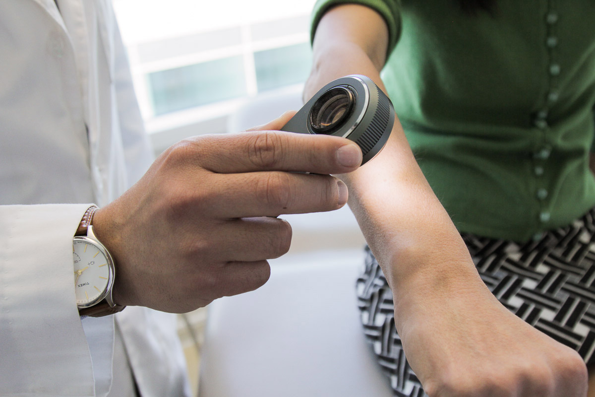
A deep learning algorithm outperforms some board-certified dermatologists in diagnosis of skin cancer

A dermatologist uses a dermatoscope, a type of handheld microscope, to look at skin. Stanford AI scientists have created a deep convolutional neural network algorithm for skin cancer that matched the performance of board-certified dermatologists. (credit: Matt Young)

Deep learning has been touted for its potential to enhance the diagnosis of diseases, and now a team of researchers at Stanford has developed a deep-learning algorithm that may make this vision a reality for skin cancer.*

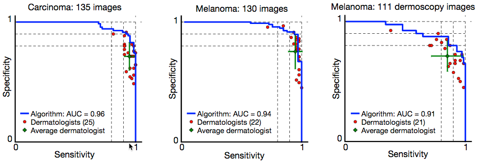
The researchers, led by Dr. Sebastian Thrun, an adjunct professor at the Stanford Artificial Intelligence Laboratory, reported in the January 25 issue of Nature that their deep convolutional neural network (CNN) algorithm performed as well or better than 21 board-certified dermatologists at diagnosing skin cancer. (See “Skin cancer classification performance of the CNN (blue) and dermatologists (red)” figure below.)

Diagnosing skin cancer begins with a visual examination. A dermatologist usually looks at the suspicious lesion with the naked eye and with the aid of a dermatoscope, which is a handheld microscope that provides low-level magnification of the skin. If these methods are inconclusive or lead the dermatologist to believe the lesion is cancerous, a biopsy is the next step. This deep learning algorithm may help dermatologists decide which skin lesions to biopsy.

“My main eureka moment was when I realized just how ubiquitous smartphones will be,” said Stanford Department of Electrical Engineering’s Andre Esteva, co-lead author of the study. “Everyone will have a supercomputer in their pockets with a number of sensors in it, including a camera. What if we could use it to visually screen for skin cancer? Or other ailments?”

It is projected that there will be 6.3 billion smartphone subscriptionst by the year 2021, according to Ericsson Mobility Report (2016), which could potentially provide low-cost universal access to vital diagnostic care.

Creating the deep convolutional neural network (CNN) algorithm

Deep CNN classification technique. Data flow is from left to right: an image of a skin lesion (for example, melanoma) is sequentially warped into a probability distribution over clinical classes of skin disease using Google Inception v3 CNN architecture pretrained on the ImageNet dataset (1.28 million images over 1,000 generic object classes) and fine-tuned on the team’s own dataset of 129,450 skin lesions comprising 2,032 different diseases. (credit: Andre Esteva et al./Nature)

Rather than building an algorithm from scratch, the researchers began with an algorithm developed by Google that was already trained to identify 1.28 million images from 1,000 object categories. It was designed primarily to be able to differentiate cats from dogs, but the researchers needed it to differentiate benign and malignant lesions. So they collaborated with dermatologists at Stanford Medicine, as well as Helen M. Blau, professor of microbiology and immunology at Stanford and co-author of the paper.

The algorithm was trained with nearly 130,000 images representing more than 2,000 different diseases with an associated disease label, allowing the system to overcome variations in angle, lighting, and zoom. The algorithm was then tested against 1,942 images of skin that were digitally annotated with biopsy-proven diagnoses of skin cancer. Overall, the algorithm identified the vast majority of cancer cases with accuracy rates that were similar to expert clinical dermatologists.

However, during testing, the researchers used only high-quality, biopsy-confirmed images provided by the University of Edinburgh and the International Skin Imaging Collaboration Project that represented the most common and deadliest skin cancers — malignant carcinomas and malignant melanomas.

Skin cancer classification performance of the CNN (blue) and dermatologists (red).** (credit: Andre Esteva et al./Nature)

The 21 dermatologists were asked whether, based on each image, they would proceed with biopsy or treatment, or reassure the patient. The researchers evaluated success by how well the dermatologists were able to correctly diagnose both cancerous and non-cancerous lesions in more than 370 images.***

However, Susan Swetter, professor of dermatology and director of the Pigmented Lesion and Melanoma Program at the Stanford Cancer Institute and co-author of the paper, notes that “rigorous prospective validation of the algorithm is necessary before it can be implemented in clinical practice, by practitioners and patients alike.”

* Every year there are about 5.4 million new cases of skin cancer in the United States, and while the five-year survival rate for melanoma detected in its earliest states is around 97 percent, that drops to approximately 14 percent if it’s detected in its latest stages.

** “Skin cancer classification performance of the CNN and dermatologists. The deep learning CNN outperforms the average of the dermatologists at skin cancer classification using photographic and
dermoscopic images. Our CNN is tested against at least 21 dermatologists at keratinocyte carcinoma and melanoma recognition. For each test, previously unseen, biopsy-proven images of lesions are displayed, and dermatologists are asked if they would: biopsy/treat the lesion or reassure the patient. Sensitivity, the true positive rate, and specificity, the true negative rate, measure performance. A dermatologist outputs a single prediction per image and is thus represented by a single red point. The green points are the average of the dermatologists for each task, with error bars denoting one standard deviation.” — Andre Esteva et al./Nature

*** The algorithm’s performance was measured through the creation of a sensitivity-specificity curve, where sensitivity represented its ability to correctly identify malignant lesions and specificity represented its ability to correctly identify benign lesions. It was assessed through three key diagnostic tasks: keratinocyte carcinoma classification, melanoma classification, and melanoma classification when viewed using dermoscopy. In all three tasks, the algorithm matched the performance of the dermatologists with the area under the sensitivity-specificity curve amounting to at least 91 percent of the total area of the graph. An added advantage of the algorithm is that, unlike a person, the algorithm can be made more or less sensitive, allowing the researchers to tune its response depending on what they want it to assess. This ability to alter the sensitivity hints at the depth and complexity of this algorithm. The underlying architecture of seemingly irrelevant photos —  including cats and dogs — helps it better evaluate the skin lesion images.


Abstract of Dermatologist-level classification of skin cancer with deep neural networks

Skin cancer, the most common human malignancy, is primarily diagnosed visually, beginning with an initial clinical screening and followed potentially by dermoscopic analysis, a biopsy and histopathological examination. Automated classification of skin lesions using images is a challenging task owing to the fine-grained variability in the appearance of skin lesions. Deep convolutional neural networks (CNNs) show potential for general and highly variable tasks across many fine-grained object categories. Here we demonstrate classification of skin lesions using a single CNN, trained end-to-end from images directly, using only pixels and disease labels as inputs. We train a CNN using a dataset of 129,450 clinical images—two orders of magnitude larger than previous datasets—consisting of 2,032 different diseases. We test its performance against 21 board-certified dermatologists on biopsy-proven clinical images with two critical binary classification use cases: keratinocyte carcinomas versus benign seborrheic keratoses; and malignant melanomas versus benign nevi. The first case represents the identification of the most common cancers, the second represents the identification of the deadliest skin cancer. The CNN achieves performance on par with all tested experts across both tasks, demonstrating an artificial intelligence capable of classifying skin cancer with a level of competence comparable to dermatologists. Outfitted with deep neural networks, mobile devices can potentially extend the reach of dermatologists outside of the clinic. It is projected that 6.3 billion smartphone subscriptions will exist by the year 2021 and can therefore potentially provide low-cost universal access to vital diagnostic care.

A 3D bioprinter that prints fully functional human skin

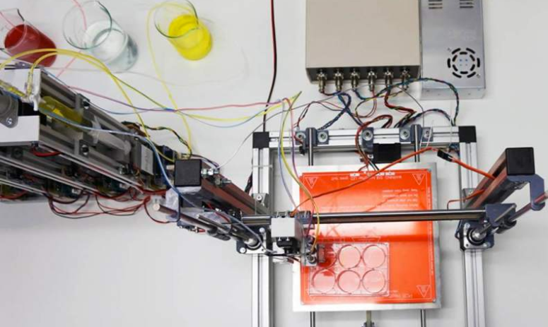
Skin-producing bioprinter (credit: Universidad Carlos III de Madrid)

A prototype 3D bioprinter that can create totally functional human skin has been developed by scientists from Universidad Carlos III de Madrid (UC3M) and BioDan Group in Spain. The skin has been used to treat burns as well as traumatic and surgical wounds in a large number of patients in Spain, according to the scientists.

The system provides two processes.

Autologous skin (from the patient’s own cells to generate human collagen) for therapeutic use, such as in the treatment of severe burns, instead of the animal collagen used in other methods.  The researchers have applied for approval by various European regulatory agencies to guarantee that the skin that is produced is adequate for use in transplants on burn patients and on those with other skin problems.

3D skin bioprinter in operation (credit: Universidad Carlos III de Madrid)

The 3D-printed skin replicates human bilayered skin, using “bioinks” (biological components) containing human plasma as well as primary human fibroblasts and keratinocytes obtained from skin biopsies. These are controlled by a computer, which deposits them on a print bed in an orderly manner to then produce the skin.

The researchers were able to generate 100 cm2 of printed skin in less than 35 minutes (including the 30 min required for fibrin gelation).

Allogeneic skin (from a stock of cells), done on a large scale for industrial processes. This skin can be used to test pharmaceutical products, cosmetics, and consumer chemical products where current regulations require testing that does not use animals.

“This method of bioprinting allows skin to be generated in a standardized, automated way, and the process is less expensive than manual production,” says Alfredo Brisac, CEO of BioDan Group, the Spanish bioengineering firm specializing in regenerative medicine that is collaborating on this research and commercializing this technology.

The research was published online in the journal Biofabrication.


UC3M | Científicos españoles crean una bioimpresora 3D de piel humana


Abstract of 3D bioprinting of functional human skin: production and in vivo analysis

Significant progress has been made over the past 25 years in the development of in vitro-engineered substitutes that mimic human skin, either to be used as grafts for the replacement of lost skin, or for the establishment of in vitro human skin models. In this sense, laboratory-grown skin substitutes containing dermal and epidermal components offer a promising approach to skin engineering. In particular, a human plasma-based bilayered skin generated by our group, has been applied successfully to treat burns as well as traumatic and surgical wounds in a large number of patients in Spain. There are some aspects requiring improvements in the production process of this skin; for example, the relatively long time (three weeks) needed to produce the surface required to cover an extensive burn or a large wound, and the necessity to automatize and standardize a process currently performed manually. 3D bioprinting has emerged as a flexible tool in regenerative medicine and it provides a platform to address these challenges. In the present study, we have used this technique to print a human bilayered skin using bioinks containing human plasma as well as primary human fibroblasts and keratinocytes that were obtained from skin biopsies. We were able to generate 100 cm2, a standard P100 tissue culture plate, of printed skin in less than 35 min (including the 30 min required for fibrin gelation). We have analysed the structure and function of the printed skin using histological and immunohistochemical methods, both in 3D in vitro cultures and after long-term transplantation to immunodeficient mice. In both cases, the generated skin was very similar to human skin and, furthermore, it was indistinguishable from bilayered dermo-epidermal equivalents, handmade in our laboratories. These results demonstrate that 3D bioprinting is a suitable technology to generate bioengineered skin for therapeutical and industrial applications in an automatized manner.

A ‘smart’ patch that automatically delivers insulin when needed

Tiny, painless microneedles on a patch can deliver insulin in response to rising glucose levels (credit: American Chemical Society)

A team of scientists has invented a replacement for daily glucose-level finger-pricking and insulin shots: a painless “smart” patch that monitors blood glucose and releases insulin when levels climb too high.

The report on the device, which has only been tested on mice so far, appears in the journal ACS Nano.

People with Type 1 diabetes don’t make insulin — a hormone that regulates blood glucose (sugar). Those with Type 2 diabetes can’t use insulin effectively. Either way, glucose builds up in the blood, which can lead to a host of health problems, including heart disease, stroke, blindness and amputation of toes, feet or legs.

To avoid these outcomes, people with Type 1 or advanced Type 2 diabetes regularly prick their fingers to measure blood-sugar levels, and some patients must inject themselves with insulin when needed. But sometimes, despite a person’s vigilance, glucose levels can still get out of whack.

A skin patch with painless microneedles

Self-assembly of block copolymer into vesicles loaded with insulin and glucose oxidase. The vesicles (engineered pouches) are dissociated to release insulin in the presence of a hyperglycemic state. (credit: American Chemical Society)

So Zhen Gu and colleagues* decided to invent a simpler, more effective, shot-free way to manage diabetes: a skin patch covered in painless microneedles that are loaded with tiny insulin-carrying pouches. The pouches (vesicles) are engineered to break apart rapidly and release the insulin in response to rising glucose levels.

Diabetic mice wearing the patch maintained consistent concentrations of insulin in their blood. When these mice received a shot of glucose, their blood sugar levels spiked initially, but then fell to normal levels within two hours.

Another automated approach: Insulin Pump Therapy system: 1. Insulin pump. 2: Flexible tubing delivers insulin from the pump reservoir to the infusion set. 3. A tiny tube called a cannula is inserted under your skin to deliver insulin. 4. Insulin in the blood (credit: Medtronics)

The authors acknowledge funding from the American Diabetes AssociationNational Institutes of Health, and the National Science Foundation.

* Affiliated with the University of North Carolina at Chapel Hill, North Carolina State University, and State Key Laboratory of Polymer Chemistry and Physics, Changchun Institute of Applied Chemistry, Chinese Academy of Sciences.


Abstract of H2O2-Responsive Vesicles Integrated with Transcutaneous Patches for Glucose-Mediated Insulin Delivery

A self-regulated “smart” insulin administration system would be highly desirable for diabetes management. Here, a glucose-responsive insulin delivery device, which integrates H2O2-responsive polymeric vesicles (PVs) with a transcutaneous microneedle-array patch was prepared to achieve a fast response, excellent biocompatibility, and painless administration. The PVs are self-assembled from block copolymer incorporated with polyethylene glycol (PEG) and phenylboronic ester (PBE)-conjugated polyserine (designated mPEG-b-P(Ser-PBE)) and loaded with glucose oxidase (GOx) and insulin. The polymeric vesicles function as both moieties of the glucose sensing element (GOx) and the insulin release actuator to provide basal insulin release as well as promote insulin release in response to hyperglycemic states. In the current study, insulin release responds quickly to elevated glucose and its kinetics can be modulated by adjusting the concentration of GOx loaded into the microneedles. In vivo testing indicates that a single patch can regulate glucose levels effectively with reduced risk of hypoglycemia.

Wearable sensors can alert you when you are getting sick, Stanford study shows

Current versions of three of the devices used for heart-rate and peripheral capillary oxygen saturation measurements in the study (credits left to right: Scanadu, iHealth, and Masimo)

Fitness monitors and other wearable biosensors can tell when your heart rate, activity, skin temperature, and other measures are abnormal, suggesting possible illness, including the onset of infection, inflammation, and even insulin resistance, according to a study by researchers at the Stanford University School of Medicine.

The team collected nearly 2 billion measurements from 60 people, including continuous data from each participant’s wearable biosensor devices* and periodic data from laboratory tests of their blood chemistry, gene expression, and other measures, and related this data to a range of normal (baseline) values for each person in the study compared to when they were ill.**

Participants wore between one and seven commercially available activity monitors and other monitors that collected more than 250,000 measurements a day. The team collected data on weight; heart rate; oxygen in the blood; skin temperature; activity, including sleep, steps, walking, biking and running; calories expended; acceleration; and even exposure to gamma rays and X-rays.

There were three participant groups: Participant #1 wore seven portable devices for large segments of this study; 43 individuals wore Intel’s Basis to measure activity (steps), heart rate, sleep, and skin temperature, with data securely uploaded to the cloud; and 16 individuals wore either iHealth or Masimo finger devices for sensing heart rate and SpO2 (peripheral capillary oxygen saturation).

Identifying health problems in advance

Wearable devices used by participant 1 (credit: Xiao Li/PLOS Biology)

The study, led by Michael Snyder, PhD, professor and chair of genetics, and senior author of the study, was published online Jan. 12 in open-access PLOS Biology. It demonstrated that, given a baseline range of values for each person, it is possible to monitor deviations from normal and associate those deviations with environmental conditions, illness, or other factors that affect health. Distinctive patterns of deviation from normal seem to correlate with particular health problems. Algorithms designed to pick up on these patterns of change could potentially contribute to clinical diagnostics and research.

The results of the current study raise the possibility of identifying inflammatory disease in individuals who may not even know they are getting sick.

For example, in several participants, higher-than-normal readings for heart rate and skin temperature correlated with increased levels of C reactive protein in blood tests. C reactive protein is an immune system marker for inflammation and often indicative of infection, autoimmune diseases, developing cardiovascular disease or even cancer. Snyder’s own data revealed four separate bouts of illness and inflammation, including a Lyme disease infection and another that he was unaware of until he saw his sensor data and an increased level of C reactive protein.

The wearable devices could also help distinguish participants with insulin resistance, a precursor for Type 2 diabetes. Of 20 participants who received glucose tests, 12 were insulin-resistant. The team designed and tested an algorithm combining participants’ daily steps, daytime heart rate and the difference between daytime and nighttime heart rate. The algorithm was able to process the data from just these few simple measures to predict which individuals in the study were likely to be insulin-resistant.

The study also revealed that declines in blood-oxygen levels during airplane flights were correlated with fatigue. Fortunately, the study showed that people tend to adapt on long flights; oxygen levels in their blood go back up, and they generally feel less fatigued as the hours go by.

The future of wearable devices: monitoring human health continuously

During a visit to the doctor, patients normally have their blood pressure and body temperature measured, but such data is typically collected only every year or two and often ignored unless the results are outside of normal range for entire populations. But biomedical researchers envisage a future in which human health is monitored continuously.

“We have more sensors on our cars than we have on human beings,” said Snyder. In the future, he said, he expects the situation will be reversed and people will have more sensors than cars do. Already, consumers have purchased millions of wearable devices, including more than 50 million smart watches and 20 million other fitness monitors. Most monitors are used to track activity, but they could easily be adjusted to more directly track health measures, Snyder said.

The work is an example of Stanford Medicine’s focus on “precision health,” whose goal is to anticipate and prevent disease in the healthy and to precisely diagnose and treat disease in the ill. With a precision health approach, every person could know his or her normal baseline for dozens of measures. Automatic data analysis could spot patterns of outlier data points and flag the onset of ill health, providing an opportunity for intervention, prevention or cure.

Researcher Elizabeth Colbert, of the Veterans Affairs Palo Alto Health Care System, is also a co-author. This research was funded by the National Institutes of Health, a gift from Bert and Candace Forbes, and Stanford’s Department of Genetics.

* “After evaluating more than 400 available wearable devices at the beginning of the study, we selected [seven] for participants to use. The criteria for selection [were] (1) ability to access the raw data from the manufacturer, (2) cost, (3) overlap in measurement of at least one component with another device to assist in reproducibility, and (4) ease of use, reasonable accuracy, and had a direct interface for raw data. These devices collectively measure (a) three physiological parameters, including heart rate, peripheral capillary oxygen saturation, and skin temperature, (b) six activity-related parameters, including sleep, steps, walking, biking, running, calories, and acceleration forces caused by movement, (c) weight, and (d) total gamma and X-ray radiation exposure.” — PLOS Biology paper authors

** “In this work, we investigate the use of portable devices to (1) easily and accurately record physiological measurements in individuals in real time (or at high frequency), (2) quantify daily patterns and reveal interesting physiological responses to different circadian cycles and environmental conditions, (3) identify personalized baseline norms and differences among individuals, (4) detect differences in health states among individuals (e.g., people with diabetes versus people without diabetes), and (5) detect inflammatory responses and assist in medical diagnosis at the early phase of disease development, thereby potentially impacting medical care.” — PLOS Biology paper authors


Abstract of Digital Health: Tracking Physiomes and Activity Using Wearable Biosensors Reveals Useful Health-Related Information

A new wave of portable biosensors allows frequent measurement of health-related physiology. We investigated the use of these devices to monitor human physiological changes during various activities and their role in managing health and diagnosing and analyzing disease. By recording over 250,000 daily measurements for up to 43 individuals, we found personalized circadian differences in physiological parameters, replicating previous physiological findings. Interestingly, we found striking changes in particular environments, such as airline flights (decreased peripheral capillary oxygen saturation [SpO2] and increased radiation exposure). These events are associated with physiological macro-phenotypes such as fatigue, providing a strong association between reduced pressure/oxygen and fatigue on high-altitude flights. Importantly, we combined biosensor information with frequent medical measurements and made two important observations: First, wearable devices were useful in identification of early signs of Lyme disease and inflammatory responses; we used this information to develop a personalized, activity-based normalization framework to identify abnormal physiological signals from longitudinal data for facile disease detection. Second, wearables distinguish physiological differences between insulin-sensitive and -resistant individuals. Overall, these results indicate that portable biosensors provide useful information for monitoring personal activities and physiology and are likely to play an important role in managing health and enabling affordable health care access to groups traditionally limited by socioeconomic class or remote geography.

Intricate microdevices that can be safely implanted

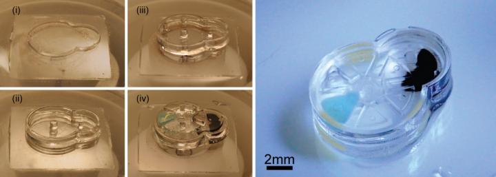
Fabrication and assembly of an iMEMS microdevice. Left: layer-by-layer fabrication of support structures and assembly of gear components. Right: the complete device after the layers have been sealed, with ferromagnetic iron material (black) to enable external magnetic control. (credit: SauYin Chin/Columbia Engineering)

Columbia Engineering researchers have invented a technique for manufacturing complex microdevices with three-dimensional, freely moving parts made from biomaterials that can safely be implanted in the body. Potential applications include a drug-delivery system to provide tailored drug doses for precision medicine, catheters, stents, cardiac pacemakers, and soft microbotics.

Most current implantable microdevices have static components rather than moving parts and, because they require batteries or other toxic electronics, they have limited biocompatibility.

The new technique stacks a soft biocompatible hydrogel material in layers, using a fast manufacturing method the researchers call “implantable microelectromechanical systems” (iMEMS).

iMEMS drug-delivery system. The payload delivery system was tested in a bone cancer mouse model, finding that the triggering of releases of doxorubicin from the device over 10 days showed high treatment efficacy and low toxicity, at 1/10th of the standard systemic chemotherapy dose. The device contains iron nanoparticle–doped components, which respond to external magnetic actuation. Actuation of the device triggers release of payloads from reservoirs. (credit: Sau Yin Chin et al./Science Robotics)

“Our iMEMS platform enables development of biocompatible implantable microdevices with a wide range of intricate moving components that can be wirelessly controlled on demand, and solves issues of device powering and biocompatibility,” says Biomedical Engineering Professor Sam Sia, senior author of an open-access paper published online January 4, 2017, in Science Robotics).

The researchers were able to trigger the iMEMS device to release payloads over days to weeks after implantation, with precise actuation by using magnetic forces to induce gear movements that, in turn, bend structural beams made of hydrogels with highly tunable properties. (Magnetic iron particles are commonly used and are FDA-approved for human use as contrast agents.)

Batteryless implantable medical devices or sensors

Sia’s iMEMS technique addresses several issues in building biocompatible microdevices, micromachines, and microrobots: how to power small robotic devices without using toxic batteries; how to make small, biocompatible, moveable components that are not silicon, which has limited biocompatibility; and how to communicate wirelessly once implanted (radio-frequency microelectronics require power, are relatively large, and are not biocompatible).

The researchers developed a “locking mechanism” for precise actuation and movement of freely moving parts, which can function as valves, manifolds, rotors, pumps, and drug delivery systems. They were able to tune the biomaterials within a wide range of mechanical and diffusive properties and to control them after implantation without a sustained power supply, such as a toxic battery.

“We can make small implantable devices, sensors, or robots that we can talk to wirelessly. Our iMEMS system could bring the field a step closer to developing soft miniaturized robots that can safely interact with humans and other living systems,” said Sia.

The team developed a drug delivery system and tested it on mice with bone cancer. The iMEMS system delivered chemotherapy adjacent to the cancer, and limited tumor growth while showing less toxicity than with chemotherapy administered throughout the body.

The study was supported by the National Science Foundation, NIH, and the Agency for Science, Technology and Research (Singapore).

* The team used light to polymerize sheets of gel and incorporated a stepper mechanization to control the z-axis and pattern the sheets layer by layer, giving them three-dimensionality. Controlling the z-axis enabled the researchers to create composite structures within one layer of the hydrogel while managing the thickness of each layer throughout the fabrication process. They were able to stack multiple layers that are precisely aligned and, because they could polymerize a layer at a time, one right after the other, the complex structure was built in under 30 minutes.

Hydrogels are difficult to work with, as they are soft and not compatible with traditional machining techniques,” says Sau Yin Chin, lead author of the study, who worked with Sia. “We have tuned the mechanical properties and carefully matched the stiffness of structures that come in contact with each other within the device. Gears that interlock have to be stiff in order to allow for force transmission and to withstand repeated actuation. Conversely, structures that form locking mechanisms have to be soft and flexible to allow for the gears to slip by them during actuation, while at the same time they have to be stiff enough to hold the gears in place when the device is not actuated. We also studied the diffusive properties of the hydrogels to ensure that the loaded drugs do not easily diffuse through the hydrogel layers.”


Abstract of Additive manufacturing of hydrogel-based materials for next-generation implantable medical devices

Implantable microdevices often have static components rather than moving parts and exhibit limited biocompatibility. This paper demonstrates a fast manufacturing method that can produce features in biocompatible materials down to tens of micrometers in scale, with intricate and composite patterns in each layer. By exploiting the unique mechanical properties of hydrogels, we developed a “locking mechanism” for precise actuation and movement of freely moving parts, which can provide functions such as valves, manifolds, rotors, pumps, and delivery of payloads. Hydrogel components could be tuned within a wide range of mechanical and diffusive properties and can be controlled after implantation without a sustained power supply. In a mouse model of osteosarcoma, triggering of release of doxorubicin from the device over 10 days showed high treatment efficacy and low toxicity, at 1/10 of the standard systemic chemotherapy dose. Overall, this platform, called implantable microelectromechanical systems (iMEMS), enables development of biocompatible implantable microdevices with a wide range of intricate moving components that can be wirelessly controlled on demand, in a manner that solves issues of device powering and biocompatibility.

MRI breakthroughs include ultra-sensitive MRI magnetic field sensing, more-sensitive monitoring without chemical or radioactive labels

Highly sensitive magnetic field sensor (credit: ETH Zurich/Peter Rüegg)

Swiss researchers have succeeded in measuring changes in strong magnetic fields with unprecedented precision, they report in the open-access journal Nature Communications. The finding may find widespread use in medicine and other areas.

In their experiments, the researchers at the Institute for Biomedical Engineering, which is operated jointly by ETH Zurich and the University of Zurich, magnetized a water droplet inside a magnetic resonance imaging (MRI) scanner, a device used for medical imaging. The researchers were able to detect even the tiniest variations of the magnetic field strength within the droplet. These changes were up to 10-12 (1 trillion) times smaller than the 7 tesla field strength of the MRI scanner used in the experiment.

“Until now, it was possible only to measure such small variations in weak magnetic fields,” says Klaas Prüssmann, Professor of Bioimaging at ETH Zurich and the University of Zurich. An example of a weak magnetic field is that of the Earth, where the field strength is just a few dozen microtesla. For fields of this kind, highly sensitive measurement methods are already able to detect variations of about a trillionth of the field strength, says Prüssmann. “Now, we have a similarly sensitive method for strong fields of more than one tesla, such as those used … in medical imaging.”

The scientists based the sensing technique on the principle of nuclear magnetic resonance (NMR), which also serves as the basis for magnetic resonance imaging and the spectroscopic methods that biologists use to elucidate the 3D structure of molecules, but with 1000 times greater sensitivity than current NMR methods.

Ultra-sensitive recordings of heart contractions in an MRI machine

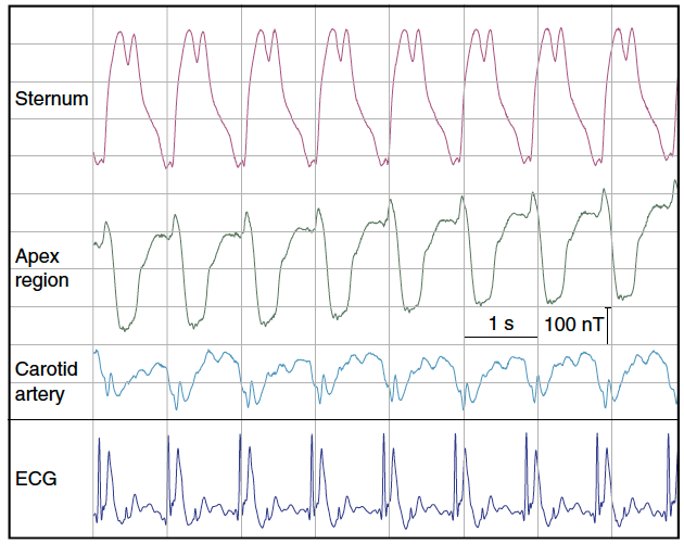
Real-time magnetic field recordings of cardiac activity. Magnetic field dynamics generated by the beating human heart in a background of 7 tesla, recorded at three different positions on the chest and neck, along with simultaneous electrocardiogram (ECG). (credit: Simon Gross et al./Nature Communications)

The scientists carried out an experiment in which they positioned their sensor in front of the chest of a volunteer test subject inside an MRI scanner. They were able to detect periodic changes in the magnetic field, which pulsated in time with the heartbeat. The measurement curve is similar to an electrocardiogram (ECG), but measures a mechanical process (the contraction of the heart) rather than electrical conduction.

“We are in the process of analyzing and refining our magnetometer measurement technique in collaboration with cardiologists and signal processing experts,” says Prüssmann. “Ultimately, we hope that our sensor will be able to provide information on heart disease — and do so non-invasively and in real time.”

The new measurement technique could also be used in the development of new contrast agents for magnetic resonance imaging and improved nuclear magnetic resonance (NMR) spectroscopy for applications in biological and chemical research.

A radiation-free approach to imaging molecules in the brain

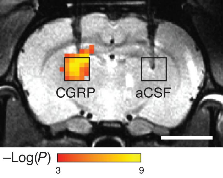
Scientists hoping to see molecules that control brain activity have devised a probe that lets them image such molecules without using chemical or radioactive labels. The sensors consist of proteins that detect a particular target, which causes them to dilate blood vessels, producing a change in blood flow that can be imaged with magnetic resonance imaging (MRI) or other techniques. (credit: Mitul Desai et al./ Nature Communications)

In a related development, MIT scientists hoping to get a glimpse of molecules that control brain activity have devised a new sensor that allows them to image these molecules without using any chemical or radioactive labels (which feature low resolution and can’t be easily used to watch dynamic events).

The new sensors consist of enzymes called proteases designed to detect a particular target, which causes them to dilate blood vessels in the immediate area. This produces a change in blood flow that can be imaged with magnetic resonance imaging (MRI) or other imaging techniques.*

A peptide called calcitonin gene-related peptide (CGRP) acts on a receptor in smooth muscle cells (left) to induce cAMP production, resulting in relaxation of vascular smooth muscle cells and consequent vasodilation (middle). That induces haemodynamic effects visible by MRI and other imaging methods (right). (credit: Mitul Desai et al./ Nature Communications)

“This is an idea that enables us to detect molecules that are in the brain at biologically low levels, and to do that with these imaging agents or contrast agents that can ultimately be used in humans,” says Alan Jasanoff, an MIT professor of biological engineering and brain and cognitive sciences. “We can also turn them on and off, and that’s really key to trying to detect dynamic processes in the brain.”

Monitoring neurotransmitters at 100 times lower levels

In a paper appearing in the Dec. 2 issue of open-access Nature Communications, Jasanoff and his colleagues explain that they used proteases (sometimes used as biomarkers to diagnose diseases such as cancer and Alzheimer’s disease) to demonstrate the validity of their approach. But now they’re working on adapting these imaging agents to monitor neurotransmitters, such as dopamine and serotonin, which are critical to cognition and processing emotions.

“What we want to be able to do is detect levels of neurotransmitter that are 100-fold lower than what we’ve seen so far. We also want to be able to use far less of these molecular imaging agents in organisms. That’s one of the key hurdles to trying to bring this approach into people,” Jasanoff says.

“Many behaviors involve turning on genes, and you could use this kind of approach to measure where and when the genes are turned on in different parts of the brain,” Jasanoff says.

His lab is also working on ways to deliver the peptides without injecting them, which would require finding a way to get them to pass through the blood-brain barrier. This barrier separates the brain from circulating blood and prevents large molecules from entering the brain.

Jeff Bulte, a professor of radiology and radiological science at the Johns Hopkins School of Medicine, described the technique as “original and innovative,” while adding that its safety and long-term physiological effects will require more study.

“It’s interesting that they have designed a reporter without using any kind of metal probe or contrast agent,” says Bulte, who was not involved in the research. “An MRI reporter that works really well is the holy grail in the field of molecular and cellular imaging.”

The research was funded by the National Institutes of Health BRAIN Initiative, the MIT Simons Center for the Social Brain, and fellowships from the Boehringer Ingelheim Fonds and the Friends of the McGovern Institute.

* To make their probes, the researchers modified a naturally occurring peptide called calcitonin gene-related peptide (CGRP), which is active primarily during migraines or inflammation. The researchers engineered the peptides so that they are trapped within a protein cage that keeps them from interacting with blood vessels. When the peptides encounter proteases in the brain, the proteases cut the cages open and the CGRP causes nearby blood vessels to dilate. Imaging this dilation with MRI allows the researchers to determine where the proteases were detected.

Another possible application for this type of imaging is to engineer cells so that the gene for CGRP is turned on at the same time that a gene of interest is turned on. That way, scientists could use the CGRP-induced changes in blood flow to track which cells are expressing the target gene, which could help them determine the roles of those cells and genes in different behaviors. Jasanoff’s team demonstrated the feasibility of this approach by showing that implanted cells expressing CGRP could be recognized by imaging.


Abstract of Dynamic nuclear magnetic resonance field sensing with part-per-trillion resolution

High-field magnets of up to tens of teslas in strength advance applications in physics, chemistry and the life sciences. However, progress in generating such high fields has not been matched by corresponding advances in magnetic field measurement. Based mostly on nuclear magnetic resonance, dynamic high-field magnetometry is currently limited to resolutions in the nanotesla range. Here we report a concerted approach involving tailored materials, magnetostatics and detection electronics to enhance the resolution of nuclear magnetic resonance sensing by three orders of magnitude. The relative sensitivity thus achieved amounts to 1 part per trillion (10−12). To exemplify this capability we demonstrate the direct detection and relaxometry of nuclear polarization and real-time recording of dynamic susceptibility effects related to human heart function. Enhanced high-field magnetometry will generally permit a fresh look at magnetic phenomena that scale with field strength. It also promises to facilitate the development and operation of high-field magnets.


Abstract of Molecular imaging with engineered physiology

In vivo imaging techniques are powerful tools for evaluating biological systems. Relating image signals to precise molecular phenomena can be challenging, however, due to limitations of the existing optical, magnetic and radioactive imaging probe mechanisms. Here we demonstrate a concept for molecular imaging which bypasses the need for conventional imaging agents by perturbing the endogenous multimodal contrast provided by the vasculature. Variants of the calcitonin gene-related peptide artificially activate vasodilation pathways in rat brain and induce contrast changes that are readily measured by optical and magnetic resonance imaging. CGRP-based agents induce effects at nanomolar concentrations in deep tissue and can be engineered into switchable analyte-dependent forms and genetically encoded reporters suitable for molecular imaging or cell tracking. Such artificially engineered physiological changes, therefore, provide a highly versatile means for sensitive analysis of molecular events in living organisms.

Immune cells in covering of brain discovered; may play critical role in battling neurological diseases

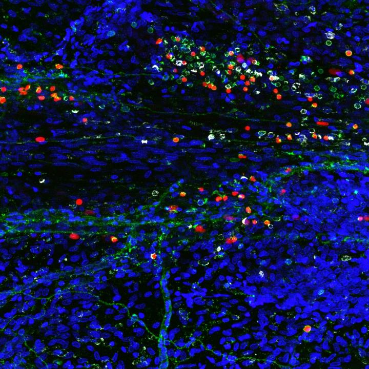
A composite image showing newly discovered immune cells in the brain (credit: Sachin Gadani | University of Virginia School of Medicine)

University of Virginia School of Medicine researchers have discovered a rare and powerful type of immune cell in the meninges (protective covering) of the brain that are activated in response to central nervous system injury — suggesting that these cells may play a critical role in battling Alzheimer’s, multiple sclerosis, meningitis, and other neurological diseases, and in supporting healthy mental functioning.

By harnessing the power of the cells, known as “type 2 innate lymphocytes” (ILC2s), doctors may be able to develop new treatments for neurological diseases, traumatic brain injury, and spinal cord injuries, as well as migraines, the researchers suggest. They also suspect the cells may be the missing link connecting the brain and the microbiota in our guts, a relationship that has been shown to be important in the development of Parkinson’s disease.

Important immune roles

ILC2 cells have previously been found in the gut, lung, and skin, the body’s barriers to disease. Their discovery by UVA researcher Jonathan Kipnis, PhD, in the meninges, the membranes surrounding the brain, comes as a surprise. They were found along the same vessels discovered by the Kipnis lab last year, which showed that the brain and the immune system are directly connected.

“This all comes down to immune system and brain interaction,” said Kipnis, chairman of UVA’s Department of Neuroscience. These where previously believed to be not communicating, but not only are these [immune] cells present in the areas near the brain, they are integral to its function, Kipnis said.

Immune cells play several important roles within the body, including guarding against pathogens, triggering allergic reactions, and responding to spinal cord injuries. But its their role in the gut that makes Kipnis suspect they may also be serving as a vital communicator between the brain’s immune response and our microbiomes (microbes in the body). That could be very important, because our intestinal flora is critical for maintaining our health and well being.

“These cells are potentially the mediator between the gut and the brain. They are the main responder to microbiota changes in the gut,” Kipnis said. “They may go from the gut to the brain, or they may just produce something that will impact those cells. We know the brain responds to things happening in the gut. Is it logical that these will be the cells that connect the two? Potentially.”

The findings have been published online by the Journal of Experimental Medicine. The work was supported by a National Institutes of Health grant.


Abstract of Characterization of meningeal type 2 innate lymphocytes and their response to CNS injury

The meningeal space is occupied by a diverse repertoire of immune cells. Central nervous system (CNS) injury elicits a rapid immune response that affects neuronal survival and recovery, but the role of meningeal inflammation remains poorly understood. Here, we describe type 2 innate lymphocytes (ILC2s) as a novel cell type resident in the healthy meninges that are activated after CNS injury. ILC2s are present throughout the naive mouse meninges, though are concentrated around the dural sinuses, and have a unique transcriptional profile. After spinal cord injury (SCI), meningeal ILC2s are activated in an IL-33–dependent manner, producing type 2 cytokines. Using RNAseq, we characterized the gene programs that underlie the ILC2 activation state. Finally, addition of wild-type lung-derived ILC2s into the meningeal space of IL-33R−/− animals partially improves recovery after SCI. These data characterize ILC2s as a novel meningeal cell type that responds to SCI and could lead to new therapeutic insights for neuroinflammatory conditions.

Using graphene to detect brain cancer cells

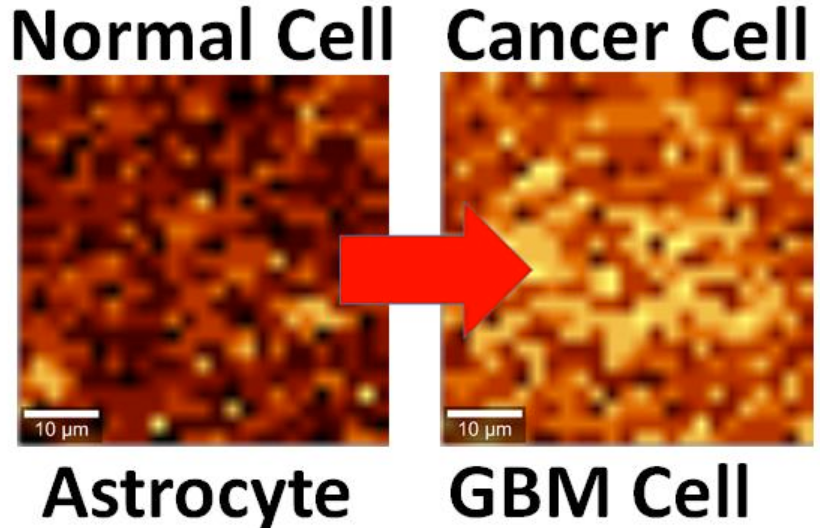
Brain cell culture. Left: Normal astrocyte brain cell; Right: cancerous Glioblastoma Multiforme (GBM) version, imaged by Raman spectrography. (credit: B. Keisham et al./ACS Appl. Mater. Interfaces)

By interfacing brain cells with graphene, University of Illinois at Chicago researchers have differentiated a single hyperactive Glioblastoma Multiforme cancerous astrocyte cell from a normal cell in the lab — pointing the way to developing a simple, noninvasive tool for early cancer diagnosis.

In the study, reported in the journal ACS Applied Materials & Interfaces, the researchers looked at lab-cultured human brain astrocyte cells taken from a mouse model. They compared normal astrocytes to their cancerous counterpart, highly malignant brain tumor glioblastoma multiforme.

Illustration showing an astrocyte cell taken from a mouse brain draped over graphene (credit: B. Keisham et al./ACS Appl. Mater. Interfaces)

In a lab analysis, the cell is draped over graphene, explains Vikas Berry, associate professor and head of chemical engineering at UIC, who led the research along with Ankit Mehta, assistant professor of clinical neurosurgery in the UIC College of Medicine.

“The electric field around the cancer cell pushes away electrons in graphene’s electron cloud,” he said, which changes the vibration energy of the carbon atoms [in the graphene]. The change in vibration energy (resulting from the cancerous condition) can be pinpointed by Raman spectroscopy with a resolution of 300 nanometers, allowing for determining the activity of a single cell. (Raman spectroscopy is a highly sensitive method commonly used in chemistry to identify molecules by how they scatter laser light.)

“Graphene is the thinnest known material and is very sensitive to whatever happens on its surface,” Berry said. The nanomaterial is composed of a single layer of carbon atoms linked in a hexagonal chicken-wire pattern, and all the atoms share a cloud of electrons moving freely about the surface.

Patient biopsies planned

The technique is now being studied in a mouse model of cancer, with results that are “very promising,” Berry said. Experiments with patient biopsies would be further down the road. “Once a patient has brain tumor surgery, we could use this technique to see if the tumor relapses,” Berry said. “For this, we would need a cell sample we could interface with graphene and look to see if cancer cells are still present.”

The same technique may also work to differentiate between other types of cells or the activity of cells. “We may be able to use it with bacteria to quickly see if the strain is Gram-positive or Gram-negative,” Berry said. “We may be able to use it to detect sickle cells.”

Earlier this year, Berry and other coworkers introduced nanoscale ripples in graphene, causing it to conduct differently in perpendicular directions, useful for electronics. They wrinkled the graphene by draping it over a string of rod-shaped bacteria, then vacuum-shrinking the germs. “We took the earlier work and sort of flipped it over,” Berry said. “Instead of laying graphene on cells, we laid cells on graphene and studied graphene’s atomic vibrations.”

Funding was provided by UIC.


Abstract of Cancer Cell Hyperactivity and Membrane Dipolarity Monitoring via Raman Mapping of Interfaced Graphene: Toward Non-Invasive Cancer Diagnostics

Ultrasensitive detection, mapping, and monitoring of the activity of cancer cells is critical for treatment evaluation and patient care. Here, we demonstrate that a cancer cell’s glycolysis-induced hyperactivity and enhanced electronegative membrane (from sialic acid) can sensitively modify the second-order overtone of in-plane phonon vibration energies (2D) of interfaced graphene via a hole-doping mechanism. By leveraging ultrathin graphene’s high quantum capacitance and responsive phononics, we sensitively differentiated the activity of interfaced Glioblastoma Multiforme (GBM) cells, a malignant brain tumor, from that of human astrocytes at a single-cell resolution. GBM cell’s high surface electronegativity (potential ∼310 mV) and hyperacidic-release induces hole-doping in graphene with a 3-fold higher 2D vibration energy shift of approximately 6 ± 0.5 cm–1 than astrocytes. From molecular dipole-induced quantum coupling, we estimate that the sialic acid density on the cell membrane increases from one molecule per ∼17 nm2 to one molecule per ∼7 nm2. Furthermore, graphene phononic response also identified enhanced acidity of cancer cell’s growth medium. Graphene’s phonon-sensitive platform to determine interfaced cell’s activity/chemistry will potentially open avenues for studying activity of other cancer cell types, including metastatic tumors, and characterizing different grades of their malignancy.

How diabetes drug metformin prevents, suppresses cancer growth

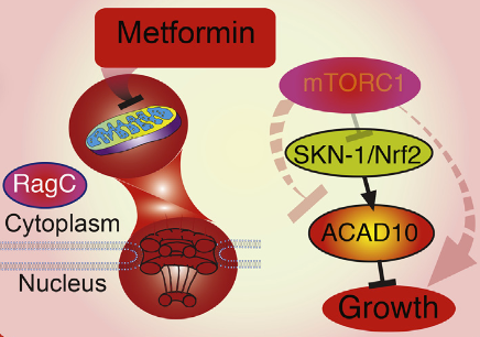
Metformin growth inhibition process (credit: Lianfeng Wu et al./Cell)

A team of Massachusetts General Hospital (MGH) and Harvard Medical School investigators has identified a pathway that appears to underlie the apparent ability of the diabetes drug metformin to both block the growth of human cancer cells and extend the lifespan of the C.elegans roundworm.

That finding implies that this single genetic pathway may play an important role in a wide range of organisms — including humans.

“We found that metformin reduces the traffic of molecules into and out of the nucleus — the ‘information center’ of the cell,” says Alexander Soukas, MD, PhD, of the MGH Center for Human Genetic Research, senior author of the study, published in the Thursday, Dec. 15 issue of Cell.

“Reduced nuclear traffic translates into the ability of the drug to block cancer growth and, remarkably, is also responsible for metformin’s ability to extend lifespan,” he said. “By shedding new light on metformin’s health-promoting effects, these results offer new potential ways that we can think about treating cancer and increasing healthy aging.”

Several studies have suggested that individuals taking metformin have a reduced risk of developing certain cancers and of dying from cancers that do develop. Current clinical trials are testing the impact of metformin on cancers of the breast, prostate and pancreas; and several research groups are working to identify its molecular targets.

How Metformin lowers blood glucose — and growth

Metformin’s ability to lower blood glucose in patients with type 2 diabetes appears to result from the drug’s effects on the liver. The drug reduces the liver’s ability to produce glucose for release into the bloodstream. Metformin has been thought to block the activity of mitochondria — structures that serve as the powerhouse of the cell.

But Soukas says more recent information suggests the mechanism is more complex. The Soukas team found a genetic pathway that slows the growth and extends the lifespan of C.elegans roundworms. That suggests that the roundworm could serve as a model for investigating the drug’s effects on cancer.*

“Amazingly, this pathway operates identically, whether in the worm or in human cancer cells,” says Soukas, who is an assistant professor of Medicine at Harvard Medical School. “Determining exactly how [it] slows cell growth will provide additional insights into novel therapeutic targets for cancer and possibly ways to manipulate the pathway to promote healthy aging.”**

Support for this study includes National Institutes of Health grants, a Broad Institute SPARC Grant, and the Ellison Medical Foundation New Scholar in Aging Award.

Metformin both suppresses cancer cell growth and promotes organismal longevity through a key transcriptional target that is induced through inhibition of mitochondrial respiration and modulation of mTOR signaling. (credit: Lianfeng Wu et al./Cell)

* The Soukas team’s experiments found that metformin’s action against cancer relies on two elements of a single genetic pathway – the nuclear pore complex, which allows the passage of molecules into and out of the nucleus, and an enzyme called ACAD10.

Basically, metformin’s suppression of mitochondrial activity reduces cellular energy, restricting the traffic of molecules through the nuclear pore. This shuts off an important cellular growth molecule called mTORC1, resulting in activation of ACAD10, which both slows the growth and extends the lifespan of C.elegans.

In human melanoma and pancreatic cancer cells, the investigators confirmed that application of drugs in the metformin family induced ACAD10 expression, an effect that depended on the function of the nuclear pore complex.

Without the complete signaling pathway – from mitochondrial suppression, through nuclear pore restriction to ACAD10 expression – cancer cells were no longer sensitive to the effects of metformin-like drugs.

** “Our experiments showed two very important things: if we force the nuclear pore to remain open or if we permanently shut down ACAD10, metformin can no longer block the growth of cancer cells. That suggests that the nuclear pore and ACAD10 may be manipulated in specific circumstances to prevent or even treat certain cancers.”

The essential contribution of ACAD10 to metformin’s anticancer action is intriguing, Soukas adds, because the only published study on ACAD10 function tied a variant in the gene to the increased risk of type 2 diabetes in Pima Indians, suggesting that ACAD10 also has a role in the drug’s antidiabetes action. “What ACAD10 does is a great mystery that we are greatly interested in solving,” he says. 


Abstract of An Ancient, Unified Mechanism for Metformin Growth Inhibition in C. elegans and Cancer

Metformin has utility in cancer prevention and treatment, though the mechanisms for these effects remain elusive. Through genetic screening in C. elegans, we uncover two metformin response elements: the nuclear pore complex (NPC) and acyl-CoA dehydrogenase family member-10 (ACAD10). We demonstrate that biguanides inhibit growth by inhibiting mitochondrial respiratory capacity, which restrains transit of the RagA-RagC GTPase heterodimer through the NPC. Nuclear exclusion renders RagC incapable of gaining the GDP-bound state necessary to stimulate mTORC1. Biguanide-induced inactivation of mTORC1 subsequently inhibits growth through transcriptional induction of ACAD10. This ancient metformin response pathway is conserved from worms to humans. Both restricted nuclear pore transit and upregulation of ACAD10 are required for biguanides to reduce viability in melanoma and pancreatic cancer cells, and to extend C. elegans lifespan. This pathway provides a unified mechanism by which metformin kills cancer cells and extends lifespan, and illuminates potential cancer targets.