Trump considering libertarian reformer to head FDA

The Seasteading Institute wants to create new societies at sea, away from FDA (and other government) regulations. (credit: Seasteading Institute)

President-elect Donald Trump’s transition team is considering libertarian Silicon Valley investor Jim O’Neill, a Peter Thiel associate, to head the Food and Drug Administration, Bloomberg Politics has reported.

O’Neill, the Managing Director of Mithril Capital Management LLC, doesn’t have a medical background, but served in the George W. Bush administration as principal associate deputy secretary at the Department of Health and Human Services. He’s also a board member of the Seasteading Institute, a Thiel-backed venture to create new societies at sea, away from existing governments.

“We should reform FDA so there is approving drugs after their sponsors have demonstrated safety — and let people start using them, at their own risk, but not much risk of safety,” O’Neill said in a speech at the August 2014 Rejuvenation Biotechnology conference. “O’Neill also advocated anti-aging medicine in that speech, saying he believed it was scientifically possible to develop treatments that would reverse aging,” said Bloomberg.

O’Neill’s prospective nomination could also bring about “significant changes to medical cannabis policy and potentially address the regulations that have prevented medical cannabis research,” Mike Liszewski, the director of government affairs at Americans for Safe Access, told ATTN:.

Scott Gottlieb, M.D., a former FDA official and now at the American Enterprise Institute (AEI), is also reportedly under consideration, according to The Hill.

In a recent related announcement, Trump has selected Rep. Tom Price, M.D. (R., Ga.), a leader in the efforts to replace ObamaCare, to be his secretary of Health and Human Services. “His most frequent objection to [the Affordable Care Act] is that it interferes with the ability of patients and doctors to make medical decisions,” The New York Times notes. Price also proposes to deregulate the market for medical services, according to the AEI.


Implantable device targets cancer, other illnesses with controlled long-term drug delivery

A drug-delivery capsule directly implanted into a cancerous tumor (credit: Lyle Hood/UTSA)

A new drug-delivery system based on an tiny implantable capsule could “revolutionize” the delivery of medicine to treat cancer and a host of other diseases and ailments, according to researchers at The University of Texas at San Antonio (UTSA).

“The problem with most drug-delivery systems is that you have a specific minimum dosage of medicine that you need to take for it to be effective,” said Lyle Hood, assistant professor of mechanical engineering. “There’s also a limit to how much of the drug can be present in your system so that it doesn’t make you sick.” So a person who needs frequent doses of a specific medicine is required to take a pill every day or visit a doctor for injections.

KurzweilAI has covered a number of drug-delivery systems targeting cancer this year, based on “Nanoparticle cluster bombs,” “magnetically guided bacteria,” and “DNA-based lock-and-key pores,” for example.

What makes the UTSA system different is that it’s an implantable capsule, filled with medicinal fluid that uses about 5000 nanochannels to regulate the rate of release of the medicine, Hood explained. “This way, we have the proper amount of drugs in a person’s system to be effective, but not so much that they’ll harm that person.”

The capsule can deliver medicinal doses for several days or a few weeks, says Hood, and can be used for any kind of ailment that needs a localized delivery over several days or a few weeks. He believes this makes it especially tailored for treating cancer.

A larger version of the device, originally created by Alessandro Grattoni, chair of the Department of Nanomedicine at Houston Methodist Research Institute, can treat diseases like HIV for up to a year. “In HIV treatment, you can bombard the virus with drugs to the point that that person is no longer infectious and shows no symptoms,” Hood said. “The danger is that if that person stops taking their drugs, the amount of medicine in his or her system drops below the effective dose and the virus is able to become resistant to the treatments.”

The new capsule could provide a constant delivery of the HIV-battling drugs to prevent such an outcome. Hood noted it can also be used to deliver cortisone to damaged joints to avoid painful, frequent injections, and possibly even to pursue immunotherapy treatments for cancer patients.

“The idea behind immunotherapy is to deliver a cocktail of immune drugs to call attention to the cancer in a person’s body, so the immune system will be inspired to get rid of the cancer itself,” he added.

The current prototype of the device is permanent and injected under the skin, but the researchers are working on 3-D printing technology to make a new, fully biodegradable version of the device that could potentially be swallowed.


Abstract of Nanochannel Implants for Minimally-Invasive Insertion and Intratumoral Delivery

Novel approaches to achieve local, intratumoral drug delivery have the dual benefit of reducing systemic toxicity while enhancing efficacy for malignant cells. We have developed a new implantable system combining a next-generation BioNEMS nanofluidic membrane with parallel nanochannels that offers controlled release of biomolecules. Based on concentration-driven diffusive transport, nanochannel membranes provide a “drug agnostic” delivery mechanism. Integrating this nanotechnology within a small implantable capsule permits multipurpose functionality and compatibility with different therapeutic approaches as well as diagnostic imaging capability. A minimally-invasive, percutaneous trocar delivery mechanism enables serial implantation throughout a target tissue volume. In this manuscript, we demonstrate that this platform is capable of sustained delivery for chemotherapy, radiosensitization, immunomodulation, and imaging contrast, among others. This platform’s utility was established through release of doxorubicin, OX86, FGK45, and Magnevist. Further proof-of-concept experiments demonstrated successful in vivo implantation and intratumoral release of antibodies and contrast agents, as well as the platform’s MR-compatibility and capability as a radiopaque fiducial. These results provide strong evidence for a flexible, multifunctional nanofluidic implant capable of broadening local delivery utility in the clinic.

Smart skin patch releases blood thinners in closed-loop control system

A painless microneedle skin patch for delivering the right amount of a blood-thinning drug (credit: Yuqi Zhang)

North Carolina researchers have developed a smart skin patch designed to monitor a patient’s blood and release a blood-thinning drug, as needed, to prevent thrombosis (dangerous blood clots).

Thrombosis — one of the leading causes of cardiovascular mortalities and morbidities worldwide — occurs when blood clots disrupt the normal flow of blood in the body, which can cause severe health problems such as pulmonary embolism, heart attack, or stroke.

Current treatments often rely on the use of blood thinners, such as heparin, requiring patients to test their blood on a regular basis to ensure proper dosages. Dosage is iffy; too large a dose can cause problems such as spontaneous hemorrhaging, while doses that are too small may not prevent thrombosis.

So researchers at North Carolina State University and the University of North Carolina at Chapel Hill developed a feedback-controlled anti-coagulant smart skin patch to release just the needed level of heparin.

Schematic of skin patch that releases blood-thining heparin (to prevent blood clots) in response to thrombin (an enzyme that initiates clotting in the blood) (credit: Yuqi Zhang)

The patch incorporates an array of painless microneedles made of a material that is responsive to thrombin (an enzyme that initiates clotting in the blood). When elevated levels of thrombin enzymes in the bloodstream come into contact with the microneedle, the enzymes cause the thrombin-responsive material in the microneedles to release the right amount (and no more) of blood-thining heparin into the bloodstream to prevent blood clots.*

The patch was successfully tested in a mouse model.

“Our goal was to generate a patch that can monitor a patient’s blood and release additional drugs when necessary — effectively, a self-regulating system,” says Zhen Gu, co-corresponding author on a paper published Nov. 25, 2016 in Advanced Materials describing the work. Gu is an associate professor in the joint biomedical engineering program at NC State and UNC.

“This paper represents a good first step, and we’re now looking for funding to perform additional preclinical testing,” Gu said.

The work was supported by the Alfred P. Sloan Foundation, NIH’s Clinical and Translational Science Awards, and the National Science Foundation.

* Closed-loop heparin delivery system

A peptide is introduced as a linker during the conjugation of Heparin to the main chain of hyaluronic acid (which has been UV-modified to be sensitive to thrombin. The presence of thrombin (top) causes the peptide to be cleaved (cut), triggering heparin drug release (right). The released heparin then inhibits the coagulation activation by inactivating thrombin (bottom). In a feedback loop, that process then suppresses further release of heparin (left), minimizing the risk of undesirable spontaneous hemorrhage. (credit: Yuqi Zhang et al./Advanced Materials)


Abstract of Thrombin-Responsive Transcutaneous Patch for Auto-Anticoagulant Regulation

A thrombin-responsive closed-loop patch is developed for prolonged heparin delivery in a feedback-controlled manner. This microneedle-based patch can sense activated thrombin and subsequently releases heparin to prevent coagulation in the blood flow. This “smart” heparin patch can be transcutaneously inserted into skin without drug leakage and can sustainably regulate blood coagulation in response to thrombin.

This tiny electronic device applied to the skin can pick up heart and speech sounds

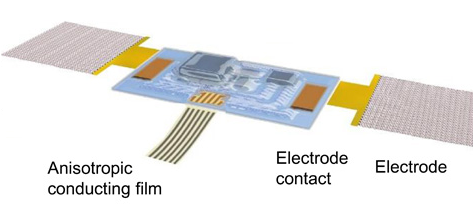
Illustration of the assembled acoustic sensor device and its interface with soft electrophysiology measurement electrodes and flexible cable for power supply and data acquisition (credit: Yuhao Liu et al./Science Advances)

Researchers from the University of Colorado Boulder and Northwestern University have developed a tiny, soft, wearable acoustic sensor that measures vibrations in the human body and can be used to monitor human heart health and recognize spoken words.

The stretchable Band-aid-like device attaches to the skin on nearly any surface of the body, using “epidermal electronics” to capture sound signals from the body.

It’s a sort of tiny, wearable stethoscope. As described in an open-access paper published Nov. 16 in Science Advances, a sister journal of Science, it can detect things like heart murmurs in cardiac patients and lung problems, and can monitor ventricular assist devices. It can also be used to pick up speech sounds (for automated speech recognition or controlling video games and other machines), and even movements in gastrointestinal tracts.

Listening in on the body

The sensor can also integrate electrodes that can record electrocardiogram (ECG) signals that measure the electrical activity of the heart as well electromyogram (EMG) signals that measure the electrical activity of muscles at rest and during contraction.

While the sensor was wired to an external data acquisition system for the tests, it can easily be converted into a wireless device, said CU Boulder Assistant Professor Jae-Woong Jeong, a lead author. Such sensors could be of use in remote, noisy places — including battlefields — producing quiet, high-quality cardiology or speech signals that can be read in real time at distant medical facilities.

Using the data from these sensors, a doctor at a hospital far away from a patient would be able to make a fast, accurate diagnosis, said Jeong.

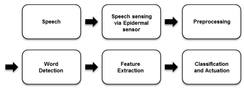
Process loop for a speech-based human-machine interface (credit: Yuhao Liu et al./Science Advances)

Vocal cord vibration signals also could be used by the military personnel or civilians to control robots, vehicles or drones. The speech recognition capabilities of the sensor also have implications for improving communication for people suffering from speech impairments, he said.

As part of the study, the team used the device to measure cardiac acoustic responses and ECG activity — including the detection of heart murmurs — in a group of elderly volunteers.

The researchers also were able to detect the acoustical signals of blood clots in a related lab experiment, said Jeong.


University of Colorado Boulder | CU Science Story — Wearable Tech


Abstract of Epidermal mechano-acoustic sensing electronics for cardiovascular diagnostics and human-machine interfaces

Physiological mechano-acoustic signals, often with frequencies and intensities that are beyond those associated with the audible range, provide information of great clinical utility. Stethoscopes and digital accelerometers in conventional packages can capture some relevant data, but neither is suitable for use in a continuous, wearable mode, and both have shortcomings associated with mechanical transduction of signals through the skin. We report a soft, conformal class of device configured specifically for mechano-acoustic recording from the skin, capable of being used on nearly any part of the body, in forms that maximize detectable signals and allow for multimodal operation, such as electrophysiological recording. Experimental and computational studies highlight the key roles of low effective modulus and low areal mass density for effective operation in this type of measurement mode on the skin. Demonstrations involving seismocardiography and heart murmur detection in a series of cardiac patients illustrate utility in advanced clinical diagnostics. Monitoring of pump thrombosis in ventricular assist devices provides an example in characterization of mechanical implants. Speech recognition and human-machine interfaces represent additional demonstrated applications. These and other possibilities suggest broad-ranging uses for soft, skin-integrated digital technologies that can capture human body acoustics.

Scientists find key protein for spinal cord repair in zebrafish


Duke University | Spinal Cord Injury and Regeneration in Zebrafish

Duke University scientists have found a protein that’s important for the ability of the freshwater zebrafish’s spinal cord to heal completely after being severed. Their study, published Nov. 4  in the journal Science, could generate new leads for what is a paralyzing and often fatal injury for humans.

Searching for the repair molecules

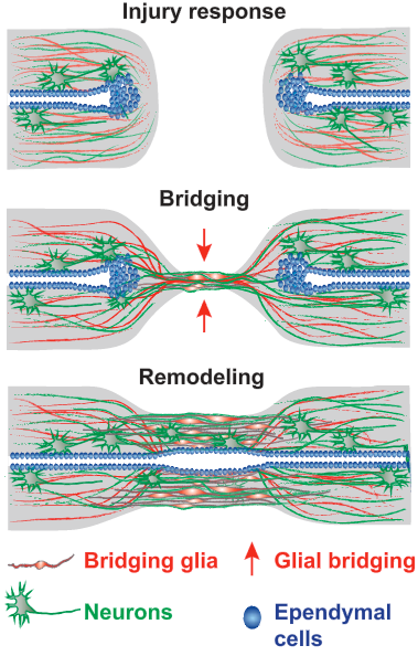
Schematic of the multistep process of spinal cord regeneration in zebrafish. (Injury response:) When the zebrafish’s severed spinal cord undergoes regeneration, a bridge forms. (Bridging:) The first cells extend projections into a distance tens of times their own length and connect across a wide gulf of the injury. Nerve cells follow. (Remodeling:) By 8 weeks, new nerve tissue has filled the gap and the animals have fully reversed their severe paralysis. (credit: Mayssa H. Mokalled et al./Science)

To understand what molecules were potentially responsible for this remarkable process, the scientists searched for the genes whose activity abruptly changed after spinal cord injury. Of dozens of genes strongly activated by injury, seven coded for proteins that are secreted from cells. One of these, called CTGF (connective tissue growth factor), was intriguing because its levels rose in the supporting cells, or glia, that formed the bridge in the first two weeks following injury.

What’s more, when they tried deleting CTGF genetically, those fish failed to regenerate.

The human CTGF protein is 87% similar in its amino acid building blocks to the zebrafish form. So when the team added the human version of CTGF to the injury site in fish, it boosted regeneration and the fish swam better by two weeks after the injury.

The second half of the CTGF protein seems to be the key to the healing, the group found. It’s a large protein, made of four smaller parts, and it has more than one function. That might make it easier to deliver and more specific as a therapy for spinal injuries.

Mouse studies next

Unfortunately, CTGF is probably not sufficient on its own for people to regenerate their own spinal cords, according to the study’s senior investigator, Kenneth Poss, professor of cell biology and director of the Regeneration Next initiative at Duke. Healing is more complex in mammals, in part because scar tissue forms around the injury. The Poss team expects studies of CTGF to move into mammals like mice to determine when they express CTGF, and in what cell types.

These experiments may reveal some answers to why zebrafish can regenerate whereas mammals cannot. It may be a matter of how the protein is controlled rather than its make-up, Poss said.

The group also plans to follow up on other proteins secreted after injury that were identified in their initial search, which may provide additional hints into the zebrafish’s secrets of regeneration.

Scientists at the Max Planck Institute for Heart and Lung Research were also involved in the research, which was supported by the National Institutes of Health, the Max Planck Society, and Duke University School of Medicine.


Abstract of Injury-induced ctgfa directs glial bridging and spinal cord regeneration in zebrafish

Unlike mammals, zebrafish efficiently regenerate functional nervous system tissue after major spinal cord injury.Whereas glial scarring presents a roadblock for mammalian spinal cord repair, glial cells in zebrafish form a bridge across severed spinal cord tissue and facilitate regeneration. We performed a genome-wide profiling screen for secreted factors that are up-regulated during zebrafish spinal cord regeneration. We found that connective tissue growth factor a (ctgfa) is induced in and around glial cells that participate in initial bridging events. Mutations in ctgfa disrupted spinal cord repair, and transgenic ctgfa overexpression and local delivery of human CTGF recombinant protein accelerated bridging and functional regeneration. Our study reveals that CTGF is necessary and sufficient to stimulate glial bridging and natural spinal cord regeneration.

Electroacupuncture lowers hypertension by activating natural opioids

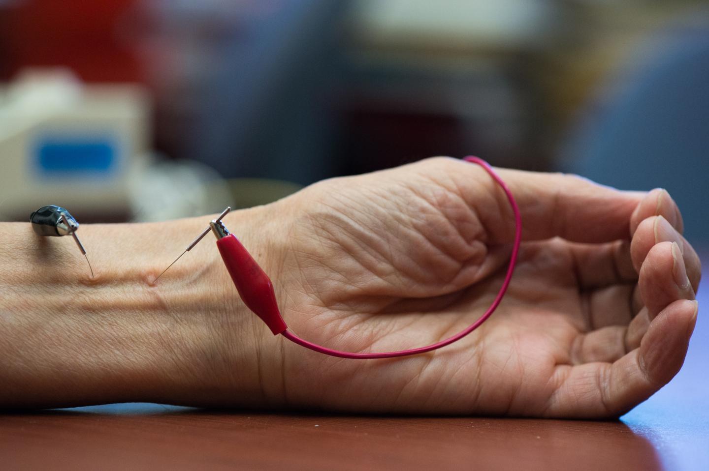
UCI study shows that repetitive electroacupuncture evokes a long-lasting action in lowering blood pressure in hypertension. (credit: Chris Nugent/UCI)

A study led by researchers at UC Irvine’s Susan Samueli Center for Integrative Medicine suggests electroacupuncture can effectively reduce hypertension in rats.

The team led by cardiology researcher Zhi-Ling Guo published evidence in Nature’s Scientific Reports (open access) to show how electroacupuncture remediates high blood pressure “by increasing the gene expression of enkephalin, one of three major opioid peptides produced by the body.”

The new study* builds on earlier reported research at the Samueli Center that tracked the efficacy of electroacupuncture to lower hypertension in humans. Patients stimulated at specific locations in the wrist had drops in their overall blood pressure scores.

The potential advantages of electroacupuncture over conventional medical therapy include few (if any) side effects, according to the researchers.

The study was supported by National Institutes of Health grants.

* In tests on rats subjected to cold exposure, UC Irvine School of Medicine cardiology researcher Zhi-Ling Guo and colleagues noted that blood pressure reductions lasted for at least three days after electroacupuncture (2 Hz, 0.1 – 0.4 mA, 0.5 ms duration) for 30 minutes twice weekly for five weeks by increasing the gene expression of enkephalin. With the onset of winter in the northern hemisphere, hospitals and clinicians typically see an uptick in deaths from stroke and heart attack associated with high blood pressure, according to a study by Canadian scientists published in Nature Scientific Reports.


Abstract of Repetitive Electroacupuncture Attenuates Cold-Induced Hypertension through Enkephalin in the Rostral Ventral Lateral Medulla

Acupuncture lowers blood pressure (BP) in hypertension, but mechanisms underlying its action are unclear. To simulate clinical studies, we performed electroacupuncture (EA) in unanesthetized rats with cold-induced hypertension (CIH) induced by six weeks of cold exposure (6 °C). EA (0.1 – 0.4 mA, 2 Hz) was applied at ST36-37 acupoints overlying the deep peroneal nerve for 30 min twice weekly for five weeks while sham-EA was conducted with the same procedures as EA except for no electrical stimulation. Elevated BP was reduced after six sessions of EA treatment and remained low 72 hrs after EA in 18 CIH rats, but not in sham-EA (n = 12) and untreated (n = 6) CIH ones. The mRNA level of preproenkephalin in the rostral ventrolateral medulla (rVLM) 72 hr after EA was increased (n = 9), compared to the sham-EA (n = 6), untreated CIH rats (n = 6) and normotensive control animals (n = 6). Microinjection of ICI 174,864, a δ-opioid receptor antagonist, into the rVLM of EA-treated CIH rats partially reversed EA’s effect on elevated BP (n = 4). Stimulation of rVLM of CIH rats treated with sham-EA using a δ-opioid agonist, DADLE, decreased BP (n = 6). These data suggest that increased enkephalin in the rVLM induced by repetitive EA contributes to BP lowering action of EA.

Boosting levels of antioxidant may help resist age-related decline

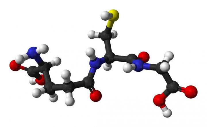
The chemical structure of glutathione, an antioxidant that may help resist the toxins that are an underlying cause of aging. (credit: Oregon State University)

Researchers at Oregon State University have found evidence in a rat study* that levels of glutathione, which helps resist the toxic stresses of everyday life, decline with age, and this sets the stage for a wide range of age-related health problems, they suggest.

The new study, published in the journal Redox Biology, also highlighted a compound called N-acetyl-cysteine (NAC), which is used in high doses in medical detoxification emergencies to help patients in a toxic crisis, such as ingestion of poisonous levels of heavy metals or acetaminophen overdose.

NAC, the researchers said, is known to boost the metabolic function of glutathione and increase its rate of synthesis. In the study, pretreatment with NAC increased glutathione levels in the older cells and largely helped offset the level of cell death.

But the researchers said that at much lower levels, NAC might also help maintain glutathione levels and prevent the routine metabolic declines associated with aging.

Aging-related decline of these detoxification pathways, the scientists say, are linked to cardiovascular disease, diabetes and cancer, some of the primary causes of death in the developed world.

“We’ve known for some time of the importance of glutathione as a strong antioxidant,” said Tory Hagen, lead author on the research and the Helen P. Rumbel Professor for Health Aging Research in the Linus Pauling Institute at OSU.

Detoxing with glutathione

“What this study pointed out was the way that cells from younger animals are far more resistant to stress than those from older animals. In young animal cells, stress doesn’t cause such a rapid loss of glutathione. The cells from older animals, on the other hand, were quickly depleted of glutathione and died twice as fast when subjected to stress.

According to Hagen, glutathione is such an important antioxidant that its existence appears to date back as far as oxygen-dependent, or aerobic life itself — about 1.5 billion years. It’s a principal compound to detoxify environmental stresses, air pollutants, heavy metals, pharmaceuticals and many other toxic insults.

“I’m optimistic there could be a role for this compound in preventing the increased toxicity we face with aging, as our abilities to deal with toxins decline,” Hagen said. “We might be able to improve the metabolic resilience that we’re naturally losing with age.”

Hagen suggested that higher levels of glutathione — boosted by NAC — might also help reduce the toxicity of some prescription drugs, cancer chemotherapies, and treat other health issues.

This research was supported by the National Institutes of Health, the National Science Foundation, and the Medical Research Foundation of Oregon.

* In this study, scientists tried to identify the resistance to toxins of young cells, compared to those of older cells. They used a toxic compound called menadione to stress the cells, and in the face of that stress, the younger cells lost significantly less of their glutathione than older cells did. The glutathione levels of young rat cells never decreased to less than 35 percent of its initial level, whereas in older rat cells glutathione levels plummeted to 10 percent of their original level.


Abstract of Glutathione maintenance mitigates age-related susceptibility to redox cycling agents

Isolated hepatocytes from young (4–6 mo) and old (24–26 mo) F344 rats were exposed to increasing concentrations of menadione, a vitamin K derivative and redox cycling agent, to determine whether the age-related decline in Nrf2-mediated detoxification defenses resulted in heightened susceptibility to xenobiotic insult. An LC50 for each age group was established, which showed that aging resulted in a nearly 2-fold increase in susceptibility to menadione (LC50 for young: 405 μM; LC50 for old: 275 μM). Examination of the known Nrf2-regulated pathways associated with menadione detoxification revealed, surprisingly, that NAD(P)H: quinone oxido-reductase 1 (NQO1) protein levels and activity were induced 9-fold and 4-fold with age, respectively (p=0.0019 and p=0.018; N=3), but glutathione peroxidase 4 (GPX4) declined by 70% (p=0.0043; N=3). These results indicate toxicity may stem from vulnerability to lipid peroxidation instead of inadequate reduction of menadione semi-quinone. Lipid peroxidation was 2-fold higher, and GSH declined by a 3-fold greater margin in old versus young rat cells given 300 µM menadione (p<0.05 and p≤0.01 respectively; N=3). We therefore provided 400 µM N-acetyl-cysteine (NAC) to hepatocytes from old rats before menadione exposure to alleviate limits in cysteine substrate availability for GSH synthesis during challenge. NAC pretreatment resulted in a >2-fold reduction in cell death, suggesting that the age-related increase in menadione susceptibility likely stems from attenuated GSH-dependent defenses. This data identifies cellular targets for intervention in order to limit age-related toxicological insults to menadione and potentially other redox cycling compounds.

Zapping deep tumors with microwave-heated photosensitizer nanoparticle

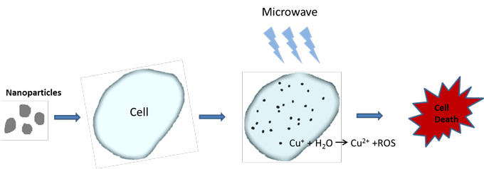
A schematic illustration of microwave-induced photodynamic therapy for cancer treatment (credit: UTA)

Physicists at The University of Texas at Arlington have invented a new photosensitizer  nanoparticle called copper-cysteamine (Cu-Cy) that when heated by microwave energy can precisely zap cancer cells deep in the body .

Photodynamic therapy kills cancer cells when a photosensitizer* nanoparticle introduced into tumor tissue is stimulated by (typically) near-infrared light, generating toxic reactive oxygen species (ROS), such as singlet oxygen, by photoexcitation. However, near-IR light cannot penetrate deeper than 10 mm in tissue while retaining enough energy to activate ROS.**

The new “microwave-induced photodynamic therapy (MIPDT)” method can “propagate through all types of tissues and target deeply situated tumors, without harming surrounding tissue,” according to Wei Chen, UTA professor of physics and lead author of the study, published in the October 2016 issue of The Journal of Biomedical Nanotechnology.***

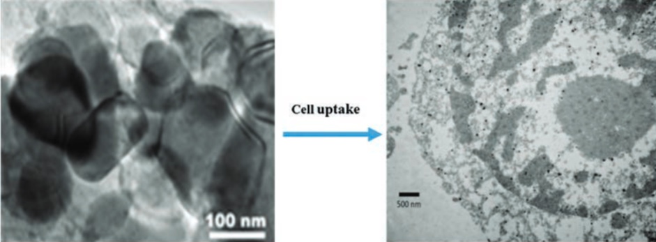
TEM images of Cu-Cy particles (left) and particles after uptake by osteosarcoma cells (right) (Mengyu Yao et al./J. Biomed. Nanotechnol.)

The new nanoparticle demonstrates “very low toxicity, is easy to make and inexpensive, and also emits intense luminescence, which means it can also be used as an imaging agent,” said Chen.

*  A photosensitizer is a molecule that can be activated by light to a high-energy state. It may then collide with oxygen and transfer its extra energy to oxygen, forming toxic singlet oxygen.

** In previous research, the researchers found that the Cu-Cy nanoparticle could be activated by X-rays to produce singlet oxygen and slow the growth of tumors. X-ray radiation, however, poses significant risks to patients and can harm healthy tissue. Other photodynamic therapy activation methods that have been explored, with limited results, include upconversion nanoparticles that can be excited at NIR and emit light in the UV-visible range, scintillation or afterglow nanoparticles, and Cerenkov light (generated in nuclear reactors).

*** Scientists from the The Guangdong Key Laboratory of Orthopaedic Technology and Implant Materials, Department of Orthopedics, Guangzhou General Hospital of Guangzhou Military Command, Guangzhou, China; and the Physics Department at Beihang University in Beijing, China, were also involved in the research. The U.S. Army Medical Research Acquisition Activity, the National Science Foundation, the Department of Homeland Security’s joint Academic Research Initiative program, the National Basic Research Program of China, the National Natural Science Foundation of China, and the five-year plan of the Chinese Military.


Abstract of A New Modality for Cancer Treatment—Nanoparticle Mediated Microwave Induced Photodynamic Therapy

Photodynamic therapy (PDT) has attracted ever-growing attention as a promising modality for cancer treatment. However, due to poor tissue penetration by light, photodynamic therapy has rarely been used for deeply situated tumors. This problem can be solved if photosensitizers are activated by microwaves (MW) that are able to penetrate deeply into tissues. Here, for the first time, we report microwave-induced photodynamic therapy and exploit copper cysteamine nanoparticles as a new type of photosensitizer that can be activated by microwaves to produce singlet oxygen for cancer treatment. Both in vitro and in vivo studies on a rat osteosarcoma cell line (UMR 106-01) have shown significant cell destruction using copper cysteamine (Cu-Cy) under microwave activation. The heating effects and the release of copper ions from Cu-Cy upon MW stimulation are the main mechanisms for the generation of reactive oxygen species that are lethal bullets for cancer destruction. The copper cysteamine nanoparticle-based microwave-induced photodynamic therapy opens a new door for treating cancer and other diseases.

First human clinical trial for nicotinamide riboside

(credit: iStock)

In the first controlled clinical trial of nicotinamide riboside (NR), a newly discovered form of Vitamin B3, researchers have shown that the compound is safe for humans and increases levels of a cell metabolite called NAD+ that is critical for cellular energy production and protection against stress and DNA damage.

Levels of NAD+ (first discovered by biochemists in 1906) diminish with age, and it has been suggested that loss of this metabolite may play a role in age-related health decline.

Studies in mice previously showed that boosting the levels of NAD+ can produce multiple health benefits, including resistance to weight gain, improved control of blood sugar and cholesterol, reduced nerve damage, and longer lifespan.* These findings have spurred people to take commercially available NR supplements designed to boost NAD+. However, these over-the-counter supplements have not undergone clinical trials to see if they work in people.

Human clinical trial shows NR is safe, boosts human NAD+ metabolism

The new research, reported Oct. 10 in the open-access journal Nature Communications, determined the time and dose-dependent effects of NR on blood NAD+ metabolism in humans. It was led by Charles Brenner, PhD, professor and Roy J. Carver Chair of Biochemistry at the University of Iowa Carver College of Medicine in collaboration with colleagues at Queens University Belfast and ChromaDex Corp., which supplied the NR used in the trial.**

The human trial involved six men and six women, all healthy. Each participant received single oral doses of 100 mg, 300 mg, or 1,000 mg of NR in a different sequence with a seven-day gap between doses. After each dose, blood and urine samples were collected and analyzed by Brenner’s lab to measure various NAD+ metabolites in a process called metabolomics.

The trial showed that the NR vitamin increased NAD+ metabolism by amounts directly related to the dose, and there were no serious side effects with any of the doses.

“This trial shows that oral NR safely boosts human NAD+ metabolism,” Brenner says. “We are excited because everything we are learning from animal systems indicates that the effectiveness of NR depends on preserving and/or boosting NAD+ and related compounds in the face of metabolic stresses. Because the levels of supplementation in mice that produce beneficial effects are achievable in people, it appears than health benefits of NR will be translatable to humans safely.”

The next step will be to study the effect of longer duration NR supplementation on NAD+ metabolism in healthy adults, but Brenner also has plans to test the effects of NR in people with diseases and health conditions, including elevated cholesterol, obesity and diabetes, and people at risk for chemotherapeutic peripheral neuropathy.

In addition to Brenner, the research team included researchers at the UI Carver College of Medicine, Queens University Belfast, and ChromaDex.

The research was funded in part by grants from the National Institutes of Health, the Biotechnology and Biological Sciences Research Council, the Roy J. Carver Trust, and ChromaDex.

** The mouse study showed that NAAD is formed from NR and confirmed that NAAD levels are a strong biomarker for increased NAD+ metabolism. The experiments also revealed more detail about NAD+ metabolic pathways.

In particular, the researchers compared the ability of all three NAD+ precursor vitamins — NR, niacin, and nicotinamide — to boost NAD+ metabolism and stimulate the activity of certain enzymes, which have been linked to longevity and health benefits. The study showed for the first time that oral NR is superior to nicotinamide, which is better than niacin in terms of the total amount of NAD+ produced at an equivalent dose. NR was also the best of the three in stimulating the activity of sirtuin enzymes. However, in this case, NR was the best at stimulating sirtuin-like activities, followed by niacin, followed by nicotinamide.

The information from the mouse study subsequently helped Brenner’s team design the formal clinical trial. In addition to showing that NR boosts NAD+ in humans without adverse effects, the trial confirmed that NAAD is a highly sensitive biomarker of NAD+ supplementation in people.

** Brenner is a consultant for ChromaDex. He also is co-founder and Chief Scientific Adviser of ProHealthspan, which sells NR supplements under the trade name Tru NIAGEN. Prior to the formal clinical trial, Brenner conducted a pilot human study — on himself. In 2004, he had discovered that NR is a natural product found in milk and that there is pathway to convert NR to NAD+ in people. More than a decade of research on NR metabolic pathways and health effects in mice and rats had convinced him that NR supplementation had real promise to improve human health and wellness.

After consulting with UI’s institutional review board, he conducted an experiment in which he took 1 gram of NR once a day for seven days, and his team analyzed blood and urine samples using mass spectrometry. The experiment showed that Brenner’s blood NAD+ increased by about 2.7 times. In addition, though he reported immediate sensitivity to flushing with the related compound niacin, he did not experience any side effects taking NR.

The biggest surprise from his metabolomic analysis was an increase in a metabolite called NAAD, which was multiplied by 45 times, from trace levels to amounts in the micromolar range that were easily detectable.

“While this was unexpected, I thought it might be useful,” Brenner says. “NAD+ is an abundant metabolite and it is sometimes hard to see the needle move on levels of abundant metabolites. But when you can look at a low-abundance metabolite that goes from undetectable to easily detectable, there is a great signal to noise ratio, meaning that NAAD levels could be a useful biomarker for tracking increases in NAD+ in human trials.”

Brenner notes this was a case of bidirectional translational science; having learned something from the initial human experiment, his team was able to return to laboratory mice to explore the unexpected NAAD finding in more detail.


Abstract of Nicotinamide riboside is uniquely and orally bioavailable in mice and humans

Nicotinamide riboside (NR) is in wide use as an NAD+ precursor vitamin. Here we determine the time and dose-dependent effects of NR on blood NAD+ metabolism in humans. We report that human blood NAD+ can rise as much as 2.7-fold with a single oral dose of NR in a pilot study of one individual, and that oral NR elevates mouse hepatic NAD+ with distinct and superior pharmacokinetics to those of nicotinic acid and nicotinamide. We further show that single doses of 100, 300 and 1,000 mg of NR produce dose-dependent increases in the blood NAD+ metabolome in the first clinical trial of NR pharmacokinetics in humans. We also report that nicotinic acid adenine dinucleotide (NAAD), which was not thought to be en route for the conversion of NR to NAD+, is formed from NR and discover that the rise in NAAD is a highly sensitive biomarker of effective NAD+ repletion.

New catheter lets doctors see inside arteries for first time

Image-guided catheter with a camera the size of a grain of salt (credit: Avinger)

A new safer catheter design that allows cardiologists to see inside arteries for the first time and remove plaque from only diseased tissue has been used by interventional cardiologists at UC San Diego Health.

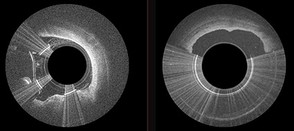
The new image-guided device, Avinger’s Pantheris, allows doctors to see and remove plaque simultaneously during an atherectomy — a minimally invasive procedure that involves cutting plaque away from the artery and clearing it out to restore blood flow.

Left: OCT imaging fiber on cutter allows for seeing the layers and disease. Center: the torque shaft, cutter window, and apposition balloon prove the direction and precision to avoid disruption of layers. Right: the cutter and nose-cone allow for capturing and removing only diseased tissue. (credit: Avinger)

The new technology treats patients suffering from the painful symptoms of peripheral artery disease (PAD), a condition caused by a build-up of plaque that blocks blood flow in the arteries of the legs and feet, preventing oxygen-rich blood from reaching the extremities. Patients with PAD frequently develop life threatening complications, including heart attack and stroke; in some severe cases, patients may even face amputation.

PAD affects nearly 20 million adults in the United States and more than 200 million globally.

“Peripheral artery disease greatly impacts quality of life, with patients experiencing cramping, numbness and discoloration of their extremities,” said Mitul Patel, MD, cardiologist at UC San Diego Health. “This new device is a significant step forward for the treatment of PAD with a more efficient approach for plaque removal and less radiation exposure to the doctor and patient.”

Pantheris allows interventional cardiologists to see the layers and disease (left) and remove just the disease (right) (credit: Avinger)

X-ray technology was previously used during similar procedures, but those images are not as clear and do not allow for visualization inside the blood vessel. The new catheter, with a fiber optic camera the size of a grain of salt on the tip, is fed through a small incision in the groin that does not require full anesthesia. Once inside, the interventional cardiologist is able to see exactly what needs to be removed without damaging the artery wall, which can cause further narrowing.

Pantheris was approved by the FDA in March 2016. So far, cardiologists at UC San Diego Health have used the new catheter on 10 patients undergoing an atherectomy procedure, with successful results.

Avinger | PML0290-D Pantheris Animation


Abstract of Utility of Image-Guided Atherectomy for Optimal Treatment of Ambiguous Lesions by Angiography

Peripheral endovascular interventions have been limited by multiple shortcomings, including difficulty in assessment of the 3-dimensional nature of obstructive plaque within the vasculature with only contrast angiography and 2-dimensional fluoroscopy. Herein, we present a case of pseudodissection seen angiographically post CTO crossing, which was accurately assessed as eccentric plaque using OCT imaging and treated using an OCT-guided directional atherectomy device, preventing bail-out stenting.