New catheter lets doctors see inside arteries for first time

Image-guided catheter with a camera the size of a grain of salt (credit: Avinger)

A new safer catheter design that allows cardiologists to see inside arteries for the first time and remove plaque from only diseased tissue has been used by interventional cardiologists at UC San Diego Health.

The new image-guided device, Avinger’s Pantheris, allows doctors to see and remove plaque simultaneously during an atherectomy — a minimally invasive procedure that involves cutting plaque away from the artery and clearing it out to restore blood flow.

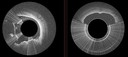
Left: OCT imaging fiber on cutter allows for seeing the layers and disease. Center: the torque shaft, cutter window, and apposition balloon prove the direction and precision to avoid disruption of layers. Right: the cutter and nose-cone allow for capturing and removing only diseased tissue. (credit: Avinger)

The new technology treats patients suffering from the painful symptoms of peripheral artery disease (PAD), a condition caused by a build-up of plaque that blocks blood flow in the arteries of the legs and feet, preventing oxygen-rich blood from reaching the extremities. Patients with PAD frequently develop life threatening complications, including heart attack and stroke; in some severe cases, patients may even face amputation.

PAD affects nearly 20 million adults in the United States and more than 200 million globally.

“Peripheral artery disease greatly impacts quality of life, with patients experiencing cramping, numbness and discoloration of their extremities,” said Mitul Patel, MD, cardiologist at UC San Diego Health. “This new device is a significant step forward for the treatment of PAD with a more efficient approach for plaque removal and less radiation exposure to the doctor and patient.”

Pantheris allows interventional cardiologists to see the layers and disease (left) and remove just the disease (right) (credit: Avinger)

X-ray technology was previously used during similar procedures, but those images are not as clear and do not allow for visualization inside the blood vessel. The new catheter, with a fiber optic camera the size of a grain of salt on the tip, is fed through a small incision in the groin that does not require full anesthesia. Once inside, the interventional cardiologist is able to see exactly what needs to be removed without damaging the artery wall, which can cause further narrowing.

Pantheris was approved by the FDA in March 2016. So far, cardiologists at UC San Diego Health have used the new catheter on 10 patients undergoing an atherectomy procedure, with successful results.

Avinger | PML0290-D Pantheris Animation


Abstract of Utility of Image-Guided Atherectomy for Optimal Treatment of Ambiguous Lesions by Angiography

Peripheral endovascular interventions have been limited by multiple shortcomings, including difficulty in assessment of the 3-dimensional nature of obstructive plaque within the vasculature with only contrast angiography and 2-dimensional fluoroscopy. Herein, we present a case of pseudodissection seen angiographically post CTO crossing, which was accurately assessed as eccentric plaque using OCT imaging and treated using an OCT-guided directional atherectomy device, preventing bail-out stenting.

Smoking leaves ‘footprint’ in DNA

(credit: American Heart Association)

Smoking leaves its “footprint” on the human genome in the form of DNA methylation, a process that affects what genes are turned on, according to new research in Circulation: Cardiovascular Genetics, an American Heart Association journal.

The new findings could provide researchers with potential targets for new therapies.

“These results are important because methylation, as one of the mechanisms of the regulation of gene expression, affects what genes are turned on, which has implications for the development of smoking-related diseases, said Stephanie J. London, M.D., Dr.P.H., last author and deputy chief of the Epidemiology Branch at the National Institute of Environmental Health Sciences, National Institutes of Health, Research Triangle Park, North Carolina. “Even after someone stops smoking, we still see the effects of smoking on their DNA.”

The good news (for some): most DNA methylation sites returned to levels seen in never-smokers within five years of quitting smoking

Smoking remains the leading preventable cause of death worldwide. Even decades after stopping, former smokers are at long-term risk of developing diseases including some cancers, chronic obstructive pulmonary disease, and stroke. While the molecular mechanisms responsible for these long-term effects remain poorly understood, previous studies linking DNA methylation sites to genes involved with coronary heart disease and pulmonary disease suggest it may play an important role.

To find out more, researchers conducted a meta-analysis of DNA methylation sites across the human genome using blood samples taken from nearly 16,000 participants from 16 groups of the Cohorts for Heart and Aging Research in Genetic Epidemiology (CHARGE) Consortium, including a group of the Framingham Heart Study that has been followed by researchers since 1971.

The researchers compared DNA methylation sites in current and former smokers to those who never smoked. They found:

  • Smoking-associated DNA methylation sites were associated with more than 7,000 genes, or one-third of known human genes.
  • For people who stopped smoking, the majority of DNA methylation sites returned to levels seen in never-smokers within five years of quitting smoking. However, some DNA methylation sites persisted even after 30 years of quitting.
  • The most statistically significant methylation sites were linked to genes enriched for association with numerous diseases caused by cigarette smoking, such as cardiovascular diseases and certain cancers, suggesting that some of these long-lasting methylation sites may be marking genes potentially important for former smokers who are still at increased risk of developing certain diseases.

The discovery of smoking-related DNA methylation sites raises the possibility of developing biomarkers to evaluate a patient’s smoking history, as well as potentially developing new treatments targeted toward these methylation sites. However, the main analysis was not designed to examine effects over long periods of time.


Abstract of Epigenetic Signatures of Cigarette Smoking

Background — DNA methylation leaves a long-term signature of smoking exposure and is one potential mechanism by which tobacco exposure predisposes to adverse health outcomes, such as cancers, osteoporosis, lung, and cardiovascular disorders.

Methods and Results — To comprehensively determine the association between cigarette smoking and DNA methylation, we conducted a meta-analysis of genome-wide DNA methylation assessed using the Illumina BeadChip 450K array on 15,907 blood derived DNA samples from participants in 16 cohorts (including 2,433 current, 6,518 former, and 6,956 never smokers). Comparing current versus never smokers, 2,623 CpG sites (CpGs), annotated to 1,405 genes, were statistically significantly differentially methylated at Bonferroni threshold of p

Conclusions — Cigarette smoking has a broad impact on genome-wide methylation that, at many loci, persists many years after smoking cessation. Many of the differentially methylated genes were novel genes with respect to biologic effects of smoking, and might represent therapeutic targets for prevention or treatment of tobacco-related diseases. Methylation at these sites could also serve as sensitive and stable biomarkers of lifetime exposure to tobacco smoke.

How to detect emotions remotely with wireless signals


MITCSAIL | EQ-Radio: Emotion Recognition using Wireless Signals

MIT researchers from have developed “EQ-Radio,” a device that can detect a person’s emotions using wireless signals.

By measuring subtle changes in breathing and heart rhythms, EQ-Radio is 87 percent accurate at detecting if a person is excited, happy, angry or sad — and can do so without on-body sensors, according to the researchers.

MIT professor and project lead Dina Katabi of MIT’s Computer Science and Artificial Intelligence Laboratory (CSAIL) envisions the system being used in health care and testing viewers’ reactions to ads or movies in real time.

Using wireless signals reflected off people’s bodies, the device measures heartbeats as accurately as an ECG monitor, with a margin of error of approximately 0.3 percent, according to the researchers. It then studies the waveforms within each heartbeat to match a person’s behavior to how they previously acted in one of the four emotion-states.

The team will present the work next month at the Association of Computing Machinery’s International Conference on Mobile Computing and Networking (MobiCom).

How it works

EQ-Radio has three components: a radio for capturing RF reflections, a heartbeat extraction algorithm, and a classification subsystem that maps the learned physiological signals to emotional states. (credit: Mingmin Zhao et al./MIT)

EQ-Radio sends wireless signals that reflect off of a person’s body and back to the device. To detect emotions, its beat-extraction algorithms break the reflections into individual heartbeats and analyze the small variations in heartbeat intervals to determine their levels of arousal and positive affect.

These measurements are what allow EQ-Radio to detect emotion. For example, a person whose signals correlate to low arousal and negative affect is more likely to tagged as sad, while someone whose signals correlate to high arousal and positive affect would likely be tagged as excited.

The exact correlations vary from person to person, but are consistent enough that EQ-Radio could detect emotions with 70 percent accuracy even when it hadn’t previously measured the target person’s heartbeat. In the future it could be used for non-invasive health monitoring and diagnostic settings.

For the experiments, subjects used videos or music to recall a series of memories that each evoked one the four emotions, as well as a no-emotion baseline. Trained just on those five sets of two-minute videos, EQ-Radio could then accurately classify the person’s behavior among the four emotions 87 percent of the time.

One of the challenges was to tune out irrelevant data. To get individual heartbeats, for example, the team had to dampen the breathing, since the distance that a person’s chest moves from breathing is much greater than the distance that their heart moves to beat.

To do so, the team focused on wireless signals that are based on acceleration rather than distance traveled, since the rise and fall of the chest with each breath tends to be much more consistent —  and, therefore, have a lower acceleration — than the motion of the heartbeat.


Abstract of Emotion Recognition using Wireless Signals

This paper demonstrates a new technology that can infer a person’s emotions from RF signals reflected off his body. EQ-Radio transmits an RF signal and analyzes its reflections off a person’s body to recognize his emotional state (happy, sad, etc.). The key enabler underlying EQ-Radio is a new algorithm for extracting the individual heartbeats from the wireless signal at an accuracy comparable to on-body ECG monitors. The resulting beats are then used to compute emotion-dependent features which feed a machine-learning emotion classifier. We describe the design and implementation of EQ-Radio, and demonstrate through a user study that its emotion recognition accuracy is on par with stateof-the-art emotion recognition systems that require a person to be hooked to an ECG monitor.

These six plant extracts could delay aging

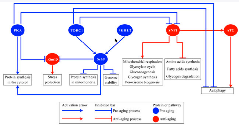
Several nutrient- and energy-sensing signaling pathways and protein kinases converge into a network that defines the rate of yeast chronological aging (credit: Vicky Lutchman et al./Oncotarget)

Six previously identified plant extracts can delay aging by affecting different signaling pathways that set the pace of growing old, researchers from Concordia University and Idunn Technologies have found, in a study recently published (open-access) in Oncotarget.

Using yeast — a favored cellular aging model — Vladimir Titorenko, a biology professor and the study’s senior author, and his colleagues conducted a screen of a library of 35 different plant extracts (PEs) to determine those that can extend yeast chronological lifespan. To do that, they monitored how the information flowing through signaling pathways was affected. They identified six aging-delaying plant extracts: Cimicifuga racemosa, Valeriana officinalis L., Passiflora incarnata L., Ginkgo biloba, Apium graveolens L., and “particularly effective”: Salix alba, commonly known as white willow bark.

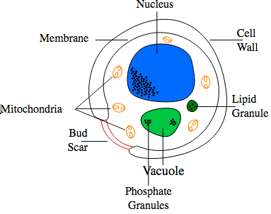
Diagram of yeast cell (credit: Frankie Robertson/CC)

Anti-aging pathways

At a cellular level, aging progresses similarly in yeast and humans. In both, the pace of aging is defined by a distinct set of chemical reactions arranged into several “signaling pathways” that regulate the rate of aging in a wide range of organisms.

“It’s known that some of these signaling pathways delay aging if activated in response to certain nutrients or hormones,” Titorenko says. “These pathways are called ‘anti-aging’ or ‘pro-longevity’ pathways. Other signaling pathways speed up aging if activated in response to certain other nutrients or hormones. These pathways are called ’pro-aging’ or ‘pro-death’ pathways.” Each of the six aging-delaying plant extracts targets a different anti-aging or pro-aging signaling pathway.

The study revealed several features of the six plant extracts as potential tools in decelerating chronic symptoms and diseases of old age:

  • They imitate the aging-delaying effects of the caloric restriction diet in yeast
  • They slow yeast aging by eliciting a mild stress response
  • They extend yeast longevity more efficiently than any lifespan-prolonging chemical compound yet described
  • They delay aging through signaling pathways implicated in age-related diseases
  • One of them delays aging via a previously unknown pathway
  • They extend longevity and delay the onset of age-related diseases in organisms other than yeast

Health Canada classifies the six plant extracts as safe for human consumption, and recommends five of them as health-improving supplements with clinically proven benefits.

The study was supported by grants from the Natural Sciences and Engineering Research Council of Canada and the Fonds québécois de la recherche sur la nature et les technologies.

UPDATE Sept. 19, 2016: the six aging-delaying plant extracts’ names added.


Abstract of Six plant extracts delay yeast chronological aging through different signaling pathways

Our recent study has revealed six plant extracts that slow yeast chronological aging more efficiently than any chemical compound yet described. The rate of aging in yeast is controlled by an evolutionarily conserved network of integrated signaling pathways and protein kinases. Here, we assessed how single-gene-deletion mutations eliminating each of these pathways and kinases affect the aging-delaying efficiencies of the six plant extracts. Our findings imply that these extracts slow aging in the following ways: 1) plant extract 4 decreases the efficiency with which the pro-aging TORC1 pathway inhibits the anti-aging SNF1 pathway; 2) plant extract 5 mitigates two different branches of the pro-aging PKA pathway; 3) plant extract 6 coordinates processes that are not assimilated into the network of presently known signaling pathways/protein kinases; 4) plant extract 8 diminishes the inhibitory action of PKA on SNF1; 5) plant extract 12 intensifies the anti-aging protein kinase Rim15; and 6) plant extract 21 inhibits a form of the pro-aging protein kinase Sch9 that is activated by the pro-aging PKH1/2 pathway.

Mayo Clinic, collaborators working to advance aging research via clinical trials

(credit: iStock)

Mayo Clinic and other members of the Geroscience Network* have developed strategies for taking new drugs to clinical trials — specifically, drugs that target processes underlying multiple age-related diseases and disabilities. And they’ve written six supporting articles that appeared Wednesday Aug. 17 in The Journals of Gerontology: Series A – Biological Sciences and Medical Sciences.

The Geroscience Network consists of 18 academic aging center, with the participation of more than 100 investigators from across the U.S. and Europe.

Aging may be a modifiable risk factor

“Aging is the largest risk factor for most chronic diseases, including stroke, heart disease, cancer, dementias, osteoporosis, arthritis, diabetes, metabolic syndrome, blindness and frailty,” said James Kirkland, M.D., Ph.D., director of the Mayo Clinic Robert and Arlene Kogod Center on Aging.

However, he said recent research suggests that aging may actually be a modifiable risk factor. “The goal of our network’s collaborative efforts is to accelerate the pace of discovery in developing interventions to delay, prevent, or treat these conditions as a group, instead of one at a time.”

Felipe Sierra, Ph.D., of the National Institute on Aging and a member of the Geroscience Network, describes the potential impact of such discoveries in his article, “Moving Geroscience into Uncharted Waters.” He notes that in addition to the direct health issues, care for the elderly currently accounts for 43 percent of the total health care spending in the U.S,, or approximately 1 trillion dollars a year, and that this number is expected to rise as baby boomers reach retirement age.

“Reducing these costs is critical for the survival of society as we know it,” he said. “A 2013 paper by Dana Goldman and colleagues calculated that a just modest increase (2.2 years) in lifespan and healthspan could reduce those expenses by 7 trillion dollars by 2050.”

This work was supported by the National Institutes of Health, the Paul Glenn Foundation, Nathan Shock Centers of Excellence for the Biology of Aging, the Connor Group, and the Noaber and Ted Nash foundations.

The first two articles cited below are open-access.

* In addition to Mayo Clinic, members of the Geroscience Network are Albert Einstein College of Medicine, Buck Institute for Research on Aging, Harvard University, Johns Hopkins University, National Institute on Aging, the Scripps Research Institute, Stanford University, the University of Alabama at Birmingham, the University of Arkansas, the University of Connecticut, the University of Michigan, the University of Minnesota, the University of Oklahoma, the University of Texas Health Science Center San Antonio, the University of Southern California, the University of Washington, and Wake Forest University as well as members from other institutions across the U.S. and Europe.

Rejuvenation Biotechnology Conference to be live-streamed Aug. 16–17

The Rejuvenation Biotechnology Conference will be live-streamed Tuesday Aug. 16, starting at 1 PM PDT, and Wednesday Aug. 17.

The 2016 Rejuvenation Biotechnology Conference is focused on taking the Rejuvenation Biotechnology Industry to the next level by addressing the question: what will it take to push emerging breakthroughs in regenerative medicine from proof-of-concept to implementation?

This year’s conference seeks to answer this critical inquiry by covering all the stages from securing funding, to production, to navigating regulation, to clinical evaluation and adoption of new treatments. Industry leading experts will present real-life examples drawn from their own work followed by an open panel discussion and Q&A.

Register here for live-streaming access.  If you want to ask a question during the live stream of the event, contact the conference via Twitter at #RejBioCon

Rejuvenation Biotechnology Conference Tuesday 16th Streaming from 1 p.m. – 6 p.m. PDT (8 p.m. – 1 a.m. GMT)

New cancer-drug delivery system uses magnetically guided bacteria to target cancerous tumors with high precision

Artist’s impression of new cancer-drug nanotransporters using more than 100 million self-propelled, flagellated bacteria carrying nanoliposomes (green discs) loaded with drugs. The bacteria are guided by magnetic fields to take the most direct path between the drug’s injection point and the area of the body intended to cure. (credit: Montréal Nanorobotics Laboratory)

Researchers from Polytechnique Montréal, Université de Montréal, and McGill University have designed a new cancer-drug-delivery nanotransporter system using more than 100 million flagellated, self-propelled bacteria that are capable of navigating through the bloodstream to administer a drug to tumors with precision.* The goal of the research is to avoid jeopardizing the integrity of organs and surrounding healthy tissues while reducing drug dosage.

In an experiment with mice reported in the journal Nature Nanotechnology, the researchers confirmed that “the drug’s propelling force was enough to travel efficiently and enter deep inside the tumors,” autonomously detecting the oxygen-depleted tumor areas and delivering the drug to them, said Professor Sylvain Martel, holder of the Canada Research Chair in Medical Nanorobotics and Director of the Polytechnique Montréal Nanorobotics Laboratory, who heads the research team’s work.

Bacteria that detect both magnetic field lines and low-oxygen concentrations

In the experiment with mice, the researchers used liposomes as drug nanocarriers, attached to Magnetococcus marinus strain MC-1 bacteria to transport the drug**. In their natural environment, MC-1 bacteria tend to swim along local magnetic field lines, which are detected by a chain of magnetic iron-oxide nanocrystals in the bacteria. The bacteria also swim towards hypoxic (low-oxygen) concentrations. (In a tumor, this hypoxic zone is created by the substantial consumption of oxygen by rapidly proliferative tumor cells. Hypoxic zones are known to be resistant to most therapies, including radiotherapy.)

Test of penetration of live MC-1 bacteria cells with and without magnetic field exposure in xenografts following a peritumoral injection. (left) Magnetotaxis directional control system used to generate the magnetic field necessary to guide the MC-1 cells towards the xenograft. The directional magnetic field B was aligned towards the center of the tumoral volume. (center) MC-1 peritumoral injection into tumor xenograft in mice and representation of the applied directional magnetic field used in this study to direct the bacteria towards the xenograft. (right) Summary of the total average numbers of injected and detected MC-1 cells for all tumors in groups I and II. The results show that a significant number of the peritumorally injected MC-1 cells that were magnetically guided — using a relatively simple static directional magnetic field — were able to penetrate the xenograft, unlike the case for bacteria that were not guided into the tumor. (credit: Ouajdi Felfoul et al./Nature Nanotechnology)

The new nanotransporters used both of these natural systems (magnetic and hypoxic-concentration-seeking). The MC-1 bacteria first moved in the direction of a computer-controlled magnetic field to the point where oxygen gradients could be detected by the bacteria, enabling the bacteria to penetrate and remain in the tumor’s active regions. (In future human use, the drug could be released from the liposomes by sensing pH or particular enzymes, or by applying ultrasonics or heat, for example, the researchers explained to KurzweilAI.)

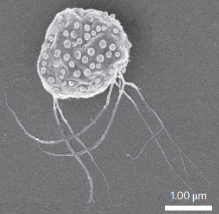
Scanning electron microscopy image of MC-1 bacterium with ~70 drug-bearing liposomes attached to the surface (credit: Ouajdi Felfoul et al./Nature Nanotechnology)

“This innovative use of nanotransporters will have an impact not only on creating more advanced engineering concepts and original intervention methods, but it also throws the door wide open to the synthesis of new vehicles for therapeutic, imaging, and diagnostic agents,” said Martel. Chemotherapy, which is so toxic for the entire human body, could make use of these natural nanotransporters to move drugs directly to the targeted area, “eliminating the harmful side effects while also boosting its therapeutic effectiveness,” he added.

The research was supported by the Consortium québécois sur la découverte du médicament (Québec consortium for drug discovery — CQDM), the Canada Research Chairs, the Natural Sciences and Engineering Research Council of Canada (NSERC), the Research Chair in Nanorobotics of Polytechnique Montréal, Mitacs, the Canada Foundation for Innovation (CFI), and the National Institutes of Health (NIH). Montréal’s Jewish General Hospital, the McGill University Health Centre (MUHC), the Institute for Research in Immunology and Cancer (IRIC), and the Rosalind and Morris Goodman Cancer Research Centre also participated.

* Only ∼2% of the total administered dose is deposited in the tumor with current delivery methods, according to a 2009 study by M. Hong et al. in J. Control. Rel., the researchers note in their Nature Nanotechnology paper.

** “Liposomes were selected as a first proof of concept because they are biocompatible, exhibit low immunogenicity and high flexibility, and protect the body from potential toxic cargo. Liposomes also shield therapeutic agents from premature degradation, control release kinetics, and may encapsulate a multitude of hydrophilic and∕or hydrophobic drug cargos, pharmaceutical ingredients, imaging agents and genetic material by virtue of their aqueous interior and lipid exterior,” the researchers note in their Nature Nanotechnology paper.


POLYTECHNIQUE MONTRÉAL | A Robotic Micro-assembly Process Inspired by the Construction of the Ancient Pyramids and Relying on Several Thousand of Flagellated Bacteria Acting as Workers


Abstract of Magneto-aerotactic bacteria deliver drug-containing nanoliposomes to tumour hypoxic regions

Oxygen-depleted hypoxic regions in the tumour are generally resistant to therapies. Although nanocarriers have been used to deliver drugs, the targeting ratios have been very low. Here, we show that the magneto-aerotactic migration behaviour of magnetotactic bacteria, Magnetococcus marinus strain MC-1, can be used to transport drug-loaded nanoliposomes into hypoxic regions of the tumour. In their natural environment, MC-1 cells, each containing a chain of magnetic iron-oxide nanocrystals, tend to swim along local magnetic field lines and towards low oxygen concentrationsbased on a two-state aerotactic sensing system. We show that when MC-1 cells bearing covalently bound drug-containing nanoliposomes were injected near the tumour in severe combined immunodeficient beige mice and magnetically guided, up to 55% of MC-1 cells penetrated into hypoxic regions of HCT116 colorectal xenografts. Approximately 70 drug-loaded nanoliposomes were attached to each MC-1 cell. Our results suggest that harnessing swarms of microorganisms exhibiting magneto-aerotactic behaviour can significantly improve the therapeutic index of various nanocarriers in tumour hypoxic regions.

No, exercise does not wipe out previous memories

2016 Rio Games Opening Ceremony promo (credit: NBC)

A week ago on KurzweilAI, we learned that prolonged sitting may increase risk of death, but that an hour of moderate exercise a day is enough to counter health risks. Now new research suggests that such exercise results in larger brain size and lowered dementia risk, while other new research suggests that the new neurons created in that exercise preserve old memories, contrary to previous research.

Exercise results in larger brain size and lowered dementia risk

UCLA researchers have found an association between low physical activity and a higher risk for dementia in older individuals, based on data from the landmark Framingham Heart Study.

The researchers found that physical activity particularly affected the size of the hippocampus, involved in short-term memory. They also found the protective effect of regular physical activity against dementia was strongest in people age 75 and older.

This suggests that regular physical activity for older adults could lead to higher brain volumes and a reduced risk for developing dementia.

The Framingham study was begun in 1948 primarily as a way to trace factors and characteristics leading to cardiovascular disease, but also examining dementia and other physiological conditions. For this study, the UCLA researchers followed an older, community-based cohort from the Framingham study for more than a decade to examine the association between physical activity and the risk for incident dementia and subclinical brain MRI markers of dementia.

The study appears in the Journals of Gerontology Series A: Biological Sciences and Medical Sciences. It was supported by an NIH/National Heart, Lung, and Blood Institute contract and training grant, the National Institute on Aging, the National Institute of Neurological Disorders and Stroke, and the American Heart Association.

New neurons created through exercise don’t cause you to forget old memories

Meanwhile, Texas A&M College of Medicine scientists have found in a study recently published in the Journal of Neuroscience that exercise causes more new neurons to be formed in a critical brain region — and contrary to an earlier study, these new neurons do not cause the individual to forget old memories.

Exercise is well known for its cognitive benefits, thought to occur because it causes neurogenesis, or the creation of new neurons, in the hippocampus, which is a key brain region for learning, memory and mood regulation. So it was a surprise in 2014 when a research study, published in the journal Science, found that exercise caused mice to forget what they’d already learned.

“It stunned the field of hippocampal neurogenesis,” said Ashok K. Shetty, PhD, a professor in the Texas A&M College of Medicine Department of Molecular and Cellular Medicine, associate director of the Institute for Regenerative Medicine, and research career scientist at the Central Texas Veterans Health Care System.

The animal models in the exercise group — in the previous study — showed far more neurogenesis than the control group, but these additional neurons seemed to erase memories that were formed before they started the exercise regimen. To test this, the researchers removed the extra neurons, and the mice suddenly were able to remember again.

Replicating the research with rats reversed the outcome

Shetty and his team decided to replicate this earlier research, using rats instead of mice. Rats are thought to be more like humans physiologically, with more-similar neuronal workings. They found that these animal models showed no such degradation in memories.

The researchers trained their animal models to complete a task over the course of four days, followed by several days of memory consolidation by performing the task over and over again. Then, half the trained animal models were put into cages with running wheels for several weeks, while the control group remained sedentary.

The rats who ran further over the course of that time had much greater neurogenesis in their hippocampus, and all rats who had access to a wheel (and therefore ran at least some), had greater neurogenesis than the sedentary group.

Importantly, despite differing levels of increased neurogenesis, both moderate runners and brisk runners (those who ran further than average) in Shetty’s study showed the same ability as the sedentary runners to recall the task they learned before they began to exercise.

This means even a large amount of running (akin to people who perform significant amount of exercise on a daily basis) doesn’t interfere with the recall of memory.

Why fidgeting can protect leg arteries and improve learning

(credit: Dave Clark/National Education Association)

But not everybody has that extra hour — or is motivated to exercise. For all of us couch/desk-bound folks, new research suggests a simple sitting exercise that may tide you over until you can go for a walk or run: fidgeting.

Ignore that teacher advice to “sit in your seat and don’t fidget!” — which the National Education Association says in this article actually improves learning — and suggests footrests using old tires to make giant rubber bands to facilitate fidgeting.

University of Missouri researchers have found that fidgeting while sitting can also protect the arteries in legs and potentially help prevent the arterial disease that may be brought on by binge TV watching or working at a computer.

In the study, the researchers compared the leg vascular function of 11 healthy young men and women before and after three hours of sitting. While sitting, the participants were asked to fidget one leg intermittently, tapping one foot for one minute and then resting it for four minutes, while the other leg remained still throughout. On average, the participants moved their feet 250 times per minute.

The researchers then measured the blood flow of the popliteal — an artery in the lower leg — and found that the fidgeting leg had a significant increase in blood flow, as expected, while the stationary leg experienced a reduction in blood flow.

Research has shown that increased blood flow and its associated shear stress — the friction of the flowing blood on the artery wall — is an important stimulus for vascular health.

While only one leg was exposed to fidgeting during the experiment, in a real-world scenario the researchers recommend tapping both legs to maximize the beneficial effects.

But not a substitute for walking and exercise

“Many of us sit for hours at a time, whether it’s binge watching our favorite TV show or working at a computer,” said Jaume Padilla, PhD, an assistant professor of nutrition and exercise physiology at MU and lead author of the study.

“We wanted to know whether a small amount of leg fidgeting could prevent a decline in leg vascular function caused by prolonged sitting. While we expected fidgeting to increase blood flow to the lower limbs, we were quite surprised to find this would be sufficient to prevent a decline in arterial function.”

But fidgeting is not a substitute for walking and exercise, which produce more overall cardiovascular benefits, the researchers caution.

“You should attempt to break up sitting time as much as possible by standing or walking,” Padilla said. “But if you’re stuck in a situation in which walking just isn’t an option, fidgeting can be a good alternative. Any movement is better than no movement.”

The study was recently published in the American Journal of Physiology Heart and Circulatory Physiology and was supported by the NIH and the Japan Society for the Promotion of Science.


Abstract of Physical Activity, Brain Volume, and Dementia Risk: The Framingham Study

Background: Several longitudinal studies found an inverse relationship between levels of physical activity and cognitive decline, dementia, and/or Alzheimer’s disease (AD), but results have been inconsistent. We followed an older, community-based cohort for over a decade to examine the association of physical activity with the risk of incident dementia and subclinical brain MRI markers of dementia.

Methods: The physical activity index (PAI) was assessed in the Framingham Study Original and Offspring cohorts, aged 60 years or older. We examined the association between PAI and risk of incident all-cause dementia and AD in participants of both cohorts who were cognitively intact and had available PAI (n = 3,714; 54% women; mean age = 70±7 years). We additionally examined the association between PAI and brain MRI in the Offspring cohort (n = 1,987).

Results: Over a decade of follow-up, 236 participants developed dementia (188 AD). Participants in the lowest quintile of PAI had an increased risk of incident dementia compared with those in higher quintiles (hazard ratio [HR] = 1.50, 95% confidence interval [CI] = 1.04–1.97, p = .028) in a multivariable-adjusted model. Secondary analysis revealed that this relation was limited to participants who were apolipoprotein (APO)E ε4 allele noncarriers (HR = 1.58, 95% CI = 1.08–2.32; p = .018) and strongest in participants aged 75 years or older. PAI was also linearly related to total brain and hippocampal volumes (β ± SE = 0.24±0.06; p < .01 and 0.004±0.001; p = .003, respectively).

Conclusion: Low physical activity is associated with a higher risk for dementia in older individuals, suggesting that a reduced risk of dementia and higher brain volumes may be additional health benefits of maintaining physical activity into old age.


Abstract of Voluntary Running Exercise-Mediated Enhanced Neurogenesis Does Not Obliterate Retrograde Spatial Memory

Running exercise (RE) improves cognition, formation of anterograde memories, and mood, alongside enhancing hippocampal neurogenesis. A previous investigation in a mouse model showed that RE-induced increased neurogenesis erases retrograde memory (Akers et al., 2014). However, it is unknown whether RE-induced forgetting is common to all species. We ascertained whether voluntary RE-induced enhanced neurogenesis interferes with the recall of spatial memory in rats. Young rats assigned to either sedentary (SED) or running exercise (RE) groups were first subjected to eight learning sessions in a water maze. A probe test (PT) conducted 24 h after the final training session confirmed that animals in either group had a similar ability for the recall of short-term memory. Following this, rats in the RE group were housed in larger cages fitted with running wheels, whereas rats in the SED group remained in standard cages. Animals in the RE group ran an average of 78 km in 4 weeks. A second PT performed 4 weeks after the first PT revealed comparable ability for memory recall between animals in the RE and SED groups, which was evidenced through multiple measures of memory retrieval function. The RE group displayed a 1.5- to 2.1-fold higher hippocampal neurogenesis than SED rats. Additionally, both moderate and brisk RE did not interfere with the recall of memory, although increasing amounts of RE proportionally enhanced neurogenesis. In conclusion, RE does not impair memory recall ability in a rat model despite substantially increasing neurogenesis.

SIGNIFICANCE STATEMENT Running exercise (RE) improves new memory formation along with an increased neurogenesis in the hippocampus. In view of a recent study showing that RE-mediated increased hippocampal neurogenesis promotes forgetfulness in a mouse model, we ascertained whether a similar adverse phenomenon exists in a rat model. Memory recall ability examined 4 weeks after learning confirmed that animals that had run a mean of 78 km and displayed a 1.5- to 2.1-fold increase in hippocampal neurogenesis demonstrated similar proficiency for memory recall as animals that had remained sedentary. Furthermore, both moderate and brisk RE did not interfere with memory recall, although increasing amounts of RE proportionally enhanced neurogenesis, implying that RE has no adverse effects on memory recall.


Abstract of Prolonged sitting-induced leg endothelial dysfunction is prevented by fidgeting

Prolonged sitting impairs endothelial function in the leg vasculature, and this impairment is thought to be largely mediated by a sustained reduction in blood flow-induced shear stress. Indeed, preventing the marked reduction of shear stress during sitting with local heating abolishes the impairment in popliteal artery endothelial function. Herein, we tested the hypothesis that sitting-induced reductions in shear stress and ensuing endothelial dysfunction would be prevented by periodic leg movement, or “fidgeting.” In 11 young, healthy subjects, bilateral measurements of popliteal artery flow-mediated dilation (FMD) were performed before and after a 3-h sitting period during which one leg was subjected to intermittent fidgeting (1 min on/4 min off) while the contralateral leg remained still throughout and served as an internal control. Fidgeting produced a pronounced increase in popliteal artery blood flow and shear rate (prefidgeting, 33.7 ± 2.6 s−1 to immediately postfidgeting, 222.7 ± 28.3 s−1; mean ± SE; P < 0.001) that tapered off during the following 60 s. Fidgeting did not alter popliteal artery blood flow and shear rate of the contralateral leg, which was subjected to a reduction in blood flow and shear rate throughout the sitting period (presit, 71.7 ± 8.0 s−1 to 3-h sit, 20.2 ± 2.9 s−1; P < 0.001). Popliteal artery FMD was impaired after 3 h of sitting in the control leg (presit, 4.5 ± 0.3% to postsit: 1.6 ± 1.1%; P = 0.039) but improved in the fidgeting leg (presit, 3.7 ± 0.6% to postsit, 6.6 ± 1.2%; P = 0.014). Collectively, the present study provides evidence that prolonged sitting-induced leg endothelial dysfunction is preventable with small amounts of leg movement while sitting, likely through the intermittent increases in vascular shear stress.

Eating more plant protein associated with lower risk of death

Some high-protein plant sources (credit: iStock)

Eating more protein from plant sources was associated with a lower risk of death, while eating more protein from animals was associated with a higher risk of death — especially among adults with at least one unhealthy behavior such as smoking, drinking, and being overweight or sedentary — according to an open-access survey article published online by JAMA Internal Medicine.

Mingyang Song, M.D., Sc.D., of Massachusetts General Hospital and Harvard Medical School, and coauthors used data from two large U.S. studies, with repeated measures of diet through food questionnaires and up to 32 years of follow-up of 131,342 participants.

The authors report:

  • After adjusting for major lifestyle and dietary risk factors, every 10 percent increment of animal protein from total calories was associated with a 2 percent higher risk of death from all causes and an 8 percent increased risk of death from cardiovascular disease death.
  • In contrast, eating more plant protein was associated with a 10 percent lower risk of death from all causes for every 3 percent increment of total calories and a 12 percent lower risk of cardiovascular death.
  • Increased mortality associated with eating more animal protein was more pronounced among study participants who were obese and those who drank alcohol heavily.
  • The association between eating more plant protein and lower mortality was stronger among study participants who smoked, drank at least 14 grams of alcohol a day, were overweight or obese, were physically inactive or were younger than 65 or older than 80.
  • Substituting 3 percent of calories from animal protein with plant protein was associated with a lower risk of death from all causes: 34 percent for replacing processed red meat, 12 percent for replacing unprocessed red meat and 19 percent for replacing eggs.

“Substitution of plant protein for animal protein, especially from processed red meat, may confer substantial health benefit. Therefore, public health recommendations should focus on improvement of protein sources,” the study concludes.

Limitations of the study

Limitations include the study’s observational design, so residual confounding (other mitigating factors) cannot be excluded, and “the moderately higher protein consumption (median, 19% of calories) in our study population compared with the general U.S. population (15%-16%), thus limiting our ability to assess the effect of the very low end of intake,” the authors note. “Other components in protein-rich foods (e.g., sodium,nitrates, and nitrites in processed red meat), in addition to protein per se, may have a critical health effect.”

Some nutritionists also advise that deficiencies in the diet of vegetarians may include vitamin B(12), vitamin D, omega-3 fatty acids, calcium, iron, and zinc, which may require supplements and fortified foods, according to a 2010 analysis in the journal Nutrition in Clinical Practice.

* They examined hazard ratios (risk) for all-cause and cause-specific mortality in relation to eating animal protein vs. plant protein. Median protein intake, measured as a percentage of calories, was 14 percent for animal protein and 4 percent for plant protein. Among 131,342 study participants, 85,013 (64.7 percent) were women and the average age of participants was 49.


Abstract of Association of Animal and Plant Protein Intake With All-Cause and Cause-Specific Mortality

Importance  Defining what represents a macronutritionally balanced diet remains an open question and a high priority in nutrition research. Although the amount of protein may have specific effects, from a broader dietary perspective, the choice of protein sources will inevitably influence other components of diet and may be a critical determinant for the health outcome.

Objective  To examine the associations of animal and plant protein intake with the risk for mortality.

Design, Setting, and Participants  This prospective cohort study of US health care professionals included 131 342 participants from the Nurses’ Health Study (1980 to end of follow-up on June 1, 2012) and Health Professionals Follow-up Study (1986 to end of follow-up on January 31, 2012). Animal and plant protein intake was assessed by regularly updated validated food frequency questionnaires. Data were analyzed from June 20, 2014, to January 18, 2016.

Main Outcomes and Measures  Hazard ratios (HRs) for all-cause and cause-specific mortality.

Results  Of the 131 342 participants, 85 013 were women (64.7%) and 46 329 were men (35.3%) (mean [SD] age, 49 [9] years). The median protein intake, as assessed by percentage of energy, was 14% for animal protein (5th-95th percentile, 9%-22%) and 4% for plant protein (5th-95th percentile, 2%-6%). After adjusting for major lifestyle and dietary risk factors, animal protein intake was weakly associated with higher mortality, particularly cardiovascular mortality (HR, 1.08 per 10% energy increment; 95% CI, 1.01-1.16; P for trend = .04), whereas plant protein was associated with lower mortality (HR, 0.90 per 3% energy increment; 95% CI, 0.86-0.95; P for trend < .001). These associations were confined to participants with at least 1 unhealthy lifestyle factor based on smoking, heavy alcohol intake, overweight or obesity, and physical inactivity, but not evident among those without any of these risk factors. Replacing animal protein of various origins with plant protein was associated with lower mortality. In particular, the HRs for all-cause mortality were 0.66 (95% CI, 0.59-0.75) when 3% of energy from plant protein was substituted for an equivalent amount of protein from processed red meat, 0.88 (95% CI, 0.84-0.92) from unprocessed red meat, and 0.81 (95% CI, 0.75-0.88) from egg.

Conclusions and Relevance  High animal protein intake was positively associated with mortality and high plant protein intake was inversely associated with mortality, especially among individuals with at least 1 lifestyle risk factor. Substitution of plant protein for animal protein, especially that from processed red meat, was associated with lower mortality, suggesting the importance of protein source.

Why prolonged sitting may increase risk of death


American Heart Association | Signs and Symptoms of  VTE

Prolonged sitting, such as watching a lot of television every day, may increase your risk of dying from a blood clot in the lung, according to a new open-access research letter published July 26 in the American Heart Association’s journal Circulation.

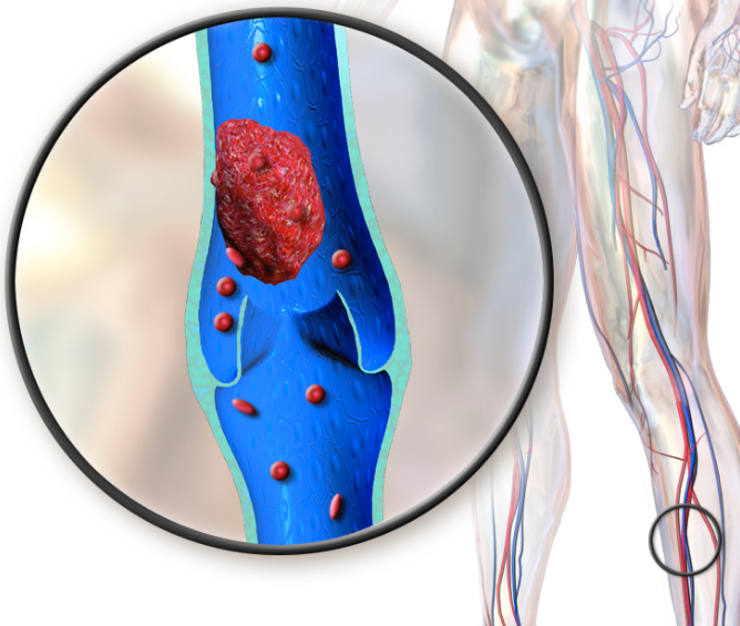
Deep vein thrombosis (credit: Blausen.com staff)

A lung blood clot (pulmonary embolism) usually begins as a clot in the leg or pelvis as a result of inactivity and slowed blood flow (deep vein thrombosis). If the clot breaks free, it can travel to a lung and become lodged in a small blood vessel, where it is especially dangerous.

From 1988 to 1990, Japanese researchers asked 86,024 participants, age 40-79, how many hours they spent watching TV. Over the next 19 years, 59 participants died of a pulmonary embolism*.

The researchers found that compared to participants who watched TV less than 2.5 hours each day, deaths from a pulmonary embolism increased by 70 percent among those who watched TV from 2.5 to 4.9 hours; by 40 percent for each additional 2 hours of daily TV watching; and 2.5 times among those who watched TV 5 or more hours.

“Pulmonary embolism occurs at a lower rate in Japan than it does in Western countries, but it may be on the rise,” said Hiroyasu Iso, M.D., Ph.D., professor of public health at Osaka University Graduate School of Medicine and study corresponding author. “The Japanese people are increasingly adopting sedentary lifestyles, which we believe is putting them at increased risk.”

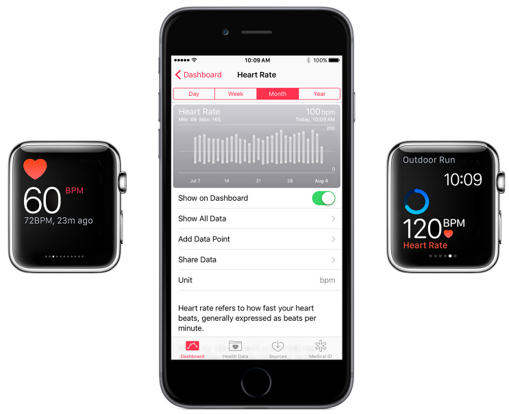
Apple Watch heart rate measure (credit: Apple)

Authors noted that the risk is likely greater than the findings suggest. Deaths from pulmonary embolism are believed to be underreported because diagnosis is difficult. The most common symptoms of pulmonary embolism — chest pain and shortness of breath — are the same as other life-threatening conditions, and diagnosis requires imaging that many hospitals are not equipped to provide.


How my Apple Watch’s heart rate monitoring saved my life (Macworld)


Other risks factors studied were obesity (the strongest link after hours sitting), diabetes, cigarette smoking, and hypertension.

The researchers said the findings may be particularly relevant to Americans, since U.S. adults watch more television — especially “binge watching” — than Japanese adults (according to another study).

The researchers advised a few simple steps to avoid a pulmonary embolism: “After an hour or so, stand up, stretch, walk around, or while you’re watching TV, tense and relax your leg muscles for 5 minutes (similar to that given to travelers on long plane flights), keep hydrated, and shed pounds if overweight.

An hour of moderate exercise a day enough to counter health risks from prolonged sitting

University of Cambridge researchers, in another new study published July 27 in Lancet, noted that recent estimates suggest that more than 5 million people die globally each year as a result of failing to meet recommended daily activity levels.

But they found that 60 to 75 minutes of moderate intensity exercise per day were sufficient to eliminate the increased risk of early death associated with sitting for over eight hours per day, based on an analysis of 16 studies, which included data from more than one million men and women.** (Three out of four people in the study failed to reach this level of daily activity, though.)

The greatest risk of early death was for those individuals who were physically inactive. They were between 28% and 59% more likely to die early compared with those who were in the most active quartile — a risk similar to that associated with smoking and obesity. In other words, lack of physical activity is a greater health risk than prolonged sitting.

The enormous economic burden of physical inactivity — a global pandemic

Another study, also published July 27 in Lancet, revealed that in 2013, physical inactivity cost INT $67.5 billion*** (in international dollars = what U.S. dollars could buy in 2013) globally in healthcare expenditure and lost productivity in 142 countries, representing 93.2 per cent of the world’s population. The study was based on the direct health-care cost, productivity losses, and disability-adjusted life years (DALYs) for five major non-communicable diseases attributable to inactivity: coronary heart disease, stroke, type 2 diabetes, breast cancer and colon cancer.

* The study, which is part of the Japan Collaborative Cohort Study funded by the Ministry of Education, Science, Sports and Culture of Japan, was conducted before computers, tablets and smartphones became popular sources of information and entertainment. The researchers believe new studies are needed to determine the effect of these new technologies on pulmonary embolism risk.

** The researchers acknowledge that there are limitations to the data analyzed, which mainly came from participants aged 45 years and older and living in western Europe, the U.S. and Australia. But the researchers asked all included studies to reanalyze their data in a harmonized manner, an approach that has never before been adopted for a study of this size and therefore also provides much more robust effect estimates compared with previous studies.

*** Cost breakdown:

$67.5bn: Total costs, including $53.8bn in direct cost (healthcare expenditure) and 13.7bn in indirect costs (productivity losses).

$31.2bn: Total loss in tax revenue through public healthcare expenditure

$12.9bn: Total amount in private sector pays for physical inactivity-related diseases (e.g. health insurance companies)

$9.7bn: Total amount households paid out-of-pocket for physical inactivity-related diseases

Type 2 Diabetes was the costliest disease, accounting for $37.6bn (70 percent) of direct costs.


Abstract of Watching Television and Risk of Mortality From Pulmonary Embolism Among Japanese Men and Women

Although case series reporting pulmonary embolism or deep vein thrombosis after prolonged television watching have been published, no prospective study has examined the association between time spent watching television and the risk of mortality from pulmonary embolism. We examined this association in a large cohort study of Japanese men and women.


Abstract of Does physical activity attenuate, or even eliminate, the detrimental association of sitting time with mortality? A harmonised meta-analysis of data from more than 1 million men and women

Background: High amounts of sedentary behaviour have been associated with increased risks of several chronic conditions and mortality. However, it is unclear whether physical activity attenuates or even eliminates the detrimental effects of prolonged sitting. We examined the associations of sedentary behaviour and physical activity with all-cause mortality.

Methods: We did a systematic review, searching six databases (PubMed, PsycINFO, Embase, Web of Science, Sport Discus, and Scopus) from database inception until October, 2015, for prospective cohort studies that had individual level exposure and outcome data, provided data on both daily sitting or TV-viewing time and physical activity, and reported effect estimates for all-cause mortality, cardiovascular disease mortality, or breast, colon, and colorectal cancer mortality. We included data from 16 studies, of which 14 were identified through a systematic review and two were additional unpublished studies where pertinent data were available. All study data were analysed according to a harmonised protocol, which categorised reported daily sitting time and TV-viewing time into four standardised groups each, and physical activity into quartiles (in metabolic equivalent of task [MET]-hours per week). We then combined data across all studies to analyse the association of daily sitting time and physical activity with all-cause mortality, and estimated summary hazard ratios using Cox regression. We repeated these analyses using TV-viewing time instead of daily sitting time.

Findings: Of the 16 studies included in the meta-analysis, 13 studies provided data on sitting time and all-cause mortality. These studies included 1 005 791 individuals who were followed up for 2–18·1 years, during which 84 609 (8·4%) died. Compared with the referent group (ie, those sitting <4 h/day and in the most active quartile [>35·5 MET-h per week]), mortality rates during follow-up were 12–59% higher in the two lowest quartiles of physical activity (from HR=1·12, 95% CI 1·08–1·16, for the second lowest quartile of physical activity [<16 MET-h per week] and sitting <4 h/day; to HR=1·59, 1·52–1·66, for the lowest quartile of physical activity [<2·5 MET-h per week] and sitting >8 h/day). Daily sitting time was not associated with increased all-cause mortality in those in the most active quartile of physical activity. Compared with the referent (<4 h of sitting per day and highest quartile of physical activity [>35·5 MET-h per week]), there was no increased risk of mortality during follow-up in those who sat for more than 8 h/day but who also reported >35·5 MET-h per week of activity (HR=1·04; 95% CI 0·99–1·10). By contrast, those who sat the least (<4 h/day) and were in the lowest activity quartile (<2·5 MET-h per week) had a significantly increased risk of dying during follow-up (HR=1·27, 95% CI 1·22–1·31). Six studies had data on TV-viewing time (N=465 450; 43 740 deaths). Watching TV for 3 h or more per day was associated with increased mortality regardless of physical activity, except in the most active quartile, where mortality was significantly increased only in people who watched TV for 5 h/day or more (HR=1·16, 1·05–1·28).

Interpretation: High levels of moderate intensity physical activity (ie, about 60–75 min per day) seem to eliminate the increased risk of death associated with high sitting time. However, this high activity level attenuates, but does not eliminate the increased risk associated with high TV-viewing time. These results provide further evidence on the benefits of physical activity, particularly in societies where increasing numbers of people have to sit for long hours for work and may also inform future public health recommendations.

Funding: None.


Abstract of The economic burden of physical inactivity: a global analysis of major non-communicable diseases

Background: The pandemic of physical inactivity is associated with a range of chronic diseases and early deaths. Despite the well documented disease burden, the economic burden of physical inactivity remains unquantified at the global level. A better understanding of the economic burden could help to inform resource prioritisation and motivate efforts to increase levels of physical activity worldwide.

Methods: Direct health-care costs, productivity losses, and disability-adjusted life-years (DALYs) attributable to physical inactivity were estimated with standardised methods and the best data available for 142 countries, representing 93·2% of the world’s population. Direct health-care costs and DALYs were estimated for coronary heart disease, stroke, type 2 diabetes, breast cancer, and colon cancer attributable to physical inactivity. Productivity losses were estimated with a friction cost approach for physical inactivity related mortality. Analyses were based on national physical inactivity prevalence from available countries, and adjusted population attributable fractions (PAFs) associated with physical inactivity for each disease outcome and all-cause mortality.

Findings: Conservatively estimated, physical inactivity cost health-care systems international $ (INT$) 53·8 billion worldwide in 2013, of which $31·2 billion was paid by the public sector, $12·9 billion by the private sector, and $9·7 billion by households. In addition, physical inactivity related deaths contribute to $13·7 billion in productivity losses, and physical inactivity was responsible for 13·4 million DALYs worldwide. High-income countries bear a larger proportion of economic burden (80·8% of health-care costs and 60·4% of indirect costs), whereas low-income and middle-income countries have a larger proportion of the disease burden (75·0% of DALYs). Sensitivity analyses based on less conservative assumptions led to much higher estimates.

Interpretation: In addition to morbidity and premature mortality, physical inactivity is responsible for a substantial economic burden. This paper provides further justification to prioritise promotion of regular physical activity worldwide as part of a comprehensive strategy to reduce non-communicable diseases.

Funding: None.