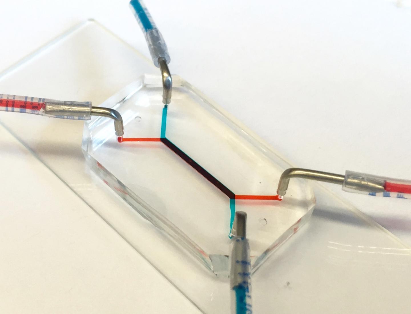
Placenta-on-a-chip models the vital mother-fetus placental barrier

The flash-drive-sized device contains two layers of human cells that model the interface between mother and fetus. Microfluidic channels on both sides of those layers allow researchers to study how molecules are transported through, or are blocked by, that interface. (credit: University of Pennsylvania)

Researchers at the University of Pennsylvania have developed the first placenta-on-a-chip that can fully model the transport of nutrients across the placental barrier — part of a nationwide effort sponsored by the March of Dimes to identify causes of preterm birth and ways to prevent it.

Prematurely born babies may experience lifelong, debilitating consequences, but the underlying mechanisms of this condition are not well understood due in part to the difficulties of experimenting with intact, living human placentae.

Like other organs-on-chips, such as ones developed to simulate lungs, intestines, and eyes, the placenta-on-a-chip provides a unique capability to mimic and study the function of that human organ in ways that have not been possible using traditional tools, which are limited by complexity, the scarcity of samples, and the limited lifespan of how long the tissue remains viable (for only a few hours after delivery), according to the researchers.

Modeling the vital placental barrier

The flash-drive-sized device contains two layers of human cells that model the interface between mother and fetus. Microfluidic channels on either side of those layers allow researchers to study how molecules are transported through, or are blocked by, that interface.
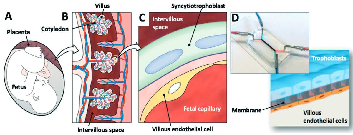
(A) Schematic of a human fetus and placenta within the uterine cavity. (B) Cross-sectional view of the placenta showing villi (vascular projections that increase the surface area of a membrane) in direct contact with maternal blood in (C),where the maternal intervillous space is separated from the fetal capillary by the maternal–fetal interface, which is composed of the syncytiotrophoblast, basal lamina, and villous endothelial cells. (D) 3-D microarchitecture of the placental barrier reconstituted within the placenta-on-a-chip, which consists of upper and lower microchannels separated by a thin, semipermeable membrane. Trophoblast cells are cultured in the upper microchannel layer and villous endothelial cells are grown in the lower microchannel layer. (credit: Cassidy Blundell et al./Lab on a Chip)

The researchers’ placenta-on-a-chip is a clear silicone device with two parallel microfluidic channels separated by a porous membrane. On one side of those pores, trophoblast cells, which are found at the placental interface with maternal blood, are grown. On the other side are endothelial cells, found on the interior of fetal blood vessels.

The layers of those two cell types mimic the placental barrier, the gatekeeper (or filter) that controls flow between the maternal and fetal circulatory systems, including nutrients that must pass, but also foreign agents like viruses that must be blocked.
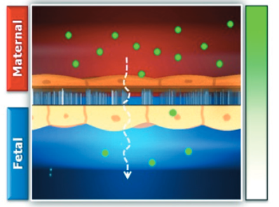
A concentration gradient of glucose (green dots) is generated across the microengineered placental barrier to drive glucose transport from the maternal to fetal compartments. (credit: Cassidy Blundell et al./Lab on a Chip)

In their new study, the Penn researchers have demonstrated that the two layers of cells continue to grow and develop while inside the chip, undergoing a process known as “syncytialization.”

“The placental cells change over the course of pregnancy,” explained Dan Huh, the Wilf Family Term Assistant Professor of Bioengineering in Penn’s School of Engineering and Applied Science. “During pregnancy, the placental trophoblast cells actually fuse with one another to form an interesting tissue called syncytium. The barrier also becomes thinner as the pregnancy progresses, and with our new model we’re able to reproduce this change.

“This process is very important because it affects placental transport and was a critical aspect not represented in our previous model.”

The Penn team validated the new model by showing glucose transfer rates across this syncytialized barrier matched those measured in perfusion studies of donated human placentae.

“The placenta is arguably the least understood organ in the human body,” Huh said, “and much remains to be learned about how transport between mother and fetus works at the tissue, cellular and molecular levels. An isolated whole organ is an not ideal platform for these types of mechanistic studies.”

Studies of preterm birth

While the placenta-on-a-chip is still in the early stages of testing, researchers at Penn and beyond are already planning to use it in studies on preterm birth. The rate of preterm birth is still about 10 to 11 percent of all pregnancies.

“Eventually,” Huh said, “we hope to leverage the unique capabilities of our model to demonstrate the potential of organ-on-a-chip technology as a new strategy to innovate basic and translational research in reproductive biology and medicine.”

The study was published in the journal Lab on a Chip. The research was supported by the March of Dimes Prematurity Research Center at the University of Pennsylvania and the National Institutes of Health.


Abstract of A microphysiological model of the human placental barrier

During human pregnancy, the fetal circulation is separated from maternal blood in the placenta by two cell layers – the fetal capillary endothelium and placental trophoblast. This placental barrier plays an essential role in fetal development and health by tightly regulating the exchange of endogenous and exogenous materials between the mother and the fetus. Here we present a microengineered device that provides a novel platform to mimic the structural and functional complexity of this specialized tissue in vitro. Our model is created in a multilayered microfluidic system that enables co-culture of human trophoblast cells and human fetal endothelial cells in a physiologically relevant spatial arrangement to replicate the characteristic architecture of the human placental barrier. We have engineered this co-culture model to induce progressive fusion of trophoblast cells and to form a syncytialized epithelium that resembles the syncytiotrophoblast in vivo. Our system also allows the cultured trophoblasts to form dense microvilli under dynamic flow conditions and to reconstitute expression and physiological localization of membrane transport proteins, such as glucose transporters (GLUTs), critical to the barrier function of the placenta. To provide a proof-of-principle for using this microdevice to recapitulate native function of the placental barrier, we demonstrated physiological transport of glucose across the microengineered maternal–fetal interface. Importantly, the rate of maternal-to-fetal glucose transfer in this system closely approximated that measured in ex vivo perfused human placentas. Our “placenta-on-a-chip” platform represents an important advance in the development of new technologies to model and study the physiological complexity of the human placenta for a wide variety of applications.

Mayo Clinic researchers discover drug combination that helps immune system attack cancer cells

Effects of combination drug treatment on tumor size in square millimeters over 67 days for multiple mice (credit: Soraya Zorro Manrique et al./Oncotarget)

Mayo Clinic researchers have developed a drug combination that could enhance the immune system’s ability to attack cancer cells. The drugs have shown a pronounced therapeutic effect against advanced and metastatic cancers in mice, according to a  study published in the July 12 edition of the online journal Oncotarget.

“Cancers can remain inconspicuous in the body for months to years before causing major problems, leading the immune system to coexist rather than to attack cancers,” explains Mayo Clinic cancer immunotherapist Peter Cohen, M.D.

The solution was to combine two drugs: toll-like receptor (TLR) agonists that can mimic invading bacteria (tricking the immune system into attacking cancer as if it were a life-threatening infection) and the chemotherapy agent cyclophosphamide. The combination resulted in permanent eradication of breast and pancreatic cancers, as well as widespread metastases.

The combined weekly treatment was very well tolerated and actually less toxic than either TLR agonists or cyclophosphamide given individually, they also found.

The drug combination also revealed an additional benefit: it activated monocytes (a type of white blood cell) to participate in the killing of cancer cells.

Mayo Clinic is continuing its research in an FDA-approved clinical trial. They are studying whether patients with advanced cancers, including pancreas, breast, colorectal, melanoma and others, respond similarly to mice when cyclophosphamide treatment is paired with the TLR agonist motolimod.


Mayo Clinic | Potential Immunotherapy Drug Combination for Targeting Advanced and Metastatic Cancers


Abstract of Definitive activation of endogenous antitumor immunity by repetitive cycles of cyclophosphamide with interspersed Toll-like receptor agonists

Many cancers both evoke and subvert endogenous anti-tumor immunity. However, immunosuppression can be therapeutically reversed in subsets of cancer patients by treatments such as checkpoint inhibitors or Toll-like receptor agonists (TLRa). Moreover, chemotherapy can leukodeplete immunosuppressive host elements, including myeloid-derived suppressor cells (MDSCs) and regulatory T-cells (Tregs). We hypothesized that chemotherapy-induced leukodepletion could be immunopotentiated by co-administering TLRa to emulate a life-threatening infection. Combining CpG (ODN 1826) or CpG+poly(I:C) with cyclophosphamide (CY) resulted in uniquely well-tolerated therapeutic synergy, permanently eradicating advanced mouse tumors including 4T1 (breast), Panc02 (pancreas) and CT26 (colorectal). Definitive treatment required endogenous CD8+ and CD4+ IFNγ-producing T-cells. Tumor-specific IFNγ-producing T-cells persisted during CY-induced leukopenia, whereas Tregs were progressively eliminated, especially intratumorally. Spleen-associated MDSCs were cyclically depleted by CY+TLRa treatment, with residual monocytic MDSCs requiring only continued exposure to CpG or CpG+IFNγ to effectively attack malignant cells while sparing non-transformed cells. Such tumor destruction occurred despite upregulated tumor expression of Programmed Death Ligand-1, but could be blocked by clodronate-loaded liposomes to deplete phagocytic cells or by nitric oxide synthase inhibitors. CY+TLRa also induced tumoricidal myeloid cells in naive mice, indicating that CY+TLRa’s immunomodulatory impacts occurred in the complete absence of tumor-bearing, and that tumor-induced MDSCs were not an essential source of tumoricidal myeloid precursors. Repetitive CY+TLRa can therefore modulate endogenous immunity to eradicate advanced tumors without vaccinations or adoptive T-cell therapy. Human blood monocytes could be rendered similarly tumoricidal during in vitro activation with TLRa+IFNγ, underscoring the potential therapeutic relevance of these mouse tumor studies to cancer patients.

How to detect early signs of Alzheimer’s with a simple eye exam before symptoms appear

Monochromatic images of normal mouse retina at four wavelengths from blue (upper left) to red (lower right). Boxes in lower right show locations typical of amyloid (a substance found in the brain associated with Alzheimer’s). (credit: Swati S. More et al./IOVS)

University of Minnesota (UMN) scientists and associates have developed new technology that can detect signs of Alzheimer’s before the onset of symptoms — early enough to give drugs a chance to work — in mice and humans by simply examining the back of their eyes.

Looking at Alzheimer’s effects through the eye is a key advantage of the new technology. “The retina of the eye is not just ‘connected’ to the brain — it is part of the central nervous system,” said Swati More, PhD, of the Center for Drug Design at UMN, co-author of a paper recently published in Investigative Ophthalmology & Visual Science (IOVS).

The brain and retina undergo similar changes due to Alzheimer’s disease, he said, but “unlike the brain, the retina is easily accessible to us. We saw changes in the retinas of Alzheimer’s mice before the typical age at which neurological signs are observed.”

Human clinical trials are set to start in July to test the technology in humans who are 40–75 years old (for more information on participating in the clinical trial, you can visit the trial website).

Optical spectra recorded from human and mouse retina samples. Upper two lines show the intensity of light for different wavelengths of light from red (right side) to blue (left side) for a normal human retina (heavy line) and for an Alzheimer’s (AD) patients retina. Comparable mouse plots, shown below (WT = normal mouse; APP1/PS1 = Alzheimer’s model mouse), show a similar pattern. (credit: Swati S. More et al./IOVS)

Early detection of Alzheimer’s is critical for two reasons. “First, effective treatments need to be administered well before patients show actual neurological signs,” said co-author Robert Vince, PhD, of the Center for Drug Design at the University of Minnesota (UMN). “Second, since there are no available early detection techniques, drugs currently cannot be tested to determine if they are effective against early Alzheimer’s disease. An early diagnostic tool like ours could help the development of drugs as well.”


Abstract of Early Detection of Amyloidopathy in Alzheimer’s Mice by Hyperspectral Endoscopy

Purpose: To describe a spectral imaging system for small animal studies based on noninvasive endoscopy of the retina, and to present time-resolved spectral changes from live Alzheimer’s mice prior to cognitive decline, corroborating our previous in vitro findings.

Methods: Topical endoscope fundus imaging was modified to use a machine vision camera and tunable wavelength system for acquiring monochromatic images across the visible to near-infrared spectral range. Alzheimer’s APP/PS1 mice and age-matched, wild-type mice were imaged monthly from months 3 through 8 to assess changes in the fundus reflection spectrum. Optical changes were fit to Rayleigh light scatter models as measures of amyloid aggregation.

Results: Good quality spectral images of the central retina were obtained. Short-wavelength reflectance from Alzheimer’s mice retinae showed significant reduction over time compared to wild-type mice. Optical changes were consistent with an increase in Rayleigh light scattering in neural retina due to soluble Aβ1–42aggregates. The changes in light scatter showed a monotonic increase in soluble amyloid aggregates over a 6-month period, with significant build up occurring at 7 months.

Conclusions: Hyperspectral imaging technique can be brought inexpensively to the study of retinal changes caused by Alzheimer’s disease progression in live small animals. A similar previous finding of reduction in the light reflection over a range of wavelengths in isolated Alzheimer’s mice retinae, was reproducible in the living Alzheimer’s mice. The technique presented here has a potential for development as an early Alzheimer’s retinal diagnostic test in humans, which will support the treatment outcome.

A host of common chemicals endanger child brain development, NIH journal reports

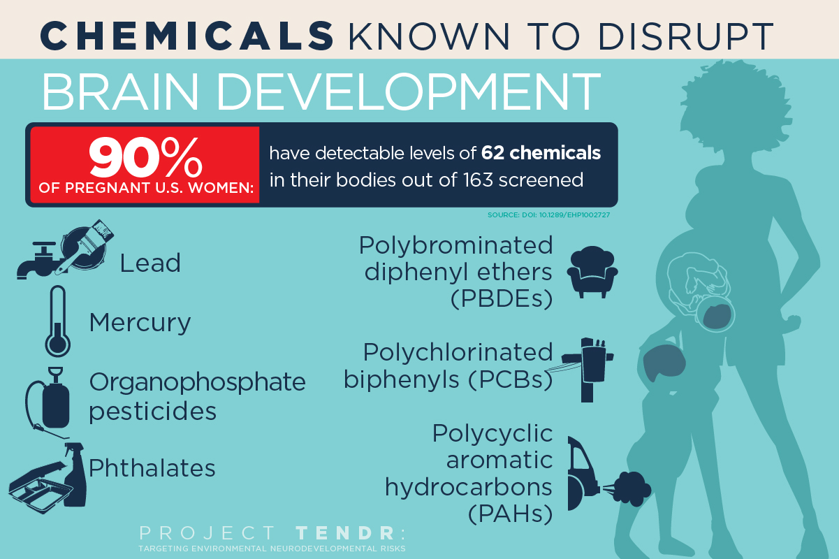
(credit: Graphic by Julie McMahon)

In a new open-access report in the NIH journal Environmental Health Perspectives, 47 scientists, health practitioners, and children’s health advocates have made a consensus statement in “Project TENDR: Targeting Environmental Neuro-Developmental Risks“ — endorsed by nine medical organizations — and issued a call to action for renewed attention to the growing evidence that many common and widely available chemicals endanger neurodevelopment in fetuses and children of all ages.

The list includes chemicals used extensively in consumer products and that have become widespread in the environment. Of most concern are lead and mercury; organophosphate pesticides used in agriculture and home gardens; phthalates, which are used in pharmaceuticals, plastics and personal care products; flame retardants known as polybrominated diphenyl ethers; and air pollutants produced by the combustion of wood and fossil fuels, said University of Illinois Comparative Biosciences professor Susan Schantz, one of dozens of individual signatories to the consensus statement.

The list provides “prime examples of toxic chemicals that can contribute to learning, behavioral, or intellectual impairment, as well as specific neurodevelopmental disorders such as ADHD or autism spectrum disorder,” according to the report.

Polychlorinated biphenyls

Polychlorinated biphenyls, once used as coolants and lubricants in transformers and other electrical equipment, also are of concern. PCBs were banned in the U.S. in 1977, but can persist in the environment for decades, she said.

“These chemicals are pervasive, not only in air and water, but in everyday consumer products that we use on our bodies and in our homes,” Schantz said. “Reducing exposures to toxic chemicals can be done, and is urgently needed to protect today’s and tomorrow’s children.”

“The human brain develops over a very long period of time, starting in gestation and continuing during childhood and even into early adulthood,” Schantz said. “But the biggest amount of growth occurs during prenatal development. The neurons are forming and migrating and maturing and differentiating. And if you disrupt this process, you’re likely to have permanent effects.”

Hormonal disrupters

Some of the chemicals of concern, such as phthalates and PBDEs, are known to interfere with normal hormone activity. For example, most pregnant women in the U.S. will test positive for exposure to phthalates and PBDEs, both of which disrupt thyroid hormone function.

“Thyroid hormone is involved in almost every aspect of brain development, from formation of the neurons to cell division, to the proper migration of cells and myelination of the axons after the cells are differentiated,” said Schantz. “It regulates many of the genes involved in nervous system development.”

Schantz and her colleagues at Illinois are studying infants and their mothers to determine whether prenatal exposure to phthalates and other endocrine disruptors leads to changes in the brain or behavior. This research, along with parallel studies in older children and animals, is a primary focus of the Children’s Environmental Health Research Center at Illinois, which Schantz directs.

Phthalates also interfere with steroid hormone activity. Studies link exposure to certain phthalates with attention deficits, lower IQ and conduct disorders in children. “Phthalates are everywhere; they’re in all kinds of different products. We’re exposed to them every day,” Schantz said.

The report criticizes current regulatory lapses that allow chemicals to be introduced into people’s lives with little or no review of their effects on fetal and child health. “For most chemicals, we have no idea what they’re doing to children’s neurodevelopment,” Schantz said. “They just haven’t been studied.

“And if it looks like something is a risk, we feel policymakers should be willing to make a decision that this or that chemical could be a bad actor and we need to stop its production or limit its use,” she said. “We shouldn’t have to wait 10 or 15 years — allowing countless children to be exposed to it in the meantime — until we’re positive it’s a bad actor.”

Project TENDR has a website with information about each of the chemicals of concern. The National Institute of Environmental Health Sciences at the National Institutes of Health and the U.S. Environmental Protection Agency fund the Children’s Environmental Health Research Center at the University of Illinois.

Project TENDR is an alliance of 48 of the nation’s top scientists, health professionals and health advocates. It was launched by Maureen Swanson of the Learning Disabilities Association of America and Irva Hertz-Picciotto of UC Davis, who brought together participants across many disciplines and sectors, including epidemiology, toxicology, exposure science, pediatrics, obstetrics and gynecology, nursing, public health, and federal and state chemical policy. Medical and scientific societies that have signed on in support include American Congress of Obstetricians and Gynecologists, American Nurses Association, Endocrine Society, National Association of Pediatric Nurse Practitioners, National Medical Association, National Hispanic Medical Association, Alliance of Nurses for Healthy Environments, Physicians for Social Responsibility and the National Council of Asian Pacific Island Physicians. TENDR’s long-term mission is to lower the incidence of neurodevelopmental disorders by reducing exposure levels to chemicals and pollutants that can contribute to these conditions, especially during fetal development and early childhood.


Steve Drake, Beckman Institute for Advanced Science and Technology | U. of I. biosciences professor Susan Schantz directs the Children’s Environmental Health Research Center at the University of Illinois, which is studying whether, and how, exposure to phthalates disrupts child brain development. Phthalates are used in some cosmetics, food packaging and products with fragrances.


Food Safety | From DDT to Glyphosate: Rachel Carson, We Need You Again


Abstract of Project TENDR: Targeting Environmental Neuro-Developmental Risks. The TENDR Consensus Statement

SUMMARY: Children in America today are at an unacceptably high risk of developing neurodevelopmental disorders that affect the brain and nervous system including autism, attention deficit hyperactivity disorder, intellectual disabilities, and other learning and behavioral disabilities. These are complex disorders with multiple causes—genetic, social, and environmental. The contribution of toxic chemicals to these disorders can be prevented. APPROACH: Leading scientific and medical experts, along with children’s health advocates, came together in 2015 under the auspices of Project TENDR: Targeting Environmental Neuro-Developmental Risks to issue a call to action to reduce widespread exposures to chemicals that interfere with fetal and children’s brain development. Based on the available scientific evidence, the TENDR authors have identified prime examples of toxic chemicals and pollutants that increase children’s risks for neurodevelopmental disorders. These include chemicals that are used extensively in consumer products and that have become widespread in the environment. Some are chemicals to which children and pregnant women are regularly exposed, and they are detected in the bodies of virtually all Americans in national surveys conducted by the U.S. Centers for Disease Control and Prevention. The vast majority of chemicals in industrial and consumer products undergo almost no testing for developmental neurotoxicity or other health effects. CONCLUSION: Based on these findings, we assert that the current system in the United States for evaluating scientific evidence and making health-based decisions about environmental chemicals is fundamentally broken. To help reduce the unacceptably high prevalence of neurodevelopmental disorders in our children, we must eliminate or significantly reduce exposures to chemicals that contribute to these conditions. We must adopt a new framework for assessing chemicals that have the potential to disrupt brain development and prevent the use of those that may pose a risk. This consensus statement lays the foundation for developing recommendations to monitor, assess, and reduce exposures to neurotoxic chemicals. These measures are urgently needed if we are to protect healthy brain development so that current and future generations can reach their fullest potential.

Prenatal exposure to acetaminophen may increase autism spectrum symptoms

Tylenol PM (left) and Tylenol (right) (credit: Ragesoss/CC)

A new study has found that paracetamol (aka acetaminophen; trade names include Tylenol and Panadol), which is used extensively during pregnancy, has a strong association with autism spectrum symptoms in boys and for both genders in relation to attention-related and hyperactivity symptoms.

The findings* were published this week in the International Journal of Epidemiology. This is the first study of its kind to report an independent association between the use of this drug in pregnancy and autism spectrum symptoms in children.

It is also the first study to report different effects on boys and girls. Comparing persistently to nonexposed children, the study has found an increase of 30 per cent in the risk of detriment to some attention functions, and an increase of two clinical symptoms of autism spectrum symptoms in boys. Boys also showed more autism spectrum symptoms when persistently exposed to paracetamol.

“Paracetamol could be harmful to neurodevelopment for several reasons,” said Co-author Dr. Jordi Júlvez, also a researcher at CREAL. “First of all, it relieves pain by acting on cannabinoid receptors in the brain.

Why boys are more likely to have autism

“Since these receptors normally help determine how neurons mature and connect with one another, paracetamol could alter these important processes. It can also affect the development of the immune system, or be directly toxic to some fetuses that may not have the same capacity as an adult to metabolize this drug, or by creating oxidative stress.”

There could also be an explanation for why boys are more likely to have autism spectrum symptoms: “The male brain may be more vulnerable to harmful influences during early life”, said Claudia Avella-Garcia. “Our differing gender results suggest that androgenic endocrine disruption, to which male brains could be more sensitive, may explain the association.”

The study concluded that the widespread exposure of infants to paracetamol in utero could increase the number of children with ADHD or autism spectrum symptoms. However, they stressed further studies should be conducted with more precise dosage measurements, and that the risks versus benefits of paracetamol use during pregnancy and early life should be assessed before treatment recommendations are made.

* Researchers in Spain recruited 2644 mother-child pairs in a birth cohort study during pregnancy. 88 per cent were evaluated when the child was one year old, and 79.9 per cent were evaluated when they were five years old. Mothers were asked about their use of paracetamol during pregnancy and the frequency of use was classified as never, sporadic, or persistent. Exact doses could not be noted due to mothers being unable to recall them exactly. 43 per cent of children evaluated at age one and 41 per cent assessed at age five were exposed to any paracetamol at some point during the first 32 weeks of pregnancy. When assessed at age five, exposed children were at higher risk of hyperactivity or impulsivity symptoms. Persistently exposed children in particular showed poorer performance on a computerised test measuring inattention, impulsivity and visual speed processing.


Abstract of Acetaminophen use in pregnancy and neurodevelopment: attention function and autism spectrum symptoms

Background: Acetaminophen is extensively used during pregnancy. But there is a lack of population-representative cohort studies evaluating its effects on a range of neuropsychological and behavioural endpoints. We aimed to assess whether prenatal exposure to acetaminophen is adversely associated with neurodevelopmental outcomes at 1 and 5 years of age.

Methods: This Spanish birth cohort study included 2644 mother-child pairs recruited during pregnancy. The proportion of liveborn participants evaluated at 1 and 5 years was 88.8% and 79.9%, respectively. Use of acetaminophen was evaluated prospectively in two structured interviews. Ever/never use and frequency of use (never, sporadic, persistent) were measured. Main neurodevelopment outcomes were assessed using Childhood Autism Spectrum Test (CAST), Conner’s Kiddie Continuous Performance Test (K-CPT) and ADHD-DSM-IV form list. Regression models were adjusted for social determinants and co-morbidities.

Results: Over 40% of mothers reported using acetaminophen. Ever-exposed offspring had higher risks of presenting more hyperactivity/impulsivity symptoms [incidence rate ratio (IRR) = 1.41, 95% confidence interval (CI) 1.01–1.98), K-CPT commission errors (IRR = 1.10, 1.03–1.17), and lower detectability scores (coefficient β = −0.75, −0.13–−0.02). CAST scores were increased in ever-exposed males (β = 0.63, 0.09–1.18). Increased effect sizes of risks by frequency of use were observed for hyperactivity/impulsivity symptoms (IRR = 2.01, 0.95–4.24) in all children, K-CPT commission errors (IRR = 1.32, 1.05–1.66) and detectability (β = −0.18, −0.36–0.00) in females, and CAST scores in males (β = 1.91, 0.44–3.38).

Conclusions: Prenatal acetaminophen exposure was associated with a greater number of autism spectrum symptoms in males and showed adverse effects on attention-related outcomes for both genders. These associations seem to be dependent on the frequency of exposure.

A smarter ‘bionic’ cardiac patch that doubles as advanced pacemaker/arrhythmia detector

(a) Schematic of the free-standing macroporous nanoelectronic scaffold with nanowire FET (field effect transistor) arrays (red dots). Inset: one nanowire FET. (b) Folded 3D free-standing scaffolds with four layers of individually addressable FET sensors. (c) Schematic of nanoelectronic scaffold/cardiac tissue resulting from the culturing of cardiac cells within the 3D folded scaffold. Inset: the nanoelectronic sensors (blue circles) innervate the 3D cell network. (credit: Xiaochuan Dai at al./Nature Nanotechnology)

Harvard researchers have designed nanoscale electronic scaffolds (support structures) that can be seeded with cardiac cells to produce a new “bionic” cardiac patch (for replacing damaged cardiac tissue with pre-formed tissue patches). It also functions as a more sophisticated pacemaker: In addition to electrically stimulating the heart, the new design can change the pacemaker stimulation frequency and direction of signal propagation.

In addition, because because its electronic components are integrated throughout the tissue (instead of being located on the surface of the skin), it could detect arrhythmia far sooner, and “operate at far lower (safer) voltages than a normal pacemaker, [which] because it’s on the surface, has to use relatively high voltages,” according to Charles Lieber, the Mark Hyman, Jr. Professor of Chemistry and Chair of the Department of Chemistry and Chemical Biology.

Early arrhythmia detection, monitoring responses to cardiac drugs

“Even before a person started to go into large-scale arrhythmia that frequently causes irreversible damage or other heart problems, this could detect the early-stage instabilities and intervene sooner,” he said. “It can also continuously monitor the feedback from the tissue and actively respond.”

The patch might also find use, Lieber said, as a tool to monitor responses to cardiac drugs, or to help pharmaceutical companies screen the effectiveness of drugs under development.

In the long term, Lieber believes, the development of nanoscale tissue scaffolds represents a new paradigm for integrating biology with electronics in a virtually seamless way.

The bionic cardiac patch can also be a unique platform to study the tissue behavior evolving during some developmental processes, such as aging, ischemia, or differentiation of stem cells into mature cardiac cells.

Although the bionic cardiac patch has not yet been implanted in animals, “we are interested in identifying collaborators already investigating cardiac patch implantation to treat myocardial infarction in a rodent model,” he said. “I don’t think it would be difficult to build this into a simpler, easily implantable system.”

Could one day deliver cardiac patch/pacemaker via injection

Using the injectable electronics technology he pioneered last year, Lieber even suggested that similar cardiac patches might one day simply be delivered by injection. “It may actually be that, in the future, this won’t be done with a surgical patch,” he said. “We could simply do a co-injection of cells with the mesh, and it assembles itself inside the body, so it’s less invasive.”

“I think one of the biggest impacts would ultimately be in the area that involves replacement of damaged cardiac tissue with pre-formed tissue patches,” Lieber said. “Rather than simply implanting an engineered patch built on a passive scaffold, our work suggests it will be possible to surgically implant an innervated patch that would now be able to monitor and subtly adjust its performance.”

In the long term, Lieber believes, the development of nanoscale tissue scaffolds represents a new paradigm for integrating biology with electronics in a virtually seamless way.

The study is described in a June 27 paper published in Nature Nanotechnology.


Abstract of Three-dimensional mapping and regulation of action potential propagation in nanoelectronics-innervated tissues

Real-time mapping and manipulation of electrophysiology in three-dimensional (3D) tissues could have important impacts on fundamental scientific and clinical studies, yet realization is hampered by a lack of effective methods. Here we introduce tissue-scaffold-mimicking 3D nanoelectronic arrays consisting of 64 addressable devices with subcellular dimensions and a submillisecond temporal resolution. Real-time extracellular action potential (AP) recordings reveal quantitative maps of AP propagation in 3D cardiac tissues, enable in situtracing of the evolving topology of 3D conducting pathways in developing cardiac tissues and probe the dynamics of AP conduction characteristics in a transient arrhythmia disease model and subsequent tissue self-adaptation. We further demonstrate simultaneous multisite stimulation and mapping to actively manipulate the frequency and direction of AP propagation. These results establish new methodologies for 3D spatiotemporal tissue recording and control, and demonstrate the potential to impact regenerative medicine, pharmacology and electronic therapeutics.

The top 10 emerging technologies of 2016

(credit: WEF)

The World Economic Forum’s annual list of this year’s breakthrough technologies, published today, includes “socially aware” openAI, grid-scale energy storage, perovskite solar cells, and other technologies with the potential to “transform industries, improve lives, and safeguard the planet.” The WEF’s specific interest is to “close gaps in investment and regulation.”

“Horizon scanning for emerging technologies is crucial to staying abreast of developments that can radically transform our world, enabling timely expert analysis in preparation for these disruptors. The global community needs to come together and agree on common principles if our society is to reap the benefits and hedge the risks of these technologies,” said Bernard Meyerson, PhD, Chief Innovation Officer of IBM and Chair of the WEF’s Meta-Council on Emerging Technologies.

The list also provides an opportunity to debate human, societal, economic or environmental risks and concerns that the technologies may pose — prior to widespread adoption.

One of the criteria used by council members during their deliberations was the likelihood that 2016 represents a tipping point in the deployment of each technology. So the list includes some technologies that have been known for a number of years, but are only now reaching a level of maturity where their impact can be meaningfully felt.

The top 10 technologies that make this year’s list are:

  1. Nanosensors and the Internet of Nanothings  — With the Internet of Things expected to comprise 30 billion connected devices by 2020, one of the most exciting areas of focus today is now on nanosensors capable of circulating in the human body or being embedded in construction materials. They could use DNA and proteins to recognize specific chemical targets, store a few bits of information, and then report their status by changing color or emitting some other easily detectable signal.
  2. Next-Generation Batteries — One of the greatest obstacles holding renewable energy back is matching supply with demand, but recent advances in energy storage using sodium, aluminum, and zinc based batteries makes mini-grids feasible that can provide clean, reliable, around-the-clock energy sources to entire villages.
  3. The Blockchain — With venture investment related to the online currency Bitcoin exceeding $1 billion in 2015 alone, the economic and social impact of blockchain’s potential to fundamentally change the way markets and governments work is only now emerging.
  4. 2D Materials — Plummeting production costs mean that 2D materials like graphene are emerging in a wide range of applications, from air and water filters to new generations of wearables and batteries.
  5. Autonomous Vehicles — The potential of self-driving vehicles for saving lives, cutting pollution, boosting economies, and improving quality of life for the elderly and other segments of society has led to rapid deployment of key technology forerunners along the way to full autonomy.
  6. Organs-on-chips — Miniature models of human organs could revolutionize medical research and drug discovery by allowing researchers to see biological mechanism behaviors in ways never before possible.
  7. Perovskite Solar Cells — This new photovoltaic material offers three improvements over the classic silicon solar cell: it is easier to make, can be used virtually anywhere and, to date, keeps on generating power more efficiently.
  8. Open AI Ecosystem — Shared advances in natural language processing and social awareness algorithms, coupled with an unprecedented availability of data, will soon allow smart digital assistants to help with a vast range of tasks, from keeping track of one’s finances and health to advising on wardrobe choice.
  9. Optogenetics — Recent developments mean light can now be delivered deeper into brain tissue, something that could lead to better treatment for people with brain disorders.
  10. Systems Metabolic Engineering — Advances in synthetic biology, systems biology, and evolutionary engineering mean that the list of building block chemicals that can be manufactured better and more cheaply by using plants rather than fossil fuels is growing every year.

To compile this list, the World Economic Forum’s Meta-Council on Emerging Technologies, a panel of global experts, “drew on the collective expertise of the Forum’s communities to identify the most important recent technological trends. By doing so, the Meta-Council aims to raise awareness of their potential and contribute to closing gaps in investment, regulation and public understanding that so often thwart progress.”

You can read 10 expert views on these technologies here or download the series as a PDF.

Unexpected discovery reveals secret of how cancer spreads in the body

Artist’s concept of primary tumor (credit: Barts Cancer Institute/QMUL)

Metastasis (spread of cancer) is one of the biggest challenges in cancer treatment. It is often not the original tumor that kills, but secondary growths. But a key question in cancer research has been how vulnerable cancer cells are able to survive once they break away from a tumor to spread around the body.

“Metastasis is currently incurable and remains one of the key targets of cancer research,” said lead researcher Stéphanie Kermorgant, PhD, from Barts Cancer Institut at Queen Mary University of London (QMUL). “Our research advances the knowledge of how two key molecules communicate and work together to help cancer cells survive during metastasis. We’re hoping that this might lead to the discovery of new drugs to block the spread of cancer within the body.”

Metastasizing cancer cells, as one enters a blood vessel (artist’s concept) (credit: Barts Cancer Institute/QMUL)

The study, published in an open-access paper in Nature Communications, examined the changes that occur in cancer cells as they break away from tumors in cell cultures in mice and zebrafish*. The research revealed a previously unknown survival mechanism in cancer cells and found that molecules known as “integrins” could be key.

Integrins are proteins on the cell surface that attach to and interact with the cell’s surroundings. “Outside-in” and “inside-out” signaling by integrins is known to help the cancer cells attach themselves to their surroundings.

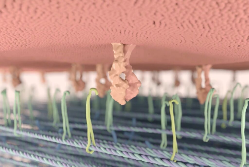
Integrins (yell0w-orange pairs) attach to cancer cell (top) to tell it about its environment and instruct the cell to attach to specific surrounding proteins (artist’s concept) (credit: Barts Cancer Institute, QMUL)

Key discovery: “inside-in” signaling

But the study suggests that when the cancer cells are floating, as they do during metastasis, the integrins switch from their adhesion role to take on an entirely new form of communication that has never been seen before: “Inside-in” signaling, in which integrins signal within the cell.

The researchers discovered that an integrin called beta-1 (β1) pairs up with another protein called c-Met and they move inside the cell together. The two proteins then travel to an unexpected location within the cell that is normally used to degrade and recycle cell material. Instead, the location is used for a new role of cell communication and the two proteins send a message to the rest of the cell to resist against death while floating during metastasis.

Using both breast and lung cells, the team found that metastases were less likely to form when β1 and c-Met were blocked from entering the cell together or were prevented from moving to the special location within the cell.

Integrins are already major targets for cancer treatment with drugs either being tested or in use in the clinic. Most integrin inhibitor drugs target their adhesive function and block them on the surface of the cancer cell. The researchers say that the limited success of these drugs could be partly explained by this newly discovered role of integrins within the cancer cell.

Keeping the integrins out of the cancer cell

But a new strategy could be to prevent the integrin from going inside the cell in the first place. The researchers hope that these insights could lead to the design of better therapies against metastasis and more effective treatment combinations that could prevent and slow both tumor growth and spread.

The research was funded by the UK Medical Research Council, Breast Cancer Now, Rosetrees Trust, British Lung Foundation, Cancer Research UK and Barts Charity.

* The team carried out part of their animal research work on zebrafish embryos to implement the principle of 3Rs (refine, reduce, replace) on their research on mice. Zebrafish provide a similar tumor microenvironment to humans, meaning fewer tests need to be carried out in mice and any future experiments in mice will have been optimized to have minimal toxicity. They are aiming to reduce the number of mice used by at least 90 per cent and ultimately use zebrafish to completely replace the use of mice.


BCIQMUL | BCI Tumor Biology: integrins and metastasis


Abstract of Beta 1-integrin–c-Met cooperation reveals an inside-in survival signalling on autophagy-related endomembranes

Receptor tyrosine kinases (RTKs) and integrins cooperate to stimulate cell migration and tumour metastasis. Here we report that an integrin influences signalling of an RTK, c-Met, from inside the cell, to promote anchorage-independent cell survival. Thus, c-Met and β1-integrin co-internalize and become progressively recruited on LC3B-positive ‘autophagy-related endomembranes’ (ARE). In cells growing in suspension, β1-integrin promotes sustained c-Met-dependent ERK1/2 phosphorylation on ARE. This signalling is dependent on ATG5 and Beclin1 but not on ATG13, suggesting ARE belong to a non-canonical autophagy pathway. This β1-integrin-dependent c-Met-sustained signalling on ARE supports anchorage-independent cell survival and growth, tumorigenesis, invasion and lung colonization in vivo. RTK–integrin cooperation has been assumed to occur at the plasma membrane requiring integrin ‘inside-out’ or ‘outside-in’ signalling. Our results report a novel mode of integrin–RTK cooperation, which we term ‘inside-in signalling’. Targeting integrin signalling in addition to adhesion may have relevance for cancer therapy.

‘Holy grail’ of breast-cancer prevention in high-risk women may be in sight

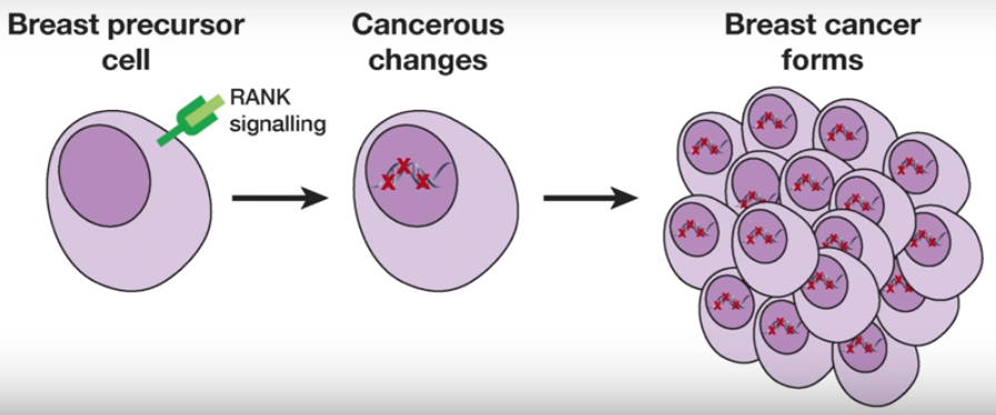
Breast cancer formation (credit: Walter and Eliza Hall Institute)

Australian researchers have discovered that an existing medication could have promise in preventing breast cancer in women carrying a faulty BRCA1 gene, who are at high risk of developing aggressive breast cancer.

Currently, many women with this mutation choose surgical removal of breast tissue and ovaries to reduce their chance of developing breast and ovarian cancer. Notably, in May 2013, actress Angelina Jolie, who reportedly had with an estimated 87 per cent risk of breast cancer and 50 per cent risk of ovarian cancer, chose to have d2ouble mastectomy with breast reconstruction.

Women with mutation have an approximately 65% cumulative risk of developing breast cancer by age 70, the researchers note, based on a 2003 combined analysis of 22 studies.

A drug option 

But now, another option may be be possible, as 16 scientists (most in Australia) report in an advance online paper in Nature Medicine this week.

The researchers discovered that pre-cancerous cells could be identified by a marker protein called RANK. A concurrent study led by an Austrian group had also identified the importance of RANK.

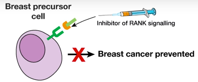
This was an important breakthrough, they said, because an inhibitor of the RANK signalling pathway was already in clinical use: the drug denosumab. The researchers suggest the drug may have potential to prevent breast cancer from developing.

If confirmed in clinical studies, this would provide a non-surgical option to prevent breast cancer in women with elevated genetic risk.

Breast cancer prevention (credit: Walter and Eliza Hall Institute)


“This is potentially a very important discovery for women who carry a faulty BRCA1 gene, who have few other options,” said  Walter and Eliza Hall Institute of Medical Research professor Geoffrey J. Lindeman. “Current cancer prevention strategies for these women include surgical removal of the breasts and/or ovaries, which can have serious impacts on people’s lives.

“To progress this work, denosumab would need to be formally tested in clinical trials in this setting as it is not approved for breast cancer prevention,” he said.

The research was published this week in Nature Medicine and was supported by The National Breast Cancer Foundation, The Qualtrough Cancer Research Fund, The Joan Marshall Breast Cancer Research Fund, the Australian Cancer Research FoundationCancer Council Victoria, the Cancer Therapeutics Cooperative Research Centre, an Amgen Preclinical Research Program Grant, the National Health and Medical Research Council, the Victorian Cancer Agency, and the Victorian Government Operational Infrastructure Support Scheme.


The Walter and Eliza Hall Institute | ‘Holy grail’ of breast cancer prevention in high-risk women may be in sight


Abstract of RANK ligand as a potential target for breast cancer prevention in BRCA1-mutation carriers

Individuals who have mutations in the breast-cancer-susceptibility gene BRCA1 (hereafter referred to as BRCA1-mutation carriers) frequently undergo prophylactic mastectomy to minimize their risk of breast cancer. The identification of an effective prevention therapy therefore remains a ‘holy grail’ for the field. Precancerous BRCA1mut/+ tissue harbors an aberrant population of luminal progenitor cells, and deregulated progesterone signaling has been implicated in BRCA1-associated oncogenesis. Coupled with the findings that tumor necrosis factor superfamily member 11 (TNFSF11; also known as RANKL) is a key paracrine effector of progesterone signaling and that RANKL and its receptor TNFRSF11A (also known as RANK) contribute to mammary tumorigenesis, we investigated a role for this pathway in the pre-neoplastic phase of BRCA1-mutation carriers. We identified two subsets of luminal progenitors (RANK+ and RANK) in histologically normal tissue of BRCA1-mutation carriers and showed that RANK+ cells are highly proliferative, have grossly aberrant DNA repair and bear a molecular signature similar to that of basal-like breast cancer. These data suggest that RANK+ and not RANK progenitors are a key target population in these women. Inhibition of RANKL signaling by treatment with denosumab in three-dimensional breast organoids derived from pre-neoplasticBRCA1mut/+ tissue attenuated progesterone-induced proliferation. Notably, proliferation was markedly reduced in breast biopsies from BRCA1-mutation carriers who were treated with denosumab. Furthermore, inhibition of RANKL in a Brca1-deficient mouse model substantially curtailed mammary tumorigenesis. Taken together, these findings identify a targetable pathway in a putative cell-of-origin population in BRCA1-mutation carriers and implicate RANKL blockade as a promising strategy in the prevention of breast cancer.

Could deep-learning systems radically transform drug discovery?

(credit: Insilico Medicine)

Scientists at Insilico Medicine have developed a new drug-discovery engine that they say is capable of predicting therapeutic use, toxicity, and adverse effects of thousands of molecules, and they plan to reveal it at the Re-Work Machine Intelligence Summit in Berlin, June 29–30.

Drug discovery takes decades, with high failure rates. Among the reasons: irreproducible experiments with poor choice of animal models and inability to translate the results from animal models directly to humans, the wide variety of diseases, and communication difficulties between scientists, managers, venture capitalists, pharmaceutical companies and regulators. And perhaps the biggest problem: the slow-paced, bureaucratic culture in the pharmaceutical industry, the researchers note.

Radically transforming pharmas with AI

Insilico Medicine says it aims to address these reasons by developing “multimodal deep-learned and parametric biomarkers,” as well as multiple drug-scoring pipelines for drug discovery and drug repurposing, and hypothesis and lead generation.

“At Insilico, we want to radically transform the pharmaceutical industry and double the number of drugs on the market, using artificial intelligence and deep understanding of pharmaceutical R&D processes,” said Polina Mamoshina*, senior research scientist at Insilico Medicine, Inc.

“We decided to start with nutraceuticals and cosmetics, but soon we will be announcing our cancer immunology concomitant drug discovery engine to boost the response rates to checkpoint inhibitors in immuno-oncology.”

“Using our drug discovery engine, we made thousands of hypotheses and narrowed these down to 800 strong molecule-disease predictions, with efficacy, toxicity, adverse effects, bioavailability and many other parameters,” said Alex Aliper, president of Insilico Medicine, Inc.

“We added many drug scoring mechanisms that further validate the initial predictions and put together a team of analysts to research and evaluate individual molecules. We are now partnering with various institutions to validate these predictions in vitro and in vivo.”

As KurzweilAI reported, earlier this month, Insilico Medicine signed an exclusive agreement with Life Extension, a major nutraceutical product vendor, to collaboratively develop a set of geroprotectors — natural products that mimic the healthy young state in multiple old tissues. The goal is to increase the rejuvenation rate of the body and slow down, or even reverse, the aging process.

Polina Mamoshina was the lead author on the paper, “Applications of Deep Learning in Biomedicine” in Molecular Pharmaceutics and contributed to another publication, “Deep learning applications for predicting pharmacological properties of drugs and drug repurposing using transcriptomic data” also in Molecular Pharmaceutics. The later paper received the Editors’ Choice Award from the American Chemical Society. She also co-authored a paper, “Deep biomarkers of human aging: Application of deep neural networks to biomarker development” in Aging, one of the highest-impact journals in aging research.