By inserting a small “coralyne” molecule into DNA, scientists were able to create a single-molecule diode (connected here by two gold electrodes), which can be used as an active element in future nanoscale circuits. The diode circuit symbol is shown on the left. (credit: University of Georgia and Ben-Gurion University)
Nanoscale electronic components can be made from single DNA molecules, as researchers at the University of Georgia and at Ben-Gurion University in Israel have demonstrated, using a single molecule of DNA to create the world’s smallest diode.
DNA double helix with base pairs (credit: National Human Genome Research Institute)
A diode is a component vital to electronic devices that allows current to flow in one direction but prevents its flow in the other direction. The development could help stimulate development of DNA components for molecular electronics.
As noted in an open-access Nature Chemistry paper published this week, the researchers designed a 11-base-pair (bp) DNA molecule and inserted a small molecule named coralyne into the DNA.*
They found, surprisingly, that this caused the current flowing through the DNA to be 15 times stronger for negative voltages than for positive voltages, a necessary feature of a diode.
Electronic elements 1,00o times smaller than current components
“Our discovery can lead to progress in the design and construction of nanoscale electronic elements that are at least 1,000 times smaller than current components,” says the study’s lead author, Bingqian Xu an associate professor in the UGA College of Engineering and an adjunct professor in chemistry and physics.
The research team plans to enhance the performance of the molecular diode and construct additional molecular devices, which may include a transistor (similar to a two-layer diode, but with one additional layer).
A theoretical model developed by Yanantan Dubi of Ben-Gurion University indicated the diode-like behavior of DNA originates from the bias voltage-induced breaking of spatial symmetry inside the DNA molecule after the coralyne is inserted.
The research is supported by the National Science Foundation.
*“We prepared the DNA–coralyne complex by specifically intercalating two coralyne molecules into a custom-designed 11-base-pair (bp) DNA molecule (5′-CGCGAAACGCG-3′) containing three mismatched A–A base pairs at the centre,” according to the authors.
UPDATE April 6, 2016 to clarify the coralyne intercalation (insertion) into the DNA molecule.
Abstract of Molecular rectifier composed of DNA with high rectification ratio enabled by intercalation
The predictability, diversity and programmability of DNA make it a leading candidate for the design of functional electronic devices that use single molecules, yet its electron transport properties have not been fully elucidated. This is primarily because of a poor understanding of how the structure of DNA determines its electron transport. Here, we demonstrate a DNA-based molecular rectifier constructed by site-specific intercalation of small molecules (coralyne) into a custom-designed 11-base-pair DNA duplex. Measured current–voltage curves of the DNA–coralyne molecular junction show unexpectedly large rectification with a rectification ratio of about 15 at 1.1 V, a counter-intuitive finding considering the seemingly symmetrical molecular structure of the junction. A non-equilibrium Green’s function-based model—parameterized by density functional theory calculations—revealed that the coralyne-induced spatial asymmetry in the electron state distribution caused the observed rectification. This inherent asymmetry leads to changes in the coupling of the molecular HOMO−1 level to the electrodes when an external voltage is applied, resulting in an asymmetric change in transmission.
Two bacterial micro machines in a bacterial cell extend and retract fibers (called pili) that pull cells forward. ATP hydrolysis in the cytoplasm generates the energy to cause an assembler-mechanism adapter protein in the inner membrane (detail shown in white rectangle) to rotate, facilitating the transfer of pilin subunits (small green balls) onto the growing pilus. The process is reversed during retraction. (credit: Yi-Wei Chang et al./Science)
Caltech researchers have used a state-of-the-art imaging technique to capture detailed 3D views of the complex mobile nanomachinery in bacteria for the first time.
Grant Jensen, a professor of biophysics and biology at Caltech and an investigator with the Howard Hughes Medical Institute (HHMI), and his colleagues used a technique called electron cryotomography to capture 3D images of intact cells with a resolution that ranges from 2 to 5 nanometers (for comparison, a whole cell can be several thousand nanometers in diameter).
(credit: Sony Pictures/Marvel Studios/HD Wallpapers)
In a paper published in the March 11 issue of the journal Science, the researchers explain they used an advanced technique to analyze bacteria-cell motility machinery — a structure called the type IVa pilus machine (T4PM). This machine enables a bacterium to move through its environment in a manner similar to how Spider-Man travels between skyscrapers.
The T4PM assembles a long fiber (the pilus) that attaches to a surface like a grappling hook and subsequently retracts, thus pulling the cell forward.
Science Magazine | How the bacterium Myxococcus xanthus makes a grappling hook for propulsion
The making of the bacteria mobility movie
First, the researchers froze the bacteria cells instantaneously to prevent water molecules from rearranging to form ice crystals. That locked the cells in place without damaging their structure. Then, using a transmission electron microscope, the researchers imaged the cells from different angles, producing a series of 2-D images. They then digitally reconstructed these images into a 3-D picture of the cell’s structures (like creating a CT scan). Jensen’s laboratory is one of only a few in the entire world that can do this type of imaging.
The researchers found that the bacteria structure is made up of several parts, including a pore on the outer membrane of the cell, four interconnected ring structures, and a stemlike structure. By systematically imaging mutants, each of which lacked one of the 10 T4PM core components, and comparing these mutants with normal M. xanthus cells, they mapped the locations of all 10 T4PM core components, providing insights into pilus assembly, structure, and function.
Strongest motor known in nature — how it works
“The machine lets M. xanthus, a predatory bacterium, move across a field to form a ‘wolf pack’ with other M. xanthus cells, and hunt together for other bacteria on which to prey,” Jensen says. “T4PMs … generate forces as high as 150 pN to pull the cell forward, making T4PMs the strongest molecular motors known,” the authors note.
Another way that bacteria move about their environment is by employing a flagellum — a long whiplike structure that extends outward from the cell. The flagellum is spun by cellular machinery, creating a sort of propeller that motors the bacterium through a substrate (see Tiny swimming ‘biobots’ propelled by heart cells or magnetic fields).
However, cells that must push through the thick mucus of the intestine, for example, need more powerful versions of these motors, compared to cells that only need enough propeller power to travel through a pool of water.
Heavy-duty vs. light-duty bacterial propellers
In a second work, published in an open-access paper in the online early edition of the Proceedings of the National Academy of Sciences (PNAS) on March 14, Jensen and his colleagues used electron cryotomography to study the differences between heavy-duty and light-duty versions of the bacterial propeller. The 3-D images they captured showed that the varying levels of propeller power among several different species of bacteria can be explained by structural differences in these tiny motors.
In order for the flagellum to act as a propeller, structures in the cell’s motor must apply torque — the force needed to cause an object to rotate—to the flagellum. The researchers found that the high-power motors have additional torque-generating protein complexes that are found at a relatively wide radius from the flagellum. This extra distance provides greater leverage to rotate the flagellum, thus generating greater torque. The strength of the cell’s motor was directly correlated with the number of these torque-generating complexes in the cell.
“These two studies establish a technique for solving the complete structures of large macromolecular complexes in situ, or inside intact cells,” Jensen says. “Other structure determination methods, such as X-ray crystallography, require complexes to be purified out of cells, resulting in loss of components and possible contamination. On the other hand, traditional 2-D imaging alone doesn’t let you see where individual protein pieces fit in the complete structure. Our electron cryotomography technique is a good solution because it can be used to look at the whole cell, providing a complete picture of the architecture and location of these structures.”
The work involving the type IVa pilus machinery was published in a Science paper by research scientists at Caltech, the Max Planck Institute for Terrestrial Microbiology, and the University of Utah. The study was funded by the National Institutes of Health (NIH), HHMI, the Max Planck Society, and the Deutsche Forschungsgemeinschaft.
Work involving the flagellum machinery in the second study was published in a PNAS paper. Additional coauthors include collaborators from Imperial College London; the University of Texas Southwestern Medical Center; and the University of Wisconsin–Madison. The study was supported by funding from the UK’s Biotechnology and Biological Sciences Research Council and from HHMI and NIH.
UPDATE April 1, 2016: Added quote from Science paper regarding forces generated by the type IVa pilus machine.
Abstract of Architecture of the type IVa pilus machine
Many bacteria, including important pathogens, move by projecting grappling-hook–like extensions called type IV pili from their cell bodies. After these pili attach to other cells or objects in their environment, the bacteria retract the pili to pull themselves forward. Chang et al. used electron cryotomography of intact cells to image the protein machines that extend and retract the pili, revealing where each protein component resides. Putting the known structures of the individual proteins in place like pieces of a three-dimensional puzzle revealed insights into how the machine works, including evidence that ATP hydrolysis by cytoplasmic motors rotates a membrane-embedded adaptor that slips pilin subunits back and forth from the membrane onto the pilus.
Abstract of Diverse high-torque bacterial flagellar motors assemble wider stator rings using a conserved protein scaffold
Although it is known that diverse bacterial flagellar motors produce different torques, the mechanism underlying torque variation is unknown. To understand this difference better, we combined genetic analyses with electron cryo-tomography subtomogram averaging to determine in situ structures of flagellar motors that produce different torques, from Campylobacter and Vibrio species. For the first time, to our knowledge, our results unambiguously locate the torque-generating stator complexes and show that diverse high-torque motors use variants of an ancestrally related family of structures to scaffold incorporation of additional stator complexes at wider radii from the axial driveshaft than in the model enteric motor. We identify the protein components of these additional scaffold structures and elucidate their sequential assembly, demonstrating that they are required for stator-complex incorporation. These proteins are widespread, suggesting that different bacteria have tailored torques to specific environments by scaffolding alternative stator placement and number. Our results quantitatively account for different motor torques, complete the assignment of the locations of the major flagellar components, and provide crucial constraints for understanding mechanisms of torque generation and the evolution of multiprotein complexes.
The duration of epigenetic responses underpinning transgenerational inheritance is determined by an active mechanism relying on the production of small RNAs and modulation of RNAi factors, dictating whether ancestral RNAi responses would be memorized or forgotten (credit: Leah Houri-Ze’evi et al./Cell)
According to epigenetics — the study of inheritable changes in gene expression not directly coded in our DNA — our life experiences may be passed on to our children and our children’s children. Studies on survivors of traumatic events have suggested that exposure to stress may indeed have lasting effects on subsequent generations.
But exactly how are these genetic “memories” passed on?
A new Tel Aviv University (TAU ) study published last week in Cell now pinpoints the precise mechanism that turns the inheritance of these environmental influences “on” and “off.”
How specific genes and small RNAs control epigenetic inheritance
Interactions between different heritable RNAi responses, and the feedback loop that times the duration of transgenerational silencing (credit: Leah Houri-Ze’evi et al./Cell)
“Until now, it has been assumed that a passive dilution or decay governs the inheritance of epigenetic responses,” said Oded Rechavi, PhD, from TAU’s Faculty of Life Sciences and Sagol School of Neuroscience. “But we showed that there is an active process that regulates epigenetic inheritance down through generations.”
The scientists discovered that specific genes, which they named “MOTEK” (Modified Transgenerational Epigenetic Kinetics), were involved in turning epigenetic transmissions on and off.
“We discovered how to manipulate the transgenerational duration of epigenetic inheritance in worms by switching ‘on’ and ‘off’ the small RNAs that worms use to regulate [these] genes,” said Rechavi.*
These switches are controlled by a feedback interaction between gene-regulating small RNAs, which are inheritable, and the MOTEK genes that are required to produce and transmit these small RNAs across generations.
The feedback determines whether epigenetic memory will continue to the progeny or not, and how long each epigenetic response will last.
The researchers are now planning to study the MOTEK genes to know exactly how these genes affect the duration of epigenetic effects, and whether similar mechanisms exist in humans.
* Rechavi and his team had previously identified a “small RNA inheritance” mechanism through which RNA molecules produced a response to the needs of specific cells and how they were regulated between generations.
“We previously showed that worms inherited small RNAs following the starvation and viral infections of their parents. These small RNAs helped prepare their offspring for similar hardships,” Dr. Rechavi said. “We also identified a mechanism that amplified heritable small RNAs across generations, so the response was not diluted. We found that enzymes called RdRPs are required for re-creating new small RNAs to keep the response going in subsequent generations.”
Most inheritable epigenetic responses in C.elegans worms were found to persist for only a few generations. This created the assumption that epigenetic effects simply “petered out” over time, through a process of dilution or decay.
“But this assumption ignored the possibility that this process doesn’t simply die out but is regulated instead,” said Dr. Rechavi, who in this study treated C.elegans worms with small RNAs that target the GFP (green fluorescent protein), a reporter gene commonly used in experiments. “By following heritable small RNAs that regulated GFP — that “silenced” its expression — we revealed an active, tuneable inheritance mechanism that can be turned ‘on’ or ‘off.’”
Abstract of A Tunable Mechanism Determines the Duration of the Transgenerational Small RNA Inheritance in C. elegans
In C. elegans, small RNAs enable transmission of epigenetic responses across multiple generations. While RNAi inheritance mechanisms that enable “memorization” of ancestral responses are being elucidated, the mechanisms that determine the duration of inherited silencing and the ability to forget the inherited epigenetic effects are not known. We now show that exposure to dsRNA activates a feedback loop whereby gene-specific RNAi responses dictate the transgenerational duration of RNAi responses mounted against unrelated genes, elicited separately in previous generations. RNA-sequencing analysis reveals that, aside from silencing of genes with complementary sequences, dsRNA-induced RNAi affects the production of heritable endogenous small RNAs, which regulate the expression of RNAi factors. Manipulating genes in this feedback pathway changes the duration of heritable silencing. Such active control of transgenerational effects could be adaptive, since ancestral responses would be detrimental if the environments of the progeny and the ancestors were different.
A schematic representation of the miniaturized gold-aluminum oxide hyperbolic metamaterial (HMM) sensor device with a fluid flow channel, showing a scanning electron microscope (SEM) image [gray inset] of the 2D subwavelength gold diffraction grating on top of the hyperbolic metamaterials layers (scale bar, 2 µm) (credit: Kandammathe Valiyaveedu Sreekanth et al./Nature Materials)
An optical sensor that’s 1 million times more sensitive than the current best available has been developed by Case Western Reserve University researchers. Based on nanostructured metamaterials, it can identify a single lightweight molecule in a highly dilute solution.
The research goal is to provide oncologists a way to detect a single molecule of an enzyme produced by circulating cancer cells. That could allow doctors to diagnose and monitor patients with certain cancers far earlier than possible today.
“The prognosis of many cancers depends on the stage of the cancer at diagnosis,” said Giuseppe “Pino” Strangi, professor of physics at Case Western Reserve and research leader. “Very early, most circulating tumor cells express proteins of a very low molecular weight, less than 500 Daltons,” Strangi explained. “These proteins are usually too small and in too low a concentration to detect with current test methods, yielding false negative results.
“With this platform, we’ve detected proteins of 244 Daltons, which should enable doctors to detect cancers earlier — we don’t know how much earlier yet,” he said. “This biosensing platform may help to unlock the next era of initial cancer detection.”
The researchers believe the sensing technology will also be useful in diagnosing and monitoring other diseases.
A biological sieve
The nanosensor, which fits in the palm of a hand, acts like a biological sieve, capable of isolating a small protein molecule weighing less than 800 quadrillionths of a nanogram from an extremely dilute solution.
To make the device so sensitive, Strangi’s team faced two long-standing barriers: Light waves cannot detect objects smaller than their own physical dimensions (about 500 nanometers, depending on wavelength). And molecules in dilute solutions float in Brownian (random) motion and are unlikely to land on the sensor’s surface.
The solution was to use a microfluidic channel to restrict the molecules’ ability to float around and a plasmon-based metamaterial made of 16 nanostructured layers of reflective and conductive gold and transparent aluminum oxide, a dielectric, each 10s of atoms thick. Light directed onto and through the layers is concentrated into a very small volume much smaller than the wavelength of light.*
“It’s extremely sensitive,” Strangi said. “When a small molecule lands on the surface, it results in a large local modification, causing the light to shift.” Depending on the size of the molecule, the reflecting light shifts different amounts. The researchers hope to learn to identify specific biomarker and other molecules for different cancers by their light shifts.
To add specificity to the sensor, the team added a layer of trap molecules — molecules that bind specifically with the molecules they hunt. In tests, the researchers used two trap molecules to catch two different biomolecules: bovine serum albumin, with a molecular weight of 66,430 Daltons, and biotin, with a molecular weight of 244 Daltons. Each produced a signature light shift.
Other researchers have reported using plasmon-based biosensors to detect biotin in solutions at concentrations ranging from more than 100 micromoles per liter to 10 micromoles per liter. This device proved 1 million times more sensitive, finding and identifying biotin at a concentration of 10 picomoles per liter.
Testing and clinical use in process
Strangi’s lab is working with other oncologists worldwide to test the device and begin moving the sensor toward clinical use.
In Cleveland, Strangi and Nima Sharifi, MD, co-leader of the Genitourinary Cancer Program for the Case Comprehensive Cancer Center, have begun testing the sensor with proteins related to prostate cancers.
“For some cancers, such as colorectal and pancreatic cancer, early detection is essential,” said Sharifi, who is also the Kendrick Family Chair for Prostate Cancer Research at Cleveland Clinic. “High sensitivity detection of cancer-specific proteins in blood should enable detection of tumors when they are at an earlier disease stage.
“This new sensing technology may help us not only detect cancers, but what subset of cancer, what’s driving its growth and spread, and what it’s sensitive to,” he said. “The sensor, for example, may help us determine markers of aggressive prostate cancers, which require treatments, or indolent forms that don’t.”
The research is published online in the journal Nature Materials.
* The top gold layer is perforated with holes, creating a grating that diffuses light shone on the surface into two dimensions. The incoming light, which is several hundreds of nanometers in wavelength, appears to be confined and concentrated in a few nanometers at the interface between the gold and the dielectric layer. As the light strikes the sensing area, it excites free electrons causing them to oscillate and generate a highly confined propagating surface wave, called a surface plasmon polariton. This propagating surface wave will in turn excite a bulk wave propagating across the sensing platform. The presence of the waves cause deep sharp dips in the spectrum of reflecting light. The combination and the interplay of surface plasmon and bulk plasmon waves are what make the sensor so sensitive. Strangi said. By exciting these waves through the eight bilayers of the metamaterial, they create remarkably sharp resonant modes. Extremely sharp and sensitive resonances can be used to detect smaller objects.
Abstract of Extreme sensitivity biosensing platform based on hyperbolic metamaterials
Optical sensor technology offers significant opportunities in the field of medical research and clinical diagnostics, particularly for the detection of small numbers of molecules in highly diluted solutions. Several methods have been developed for this purpose, including label-free plasmonic biosensors based on metamaterials. However, the detection of lower-molecular-weight (<500 Da) biomolecules in highly diluted solutions is still a challenging issue owing to their lower polarizability. In this context, we have developed a miniaturized plasmonic biosensor platform based on a hyperbolic metamaterial that can support highly confined bulk plasmon guided modes over a broad wavelength range from visible to near infrared. By exciting these modes using a grating-coupling technique, we achieved different extreme sensitivity modes with a maximum of 30,000 nm per refractive index unit (RIU) and a record figure of merit (FOM) of 590. We report the ability of the metamaterial platform to detect ultralow-molecular-weight (244 Da) biomolecules at picomolar concentrations using a standard affinity model streptavidin–biotin.
Setup for delivering spatio-temporally precise light stimulation of optogenetic proteins expressed in tadpole embryo induced tumor-like structures. (credit: Brook T. Chernet et al./Oncotarget)
Tufts University biologists have demonstrated (using a frog model*) for the first time that it is possible to prevent tumors from forming (and to normalize tumors after they have formed) by using optogenetics (light) to control bioelectrical signalling among cells.
Light/bioelectric control of tumors
Virtually all healthy cells maintain a more negative voltage in the cell interior compared with the cell exterior. But the opening and closing of ion channels in the cell membrane can cause the voltage to become more positive (depolarizing the cell) or more negative (polarizing the cell). That makes it possible to detect tumors by their abnormal bioelectrical signature before they are otherwise apparent.
The study was published online in an open-access paper in Oncotarget on March 16.
The use of light to control ion channels has been a ground-breaking tool in research on the nervous system and brain, but optogenetics had not yet been applied to cancer.
Optogenetics modulation of membrane voltage to control induced tumor-like structures. (Top) Tumor induced in tadpole embryo. (Bottom left) Control embryo not injected with light-sensitive protein is highly fluorescent, indicating relative depolarization. (Bottom right) Embryo injected with light-sensitive protein exhibits hyperpolarization, significantly lowering the incidence of tumor formation. Scale bar = 150 micrometers. (credit: Brook T. Chernet et al./Oncotarget)
The researchers first injected cells in Xenopus laevis (frog) embryos with RNA that encoded a mutant RAS oncogene known to cause cancer-like growths.
The researchers then used blue light to activate positively charged ion channels,which induced an electric current that caused the cells to go from a cancer-like depolarized state to a normal, more negative polarized state. The did the same with a green light-activated proton pump, Archaerhodopsin (Arch). Activation of both agents significantly lowered the incidence of tumor formation and also increased the frequency with which tumors regressed into normal tissue.
“Discovering new ways to specifically control this bioelectrical signaling could be an important path towards new biomedical approaches to cancer. This provides proof of principle for a novel class of therapies which use light to override the action of oncogenic mutations,” said Levin. “Using light to specifically target tumors would avoid subjecting the whole body to toxic chemotherapy or similar reagents.”
This work was supported by the G. Harold and Leila Y. Mathers Charitable Foundation.
* Frogs are a good model for basic science research into cancer because tumors in frogs and mammals share many of the same characteristics. These include rapid cell division, tissue disorganization, increased vascular growth, invasiveness and cells that have an abnormally positive internal electric voltage.
Abstract of Use of genetically encoded, light-gated ion translocators to control tumorigenesis
It has long been known that the resting potential of tumor cells is depolarized relative to their normal counterparts. More recent work has provided evidence that resting potential is not just a readout of cell state: it regulates cell behavior as well. Thus, the ability to control resting potential in vivo would provide a powerful new tool for the study and treatment of tumors, a tool capable of revealing living-state physiological information impossible to obtain using molecular tools applied to isolated cell components. Here we describe the first use of optogenetics to manipulate ion-flux mediated regulation of membrane potential specifically to prevent and cause regression of oncogene-induced tumors. Injection of mutant-KRAS mRNA induces tumor-like structures with many documented similarities to tumors, in Xenopus tadpoles. We show that expression and activation of either ChR2D156A, a blue-light activated cation channel, or Arch, a green-light activated proton pump, both of which hyperpolarize cells, significantly lowers the incidence of KRAS tumor formation. Excitingly, we also demonstrate that activation of co-expressed light-activated ion translocators after tumor formation significantly increases the frequency with which the tumors regress in a process called normalization. These data demonstrate an optogenetic approach to dissect the biophysics of cancer. Moreover, they provide proof-of-principle for a novel class of interventions, directed at regulating cell state by targeting physiological regulators that can over-ride the presence of mutations.
An artist’s interpretation of how bacteria cells on a nanoporous gold disk array are killed after exposure to near-infrared light (credit: Greggy M. Santos and Wei-Chuan Shih)
Researchers at the University of Houston have developed a new technique for killing bacteria in 5 to 25 seconds using highly porous gold nanodisks and light, according to a study published today in Optical Materials Express. The method could one day help hospitals treat some common infections without using antibiotics, which could help reduce the risk of spreading antibiotics resistance.
Gold nanoparticles are used because they absorb light strongly, converting the photons quickly into heat and reaching temperatures hot enough to destroy various types of nearby cells — including cancer and bacterial cells. Scientists create gold nanoparticles in the lab by dissolving gold, reducing the metal into smaller and smaller disconnected pieces until the size must be measured in nanometers. Once miniaturized, the particles can be crafted into various shapes.
In 2013, corresponding author Wei-Chuan Shih, a professor in the electrical and computer engineering department, and his colleagues created a new type of gold nanoparticle in the form of discs riddled with pores, lending the particles a sponge-like look that helps increase their heating efficiency while maintaining their stability, said Shih.
Zapping with light too
In the new work, the researchers set out to test the antimicrobial properties of their new nanoparticles when activated by light. They grew bacteria in the lab including E. coli and two types of heat-resistant bacteria that thrive in even the most scorching environments such as the hot springs of Yellowstone National Park.
Then, they placed the bacteria cells on surface of a single-layer coating of the tiny disks and shone near infrared light from a laser on them. Afterward, they used cell viability tests and SEM imaging to see what percentage of cells survived the procedure.
These are SEM images of (a, b and c), B. subtilis (d, e and f) and Exiguobacterium sp. AT1b (g, h and I) cells deposited on nanoporous gold disk arrays. Bacteria cells in images A, D and G were not exposed to near-infrared light. All other cells were exposed for 25 seconds. (credit: Greggy M. Santos and Wei-Chuan Shih)
Using a thermal imaging camera, the research team showed that the surface temperature of the particles reached temperatures up to 180 degrees Celsius nearly instantaneously, “delivering thermal shocks” into the surrounding array — killing all of the bacterial cells within 25 seconds, the researchers report.
E. coli proved most vulnerable to the treatment; all of its cells were dead after only five seconds of laser exposure. The other two types of bacteria required the full 25 seconds, but that’s still much quicker than traditional sterilization methods such as boiling water or using dry-heat ovens, which can take minutes to an hour to work, said Shih. And it’s “considerably shorter” than what other nanoparticle arrays have demonstrated in recent studies, the researchers write. The time needed to achieve similar levels of cell death in those studies ranges from 1 to 20 minutes.
In control trials, the researchers found that neither the gold disks nor light from the laser alone killed nearly as many cells.
Hospital use
The technique has important potential biomedical applications, said Shih. Currently, the researchers are investigating using the particles as a simple coating for catheters to help reduce the number of urinary tract infections in hospitals.
“Any sort of light activated procedure would be much easier to implement at the bedside of a patient,” instead of removing and potentially replacing the catheter every time it needs to be cleaned, he said.
Another potential application they’re exploring is integrating the nanoparticles with filter membranes in small water filters, he said, to help improve water quality.
Abstract of Photothermal inactivation of heat-resistant bacteria on nanoporous gold disk arrays
A rapid photothermal bacterial inactivation technique has been developed by irradiating near-infrared (NIR) light onto bacterial cells (Escherichia coli,Bacillus subtilis, Exiguobacterium sp. AT1B) deposited on surfaces coated with a dense, random array of nanoporous gold disks (NPGDs). With the use of cell viability tests and SEM imaging results, the complete inactivation of the pathogenic and heat-resistant bacterial model strains is confirmed within ~25 s of irradiation of the NPGD substrate. In addition to irradiation control experiments to prove the efficacy of the bacterial inactivation, thermographic imaging showed an immediate averaged temperature rise above 200 °C within the irradiation spot of the NPGD substrate. The light-gated photothermal effects on the NPGD substrate offers potential applications for antimicrobial and nanotherapeutic devices due to strong light absorption in the tissue optical window, i.e., the NIR wavelengths, and robust morphological structure that can withstand high instantaneous thermal shocks.
Researchers have reversed “primed” (developed) “epiblast” stem cells (top) from early mouse embryos using the drug MM-401, causing the treated cells (bottom) to revert to the original form of the stem cells. (credit: University of Michigan)
University of Michigan Medical School researchers have discovered a way to convert mouse stem cells (taken from an embryo) that have become “primed” (reached the stage where they can differentiate, or develop into every specialized cell in the body) to a “naïve” (unspecialized) state by simply adding a drug.
This breakthrough has the potential to one day allow researchers to avoid the ethically controversial use of human embryos left over from infertility treatments. To achieve this breakthrough, the researchers treated the primed embryonic stem cells (“EpiSC”) with a drug called MM-401* (a leukemia drug) for a short period of time.
Embryonic stem cells are able to develop into any type of cell, except those of the placenta (credit: Mike Jones/CC)
Reverting back to the embryonic state
As the research team reports in the journal Cell Stem Cell, this drug treatment caused more than half of these EpiSC cells to return to a naïve (less specialized) state as “reverted embryonic stem cells” (rESC). The researchers then bred healthy mice from those reverted rESC cells — proving that the drug-treated cells were still viable and had the ability to become any type of cell (achieved pluripotency).
This study is significant because it’s the first time scientists have been able to make stem cells revert to their original state without complications. Also, the drug leaves no trace behind, whereas genetic modification of the stem cells (used by other researchers) may block the stem cells from developing into healthy cells. The researchers only needed to treat the EpiSCs with a single drug and for just a few days.
Past attempts by other teams to return mouse EpiSC cells to the original naïve state have either resulted in a far lower proportion of cells returned to a reverted state, or have produced cells that were not viable. Those past studies also needed to use cocktails of multiple drugs given over the long term.
The work was funded by the National Institutes of Health and the Leukemia and Lymphoma Society.
Human stem cells next
Colonies of human naïve embryonic stem cells grown on mouse feeder cells (credit: Ge Guo)
Meanwhile, scientists at the University of Cambridge have also just reported (in an open-access paper in the journal Stem Cell Reports) that they have also produced stem cells directly from embryos — but they did it with human stem cells (for the first time).
“Until now it hasn’t been possible to isolate these naïve stem cells, even though we’ve had the technology to do it in mice for thirty years — leading some people to doubt it would be possible,” says Ge Guo, the study’s first author and research associate in the Stem Cell Potency group at Cambridge.
“But we’ve managed to extract the cells and grow them individually in culture. Naïve stem cells have many potential applications, from regenerative medicine to modeling human disorders.”
Jenny Nichols, PhD, joint senior author of the study, says that one of the most exciting applications of their new technique would be to study disorders that arise from cells that contain an abnormal number of chromosomes. Ordinarily, the body contains 23 pairs of identical chromosomes (22 pairs and one pair of sex chromosomes), but some children are born with additional copies, which can cause problems. For example, children with Down’s syndrome are born with three copies of chromosome 21.
“Even in many ‘normal’ early-stage embryos, we find several cells with an abnormal number of chromosomes,” explains Nichols. “Because we can separate the cells and culture them individually, we could potentially generate ‘healthy’ and ‘affected’ cell lines. This would allow us to generate and compare tissues of two models, one ‘healthy’ and one that is genetically-identical other than the surplus chromosome. This could provide new insights into conditions such as Down’s syndrome.”
The research was supported by the Medical Research Council, Biotechnology and Biological Sciences Research Council, Swiss National Science Foundation, and the Wellcome Trust.
* The drug, MM-401, specifically targets epigenetic chemical markers on histones, the protein “spools” that DNA coils around to create structures called chromatin. These epigenetic changes signal the cell’s DNA-reading machinery and tell it where to start uncoiling the chromatin in order to read it.
A gene called Mll1 is responsible for the addition of these epigenetic changes, which are like small chemical tags called methyl groups. Mll1 plays a key role in the uncontrolled explosion of white blood cells in leukemia, which is why researchers developed the drug MM-401 to interfere with this process. But Mll1 also plays a role in cell development and the formation of blood cells and other cells in later-stage embryos.
Stem cells do not turn on the Mll1 gene until they are more developed. The MM-401 drug blocks Mll1’s normal activity in developing cells so the epigenetic chemical markers are missing. These cells are then unable to continue to develop into different types of specialized cells but are still able to revert to healthy naive pluripotent stem cells.
Abstract of MLL1 Inhibition Reprograms Epiblast Stem Cells to Naive Pluripotency
The interconversion between naive and primed pluripotent states is accompanied by drastic epigenetic rearrangements. However, it is unclear whether intrinsic epigenetic events can drive reprogramming to naive pluripotency or if distinct chromatin states are instead simply a reflection of discrete pluripotent states. Here, we show that blocking histone H3K4 methyltransferase MLL1 activity with the small-molecule inhibitor MM-401 reprograms mouse epiblast stem cells (EpiSCs) to naive pluripotency. This reversion is highly efficient and synchronized, with more than 50% of treated EpiSCs exhibiting features of naive embryonic stem cells (ESCs) within 3 days. Reverted ESCs reactivate the silenced X chromosome and contribute to embryos following blastocyst injection, generating germline-competent chimeras. Importantly, blocking MLL1 leads to global redistribution of H3K4me1 at enhancers and represses lineage determinant factors and EpiSC markers, which indirectly regulate ESC transcription circuitry. These findings show that discrete perturbation of H3K4 methylation is sufficient to drive reprogramming to naive pluripotency.
Abstract of Naive Pluripotent Stem Cells Derived Directly from Isolated Cells of the Human Inner Cell Mass
Conventional generation of stem cells from human blastocysts produces a developmentally advanced, or primed, stage of pluripotency. In vitro resetting to a more naive phenotype has been reported. However, whether the reset culture conditions of selective kinase inhibition can enable capture of naive epiblast cells directly from the embryo has not been determined. Here, we show that in these specific conditions individual inner cell mass cells grow into colonies that may then be expanded over multiple passages while retaining a diploid karyotype and naive properties. The cells express hallmark naive pluripotency factors and additionally display features of mitochondrial respiration, global gene expression, and genome-wide hypomethylation distinct from primed cells. They transition through primed pluripotency into somatic lineage differentiation. Collectively these attributes suggest classification as human naive embryonic stem cells. Human counterparts of canonical mouse embryonic stem cells would argue for conservation in the phased progression of pluripotency in mammals.
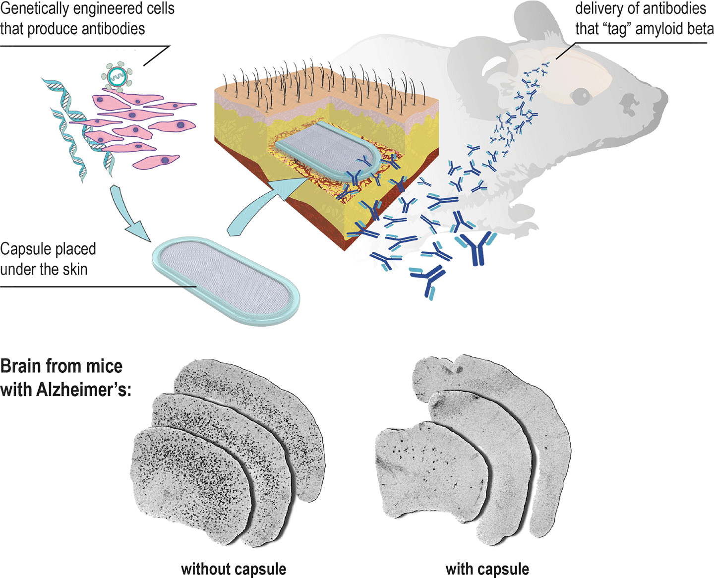
An implant that can prevent Alzheimer’s disease. A new capsule can be implanted under the skin to release antibodies that “tag” amyloid beta, signalling the patient’s immune system to clear it before it forms Alzheimer’s plaques. (credit: École polytechnique fédérale de Lausanne)
EPFL scientists have developed an implantable capsule containing genetically engineered cells that can recruit a patient’s immune system to combat Alzheimer’s disease.
Placed under the skin, the capsule releases antibody proteins that make their way to the brain and “tag” amyloid beta proteins, signalling the patient’s own immune system to attack and clear the amyloid beta proteins, which are toxic to neurons.
To be most effective, this treatment has to be given as early as possible, before the first signs of cognitive decline. Currently, this requires repeated vaccine injections, which can cause side effects. The new implant can deliver a steady, safe flow of antibodies.
Protection from immune-system rejection
Cell encapsulation device for long-term subcutaneous therapeutic antibody delivery. (B) Macroscopic view of the encapsulation device, composed of a transparent frame supporting polymer permeable membranes and reinforced with an outer polyester mesh. (C) Dense neovascularization develops around a device containing antibody-secreting C2C12 myoblasts, 8 months after implantation in the mouse subcutaneous tissue. (D and E) Representative photomicrographs showing encapsulated antibody-secreting C2C12 myoblasts surviving at high density within the flat sheet device 39 weeks after implantation. (E) Higher magnification: note that the cells produce a collagen-rich matrix stained in blue with Masson’s trichrome protocol. Asterisk: polypropylene porous membrane. Scale bars = 750 mm (B and C),100 mm (D), 50 mm (E). (credit: Aurelien Lathuiliere et al./BRAIN)
The lab of Patrick Aebischer at EPFL designed the “macroencapsulation device” (capsule) with two permeable membranes sealed together with a polypropylene frame, containing a hydrogel that facilitates cell growth. All the materials used are biocompatible and the device is reproducible for large-scale manufacturing.
The cells of choice are taken from muscle tissue, and the permeable membranes let them interact with the surrounding tissue to get all the nutrients and molecules they need. The cells have to be compatible with the patient to avoid triggering the immune system against them, like a transplant can. To do that, the capsule’s membranes shield the cells from being identified and attacked by the immune system. This protection also means that cells from a single donor can be used on multiple patients.
The researchers tested the device mice in a genetic line commonly used to simulate Alzheimer’s disease over a course of 39 weeks, showing dramatic reduction of amyloid beta plaque load in the brain. The treatment also reduced the phosphorylation of the protein tau, another sign of Alzheimer’s observed in these mice.
“The proof-of-concept work demonstrates clearly that encapsulated cell implants can be used successfully and safely to deliver antibodies to treat Alzheimer’s disease and other neurodegenerative disorders that feature defective proteins,” according to the researchers.
The work is published in the journal BRAIN. It involved a collaboration between EPFL’s Neurodegenerative Studies Laboratory (Brain Mind Institute), the Swiss Light Source (Paul Scherrer Institute), and F. Hoffmann-La Roche. It was funded by the Swiss Commission for Technology and Innovation and F. Hoffmann-La Roche Ltd.
Abstract of A subcutaneous cellular implant for passive immunization against amyloid-β reduces brain amyloid and tau pathologies
Passive immunization against misfolded toxic proteins is a promising approach to treat neurodegenerative disorders. For effective immunotherapy against Alzheimer’s disease, recent clinical data indicate that monoclonal antibodies directed against the amyloid-β peptide should be administered before the onset of symptoms associated with irreversible brain damage. It is therefore critical to develop technologies for continuous antibody delivery applicable to disease prevention. Here, we addressed this question using a bioactive cellular implant to deliver recombinant anti-amyloid-β antibodies in the subcutaneous tissue. An encapsulating device permeable to macromolecules supports the long-term survival of myogenic cells over more than 10 months in immunocompetent allogeneic recipients. The encapsulated cells are genetically engineered to secrete high levels of anti-amyloid-β antibodies. Peripheral implantation leads to continuous antibody delivery to reach plasma levels that exceed 50 µg/ml. In a proof-of-concept study, we show that the recombinant antibodies produced by this system penetrate the brain and bind amyloid plaques in two mouse models of the Alzheimer’s pathology. When encapsulated cells are implanted before the onset of amyloid plaque deposition in TauPS2APP mice, chronic exposure to anti-amyloid-β antibodies dramatically reduces amyloid-β40 and amyloid-β42 levels in the brain, decreases amyloid plaque burden, and most notably, prevents phospho-tau pathology in the hippocampus. These results support the use of encapsulated cell implants for passive immunotherapy against the misfolded proteins, which accumulate in Alzheimer’s disease and other neurodegenerative disorders.
Three bacterial motors with different torque (units of torque are piconewton nanometers, pN nm) (credit: Morgan Beeby/Imperial College London)
A new study of the exotic “motors” that bacteria use to swim reveals details of how they “swim” that may make it possible to design specific drugs that sabotage the flagella (tails) in targeted bacterial species.
Using a newly installed high-powered electron microscope, researchers at Imperial College London, led by Morgan Beeby, PhD from the Department of Life Sciences, has been able visualize these motors in unprecedented detail.
They used a method called electron cryo-tomography to rapidly freeze the bacteria to -180 degrees C — preventing ice crystals from forming that would break the structure apart and allowing the researchers to image the flash-frozen motor from all angles and build up 3D models.
Their visualizations of these motors explain the differences in swimming ability, mathematically accounting for differences in motor power. The work is published in an open-access paper in Proceedings of the National Academy of Sciences.
How bacteria motors work
L-R: Salmonella, Vibrio, Campylobacter (credit: Morgan Beeby/Imperial College London)
Bacterial flagellar motors use a clever rotational mechanism, spinning their long flagellar tail to produce a helical propeller-like motion. The team found that stronger swimmers have evolved by adding extra parts to their motors, making more powerful motors that have increased turning force, or torque.
In flagellar motors, the turning force is produced by a ring of structures called stators around the motor (similar to the structure of electric motors). The team found that Campylobacter jejuni had almost twice as many stators positioned around the motor than in Salmonella, and that these structures sat in a wider ring. More stators provide increased torque, and the increased width of the ring means individual stators exert more leverage when rotating the helical propeller.
However, not all bacteria need to be so powerful and swim through such viscous environments as stomach mucus. Instead, another bacterium the team looked at, a close relative of Vibrio cholerae, the bacterium that causes cholera, has evolved a motor with only intermediate power.
Despite motors in diverse bacteria having the same core structure, different bacteria vary widely in their swimming power. For example, Campylobacter jejuni, which causes food poisoning, can swim powerfully enough to bore through the mucus that lines the gut, an environment too thick and sticky for other bacteria to push through.
The reasons for these differences in swimming ability have remained obscure until now. By looking at distantly related bacteria from different branches of the evolutionary tree, the team speculates that the ability to alter torque in this way may have evolved up to two billion years ago.
“Entire branches of the bacterial family tree have evolved motors with different torques, leading to a diversity of species each geared to their own environment,” said Beeby. The team is now investigating how and when the evolutionary steps that altered motor torque happened.
editor’s comments: Check out the open-access paper: damn impressive engineering study! Hey, could these motor designs be genetically modified (or combined with the Drexel University method from yesterday for using electric fields to help microscopic bacteria-powered robots detect obstacles) to attack other bacteria or be sent out as scouts or on other missions? OK, just a wild and crazy idea. …
Abstract of Diverse high-torque bacterial flagellar motors assemble wider stator rings using a conserved protein scaffold
Although it is known that diverse bacterial flagellar motors produce different torques, the mechanism underlying torque variation is unknown. To understand this difference better, we combined genetic analyses with electron cryo-tomography subtomogram averaging to determine in situ structures of flagellar motors that produce different torques, from Campylobacter and Vibrio species. For the first time, to our knowledge, our results unambiguously locate the torque-generating stator complexes and show that diverse high-torque motors use variants of an ancestrally related family of structures to scaffold incorporation of additional stator complexes at wider radii from the axial driveshaft than in the model enteric motor. We identify the protein components of these additional scaffold structures and elucidate their sequential assembly, demonstrating that they are required for stator-complex incorporation. These proteins are widespread, suggesting that different bacteria have tailored torques to specific environments by scaffolding alternative stator placement and number. Our results quantitatively account for different motor torques, complete the assignment of the locations of the major flagellar components, and provide crucial constraints for understanding mechanisms of torque generation and the evolution of multiprotein complexes.
Electric fields help microscopic bacteria-powered robots detect obstacles in their environment and navigate around them to get to their destination. (credit: Drexel University)
Drexel University engineers have developed a method for using electric fields to help microscopic bacteria-powered robots detect obstacles in their environment and navigate around them. Uses include delivering medication, manipulating stem cells to direct their growth, or building a microstructure, for example.
The method is a follow-up to a 2014 report that presented a way to use the flagellated bacteria Serratia marcescens and an electric field to make a microrobot mobile. These bacteria possess a negative charge, which means they can be manipulated, in this case, with two perpendicular electric fields that turn the fluid into an electrified grid.
Serratia marcescens bacteria are the perfect candidate for use in driving microrobots because they have a natural negative charge, which means they can be manipulated with an electric field, and their flagella reduce friction while helping the robot move in a fluid environment. (credit: Drexel University)
By running a series of tests using charged particles, the team realized how the electric field changed when it encountered insulator objects. “The electric field was distorted near the corners of the obstacle,” the authors write. “Particles that passed by the first corner of the obstacles also had affected trajectories even though they had a clear space ahead to pass; this is due to the distorted electric field.”
They used this deformation in the field as input data for their steering algorithm; the robots are using electric fields both as a mode of transportation and as a means of navigation. The algorithm also uses image-tracking from a microscope-mounted camera to locate the initial starting point of the robot and its ultimate destination.
“With this level of control and input from the environment we can program the microrobot to make a series of value judgments during its journey that affect its path,” said MinJun Kim, PhD, a professor in the College of Engineering and director of Drexel’s Biological Actuation, Sensing & Transport (BAST) Lab. “If, for instance, we want the robot to avoid as many obstacles as possible, regardless of the distance traveled. Or we could set it to take the most direct, shortest route to the destination — even if it’s through the obstacles.”
The next step for Kim’s lab is to develop a system consisting of multiple bacteria-powered microrobots that can manipulate multiple live cells in vitro.
The research was recently published in IEEE Transactions on Robotics.
Abstract of Electric Field Control of Bacteria-Powered Microrobots Using a Static Obstacle Avoidance Algorithm
A bacteria-powered microrobot (BPM) is a hybrid robotic system consisting of an SU-8 microstructure with active surfaces or bacterial carpets, in which massive arrays of biomolecular flagellar motors work cooperatively. This paper suggests an obstacle-avoidance method based on a BPM’s response to electric fields. The negatively charged bacteria enable the BPM to follow electric fields. In our previous demonstration of the single BPM controllability, we observed a vast change in the control dynamics when obstructions distorted the applied electric field and affected BPM steering and control. In this paper, we demonstrate an obstacle avoidance method that takes the electric field distortion into account to navigate a BPM through multiple static obstacles in real time. We used an artificial potential field and configuration space in our algorithm to generate an objective function for the electric field distortion and collision around/with obstacles, respectively. In addition, finite-element modeling through COMSOL Multiphysics engineering software was used to simulate charged-particle trajectories in a distorted electric field. Finally, we describe the feasibility of our proposed obstacle avoidance approach through experiments and compared these data with simulation results.


")
