Scientists grow eye lens from patients’ own stem cells, restoring vision

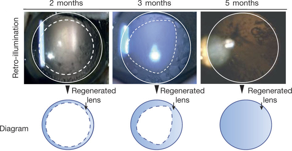
Lens regeneration in monkeys after minimally invasive surgery. Slit-lamp microscopy showed regenerating lens tissue grew from the peripheral to the central lens in a circular symmetrical pattern 2–3 months after surgery, reaching the center at 5 months post-surgery; direct illumination showed that the visual axis remained translucent. (credit: Haotian Lin et al./Nature)

Researchers at University of California, San Diego School of Medicine and Shiley Eye Institute, with colleagues in China, have developed an eye lens restoration treatment that has been tested in monkeys and in a small human clinical trial. It produced much fewer surgical complications than the current standard-of-care and resulted in regenerated lenses with superior visual function in all 12 of the pediatric cataract patients who received the new surgery.

Congenital cataracts — lens clouding that occurs at birth or shortly thereafter — is a significant cause of blindness in children.

Using the patients’ own stem cells
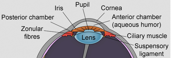
Eye lens (credit: Wikimedia Commons)

In the new research, Kang Zhang*, MD, PhD and colleagues relied on the regenerative potential of the patients’ own lens epithelial stem cells (LECs) at the site of the injury or problem, instead of creating stem cells in the lab and introducing them back into the patient (with potential hurdles like pathogen transmission and immune rejection).

After confirming the regenerative potential of LECs in animal models, the researchers developed a novel minimally invasive surgery method that preserves the integrity of the lens capsule — a membrane that helps give the lens its required shape to function — and also developed a way to stimulate LECs to grow and form a new lens with vision.

They found the new surgical technique allowed pre-existing LECs to regenerate functional lenses, producing a clear, regenerated biconvex lens in all of the patients’ eyes after three months.

Age-related cataracts next

Zhang said he and colleagues are now looking to expand their work to treating age-related cataracts. Age-related cataracts is the leading cause of blindness in the world. More than 20 million Americans suffer from cataracts, and more than 4 million surgeries are performed annually to replace the clouded lens with an artificial plastic version, called an intraocular lens.

Despite technical advances, a large portion of patients undergoing surgery are left with suboptimal vision post-surgery and are dependent upon corrective eyewear for driving a car and/or reading a book. “We believe that our new approach will result in a paradigm shift in cataract surgery and may offer patients a safer and better treatment option in the future.”

The findings are published in the March 9 online issue of Nature. Co-authors on the study include scientists at UC San Diego; Sichuan University, China; Guangzhou KangRui Biological Pharmaceutical Technology Company, China; and University of Texas Southwestern Medical Center.

Funding for this research came, in part, from the 973 Program (National Basic Research Program of China); a Major International Joint Research Project; 863 Program (State High-Tech Development Plan of China); the National Natural Science Foundation of China; the State Key Laboratory of Ophthalmology, Zhongshan Ophthalmic Center, Sun Yatsen University; Research to Prevent Blindness; and the Howard Hughes Medical Institute.

* Chief of Ophthalmic Genetics, founding director of the Institute for Genomic Medicine and co-director of Biomaterials and Tissue Engineering at the Institute of Engineering in Medicine, both at UC San Diego School of Medicine.


Abstract of Lens regeneration using endogenous stem cells with gain of visual function

The repair and regeneration of tissues using endogenous stem cells represents an ultimate goal in regenerative medicine. To our knowledge, human lens regeneration has not yet been demonstrated. Currently, the only treatment for cataracts, the leading cause of blindness worldwide, is to extract the cataractous lens and implant an artificial intraocular lens. However, this procedure poses notable risks of complications. Here we isolate lens epithelial stem/progenitor cells (LECs) in mammals and show that Pax6 and Bmi1 are required for LEC renewal. We design a surgical method of cataract removal that preserves endogenous LECs and achieves functional lens regeneration in rabbits and macaques, as well as in human infants with cataracts. Our method differs conceptually from current practice, as it preserves endogenous LECs and their natural environment maximally, and regenerates lenses with visual function. Our approach demonstrates a novel treatment strategy for cataracts and provides a new paradigm for tissue regeneration using endogenous stem cells.

Dramatic remissions in blood cancer in immunotherapy treatment trial

Recent advances in an immune-cell cancer treatment — a type of immunotherapy* using engineered immune cells to target specific molecules on cancer cells — are producing dramatic results for people with cancer, according to Stanley Riddell, MD, an immunotherapy researcher and oncologist at Seattle’s Fred Hutchinson Cancer Research Center.**

Riddell and his colleagues have refined new methods of engineering a patient’s own immune cells to better target and kill cancer cells while decreasing side effects. In laboratory and clinical trials, the researchers are seeing “dramatic responses” in patients with tumors that are resistant to conventional high-dose chemotherapy, “providing new hope for patients with many different kinds of malignancies,” Riddell said.


Fred Hutch | Immunotherapy shows great promise

Twenty-seven out of 29 patients with an advanced blood cancer who received experimental, “living” immunotherapy as part of a clinical trial experienced sustained remissions, in preliminary results of an ongoing study at Fred Hutchinson Cancer Research Center.

Boosting natural immune response

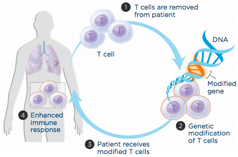
Adoptive T-cell transfer aims to boost a patient’s immune cells’ ability to recognize and attack cancer cells. (1) T cells are extracted from the patient’s blood, (2) genetically engineered to produce a molecule that recognizes cancer cells and grown in the laboratory, and (3) infused back into the patient to (4) improve immune response. (credit: LUNGevity Foundation)

The immune system produces two major types of immune reaction to protect the body: one uses antibodies secreted by B cells; the other uses T cells.

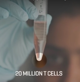
Riddell’s team takes T cells from the patient’s body, re-engineers them, and infuses them back into the patient to create an army of cancer-fighting immune cells. (credit: Fred Hutchinson Cancer Research Center)

T cells are white blood cells that detect foreign or abnormal cells — including cancerous or infected cells — and initiate a process that targets those cells for attack. But the natural immune response to a tumor is often neither potent nor persistent enough, so Riddell and associates pioneered a new way to boost this immune response using a method known as “adoptive T-cell transfer.”

With adoptive T-cell transfer, immune cells are engineered to recognize and attack the patient’s cancer cells. Researchers extract T cells from a patient’s blood and then introduce genes into those T cells so they synthesize highly potent receptors (called chimeric antigen receptors, or CARs) that can recognize and target the cancer cell.

A single treatment of a relatively small number of the re-engineered T cells only takes about 30 minutes, and within weeks, the patient goes into a complete remission. (credit: Fred Hutchinson Cancer Research Center)

They grow the T cells in a laboratory for about two weeks and then infuse the engineered cells back into the patient, where they can home in on the tumor site and destroy the cancer cells.

Sustained remission of B cell cancers

Riddell’s team has recently developed a refined version of this process that increases the effectiveness of the immune response while reducing negative side effects, such as neurological symptoms, fevers, and large decreases in blood pressure.

In a study published in the journal Nature Biotechnology, Riddell and his team describe tagging the potent T-cell receptor (with amino acid sequences called Strep-tag), and the resulting effect on human cancer cells in the laboratory and on a mouse model of lymphoma.

Those results, using the latest version of this experimental immunotherapy, suggest sustained remission in cases of B cell cancers that previously relapsed and had become resistant to treatment.***

“The results are simply astounding,” Riddell said. We are treating patients with advanced leukemia and lymphoma that have failed every conventional therapy and radiation therapy, including transplants … in a single treatment. Within weeks, the patient goes into remission.”

“In my years as a oncologist and as a research scientist, I have never seen a treatment that has that spectacular response rate in its initial testing in patients,” Riddell said. His team is initiating trials in lung, breast, sarcoma, melanoma, and soon in pancreatic cancer. The opportunities for this technology are “incredible” and the approach has the potential to also treat common cancers such as kidney and colon cancer, he said.

“We are at the precipice of a revolution in cancer treatment based on using immunotherapy.”

Funding for Riddell’s research was provided by Juno Therapeutics.

* For approximately 100 years, the main tools to treat cancer were surgery, chemotherapy, and radiation therapy. But since around 2000, doctors have had access to a type of immunotherapy based on engineered antibodies that can target specific molecules on cancer cells. For example, trastuzumab (Herceptin) can be used for some types of breast cancer and stomach cancer. The new treatment approach used by Riddell’s team is based on a new type of immunotherapy using engineered immune cells to kill cancer, rather than antibodies.

** Stanley Riddell. Engineering T cells for safe and effective cancer immunotherapy. 2016 Annual Meeting of the American Association for the Advancement of Science, Washington, D.C., February 2016.

*** Such as acute lymphoblastic leukemia, Non-Hodgkin lymphoma, and chronic lymphocytic leukemia.


Abstract of Acquisition of a CD19 negative myeloid phenotype allows immune escape of MLL-rearranged B-ALL from CD19 CAR-T cell therapy

Administration of lymphodepletion chemotherapy followed by CD19-specific chimeric antigen receptor (CAR)-modified T cells is a remarkably effective approach to treat patients with relapsed and refractory CD19+ B cell malignancies. We treated 7 patients with B-cell acute lymphoblastic leukemia (B-ALL) harboring rearrangement of the mixed lineage leukemia (MLL) gene with CD19 CAR-T cells. All patients achieved complete remission in the bone marrow by flow cytometry after CD19 CAR-T cell therapy; however, within one month of CAR-T cell infusion two of the patients developed acute myeloid leukemia that was clonally related to their B-ALL, a novel mechanism of CD19-negative immune escape. These reports have implications for the management of patients with relapsed and refractory MLL-B-ALL who receive CD19 CAR-T cell therapy.


Abstract of Inclusion of Strep-tag II in design of antigen receptors for T-cell immunotherapy

Adoptive immunotherapy with genetically engineered T cells has the potential to treat cancer and other diseases. The introduction of Strep-tag II sequences into specific sites in synthetic chimeric antigen receptors or natural T-cell receptors of diverse specificities provides engineered T cells with a marker for identification and rapid purification, a method for tailoring spacer length of chimeric receptors for optimal function, and a functional element for selective antibody-coated, microbead-driven, large-scale expansion. These receptor designs facilitate cGMP manufacturing of pure populations of engineered T cells for adoptive T-cell therapies and enable in vivo tracking and retrieval of transferred cells for downstream research applications.

CRISPR enhancements: improving the ability to delete genes

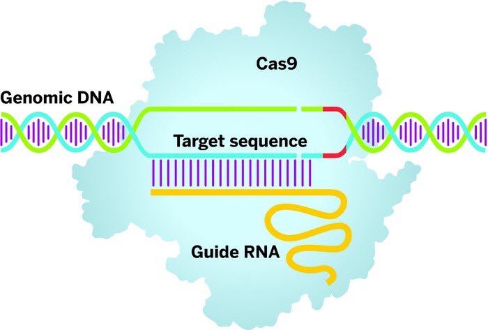
Clustered regularly interspaced short palindromic repeats (CRISPRs) technology employs a guide RNA to direct the Cas9 enzyme (light blue) to a target DNA sequence. Once there, Cas9 will bind when it finds a protospacer-adjacent motif sequence (red) in the DNA and cut both strands, priming the gene sequence for editing. (credit: Adapted from OriGene Technologies)

Scientists have found a way to improve the efficiency of the controversial gene editing technology, CRISPR/Cas9 (“CRISPR”).

Lauded as a groundbreaking technology that allows scientists to modify genes* for many different applications, CRISPR/Cas9 has hit stormy waters over the ethics of editing human embryos. Although the technology is faster and cheaper than past gene editing techniques, one of the problems cited is that the efficiency of deleting unwanted genes is low and that it gives inconsistent results — an unacceptable risk when using human embryos.

Haoquan Wu, PhD, from the Texas Tech University Health Sciences Center El Paso, has worked to improve CRISPR’s overall ability to target and eliminate genes.

In an open-access paper published in Genome Biology, senior author Wu and his team describe how they were able to significantly improve the efficiency of gene knockout (deletion) by creating structural changes to the CRISPR RNA guide molecule.**

“The extent of the improvement in knockout efficiency with these changes was striking,” Wu says. “This is going to help reduce concerns that knockout experiments might not work, and also significantly increase the efficiency of more challenging editing procedures like gene deletion.”

At this stage, the researchers are unsure why the changes to the single guide RNA enhanced CRISPR’s efficiency, although options include an enhanced ability to bind with Cas9 or improved stability of the RNA.

The TTUHSC El Paso team plans to continue studying this modified single guide RNA template to better understand why it enhances CRISPR’s functionality. They’ve also applied for a patent for their new method, which they hope will be adopted by other scientists using CRISPR.

* CRISPR/Cas9 has two main components. A small “single guide” RNA molecule (Clustered Regularly Interspaced Short Palindromic Repeats, CRISPR) directs the Cas9 enzyme to a specific sequence of DNA. The Cas9 then acts like molecular scissors to cut that specific piece of DNA. The DNA is then ready for editing.

** Some technical aspects of CRSIPR-Cas9 are inefficient, including total gene knockout. To improve the efficiency, the researchers optimized the structure of the small-guide RNA by tweaking the sequence and making it slightly longer. The optimized small-guide RNA was far more efficient at producing knockouts — 18 of 24 experiments produced greater than 50 per cent knockout efficiency compared with only four for the original small-guide RNA structure.


Abstract of Optimizing sgRNA structure to improve CRISPR-Cas9 knockout efficiency

Background: Single-guide RNA (sgRNA) is one of the two key components of the clustered regularly interspaced short palindromic repeats (CRISPR)-Cas9 genome-editing system. The current commonly used sgRNA structure has a shortened duplex compared with the native bacterial CRISPR RNA (crRNA)–transactivating crRNA (tracrRNA) duplex and contains a continuous sequence of thymines, which is the pause signal for RNA polymerase III and thus could potentially reduce transcription efficiency.

Results: Here, we systematically investigate the effect of these two elements on knockout efficiency and showed that modifying the sgRNA structure by extending the duplex length and mutating the fourth thymine of the continuous sequence of thymines to cytosine or guanine significantly, and sometimes dramatically, improves knockout efficiency in cells. In addition, the optimized sgRNA structure also significantly increases the efficiency of more challenging genome-editing procedures, such as gene deletion, which is important for inducing a loss of function in non-coding genes.

Conclusions: By a systematic investigation of sgRNA structure we find that extending the duplex by approximately 5 bp combined with mutating the continuous sequence of thymines at position 4 to cytosine or guanine significantly increases gene knockout efficiency in CRISPR-Cas9-based genome editing experiments.

Human-skin discovery suggests new anti-aging treatments

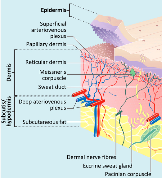
Layers in hairless skin (credit: Madhero88 and M.Komorniczak/Creative Commons)

For the first time, researchers have reported decreases in levels of a key molecule in aging human skin, which could lead to developing new anti-aging treatments and screening new compounds.

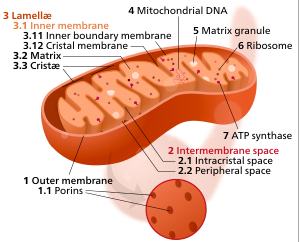
Components of a typical mitochondrion (credit: Kelvinsong/Creative Commons)

Scientists have known for some time that major structures in the cell called mitochondria (which generate and control most of the cell’s supply of energy) are somehow involved in aging, but the exact role of the mitochondria has remained unclear.

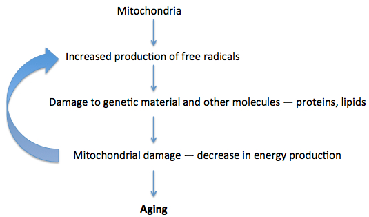
The longstanding “mitochondrial free radical theory of aging,” originally proposed by Professor Denham Harman in 1972, is currently the most widely accepted theory of aging. It proposes that mitochondria contribute to aging by producing free radicals — chemicals that can damage our genetic material and other molecules and so accelerate aging. Free-radical production increases lead to a cycle of further damage and further increases in free radicals.

Mitochondrial free radical theory of aging (credit: Julie Milland)

However, Mark Birch-Machin, PhD, professor of Molecular Dermatology at Newcastle University’s Institute of Cellular Medicine and senior author of an open-access article in the Journal of Investigative Dermatology, now reports a new research finding: a protein in mitochondria called mitochondrial complex II (a key complex for production of energy) decreases with age in human skin. (Further studies are needed to understand whether that decrease actually causes skin aging, or is a result of skin aging, or is otherwise correlated.)

The researchers also found that levels of mitochondrial complex II decrease in the deeper layers of aging human skin.*

Anti-aging treatments and cosmetic products

Birch-Machin says this new discovery brings experts a step closer to developing anti-aging treatments and cosmetic products that could counteract this decrease in mitochondrial complex II in aging human skin, and treatment could even be “tailored to differently aged and differently pigmented skin.”

The findings may also lead to a greater understanding of aging in other parts of the body, and this could pave the way for drug development in a number of age-related diseases, including cancer.

Funding was provided by the North Eastern Skin Research Fund, the NIHR Biomedical Research Centre at Newcastle University and Newcastle Upon Tyne Hospitals NHS Foundation Trust, and Newcastle University’s Faculty of Medical Sciences.

* To test the activity of mitochondrial complex II, skin samples were taken from a sun-protected area of skin from 27 people aged six to 72 years. The mitochondrial complex II activity was measured in keratinocyte cells from the upper layer of skin (epidermis) and in fibroblast cells from the deeper levels of skin (dermis).  The researchers found that mitochondrial complex II activity significantly decreased with age in the fibroblast cells from the deeper skin layer, but not in the keratinocytes from the upper layer of skin. The mitochondrial complex II activity decreased because less of the protein was being synthesized and the decrease was only observed in those cells that had stopped multiplying.


Abstract of Age-Dependent Decrease of Mitochondrial Complex II Activity in Human Skin Fibroblasts

The mitochondrial theory of aging remains one of the most widely accepted aging theories and implicates mitochondrial electron transport chain dysfunction with subsequent increasing free radical generation. Recently, complex II of the electron transport chain appears to be more important than previously thought in this process, suggested predominantly by nonhuman studies. We investigated the relationship between complex II and aging using human skin as a model tissue. The rate of complex II activity per unit of mitochondria was determined in fibroblasts and keratinocytes cultured from skin covering a wide age range. Complex II activity significantly decreased with age in fibroblasts (P = 0.015) but not in keratinocytes. This was associated with a significant decline in transcript expression (P = 0.008 and P = 0.001) and protein levels (P = 0.0006 and P = 0.005) of the succinate dehydrogenase complex subunit A and subunit B catalytic subunits of complex II, respectively. In addition, there was a significant decrease in complex II activity with age (P = 0.029) that was specific to senescent skin cells. There was no decrease in complex IV activity with increasing age, suggesting possible locality to complex II.

Engineered swarmbots rely on peers for survival

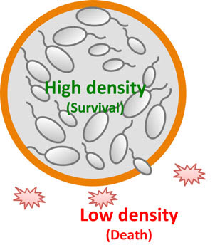
Design and modeling of safeguard control in microbial swarmbots exhibit collective survival. Bacteria confined in the microbial swarmbot can maintain a high local density and survive. Cells escaping the swarmbot will have a reduced density due to a larger extra-capsule environment. If their density drops below their survival threshold, they will die, leading to safeguard control. (credit: Shuqiang Huang et al./Molecular Systems Biology)

Duke University researchers have engineered microbes as “swarmbots” designed to only survive in a crowd.

The system could be used as a safeguard to stop genetically modified organisms (created with tools such as CRISPR) from escaping into the surrounding environment.

Collective survival

“Other labs have addressed this issue by making cells rely on unnatural amino acids for survival or by introducing a ‘kill switch’ that is activated by some chemical,” said Lingchong You, the Paul Ruffin Scarborough Associate Professor of Engineering at Duke University. “Ours is the first example that uses collective survival as a way of intrinsically realizing this safeguard.*

“In general, this concept does not depend on the use of antibiotics,” said You. “We’re using non-pathogenic E. coli, but we hope to demonstrate that the same concept can be established with a probiotic strain of bacteria.” Another method would be to insert a contained population of bacteria that could help the body respond to intruders.

“This is the foundation,” said You. “Once we’ve established the platform, then we have the freedom to introduce whatever proteins we choose and allow these cells to engage in many different applications.”

The approach could also be used to reliably program colonies of bacteria to respond to changes in their surrounding environment, such as releasing specific molecules on cue.

Terrorism implications

On Feb. 9, James R. Clapper, U.S. Director of National Intelligence warned that “given the broad distribution, low cost, and accelerated pace of development of this dual-use technology, its deliberate or unintentional misuse might lead to far-reaching economic and national security implications.” It’s not clear if the swarmbot system could address that concern.

Research in genome editing conducted by countries with different regulatory or ethical standards than those of Western countries probably increases the risk of the creation of potentially harmful biological agents or products. Given the broad distribution, low cost, and accelerated pace of development of this dual-use technology, its deliberate or unintentional misuse might lead to far-reaching economic and national security implications.

The swarmbot system is described online, February 29, 2016, in an open-access paper in Molecular Systems Biology.

This work was supported by the National Science Foundation, the National Institutes of Health, the Army Research Office, and a David and Lucile Packard Fellowship.

* In the experiment, You and his colleagues engineered a non-pathogenic strain of E. coli to produce a chemical called AHL. They also modified the cells so that, in high enough concentrations, AHL causes them to produce an antidote to antibiotics. When the population of E. coli is dense enough, the antidote keeps them alive, even in the presence of antibiotics that would otherwise kill them.

The researchers then confined a sufficiently large number of the bacteria to a capsule and bathed it in antibiotics. As long as the E. coli remained inside their container where their density was high, they all survived. But if individual bacteria escaped, they were quickly killed off by the antibiotic.


Duke University | Swarmbots


Abstract of Coupling spatial segregation with synthetic circuits to control bacterial survival

Engineered bacteria have great potential for medical and environmental applications. Fulfilling this potential requires controllability over engineered behaviors and scalability of the engineered systems. Here, we present a platform technology, microbial swarmbot, which employs spatial arrangement to control the growth dynamics of engineered bacteria. As a proof of principle, we demonstrated a safeguard strategy to prevent unintended bacterial proliferation. In particular, we adopted several synthetic gene circuits to program collective survival in Escherichia coli: the engineered bacteria could only survive when present at sufficiently high population densities. When encapsulated by permeable membranes, these bacteria can sense the local environment and respond accordingly. The cells inside the microbial swarmbot capsules will survive due to their high densities. Those escaping from a capsule, however, will be killed due to a decrease in their densities. We demonstrate that this design concept is modular and readily generalizable. Our work lays the foundation for engineering integrated and programmable control of hybrid biological–material systems for diverse applications.

Engineered ‘mini-organs’ produce insulin in mice

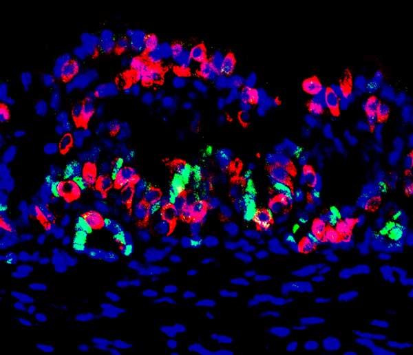
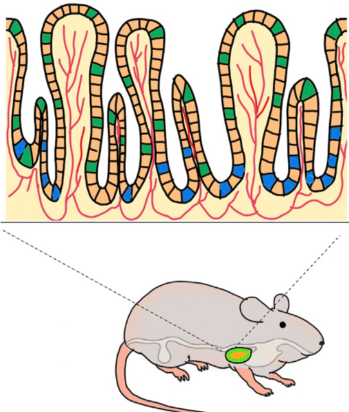
This mouse mini-organ contains stomach cells (red) that were reprogrammed to be able to produce insulin, along with some stem cells (green) that are able to replenish insulin-producing cells that are later destroyed after the mini-organ is transplanted into the mouse to supply insulin; the nuclei of all cells are shown in blue. (credit: Chaiyaboot Ariyachet)

Harvard University scientists have made major progress in dealing with a long-standing hurdle in treating diabetic patients.

People with diabetes have high blood sugar because their pancreatic beta cells (which store and release insulin) are not producing enough insulin. In type 1 diabetes (“juvenile diabetes”), the beta cells are even destroyed. In most cases, physicians treat type 1 diabetes with insulin injections, but people with complications may require pancreas implants or pancreatic islets from another person to replace those defective beta cells.

Those are limited. When effective sources of insulin-producing cells become available in the future, replacing beta cells with transplants would be preferable because that may enable blood sugar to be better controlled without the need for lifetime injections. The problem: those insulin-producing transplants tend to be destroyed by the ongoing diabetic disease process of the person (or animal) receiving the transplant.

Ideally, for a successful transplant to survive, transplanted cells or tissues need to somehow replenish or regenerate themselves.

Reprogramming stomach cells

In the new Harvard study, published online in an open access paper in Cell Stem Cell, the researchers have finally worked out a way to do that. They discovered that the lower stomach contains cells that can be reprogrammed to act as beta cells, and thus produce insulin. The stomach and intestine also naturally contain large numbers of stem cells that can multiply to continue to replenish the dying insulin-producing cells.

The report’s senior author, Qiao Zhou, PhD, associate professor at Harvard University’s Department of Stem Cell and Regenerative Biology, made the discovery after first genetically engineering mice to express three genes that can turn other cell types into insulin-producing beta-like cells. The researchers then looked for the tissue in the body that could be most easily reprogrammed to produce insulin.

“We looked all over, from the nose to the tail of the mouse,” says Zhou. “We discovered, surprisingly, that some of the cells in the pylorus region of the stomach (connecting the stomach to the small intestine) are most amenable to conversion to beta cells. This tissue appears to be the best starting material.”*

The team found that these stomach cells were naturally similar to pancreatic beta cells and could produce insulin and reverse high blood sugar in mice for up to six months.**

Creating “mini-stomachs” for humans containing insulin-producing cells for transplantation

Engineering a gastric mini-organ. An implanted bio-scafford induces beta-like cells (green) and gastric progenitor stem cells (blue). Blood vessels are shown in red. (credit: Chaiyaboot Ariyachet/Cell Stem Cell)

These initial experiments involved genetic engineering. But humans cannot yet be genetically engineered, so the researchers needed to find a version of this method that works for humans.

So the researchers took samples of the stomach tissue containing stem cells from the mice and grew them into “mini-organs” — tiny ball-like mini-stomachs that contain insulin-producing cells (that function like beta cells), as well as stem cells that can multiply to replace the insulin-producing cells as they later die after transplantation.

They transplanted these mini-organs into a membrane in the mouse’s abdomen. To test their function, the researchers then destroyed the normal insulin-producing pancreatic beta cells in the mice. They confirmed that the mini-organs compensated for the resulting loss of insulin, maintaining glucose at normal levels in five of the 22 experimental animals (this was the team’s expected success rate).

What is potentially really great about this approach (once it’s developed for humans) is that a physician will be able to create a patient-specific therapy by simply doing a biopsy (snipping out tissue for analysis) from the patient, grow the cells in the lab (as with the mice), reprogram those cells to produce insulin — and then transplant these cells directly into the patient.**

“That’s what we’re working on now,” says Zhou. “We’re very excited.”

Research funding was provided by the National Institute of Health, the HSCI, the Timothy Murphy Fund, the Children’s Hospital Boston Intellectual and Developmental Disabilities Research Center, and the Harvard Digestive Diseases Center.

* When engineered using reprogramming factors, cells in the pylorus region of the stomach were the most responsive to high glucose levels and produced insulin to normalize blood sugar. The researchers destroyed pancreatic beta cells in the mice, forcing their bodies to rely only on insulin produced by the reprogrammed stomach cells. Mice without tissue reprogramming died within eight weeks. But mice with reprogrammed cells maintained insulin and glucose levels in their blood for up to six months, which was as long as the experiment continued. The pyloric stomach also has the advantage of containing stem cells that, once reprogrammed, can replenish the insulin-producing cell population.

** These cells from the stomach are actually “beta-like cells” for producing insulin; that is, they do the same job as pancreatic beta cells. In the Cell Stem Cell paper, they are referred to as “insulin+ (insulin-positive)” cells.


Abstract of Reprogrammed Stomach Tissue as a Renewable Source of Functional β Cells for Blood Glucose Regulation

The gastrointestinal (GI) epithelium is a highly regenerative tissue with the potential to provide a renewable source of insulin+ cells after undergoing cellular reprogramming. Here, we show that cells of the antral stomach have a previously unappreciated propensity for conversion into functional insulin-secreting cells. Native antral endocrine cells share a surprising degree of transcriptional similarity with pancreatic β cells, and expression of β cell reprogramming factors in vivo converts antral cells efficiently into insulin+ cells with close molecular and functional similarity to β cells. Induced GI insulin+ cells can suppress hyperglycemia in a diabetic mouse model for at least 6 months and regenerate rapidly after ablation. Reprogramming of antral stomach cells assembled into bioengineered mini-organs in vitro yielded transplantable units that also suppressed hyperglycemia in diabetic mice, highlighting the potential for development of engineered stomach tissues as a renewable source of functional β cells for glycemic control.

Regenerative medicine scientists ‘print’ replacement tissue

Completed ear and jaw bone structures printed with the Integrated Tissue-Organ Printing System (credit: Wake Forest Baptist Medical Center)

Using a sophisticated, custom-designed 3D printer, regenerative medicine scientists at Wake Forest Baptist Medical Center have proved that it is feasible to print living tissue structures to replace injured or diseased tissue in patients.

Reporting in Nature Biotechnology, the scientists said they printed ear, bone and muscle structures. When implanted in animals, the structures matured into functional tissue and developed a system of blood vessels. Most importantly, these early results indicate that the structures have the right size, strength and function for use in humans.

“This novel tissue and organ printer is an important advance in our quest to make replacement tissue for patients,” said Anthony Atala, M.D., director of the Wake Forest Institute for Regenerative Medicine (WFIRM) and senior author on the study. “It can fabricate stable, human-scale tissue of any shape. With further development, this technology could potentially be used to print living tissue and organ structures for surgical implantation.”

With funding from the Armed Forces Institute of Regenerative Medicine, a federally funded effort to apply regenerative medicine to battlefield injuries, Atala’s team aims to implant bioprinted muscle, cartilage and bone in patients in the future.

Designing a 3D bioprinter

Tissue engineering is a science that aims to grow replacement tissues and organs in the laboratory to help solve the shortage of donated tissue available for transplants. The precision of 3D printing makes it a promising method for replicating the body’s complex tissues and organs. However, current printers based on jetting, extrusion and laser-induced forward transfer cannot produce structures with sufficient size or strength to implant in the body.

The Integrated Tissue and Organ Printing System (ITOP), developed over a 10-year period by scientists at the Institute for Regenerative Medicine, overcomes these challenges. The system deposits both bio-degradable, plastic-like materials to form the tissue “shape” and water-based gels that contain the cells. In addition, a strong, temporary outer structure is formed. The printing process does not harm the cells.*

A major challenge of tissue engineering is ensuring that implanted structures live long enough to integrate with the body. The Wake Forest Baptist scientists addressed this in two ways. They optimized the water-based “ink” that holds the cells so that it promotes cell health and growth and they printed a lattice of micro-channels throughout the structures. These channels allow nutrients and oxygen from the body to diffuse into the structures and keep them live while they develop a system of blood vessels.

It has been previously shown that tissue structures without ready-made blood vessels must be smaller than 200 microns (0.007 inches) for cells to survive. In these studies, a baby-sized ear structure (1.5 inches) survived and showed signs of vascularization at one and two months after implantation.

“Our results indicate that the bio-ink combination we used, combined with the micro-channels, provides the right environment to keep the cells alive and to support cell and tissue growth,” said Atala.

Another advantage of the ITOP system is its ability to use data from CT and MRI scans to “tailor-make” tissue for patients. For a patient missing an ear, for example, the system could print a matching structure.

The research was supported, in part, by grants from the Armed Forces Institute of Regenerative Medicine, the Telemedicine and Advanced Technology Research Center at the U.S. Army Medical Research and Material Command, and the Defense Threat Reduction Agency.

* Several proof-of-concept experiments demonstrated the capabilities of ITOP. To show that ITOP can generate complex 3D structures, printed, human-sized external ears were implanted under the skin of mice. Two months later, the shape of the implanted ear was well-maintained and cartilage tissue and blood vessels had formed.

To demonstrate the ITOP can generate organized soft tissue structures, printed muscle tissue was implanted in rats. After two weeks, tests confirmed that the muscle was robust enough to maintain its structural characteristics, become vascularized and induce nerve formation.

And, to show that construction of a human-sized bone structure, jaw bone fragments were printed using human stem cells. The fragments were the size and shape needed for facial reconstruction in humans. To study the maturation of bioprinted bone in the body, printed segments of skull bone were implanted in rats. After five months, the bioprinted structures had formed vascularized bone tissue.

Ongoing studies will measure longer-term outcomes.


Abstract of A 3D bioprinting system to produce human-scale tissue constructs with structural integrity

A challenge for tissue engineering is producing three-dimensional (3D), vascularized cellular constructs of clinically relevant size, shape and structural integrity. We present an integrated tissue–organ printer (ITOP) that can fabricate stable, human-scale tissue constructs of any shape. Mechanical stability is achieved by printing cell-laden hydrogels together with biodegradable polymers in integrated patterns and anchored on sacrificial hydrogels. The correct shape of the tissue construct is achieved by representing clinical imaging data as a computer model of the anatomical defect and translating the model into a program that controls the motions of the printer nozzles, which dispense cells to discrete locations. The incorporation of microchannels into the tissue constructs facilitates diffusion of nutrients to printed cells, thereby overcoming the diffusion limit of 100–200 μm for cell survival in engineered tissues. We demonstrate capabilities of the ITOP by fabricating mandible and calvarial bone, cartilage and skeletal muscle. Future development of the ITOP is being directed to the production of tissues for human applications and to the building of more complex tissues and solid organs.

Cancer cells in 3D

A spheroid of many lung cancer cells illustrates a diversity of behaviors. (credit: Welf and Driscoll et al./Developmental Cell)

Cancer cells don’t live on glass slides. Yet the vast majority of images related to cancer biology come from the cells being photographed on flat, two-dimensional surfaces — images sometimes used to draw conclusions about the behavior of cells that normally reside in a more complex environment.

Now a new high-resolution microscope, presented (open access) February 22 in Developmental Cell, makes it possible to visualize cancer cells in 3D and record how they are signaling to other parts of their environment — revealing previously unappreciated biology of how cancer cells survive and disperse within living things. Based on ”microenvironmental selective plane illumination microscopy” (meSPIM),  the new microscope is designed to image cells in microenvironments free of hard surfaces near the sample.

“There is clear evidence that the environment strongly affects cellular behavior — thus, the value of cell culture experiments on glass must at least be questioned,” says senior author Reto Fiolka, an optical scientist at the University of Texas Southwestern Medical Center. “Our microscope is one tool that may bring us a deeper understanding of the molecular mechanisms that drive cancer cell behavior, since it enables high-resolution imaging in more realistic tumor environments.”

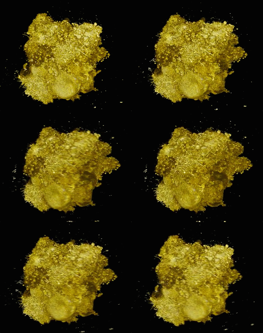
This image shows the extracted surfaces of two cancer cells. (Left) A lung cancer cell colored by actin intensity near the cell surface. Actin is a structural molecule that is integral to cell movement. (Right) A melanoma cell colored by PI3-kinase activity near the cell surface. PI3K is a signaling molecule that is key to many cell processes. (credit: Welf and Driscoll et al./Developmental Cell)

Hidden protrusions from cancer cells

In their study, Fiolka and colleagues, including co-senior author Gaudenz Danuser, and co-first authors Meghan Driscoll and Erik Welf, also of UT Southwestern, used their microscope to image different kinds of skin cancer cells from patients. They found that in a 3D environment (where cells normally reside), unlike a glass slide, multiple melanoma cell lines and primary melanoma cells (from patients with varied genetic mutations) form many small protrusions called blebs.

One hypothesis is that this blebbing may help the cancer cells survive or move around and could thus play a role in skin cancer cell invasiveness or drug resistance in patients.

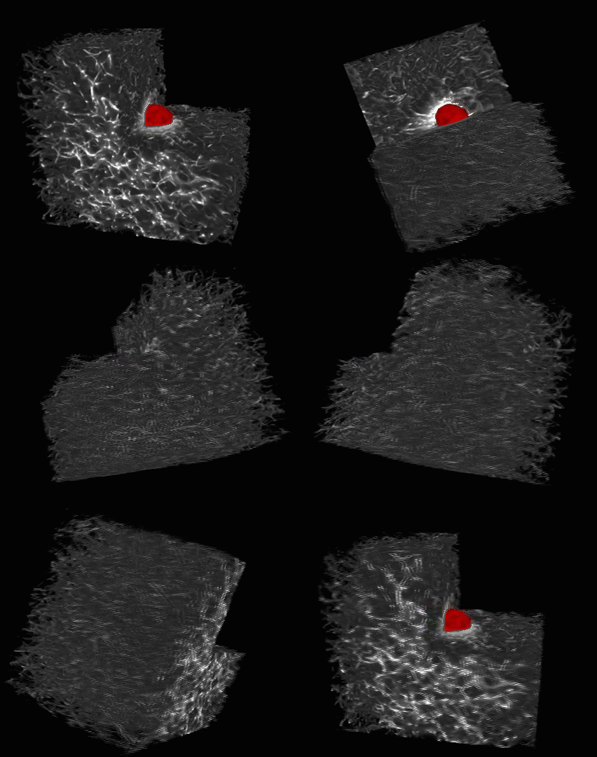
This is a melanoma cell (red) embedded in a 3-D collagen matrix (white). A 100 x 100 x 100 μm cube is shown, with one corner cut away to show the interaction of the cell with the collagen. (credit: Welf and Driscoll et al./Developmental Cell)

The researchers say that this is a first step toward understanding 3D biology in tumor microenvironments. But since these kinds of images may be too complicated to interpret by the naked eye alone, the next step will be to develop powerful computer platforms to extract and process the information.

The microscope control software and image analytical code are freely available to the scientific community.

The authors were supported by the Cancer Prevention Research Institute of Texas and the National Institutes of Health.


Abstract of Quantitative Multiscale Cell Imaging in Controlled 3D Microenvironments

The microenvironment determines cell behavior, but the underlying molecular mechanisms are poorly understood because quantitative studies of cell signaling and behavior have been challenging due to insufficient spatial and/or temporal resolution and limitations on microenvironmental control. Here we introduce microenvironmental selective plane illumination microscopy (meSPIM) for imaging and quantification of intracellular signaling and submicrometer cellular structures as well as large-scale cell morphological and environmental features. We demonstrate the utility of this approach by showing that the mechanical properties of the microenvironment regulate the transition of melanoma cells from actin-driven protrusion to blebbing, and we present tools to quantify how cells manipulate individual collagen fibers. We leverage the nearly isotropic resolution of meSPIM to quantify the local concentration of actin and phosphatidylinositol 3-kinase signaling on the surfaces of cells deep within 3D collagen matrices and track the many small membrane protrusions that appear in these more physiologically relevant environments.

New method enables discovery of 3D structures for molecules important to medicine

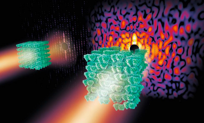
If you zap a crystal (green, left) containing highly ordered protein molecules with X-rays, the X-rays scatter and produce useful regular patterns of spots known as Bragg peaks (red dots). But if the protein in the crystal is less ordered or disordered (right), the X-rays produce some spots along with patterns of light and shade known as a continuous diffraction pattern that’s not useful. (credit: Eberhard Reimann/DESY)

Researchers have overcome a long-standing technical barrier to imaging 3D structures of thousands of molecules important to medicine and biology.

The 3D structures of many protein molecules have been discovered using a technique called X-ray crystallography, but the method relies on scientists being able to produce highly ordered crystals containing the protein molecules in a regular arrangement. When X-rays are shone on highly ordered crystals, the X-rays scatter and produce regular patterns of spots called Bragg peaks (see figure above, left). High-quality Bragg peaks contain the information to produce high-resolution 3D structures of proteins.

Unfortunately, many important and complex biomolecules do not form highly ordered crystals; instead, the protein arrangements are slightly disordered. When X-rays are shone on these more disordered crystals, a smaller number of Bragg peaks are produced, along with a vague pattern of light and shadow known as a continuous diffraction pattern (right).

In the past, scientists discarded these less-than-perfect crystals. Unfortunately, many of the molecules forming disordered crystals are important molecular complexes such as those that span cell membranes.

X-raying crystal patterns to detect hidden protein structures

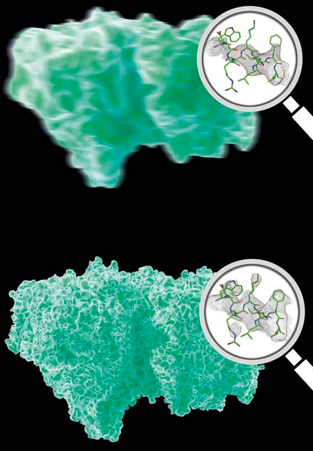
Analysis of Bragg peaks alone (top) reveals far less details than analysis of the high-res continuous diffraction pattern (bottom). Magnifying glasses show actual data. (credit: DESY, Eberhard Reimann)

So a team led by Professor Henry Chapman from the Center for Free-Electron Laser Science at DESY in Hamburg, Germany turned to the world’s most powerful X-ray laser: the SLAC LCLS at Stanford University.

Kartik Ayyer, PhD., lead author of the article in Nature, explains that the method uses an approach similar to that used to image a single molecule.

“If you would shoot X-rays on a single molecule, it would produce a continuous diffraction pattern free of any Bragg spots,” he says. “The pattern would be extremely weak, however, and very difficult to measure. But the ‘background’ in our crystal analysis is like accumulating many shots from individually aligned single molecules. We essentially just use the crystal as a way to get a lot of single molecules, aligned in common orientations, into the beam.”

As the model protein, the researchers crystallized photosystem II (PSII), a large membrane–protein complex of photosynthesis that plants use to produce oxygen for life on Earth.

After exposing the crystal to X-rays, the researchers first analyzed the Bragg peaks of PSII to produce a low-resolution outline of the 3D structure (figure above, top). They then improved this data, using an algorithm, to analyze the continuous diffraction pattern and produced a higher-resolution 3D structure (figure, bottom).

This novel method means that imperfect crystals containing a slightly disordered protein arrangement can now be used to “directly view large protein complexes in atomic detail,” says Chapman. “This kind of continuous diffraction has actually been seen for a long time from many different poorly diffracting crystals,” says Chapman. “It wasn’t understood that you can get structural information from it and so analysis techniques suppressed it.

“We’re going to be busy to see if we can solve [additional] structures of molecules from old discarded data.”


Abstract of Macromolecular diffractive imaging using imperfect crystals

The three-dimensional structures of macromolecules and their complexes are mainly elucidated by X-ray protein crystallography. A major limitation of this method is access to high-quality crystals, which is necessary to ensure X-ray diffraction extends to sufficiently large scattering angles and hence yields information of sufficiently high resolution with which to solve the crystal structure. The observation that crystals with reduced unit-cell volumes and tighter macromolecular packing often produce higher-resolution Bragg peaks suggests that crystallographic resolution for some macromolecules may be limited not by their heterogeneity, but by a deviation of strict positional ordering of the crystalline lattice. Such displacements of molecules from the ideal lattice give rise to a continuous diffraction pattern that is equal to the incoherent sum of diffraction from rigid individual molecular complexes aligned along several discrete crystallographic orientations and that, consequently, contains more information than Bragg peaks alone. Although such continuous diffraction patterns have long been observed—and are of interest as a source of information about the dynamics of proteins—they have not been used for structure determination. Here we show for crystals of the integral membrane protein complex photosystem II that lattice disorder increases the information content and the resolution of the diffraction pattern well beyond the 4.5-ångström limit of measurable Bragg peaks, which allows us to phase the pattern directly. Using the molecular envelope conventionally determined at 4.5 ångströms as a constraint, we obtain a static image of the photosystem II dimer at a resolution of 3.5 ångströms. This result shows that continuous diffraction can be used to overcome what have long been supposed to be the resolution limits of macromolecular crystallography, using a method that exploits commonly encountered imperfect crystals and enables model-free phasing.

US could see substantial impact of Zika virus, warns researcher

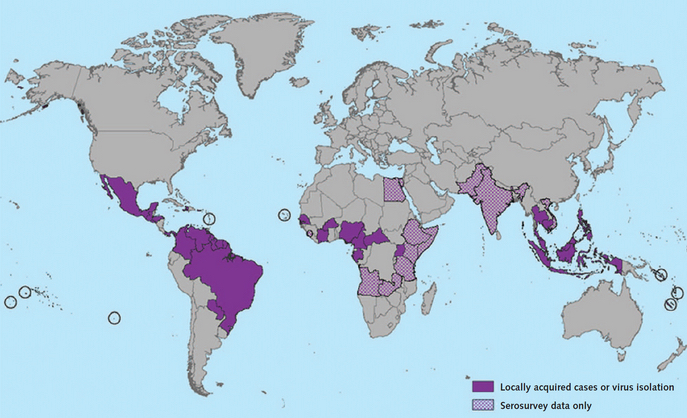
Countries that have past or current evidence of Zika virus transmission (as of January 2016) (credit: CDC)

A researcher at Boston Medical Center (BMC) and the Boston University School of Public Health (BUSPH) warns that Zika virus could spread quickly to the U.S. There is currently no vaccine or cure.

WHO director general Margaret Chan, M.D., declared on Feb. 1 that the recent cluster of microcephaly and other neurological abnormalities reported in Latin America, following a similar cluster in French Polynesia in 2014, constitutes “a public health emergency of international concern.”

The mosquito-borne virus is believed to cause microcephaly in infants who are exposed in utero and causes rash and flu-like symptoms in adults and children who have been infected.

An outbreak in French Polynesia in 2013 was responsible for 19,000 suspected cases, and since October 2015, nearly 4,000 cases of Zika virus-related microcephaly have been reported in Brazil. Microcephaly is abnormal smallness of the head, a congenital condition associated with incomplete brain development and a range of neurological complications.

The findings are published online (open access) in advance of print in the Annals of Internal Medicine.

Rapidly emerging in the Western Hemisphere via mosquitos or sexual activity

Zika virus has been rapidly emerging in the Western Hemisphere in the last few months, and as of Jan. 22, 2016, there were 20 countries and territories in the Americas with Zika virus in circulation. Currently, it can be found in Central America, the Caribbean and Mexico, and transmission has occurred in travelers to these areas returning to non-endemic countries, including the U.S., Canada, Japan, Western Europe, and Israel.

“At this time, we believe that Zika virus is primarily transmitted via infected mosquitoes, and therefore people living in or traveling to impacted areas are strongly encouraged to protect themselves against mosquitoes by using an effective insect repellent (containing DEET or picaridin),” said senior author Davidson Hamer, MD, director of the Travel Clinic at BMC, and professor of global health and medicine at the Boston University School of Public Health and School of Medicine.

“However, there is some evidence to suggest that Zika virus could be transmitted via blood transfusion and sexual activity, so researchers are trying to determine if these are meaningful pathways to transmission.” There is also evidence of mother-to-child transmission, which appears to be responsible for the surge in cases of microcephaly being seen in Brazil.

Hamer and his co-author, Lin Chen, MD, of the Mt. Auburn Hospital Travel Clinic, say there is substantial risk of introduction of the Zika virus in the U.S., given the presence of the mosquito species that carry the virus, Aedes aegypti and Ae. albopictus, in many states.

“If you are pregnant, put off travel to the endemic areas,” Hamer said. “If you absolutely must go, be sure to protect yourself against mosquitoes. For those who are not pregnant, it’s still a good idea to delay travel so that you don’t risk getting infected and transferring the virus back home — there are many unknowns about its transmission, so there is still a risk.”

In 2007, the first case was detected in a human, leading to an outbreak on an island in Micronesia. An estimated 73 percent of the island residents age 3 or older became infected; however, about 80 percent of these cases did not present significant symptoms.

Zika virus is generally mild and typically resolves itself within a week. Symptoms can include rash, conjunctivitis, muscle and joint pain, headache, joint swelling, dizziness and vomiting. However, neurological and autoimmune complications have been linked to the French Polynesia outbreak, particularly development of Guillain-Barre syndrome, a neurological illness that may result in temporary paralysis. Microcephaly has been reported in thousands of cases in Brazil, and recently in a newborn in Hawaii.

There currently is no vaccine or cure for the Zika virus.


Abstract of Zika Virus: Rapid Spread in the Western Hemisphere

Zika virus, a mosquito-borne flavivirus that causes febrile illness associated with rash, has been rapidly emerging in the Western Hemisphere over the past few months. The virus was rarely identified until outbreaks occurred on Yap Island in the Federated States of Micronesia in 2007, French Polynesia in 2013, and Easter Island in 2014. It was initially detected in Brazil in 2015, in the northeast, and was subsequently identified in other states and several South American countries, including Colombia, Ecuador, Suriname, Venezuela, French Guyana, and Paraguay. Local transmission has been documented in Central America (Panama, El Salvador, Honduras, and Guatemala), the Caribbean (Martinique, Puerto Rico, Dominican Republic, and Haiti), and Mexico. Transmission has also occurred in travelers returning from the infected regions to nonendemic countries, including the United States, Canada, Japan, and Western Europe. As of 22 January 2016, a total of 20 countries and territories in the Americas have Zika virus circulation. The explosive spread mirrors the emergence of chikungunya, which was first detected in the Americas (St. Martin) in 2013 and rapidly disseminated throughout the region.