Machine-learning technique uncovers unknown features of multi-drug-resistant pathogen

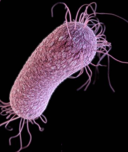
According to the CDC, Pseudomonas aeruginosa is a common cause of healthcare-associated infections, including pneumonia, bloodstream infections, urinary tract infections, and surgical site infections. Some strains of P. aeruginosa have been found to be resistant to nearly all or all antibiotics. (illustration credit: CDC)

A new machine-learning technique can uncover previously unknown features of organisms and their genes in large datasets, according to researchers from the Perelman School of Medicine at the University of Pennsylvania and the Geisel School of Medicine at Dartmouth University.

For example, the technique learned to identify the characteristic gene-expression patterns that appear when a bacterium is exposed in different conditions, such as low oxygen and the presence of antibiotics.

The technique, called “ADAGE” (Analysis using Denoising Autoencoders of Gene Expression), uses a “denoising autoencoder” algorithm, which learns to identify recurring features or patterns in large datasets — without being told what specific features to look for (that is, “unsupervised.”)*

Last year,  Casey Greene, PhD, an assistant professor of Systems Pharmacology and Translational Therapeutics at Penn, and his team published, in an open-access paper in the American Society for Microbiology’s mSystems, the first demonstration of ADAGE in a biological context: an analysis of two gene-expression datasets of breast cancers.

Tracking down gene patterns of a multi-drug-resistant bacterium

The new study, published Jan. 19 in an open-access paper in mSystems, was more ambitious. It applied ADAGE to a dataset of 950 gene-expression arrays publicly available at the time for the multi-drug-resistant bacterium Pseudomonas aeruginosa. This bacterium is a notorious pathogen in the hospital and in individuals with cystic fibrosis and other chronic lung conditions; it’s often difficult to treat due to its high resistance to standard antibiotic therapies.

The data included only the identities of the roughly 5,000 P. aeruginosa genes and their measured expression levels in each published experiment. The goal was to see if this “unsupervised” learning system could uncover important patterns in P. aeruginosa gene expression and clarify how those patterns change when the bacterium’s environment changes — for example, when in the presence of an antibiotic.

Even though the model built with ADAGE was relatively simple — roughly equivalent to a brain with only a few dozen neurons — it had no trouble learning which sets of P. aeruginosa genes tend to work together or in opposition. To the researchers’ surprise, the ADAGE system also detected differences between the main laboratory strain of P. aeruginosa and strains isolated from infected patients. “That turned out to be one of the strongest features of the data,” Greene said.

“We expect that this approach will be particularly useful to microbiologists researching bacterial species that lack a decades-long history of study in the lab,” said Greene. “Microbiologists can use these models to identify where the data agree with their own knowledge and where the data seem to be pointing in a different direction … and to find completely new things in biology that we didn’t even know to look for.”

Support for the research came from the Gordon and Betty Moore Foundation, the William H. Neukom Institute for Computational Science, the National Institutes of Health, and the Cystic Fibrosis Foundation.

* In 2012, Google-sponsored researchers applied a similar method to randomly selected YouTube images; their system learned to recognize major recurring features of those images — including cats of course.


Abstract of ADAGE-Based Integration of Publicly Available Pseudomonas aeruginosa Gene Expression Data with Denoising Autoencoders Illuminates Microbe-Host Interactions

The increasing number of genome-wide assays of gene expression available from public databases presents opportunities for computational methods that facilitate hypothesis generation and biological interpretation of these data. We present an unsupervised machine learning approach, ADAGE (analysis using denoising autoencoders of gene expression), and apply it to the publicly available gene expression data compendium for Pseudomonas aeruginosa. In this approach, the machine-learned ADAGE model contained 50 nodes which we predicted would correspond to gene expression patterns across the gene expression compendium. While no biological knowledge was used during model construction, cooperonic genes had similar weights across nodes, and genes with similar weights across nodes were significantly more likely to share KEGG pathways. By analyzing newly generated and previously published microarray and transcriptome sequencing data, the ADAGE model identified differences between strains, modeled the cellular response to low oxygen, and predicted the involvement of biological processes based on low-level gene expression differences. ADAGE compared favorably with traditional principal component analysis and independent component analysis approaches in its ability to extract validated patterns, and based on our analyses, we propose that these approaches differ in the types of patterns they preferentially identify. We provide the ADAGE model with analysis of all publicly available P. aeruginosa GeneChip experiments and open source code for use with other species and settings. Extraction of consistent patterns across large-scale collections of genomic data using methods like ADAGE provides the opportunity to identify general principles and biologically important patterns in microbial biology. This approach will be particularly useful in less-well-studied microbial species.


Abstract of Unsupervised feature construction and knowledge extraction from genome-wide assays of breast cancer with denoising autoencoders

Big data bring new opportunities for methods that efficiently summarize and automatically extract knowledge from such compendia. While both supervised learning algorithms and unsupervised clustering algorithms have been successfully applied to biological data, they are either dependent on known biology or limited to discerning the most significant signals in the data. Here we present denoising autoencoders (DAs), which employ a data-defined learning objective independent of known biology, as a method to identify and extract complex patterns from genomic data. We evaluate the performance of DAs by applying them to a large collection of breast cancer gene expression data. Results show that DAs successfully construct features that contain both clinical and molecular information. There are features that represent tumor or normal samples, estrogen receptor (ER) status, and molecular subtypes. Features constructed by the autoencoder generalize to an independent dataset collected using a distinct experimental platform. By integrating data from ENCODE for feature interpretation, we discover a feature representing ER status through association with key transcription factors in breast cancer. We also identify a feature highly predictive of patient survival and it is enriched by FOXM1 signaling pathway. The features constructed by DAs are often bimodally distributed with one peak near zero and another near one, which facilitates discretization. In summary, we demonstrate that DAs effectively extract key biological principles from gene expression data and summarize them into constructed features with convenient properties.

New acoustic-tweezer design allows for 3D bioprinting

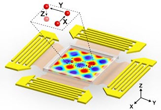
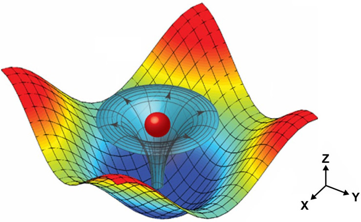
Illustration of a particle (red sphere) trapped by the 3D trapping node created by two superimposed, orthogonal (at right angles), standing surface acoustic waves and induced acoustic streaming (credit: Carnegie Mellon University)

A team of researchers at three universities has developed a way to use “acoustic tweezers” (which use ultrasonic surface acoustic waves, or SAWs, to trap and manipulate micrometer-scale particles and biological cells — see “Acoustic tweezers manipulate cellular-scale objects with ultrasound“) to non-invasively pick up and move single cells in three mutually orthogonal axes of motion (three dimensions).

The new 3D acoustic tweezers can pick up single cells or entire cell assemblies and deliver them to desired locations to create 2D and 3D cell patterns, or print* the cells into complex shapes — a promising new method for “3D bioprinting” in biological tissues, the researchers say in an open-access paper in  the Proceedings of the National Academy of Sciences (PNAS).

The new method, developed by researchers at Carnegie Mellon UniversityPennsylvania State University and MIT, offers the potential to accurately print 3D multicellular architectures for applications in biomanufacturing, tissue engineering, regenerative medicine, neuroscience, and cancer metastasis research.

Multicellular structures within living things are complex and delicate, which makes recreating or repairing these structures a daunting task. For example, the human heart contains more than 2 billion muscle cells. Each of these cells must properly interact with one another and with their environment to ensure that the heart functions properly. If those cells aren’t placed correctly, or are damaged, it could potentially result in any of a variety of heart conditions.

A 3D trapping node

Illustration of planar surface acoustic wave generators used to generate 3D nodes surrounding a microfluidic experimental area (center). The inset indicates a single particle within a “3-D trapping node” that is independently manipulated along x, y or z axes. (credit: Carnegie Mellon University)

Researchers have been using a combination of approaches for recreating the complex, multicellular architecture of biological tissue and to separate, align, pattern and transport single cells, and the approaches are renowned for their ability to gently manipulate cells without causing any cellular damage.

But they have yet to develop a single method that has the high level of precision, versatility, multiple dimensionality, and single cell resolution needed to form complex multicellular structures while maintaining cell viability, integrity and function and without the need for invasive contact, tagging, or biochemical labeling in regenerative medicine, neuroscience, tissue engineering, bio-manufacturing, and cancer metastasis.

The new microfluidic device developed by the team has now allowed the researchers to manipulate where the waves would meet along each of the three axes. At these meeting points, the waves formed a 3D trapping node that captured individual cells. The researchers could then further manipulate the acoustic waves to move and place cells.

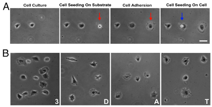
Printing living cells with 3D acoustic tweezers. (A) Single-cell printing. After previously deposited 3T3 (mouse embryonic fibroblast) cells were attached to the substrate, another single cell was picked up, transported, and dropped at the desired location on the substrate (indicated by red arrow) or on top of another cell (indicated by blue arrow). The single cell adhered and spread along the surface. (B) Cell culture patterns arranged to form the characters “3,” “D,” “A,” and “T” by printing single HeLa S3 cells (a human immortal cell line) using 3D acoustic tweezers. (Scale bar: 20 micrometers) (credit: Feng Guo et al./PNAS)

To demonstrate how their acoustic tweezers technique could be used for live cell printing, the researchers used the microfluidic device to pick up cells and deposit them in a pre-selected pattern, with an elegant level of control over cell spacing and geometry. That suggested that the device has the potential to effectively create 3D tissue-like structures, including those with complex geometries.

* Typically, cells are printed in a substrate or matrix to form tissue for later implantation (see “Robust ‘spider silk’ matrix guides cardiac tissue regeneration“).


Abstract of Three-dimensional manipulation of single cells using surface acoustic waves

The ability of surface acoustic waves to trap and manipulate micrometer-scale particles and biological cells has led to many applications involving “acoustic tweezers” in biology, chemistry, engineering, and medicine. Here, we present 3D acoustic tweezers, which use surface acoustic waves to create 3D trapping nodes for the capture and manipulation of microparticles and cells along three mutually orthogonal axes. In this method, we use standing-wave phase shifts to move particles or cells in-plane, whereas the amplitude of acoustic vibrations is used to control particle motion along an orthogonal plane. We demonstrate, through controlled experiments guided by simulations, how acoustic vibrations result in micromanipulations in a microfluidic chamber by invoking physical principles that underlie the formation and regulation of complex, volumetric trapping nodes of particles and biological cells. We further show how 3D acoustic tweezers can be used to pick up, translate, and print single cells and cell assemblies to create 2D and 3D structures in a precise, noninvasive, label-free, and contact-free manner.

A new technique for super-resolution digital microscopy

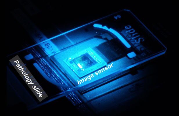
The image sensor of the wavelength scanning super-resolution apparatus collects a “stack” of images of the sample (credit: Ozcan Lab)

Researchers from the California NanoSystems Institute at UCLA have created a new technique using lens-free holograms that greatly enhances digital microscopy images, which are sometimes blurry and pixelated.

The new technique, called “wavelength scanning pixel super-resolution,” uses a device that captures a stack of digital images of the same specimen, each with a slightly different wavelength of light. Then, researchers apply a newly devised algorithm that divides the pixels in each captured image into a number of smaller pixels, resulting in a much higher-resolution digital image of the specimen.

The research team was led by Aydogan Ozcan, Chancellor’s Professor of Electrical Engineering and Bioengineering at the UCLA Henry Samueli School of Engineering and Applied Science. The study appears in an open-access paper in the journal Light: Science and Applications, published by the Nature Publishing Group.

Raw data is transformed into a super-resolution image (credit: Ozcan Lab)

Faster, more-accessible diagnosis

“These results mean we can see and inspect large samples with finer details at the sub-micron [nanoscale] level,” Ozcan said. “We have applied this method to lens-based conventional microscopes, as well as our lensless on-chip microscopy systems that create microscopic images using holograms, and it works across all these platforms.”

The benefits of this new method are wide-ranging, but especially significant in pathology, where rapid microscopic imaging of large numbers of tissue or blood cells is key to diagnosing diseases such as cancer. The specimens used in the study were blood samples, used to screen for various diseases, and Papanicolaou tests, which are used to screen for cervical cancer.

Ozcan said that wavelength scanning super-resolution works on both colorless and dye-stained samples. The entire apparatus fits on a desktop, so its size and convenience could be of great benefit to doctors and scientists using microscopes in resource-limited settings such as clinics in developing countries.

The research was supported by the Presidential Early Career Award for Scientists and Engineers, the Army Research Office, the National Science Foundation, the Office of Naval Research and the Howard Hughes Medical Institute.


Abstract of Pixel super-resolution using wavelength scanning

Undersampling and pixelation affect a number of imaging systems, limiting the resolution of the acquired images, which becomes particularly significant for wide-field microscopy applications. Various super-resolution techniques have been implemented to mitigate this resolution loss by utilizing sub-pixel displacements in the imaging system, achieved, for example, by shifting the illumination source, the sensor-array and/or the sample, followed by digital synthesis of a smaller effective pixel by merging these subpixel-shifted low-resolution images. Herein, we introduce a new pixel super-resolution method that is based on wavelength scanning and demonstrate that as an alternative to physical shifting/displacements, wavelength diversity can be used to boost the resolution of a wide-field imaging system and significantly increase its space-bandwidth product. We confirmed the effectiveness of this new technique by improving the resolution of lens-free as well as lens-based microscopy systems and developed an iterative algorithm to generate high-resolution reconstructions of a specimen using undersampled diffraction patterns recorded at a few wavelengths covering a narrow spectrum (10-30 nm). When combined with a synthetic-aperture-based diffraction imaging technique, this wavelength-scanning super-resolution approach can achieve a half-pitch resolution of 250 nm, corresponding to a numerical aperture of approximately 1.0, across a large field of view (>20 mm2 ). We also demonstrated the effectiveness of this approach by imaging various biological samples, including blood and Papanicolaou smears. Compared with displacement-based super-resolution techniques, wavelength scanning brings uniform resolution improvement in all directions across a sensor array and requires significantly fewer measurements. This technique would broadly benefit wide-field imaging applications that demand larger space-bandwidth products.

How cancer cells form tumors by reaching out with ‘cables’ and grabbing cells


University of Iowa | Cancer cells’ motion and accretion into tumors

Two University of Iowa studies have recorded the movements of cancerous human breast tissue cells in real time and in 3D — the first time cancer cells’ motion and accretion into tumors has been continuously tracked, the researchers believe.

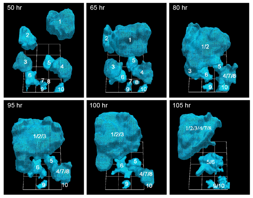
The team discovered that cancerous cells, moving at move at 92 micrometers per hour (about twice the speed of healthy cells), actively recruit healthy cells into tumors by extending a kind of cable to grab their neighbors — both cancerous and healthy — and reel them in. Surprisingly, as little as five percent of cancerous cells are needed to form the tumors, a ratio previously unknown.

“It’s not like things sticking to each other,” said David Soll, biology professor at the UI and corresponding author on the open-access paper, published in the American Journal of Cancer Research. “It’s that these cells go out and actively recruit. It’s complicated stuff, and it’s not passive. No one had a clue that there were specialized cells in this process, and that it’s a small number that pulls all the rest in.”

The findings could lead to a more precise identification of tumorigenic cells (those that form tumors) and to testing which antibodies would be best equipped to eliminate them.*

How cancer cells “know” what to do

The question is: how do these cells know what to do. Soll hypothesizes they’re reaching back to a primitive past, when these cells were programmed to form embryos. If true, perhaps the cancerous cells — masquerading as embryo-forming cells — recruit other cells to make tissue that then forms the layered, self-sustaining architecture needed for a tumor to form and thrive. “It’s as if it’s building its own defenses against the body’s efforts to defeat them.”

University of Iowa researchers have documented how cancerous tumors form by tracking in real time the movement of individual cells in 3-D. They report that just 5 percent of cancer cells are needed to form tumors, a ratio that heretofore had been unknown. (credit: Soll Laboratory)

In the AJCR paper, the researchers found support for their previous observation that tumorigenic cell lines and fresh tumor cells possess the unique capacity to form tumors by the active formation of cellular cables.

The finding lends more weight to the idea that tumors are created concurrently, in multiple locations, by individual clusters of cells that employ the cancer-cell cables to draw in more cells and enlarge themselves. Some have argued that tumors come about more by cellular changes within the masses, known as the “cancer stem cell theory.”

The Developmental Studies Hybridoma Bank funded the study.

* Soll’s Monoclonal Antibody Research Institute and the Developmental Studies Hybridoma Bank, created by the National Institutes of Health as a national resource, directed by Soll and housed at the UI, together contain one of the world’s largest collections of antibodies that could be used for the anti-cancer testing, based on the new findings.


Abstract of Mediated coalescence: a possible mechanism for tumor cellular heterogeneity

Recently, we demonstrated that tumorigenic cell lines and fresh tumor cells seeded in a 3D Matrigel model, first grow as clonal islands (primary aggregates), then coalesce through the formation and contraction of cellular cables. Non-tumorigenic cell lines and cells from normal tissue form clonal islands, but do not form cables or coalesce. Here we show that as little as 5% tumorigenic cells will actively mediate coalescence between primary aggregates of majority non-tumorigenic or non-cancerous cells, by forming cellular cables between them. We suggest that this newly discovered, specialized characteristic of tumorigenic cells may explain, at least in part, why tumors contain primarily non-tumorigenic cells.

Researchers pinpoint ‘limbo’ noisy place where cancer cells may emerge

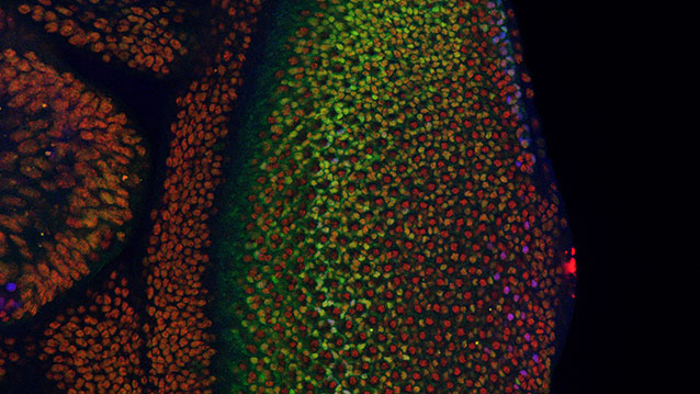
The fruit fly’s eye is an intricate pattern of many different specialized cells, and scientists use it as a workhorse to study what goes wrong in human cancer. In a new study of the fly’s eye, Northwestern University researchers have gained insight into how developing cells normally switch to a restricted, or specialized, state and how that process might go wrong in cancer. (credit: Northwestern University)

In a study involving the fruit fly equivalent of an oncogene implicated in many human leukemias, Northwestern University researchers have gained insight into how developing cells normally switch to a restricted, or specialized, state and how that process might go wrong in cancer.

The fruit fly’s eye is an intricate pattern of many different specialized cells, such as light-sensing neurons and cone cells. Because flies share with humans many of the same cancer-causing genes, scientists use the precisely made compound eye of Drosophila melanogaster (the common fruit fly) as a workhorse to study what goes wrong in human cancer.

A multidisciplinary team co-led by biologist Richard W. Carthew and engineer Luís A.N. Amaral studied normal cell behavior in the developing eye. The researchers were surprised to discover that the levels of an important protein called Yan start fluctuating wildly when the cell is switching from a more primitive, stem-like state to a more specialized state. If the levels don’t or can’t fluctuate, the cell doesn’t switch and move forward.

“This mad fluctuation, or noise, happens at the time of cell transition,” said Carthew, professor of molecular biosciences in Northwestern’s Weinberg College of Arts and Sciences. “For the first time, we see there is a brief time period as the developing cell goes from point A to point B. The noise is a state of ‘in between’ and is important for cells to switch to a more specialized state. This limbo might be where normal cells take a cancerous path.”

When the noise “off” switch doesn’t work

The researchers also found that a molecular signal received by a cell receptor called EGFR is important for turning the noise off. If that signal is not received, the cell remains in an uncontrolled state.

By pinpointing this noise and its “off” switch as important points in the normal process of cell differentiation, the Northwestern researchers provide targets for scientists studying how cells can go out of control and transform into cancer cells.

The study was published in an open-access paper and cover story Jan. 14 by the online life sciences and biomedicine journal eLife.

The “noisy” protein the Northwestern researchers studied is called Yan in the fly and Tel-1 in humans. (The protein is a transcription factor.) The Tel-1 protein instructs cells to turn into white blood cells; the gene that produces the protein, oncogene Tel-1, is frequently mutated in leukemia.

The EGFR protein that turns off the noise in flies is called Her-2 in humans. Her-2 is an oncogene that plays an important role in human breast cancer.

“On the surface, flies and humans are very different, but we share a remarkable amount of infrastructure,” said Carthew, a member of the Robert H. Lurie Comprehensive Cancer Center of Northwestern University. “We can use fruit fly genetics to understand how humans work and how things go wrong in cancer and other diseases.”

Fruit fly cells are small and closely packed together, making study of them challenging. Carthew and Amaral’s team of biologists, chemical and biological engineers, computer scientists and chemists together figured out how to identify and analyze thousands and thousands of individual cells in the flies’ eyes.

“In the past, people have built models of regulatory networks that control cell differentiation mostly by genetically perturbing one or two components of the network at a time and then compiling those results into models,” said Amaral, professor of chemical and biological engineering at the McCormick School of Engineering. “We instead measured the retina as it developed and found the unexpected behavior of the key regulatory factors Yan and EGFR.”

Nicolás Peláez, first author of the study and a Ph.D. candidate in interdisciplinary biological sciences working with Amaral and Carthew, built new tools to study this strange feature of noise in developing flies. His methods enabled the researchers to easily measure both the concentration of the Yan protein and its fluctuation (noise).

It takes 15 to 20 hours for a fruit fly cell to go from being an unrestricted cell to a restricted cell, Carthew said. Peláez determined the Yan protein is noisy, or fluctuating, for six to eight of those hours.

“Studying the dynamics of molecules regulating fly-eye patterning can inform us about human disease,” Peláez said. “Using model organisms such as fruit flies will help us understand quantitatively the basic biological principles governing differentiation in complex animals.”


Abstract of Dynamics and heterogeneity of a fate determinant during transition towards cell differentiation

Yan is an ETS-domain transcription factor responsible for maintaining Drosophila eye cells in a multipotent state. Yan is at the core of a regulatory network that determines the time and place in which cells transit from multipotency to one of several differentiated lineages. Using a fluorescent reporter for Yan expression, we observed a biphasic distribution of Yan in multipotent cells, with a rapid inductive phase and slow decay phase. Transitions to various differentiated states occurred over the course of this dynamic process, suggesting that Yan expression level does not strongly determine cell potential. Consistent with this conclusion, perturbing Yan expression by varying gene dosage had no effect on cell fate transitions. However, we observed that as cells transited to differentiation, Yan expression became highly heterogeneous and this heterogeneity was transient. Signals received via the EGF Receptor were necessary for the transience in Yan noise since genetic loss caused sustained noise. Since these signals are essential for eye cells to differentiate, we suggest that dynamic heterogeneity of Yan is a necessary element of the transition process, and cell states are stabilized through noise reduction.

DNA ‘lock and key’ allows for precision drug delivery to target cancer and other cells

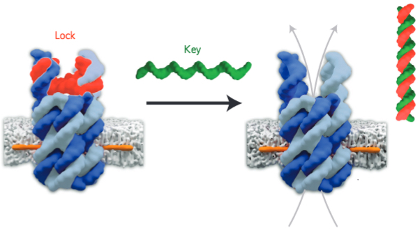
DNA-based lock-and-key pore design allows for precision delivery of drugs to cancer and other cells (credit: Stefan Howorka and Jonathan Burns/UCL)

Scientists at University College London (UCL) and Nanion Technologies in Munich have developed synthetic DNA-based pores that control which molecules can pass through a cell’s wall, achieving more precise drug delivery.

Therapeutics, including anti-cancer drugs, are ferried around the body in nanoscale carriers called vesicles, targeted to different tissues using biological markers. The new DNA-based pore design is intended to improve that process.

DNA Lock-and-key drug delivery

In operation, a drug-delivery vesicle (white in diagram above) carries the drug to a target cell for release.

The pore is designed as an open barrel made of six DNA-strand staves (blue and gray). The pore is kept closed by a DNA-strand “lock” (red) until the vesicle is prepared to release a drug. At that time, a “key” DNA-strand key (green) is released to hybridize (combine) with the lock DNA strand (forming the red-green helix on right), causing the pore to open and release the drug.*

The pore’s 2-nm-wide channel allows for selectively releasing small organic molecules, which include many medically important drug compounds.

This design for releasing drugs from vesicles improves on previous designs, in which drug release is triggered by temperature-induced leaky vesicle walls or with inserted peptide channels, both of which are less rigid and predictable than the new DNA mechanism.

The study was published Monday (Jan. 11) in Nature Nanotechnology. According to lead author Stefan Howorka, PhD., of UCL Chemistry, the researchers plan to test the synthetic pores in release of a variety of pharmaceutically active biomolecules, including anti-cancer drugs.

The research was funded by the Biotechnology and Biological Sciences Research Council (BBSRC), Leverhulme Trust and UCL Chemistry.

* Drugs are usually hydrophilic (water-loving), so pores are also designed to be hydrophilic, but that makes it difficult to attach pores to a vesicle membrane (which is hydrophobic, or fat-hating), so a hydrophobic cholesterol-based membrane (orange) was used to anchor the pore (blue and gray) to the membrane.


Abstract of A biomimetic DNA-based channel for the ligand-controlled transport of charged molecular cargo across a biological membrane

Biological ion channels are molecular gatekeepers that control transport across cell membranes. Recreating the functional principle of such systems and extending it beyond physiological ionic cargo is both scientifically exciting and technologically relevant to sensing or drug release. However, fabricating synthetic channels with a predictable structure remains a significant challenge. Here, we use DNA as a building material to create an atomistically determined molecular valve that can control when and which cargo is transported across a bilayer. The valve, which is made from seven concatenated DNA strands, can bind a specific ligand and, in response, undergo a nanomechanical change to open up the membrane-spanning channel. It is also able to distinguish with high selectivity the transport of small organic molecules that differ by the presence of a positively or negatively charged group. The DNA device could be used for controlled drug release and the building of synthetic cell-like or logic ionic networks.

Single-molecule detection of contaminants, explosives or diseases now possible

Artistic illustration showing an ultrasensitive detection platform termed “slippery liquid infused porous surface-enhanced Raman scattering” (SLIPSERS). An aqueous or oil droplet containing gold nanoparticles and captured analytes is allowed to evaporate on a slippery substrate, leading to the formation of a highly compact nanoparticle aggregate for use in surface-enhanced Raman scattering (SERS) detection. (credit: Shikuan Yang, Birgitt Boschitsch Stogin and Tak-Sing Wong/Penn State)

Penn State researchers have invented a way to detect single molecules of a number of chemical and biological species from gaseous, liquid or solid samples, with applications in analytical chemistry, molecular diagnostics, environmental monitoring and national security.

The invention is called SLIPSERS, an acronym combining “slippery liquid-infused porous surfaces” (SLIPS) and surface enhanced Raman scattering (SERS).*

“Being able to identify a single molecule is already very difficult. Being able to detect those molecules in all three phases [air, liquid, or bound to a solid] — that is really challenging,” said Tak-Sing Wong, assistant professor of mechanical engineering and the Wormley Family Early Career Professor in Engineering.

Although there are other techniques that allow researchers to concentrate molecules on a surface, those techniques mostly work with water as the medium. SLIPS can be used with any organic liquids, which makes it useful for environmental detection in soil samples, for example.

One of the researchers’ next steps will be to detect biomarkers in blood for disease diagnosis at very early stages of cancer when the disease is more easily treatable.

“Although the SLIPS technology is patented and licensed, the team has not sought patent protection on their SLIPSER work. “We believe that offering this technology to the public will get it developed at a much faster pace,” said Professor Wong. “This is a powerful platform that we think many people will benefit from.”

Their work appears an open-access online paper in Proceedings of the National Academy of Sciences (PNAS). It was funded by the National Science Foundation.

* Raman spectroscopy is a well-known method of analyzing materials in a liquid form using a laser to interact with the vibrating molecules in the sample, generating scattered light. But the molecule’s unique vibration shifts the frequency of the photons in the laser light beam up or down in a way that is characteristic of only that type of molecule, allowing it be uniquely identified.

However, the Raman signal is typically very weak and has to be enhanced in some way for detection. In the 1970s, researchers found that chemically roughening the surface of a silver substrate concentrated the Raman signal of the material adsorbed on the silver, and SERS was born.

Wong developed SLIPS as a post-doctoral researcher at Harvard University. SLIPS is composed of a surface coated with regular arrays of nanoscale posts infused with a liquid lubricant that does not mix with other liquids. The small spacing of the nanoposts traps the liquid between the posts and the result is a slippery surface that nothing adheres to.

“The problem,” Wong said, “is that trying to find a few molecules in a liquid medium is like trying to find a needle in a haystack. But if we can develop a process to gradually shrink the size of this liquid volume, we can get a better signal. To do that we need a surface that allows the liquid to evaporate uniformly until it gets to the micro or nanoscale. Other surfaces can’t do that, and that is where SLIPS comes in.”

The researchers assemble the gold nanoparticles so they have nanoscale gaps, called SERS “hot spots.” Using a laser with the right wavelength, the electrons will oscillate and a strong magnetic field will form in the gap area. This gives us very strong SERS signals of the molecules located within the gaps.”

If a droplet of liquid is placed on any normal surface, it will begin to shrink from the top down. When the liquid evaporates, the target molecules are left in random configurations with weak signals. But if all the molecules can be clustered among the gold nanoparticles, they will produce a very strong Raman signal.


Abstract of Ultrasensitive surface-enhanced Raman scattering detection in common fluids

Many analytes in real-life samples, such as body fluids, soil contaminants, and explosives, are dispersed in liquid, solid, or air phases. However, it remains a challenge to create a platform to detect these analytes in all of these phases with high sensitivity and specificity. Here, we demonstrate a universal platform termed slippery liquid-infused porous surface-enhanced Raman scattering (SLIPSERS) that enables the enrichment and delivery of analytes originating from various phases into surface-enhanced Raman scattering (SERS)-sensitive sites and their subsequent detection down to the subfemtomolar level (<10−15 mol⋅L−1). Based on SLIPSERS, we have demonstrated detection of various chemicals, biological molecules, and environmental contaminants with high sensitivity and specificity. Our platform may lead to ultrasensitive molecular detection for applications related to analytical chemistry, diagnostics, environmental monitoring, and national security.

New genes associated with extreme longevity identified

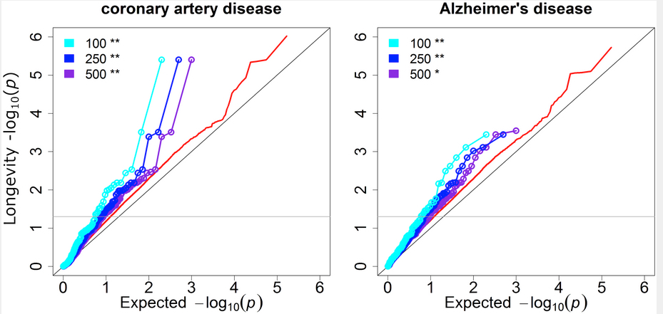
Disease GWAS show substantial genetic overlap with longevity. Shown are results for coronary artery disease and Alzheimer’s disease. The y axis is the observed P values for longevity, and the x axis is the expected P values under the null hypothesis that the disease is independent of longevity. The cyan, blue and purple lines show the P values for longevity of the top 100, 250, and 500 disease SNPs from independent genetic loci, respectively. Red lines show the background distribution of longevity P values for all independent genetic loci tested in both the longevity and disease GWAS. The grey horizontal line corresponds to the threshold for nominal significance (P< = 0.05) for longevity. Significance of enrichment was determined with the hypergeometric test. (credit: Kristen Fortney et al./PLOS Genetics)

What’s the secret of centenarians who have health and diet habits similar to the average person but have remained active and alert at very old ages?

Genes. That’s according to scientists at Stanford University and the University of Bologna, who have written a new report published in PLOS Genetics, based on their finding of several disease variants that may be absent in centenarians compared to the general population.

Genetic studies so far have only identified a single gene (APOE, known to be involved in Alzheimer’s disease) that is different in centenarians versus normal agers.

Finding additional longevity genes

To find the additional longevity genes, the authors first developed a new statistical method called informed GWAS (genome-wide association studies), which uses knowledge from 14 diseases to narrow down the search genes associated with longevity.

Using iGWAS, the scientists found eight SNPs (single nucleotide polymorphisms — molecular variations at different locations on the gene) that are significant for the centenarians they studied, and they were able to validate four of these in replication studies of long-lived subjects.

The four “longevity loci” (gene locations) along with the APOE gene may provide clues about physiological mechanisms for successful aging. These loci are known to be involved in various processes including cell senescence, autoimmunity, and cell signaling, and also with Alzheimer’s disease.

The incidence of nearly all diseases increases with age, so understanding genetic factors for successful aging could have a large impact on health. Future work may lead to a better understanding of how these genes promote successful aging and could identify additional longevity genes by recruiting more centenarians for analysis.


Abstract of Genome-Wide Scan Informed by Age-Related Disease Identifies Loci for Exceptional Human Longevity

We developed a new statistical framework to find genetic variants associated with extreme longevity. The method, informed GWAS (iGWAS), takes advantage of knowledge from large studies of age-related disease in order to narrow the search for SNPs associated with longevity. To gain support for our approach, we first show there is an overlap between loci involved in disease and loci associated with extreme longevity. These results indicate that several disease variants may be depleted in centenarians versus the general population. Next, we used iGWAS to harness information from 14 meta-analyses of disease and trait GWAS to identify longevity loci in two studies of long-lived humans. In a standard GWAS analysis, only one locus in these studies is significant (APOE/TOMM40) when controlling the false discovery rate (FDR) at 10%. With iGWAS, we identify eight genetic loci to associate significantly with exceptional human longevity at FDR < 10%. We followed up the eight lead SNPs in independent cohorts, and found replication evidence of four loci and suggestive evidence for one more with exceptional longevity. The loci that replicated (FDR < 5%) includedAPOE/TOMM40 (associated with Alzheimer’s disease), CDKN2B/ANRIL (implicated in the regulation of cellular senescence), ABO (tags the O blood group), and SH2B3/ATXN2 (a signaling gene that extends lifespan in Drosophila and a gene involved in neurological disease). Our results implicate new loci in longevity and reveal a genetic overlap between longevity and age-related diseases and traits, including coronary artery disease and Alzheimer’s disease. iGWAS provides a new analytical strategy for uncovering SNPs that influence extreme longevity, and can be applied more broadly to boost power in other studies of complex phenotypes.

MIT uses forests of carbon nanotubes with antibodies to capture hard-to-detect molecules

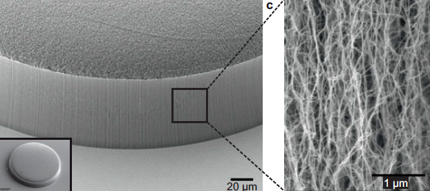
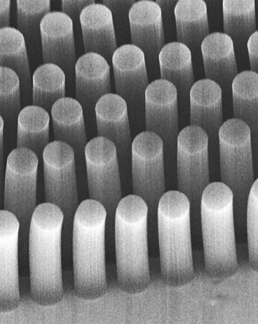
Scanning electron microscope image of carbon nanotubes showing textured porosity (credit: Allison L. Yost et al./Microsystems & Nanoengineering)

Engineers at MIT have devised a new technique for trapping hard-to-detect molecules, using forests of coated carbon nanotubes.

The team modified a simple microfluidic channel with an array of vertically aligned carbon nanotubes — rolled lattices of carbon atoms that resemble tiny tubes of chicken wire.

Carbon-nanotube posts can trap cancer and other cells as they flow through a microfluidic device (credit: Brian Wardle)

The researchers had previously devised a method for standing carbon nanotubes on their ends, like trees in a forest (see “Trapping cancer cells with carbon nanotubes“). This 3-D array of permeable carbon nanotubes allows fluid to flow through a microfluidic device.

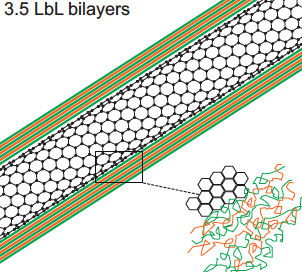
Now, in a study published this week in the Journal of Microengineering and Nanotechnology, the researchers have given the nanotube array the additional ability to trap specific particles. To do this, the team coated the array, layer by layer, with polymers of alternating electric charge.

Depending on the number of layers deposited, the researchers can create thicker or thinner nanotubes and thereby tailor the porosity of the forest to capture larger or smaller particles of interest. The nanotubes’ polymer coating can also be chemically manipulated to bind specific bioparticles flowing through the forest.

The combination of carbon nanotubes and multilayer coatings may help finely tune microfluidic devices to capture extremely small and rare particles, such as certain viruses and proteins, says Brian Wardle, professor of aeronautics and astronautics at MIT.

“There are smaller bioparticles that contain very rich amounts of information that we don’t currently have the ability to access in point-of-care [medical testing] devices like microfluidic chips,” says Wardle, who is a co-author on the paper. “Carbon nanotube arrays could actually be a platform that could target that size of bioparticle.”

What’s more, Wardle says, a three-dimensional forest of carbon nanotubes would provide much more surface area on which target molecules may interact, compared with the two-dimensional surfaces in conventional microfluidics.

Capturing specific particles of interest

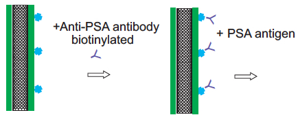
To test this idea, the researchers used an established technique to treat the surface of the nanotubes with antibodies that bind to prostate specific antigen (PSA), a common experimental target.

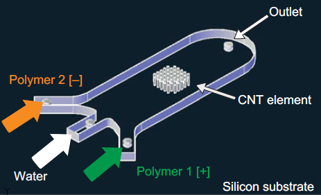
A 3-D array of carbon nanotubes on a microfluidic device coated with successive layers of alternately charged polymer solutions (credit: Allison L. Yost et al./Microsystems & Nanoengineering)

The team integrated a 3-D array of carbon nanotubes into a microfluidic device by using chemical vapor deposition and photolithography to grow and pattern carbon nanotubes onto silicon wafers. They then grouped the nanotubes into a cylinder-shaped forest, measuring about 50 micrometers tall and 1 millimeter wide, and centered the array within a 3 millimeter-wide, 7-millimeter long microfluidic channel.

Polyelectrolyte multilayer (PEM) film deposition on carbon-nanotube surface (credit: Allison L. Yost et al./Microsystems & Nanoengineering)

The researchers coated the nanotubes in successive layers of alternately charged polymer solutions to create distinct, binding layers around each nanotube. To do so, they flowed each solution through the channel and found they were able to create a more uniform coating with a gap between the top of the nanotube forest and the roof of the channel. Such a gap allowed solutions to flow over, then down into the forest, coating each individual nanotube.

Carbon nanotube treated with antibodies for PSA capture (credit: Allison L. Yost et al./Microsystems & Nanoengineering)

After coating the nanotube array in layers of polymer solution, the researchers demonstrated that the array could be primed to detect a given molecule by treating it with antibodies that typically bind to prostate specific antigen (PSA). They pumped in a solution containing small amounts of PSA and found that the array captured the antigen effectively, throughout the forest, rather than just on the outer surface of a typical microfluidic element.

The polymer-coated arrays captured 40 percent more antigens, compared with arrays lacking the polymer coating.

Wardle says that the nanotube array is extremely versatile. The carbon nanotubes can be manipulated mechanically, electrically, and optically and the polymer coatings can be chemically altered to capture a wide range of particles. He says an immediate target may be biomarkers called exosomes, which are less than 100 nanometers wide and can be important signals of a disease’s progression.

“This type of device actually has all the characteristics and functionality that would allow you to go after bioparticles like exosomes and things that really truly are nanometer scale,” he noted.

This research was funded in part by the National Science Foundation.


Abstract of Layer-by-layer functionalized nanotube arrays: A versatile microfluidic platform for biodetection

We demonstrate the layer-by-layer (LbL) assembly of polyelectrolyte multilayers (PEM) on three-dimensional nanofiber scaffolds. High porosity (99%) aligned carbon nanotube (CNT) arrays are photolithographically patterned into elements that act as textured scaffolds for the creation of functionally coated (nano)porous materials. Nanometer-scale bilayers of poly(allylamine hydrochloride)/poly(styrene sulfonate) (PAH/SPS) are formed conformally on the individual nanotubes by repeated deposition from aqueous solution in microfluidic channels. Computational and experimental results show that the LbL deposition is dominated by the diffusive transport of the polymeric constituents, and we use this understanding to demonstrate spatial tailoring on the patterned nanoporous elements. A proof-of-principle application, microfluidic bioparticle capture using N-hydroxysuccinimide-biotin binding for the isolation of prostate-specific antigen (PSA), is demonstrated.

Genetic ‘intelligence networks’ discovered in the brain

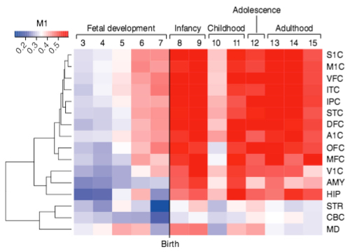
Color-coded heatmap of gradient of expression of the M1 gene network, spanning fetal development to late adulthood and expressed in distinct cortical regions (listed on right, such as primary somatosensory cortex, S1C). Most of the genes in this network express in cortical regions (indicated by red), except for the V1C (primary visual cortex), STR (striatum), CBC (cerebellar cortex), and MD (mediodorsal nucleus of thalamus) brain areas. (credit: Michael R. Johnson et al./Nature Neuroscience)

Scientists from Imperial College London have identified two clusters (“gene networks”) of genes that are linked to human intelligence. Called M1 and M3, these gene networks appear to influence cognitive function, which includes memory, attention, processing speed and reasoning.

Importantly, the scientists have discovered that these two networks are likely to be under the control of master regulator switches. The researcher want to identify those switches and see if they can manipulate them, and ultimately find out if this knowledge of gene networks could allow for boosting cognitive function.

“We know that genetics plays a major role in intelligence but until now, haven’t known which genes are relevant,” said Michael Johnson, lead author of the study from the Imperial College London Department of Medicine. Johnson says the genes they have found so far are likely to share a common regulation, which means it may be possible to manipulate a whole set of genes linked to human intelligence.

Combining data from brain samples, genomic information, and IQ tests

In the study, published in the journal Nature Neuroscience, the international team of researchers looked at samples of human brain from patients who had undergone neurosurgery for epilepsy. The investigators analyzed thousands of genes expressed in the human brain, and then combined these results with genetic information from healthy people who had undergone IQ tests and from people with neurological disorders such as autism spectrum disorder and intellectual disability.

Then they conducted various computational analyses and comparisons to identify the gene networks influencing healthy human cognitive abilities. Remarkably, they found that some of the same genes that influence human intelligence in healthy people cause impaired cognitive ability and epilepsy when mutated. And they found that genes that make new memories or sensible decisions when faced with lots of complex information also overlap with those that cause severe childhood onset epilepsy or intellectual disability.

“This study shows how we can use large genomic datasets to uncover new pathways for human brain function in both health and disease,” Johnson said. “Eventually, we hope that this sort of analysis will provide new insights into better treatments for neurodevelopmental diseases such as epilepsy, and ameliorate or treat the cognitive impairments associated with these devastating diseases.”


Abstract of Systems genetics identifies a convergent gene network for cognition and neurodevelopmental disease

Genetic determinants of cognition are poorly characterized, and their relationship to genes that confer risk for neurodevelopmental disease is unclear. Here we performed a systems-level analysis of genome-wide gene expression data to infer gene-regulatory networks conserved across species and brain regions. Two of these networks, M1 and M3, showed replicable enrichment for common genetic variants underlying healthy human cognitive abilities, including memory. Using exome sequence data from 6,871 trios, we found that M3 genes were also enriched for mutations ascertained from patients with neurodevelopmental disease generally, and intellectual disability and epileptic encephalopathy in particular. M3 consists of 150 genes whose expression is tightly developmentally regulated, but which are collectively poorly annotated for known functional pathways. These results illustrate how systems-level analyses can reveal previously unappreciated relationships between neurodevelopmental disease–associated genes in the developed human brain, and provide empirical support for a convergent gene-regulatory network influencing cognition and neurodevelopmental disease.