Magnetic nanoparticles combat biofilms, a source of chronic bacterial infections

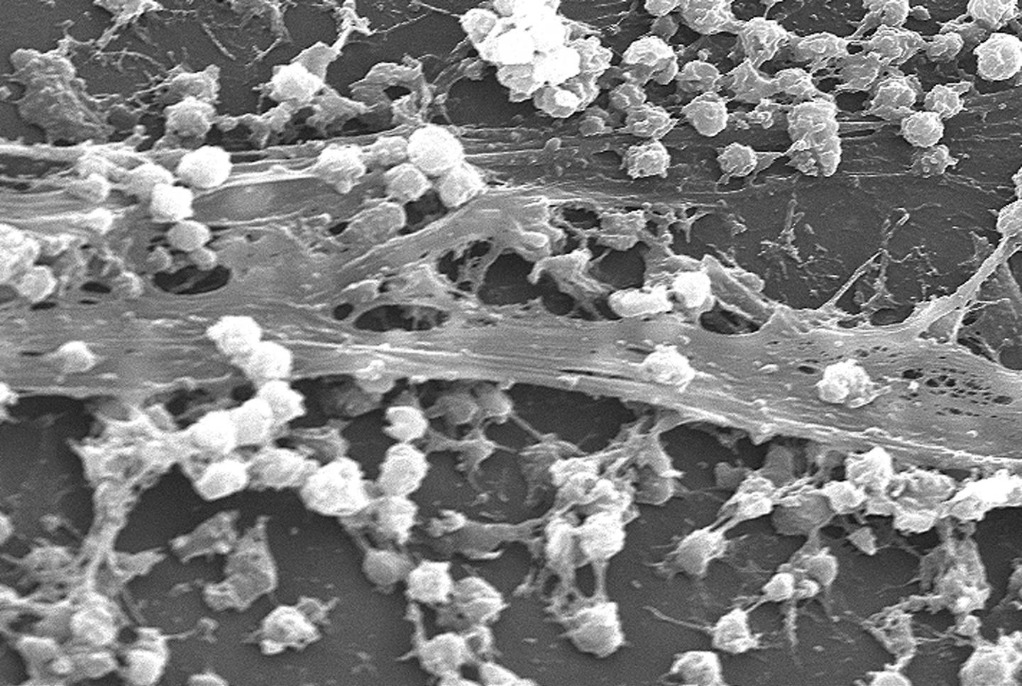
Staphylococcus aureus bacterial biofilm on an indwelling catheter (credit: CDC)

A solution for biofilms* — a scourge of infections in hospitals and kitchens formed by bacteria that stick to each other on living tissue and medical instruments — has been developed by University of New South Wales researchers: Injecting iron oxide nanoparticles into the biofilms, and using an applied magnetic field to heat them, triggering them into dispersing.
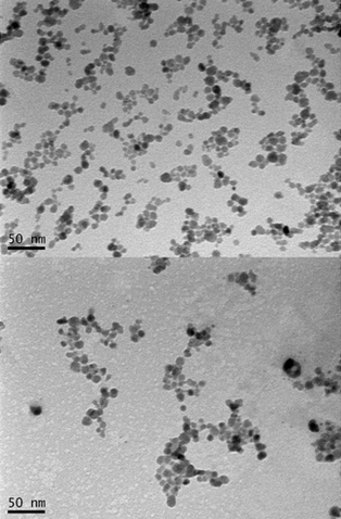
Transmission electron microscopy (TEM) micrographs of dried nanoparticles before (top) and after (bottom) conjugation with polymer (credit: Thuy-Khanh Nguyen et al./Scientific Reports)

“Chronic biofilm-based infections are often extremely resistant to antibiotics and many other conventional antimicrobial agents, and have a high capacity to evade the body’s immune system,” said Associate Professor Cyrille Boyer of the School of Chemical Engineering and deputy director of Australian Centre for NanoMedicine. “Our study points to a pathway for the non-toxic dispersal of biofilms in infected tissue, while also greatly improving the effect of antibiotic therapies.”

When biofilms want to colonize a new site, they disperse into individual cells, reducing the protective action of the biofilm. It is this process the UNSW team sought to trigger. They achieved this using iron oxide nanoparticles coated with polymers that help stabilize and maintain the nanoparticles in a dispersed state, making them an ideal non-toxic tool for treating biofilm infection.

Once dispersed, the bacteria are easier to deal with, creating the potential to remove recalcitrant, antimicrobial-tolerant biofilm infections with antimicrobial agents.

The discovery of how to dislodge biofilms by the UNSW Faculty of Engineering team was made using the opportunistic human pathogen Pseudomonas aeruginosa. This is a model organism whose response to the technique the researchers believe will apply to most other bacteria.

“The use of these polymer-coated iron oxide nanoparticles to disperse biofilms may have broad applications across a range of clinical and industrial settings,” said Boyer.

The research appears in an open-access paper today (Dec. 21) in Nature’s Scientific Reports.

* Biofilms have been linked to 80% of infections, forming on living tissues (e.g. respiratory, gastrointestinal and urinary tracts, oral cavities, eyes, ears, wounds, heart and cervix) or dwelling in medical devices (e.g. dialysis catheters, prosthetic implants and contact lenses).

The formation of biofilms is a growing and costly problem in hospitals, creating infections that are more difficult to treat — leading to chronic inflammation, impaired wound healing, rapidly acquired antibiotic resistance and the spread of infectious embolisms in the bloodstream.

They also cause fouling and corrosion of wet surfaces, and the clogging of filtration membranes in sensitive equipment, even posing a threat to public health by acting as reservoirs of pathogens in distribution systems for drinking water.

In general, bacteria have two life forms during growth and proliferation: planktonic, where bacteria exist as single, independent cells; or aggregated together in colonies as biofilms, where bacteria grow in a slime-like polymer matrix that protects them from the environment around them.

Acute infections mostly involve planktonic bacteria, which are usually treatable with antibiotics. However, when bacteria have had enough time to form a biofilm — within a human host or non-living material such as dialysis catheters – an infection can often become untreatable and develop into a chronic state.


UNSW | 2015 Malcolm McIntosh Prize for Physical Scientist of the Year


Abstract of Iron oxide nanoparticle-mediated hyperthermia stimulates dispersal in bacterial biofilms and enhances antibiotic efficacy

The dispersal phase that completes the biofilm lifecycle is of particular interest for its potential to remove recalcitrant, antimicrobial tolerant biofilm infections. Here we found that temperature is a cue for biofilm dispersal and a rise by 5 °C or more can induce the detachment of Pseudomonas aeruginosa biofilms. Temperature upshifts were found to decrease biofilm biomass and increase the number of viable freely suspended cells. The dispersal response appeared to involve the secondary messenger cyclic di-GMP, which is central to a genetic network governing motile to sessile transitions in bacteria. Furthermore, we used poly((oligo(ethylene glycol) methyl ether acrylate)-block-poly(monoacryloxy ethyl phosphate)-stabilized iron oxide nanoparticles (POEGA-b-PMAEP@IONPs) to induce local hyperthermia in established biofilms upon exposure to a magnetic field. POEGA-b-PMAEP@IONPs were non-toxic to bacteria and when heated induced the detachment of biofilm cells. Finally, combined treatments of POEGA-b-PMAEP@IONPs and the antibiotic gentamicin reduced by 2-log the number of colony-forming units in both biofilm and planktonic phases after 20 min, which represent a 3.2- and 4.1-fold increase in the efficacy against planktonic and biofilm cells, respectively, compared to gentamicin alone. The use of iron oxide nanoparticles to disperse biofilms may find broad applications across a range of clinical and industrial settings.

ASCB Celldance 2015 premieres three videos featuring live cell imaging

ASCB’s Celldance Studios released Monday (Dec. 14) three new short videos made by cell scientists, featuring dramatic live cell imaging.

The videos, which take advantage of accelerating advances in super-resolution imaging, fluorescent tagging, and Big Data manipulation, where made in the labs of Douglas Robinson at John Hopkins University, John Condeelis at Albert Einstein College of Medicine, and Satyajit Mayor at the National Centre for the Biological Sciences (NCBS) in India.

The videos were announced at the 2015 American Society for Cell Biology annual meeting.


Edison Leung et al., Albert Einstein College of Medicine for ASCB Celldance 2015 | Spying on Cancer Cell Invasion

Edison Leung says his cancer research lab makes movies of all genres—horror, action, thriller, and war—all shot inside cancer tumors. Working alongside Allison Harney in the Einstein lab of John Condeelis, Leung’s Celldance video shows metastasizing cancer cells, helped by the body’s own immune cells called macrophages, break through a blood vessel wall and escape to form new tumors. Through live cell imaging, Leung’s video captures the moment the cancer cell and the macrophage work as a team to break through the vessel wall of a mouse.


Satyajit Mayor, National Centre for Biological Science, India, for ASCB Celldance 2015 | At the Cell’s Edge

In this video made the Mayor lab in Bangalore, researchers give a detailed account of their exploration of the churning lipids and proteins on the cell surface, illustrated by startling live cell videos, high-tech simulations, and low-tech white boards. “At the Cell’s Edge” paints the cell membrane as a restless nanoscale seascape.


Douglas Robinson, Johns Hopkins University for ASCB Celldance 2015 | Shape Shifting Cells

The Robinson lab video is a visual extravaganza of high-resolution microscopy, mathematical representations, animation, and live action. It starts with a basic question: why are cancer cells softer than normal cells? It ends with a potential drug that can turn hardness against pancreatic cancer cells. Their cell story walks us through the stages of discovery — the shape and hardness of cells, cell cannibalism where the soft (cancer) eat the hard (normal), an amoeba model to see the proteins that stiffen cells, and the identification of 4HAP, a small protein that attacks pancreatic cancer cells.

New microscope creates near-real-time videos of nanoscale processes


MIT | Microscope creates near-real-time videos of nanoscale processes

MIT engineers have designed an atomic force microscope (AFM) that scans images 2,000 times faster than existing commercial models. Operating at near-real-time-video speed, it can capture structures as small as a fraction of a nanometer from single strands of DNA down to individual hydrogen bonds.

Existing AFMs have similar spatial resolution but function at slow speeds.

In one dramatic demonstration of the instrument’s capabilities (see video), the researchers scanned a 70- by-70-micrometers sample of calcite as it was first immersed in deionized water and later exposed to sulfuric acid. Over a period of several seconds, the team observed the acid eating away at the calcite, expanding existing nanometer-sized pits in the material that quickly merged and led to a layer-by-layer removal of calcite along the material’s crystal pattern.

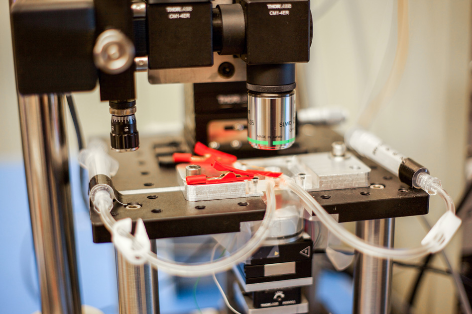
The new MIT high-speed microscope produces images of chemical processes taking place at the nanoscale at a rate that is close to real-time video. This closeup shot of the microscope shows transparent tubes used to inject various liquids into the imaging environment. This liquid can be water, acid, buffer solution for live bacteria, cells, or electrolytes in an electrochemical process. Researchers use one as an inlet and the other as an outlet to circulate and refresh the solutions throughout the experiment. (credit: Jose-Luis Olivares/MIT)

Kamal Youcef-Toumi, a professor of mechanical engineering at MIT, says the instrument’s sensitivity and speed will enable scientists to watch atomic-sized processes play out as high-resolution “movies” for the first time.

“People can see, for example, condensation, nucleation, dissolution, or deposition of material, and how these happen in real-time — things that people have never seen before,” Youcef-Toumi says. “This is fantastic to see these details emerging. And it will open great opportunities to explore all of this world that is at the nanoscale.”

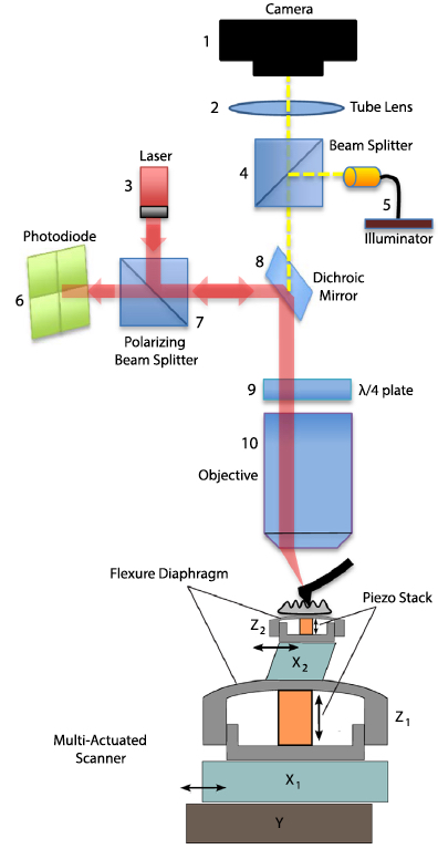
A schematic of the AFM (credit: I. Soltani Bozchalooi et al./Ultramicroscopy)

The group’s design and images, which are based on the PhD work of Iman Soltani Bozchalooi, now a postdoc in the Department of Mechanical Engineering, are published in the journal Ultramicroscopy.

Atomic force microscopes typically scan samples using an ultrafine probe, or needle, that skims along the surface of a sample, tracing its topography, similarly to how a blind person reads Braille. Samples sit on a movable platform, or scanner, that moves the sample laterally and vertically beneath the probe.

Because AFMs scan incredibly small structures, the instruments have to work slowly, line by line, to avoid any sudden movements that could alter the sample or blur the image. Such conventional microscopes typically scan only about one to two lines per second.

To speed up the scanning process, scientists have built platforms that scan samples more quickly, but over a smaller area, and the platforms don’t allow scientists to zoom out to see a wider view or study larger features.

Synchronized Scanners

The main innovation of the new design is a multi-actuated scanner. The sample platform incorporates both a smaller, speedier scanner and a larger, slower scanner for every direction, which work together as one system to scan a wide 3-D region at high speed.

The microscope operates at about eight to 10 frames per second and can scan across hundreds of microns and image features that are several microns high.

“We want to go to real video, which is at least 30 frames per second,” Youcef-Toumi says. “Hopefully we can work on improving the instrument and controls so that we can do video-rate imaging while maintaining its large range and keeping it user-friendly.”


Abstract of Design and control of multi-actuated atomic force microscope for large-range and high-speed imaging
This paper presents the design and control of a high-speed and large-range atomic force microscopy (AFM). A multi-actuation scheme is proposed where several nano-positioners cooperate to achieve the range and speed requirements. A simple data-based control design methodology is presented to effectively operate the AFM scanner components. The proposed controllers compensate for the coupled dynamics and divide the positioning responsibilities between the scanner components. As a result, the multi-actuated scanner behavior is equivalent to that of a single XYZ positioner with large range and high speed. The scanner of the designed AFM is composed of five nano-positioners, features 6 μm out-of-plane and 120 μm lateral ranges and is capable of high-speed operation. The presented AFM has a modular design with laser spot size of 3.5 μm suitable for small cantilever, an optical view of the sample and probe, a conveniently large waterproof sample stage and a 20 MHz data throughput for high resolution image acquisition at high imaging speeds. This AFM is used to visualize etching of calcite in a solution of sulfuric acid. Layer-by-layer dissolution and pit formation along the crystalline lines in a low pH environment is observed in real time.


Abstract of Multi-actuation and PI control: A simple recipe for high-speed and large-range atomic force microscopy

High speed atomic force microscopy enables observation of dynamic nano-scale processes. However, maintaining a minimal interaction force between the sample and the probe is challenging at high speed specially when using conventional piezo-tubes. While rigid AFM scanners are operational at high speeds with the drawback of reduced tracking range, multi-actuation schemes have shown potential for high-speed and large-range imaging. Here we present a method to seamlessly incorporate additional actuators into conventional AFMs. The equivalent behavior of the resulting multi-actuated setup resembles that of a single high-speed and large-range actuator with maximally flat frequency response. To achieve this, the dynamics of the individual actuators and their couplings are treated through a simple control scheme. Upon the implementation of the proposed technique, commonly used PI controllers are able to meet the requirements of high-speed imaging. This forms an ideal platform for retroactive enhancement of existing AFMs with minimal cost and without compromise on the tracking range. A conventional AFM with tube scanner is retroactively enhanced through the proposed method and shows an order of magnitude improvement in closed loop bandwidth performance while maintaining large range. The effectiveness of the method is demonstrated on various types of samples imaged in contact and tapping modes, in air and in liquid.

New mass spectral imaging instrument maps cells’ composition in 3-D at more than 100 times higher resolution

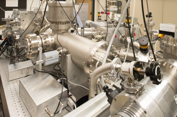
A mass spectral imaging instrument instrument developed at Colorado State University (credit: William Cotton/Colorado State University)

A one-of-a-kind mass spectral imaging instrument built at Colorado State University (CSU) lets scientists map cellular composition in three dimensions at a nanoscale image resolution of 75 nanometers wide and 20 nanometers deep — more than 100 times higher resolution than was earlier possible, according to the scientists.

The instrument may be able to observe how well experimental drugs penetrate and are processed by cells as new medications are developed to combat disease, customize treatments for specific cell types in specific conditions, identify the sources of pathogens propagated for bioterrorism, or investigate new ways to overcome antibiotic resistance among patients with surgical implants, according to professor Dean Crick of the CSU Mycobacteria Research Laboratories.

Crick’s primary research interest is tuberculosis, an infectious respiratory disease that contributes to an estimated 1.5 million deaths around the world each year. “We’ve developed a much more refined instrument,” Crick said. “It’s like going from using a dull knife to using a scalpel. You could soak a cell in a new drug and see how it’s absorbed, how quickly, and how it affects the cell’s chemistry.”

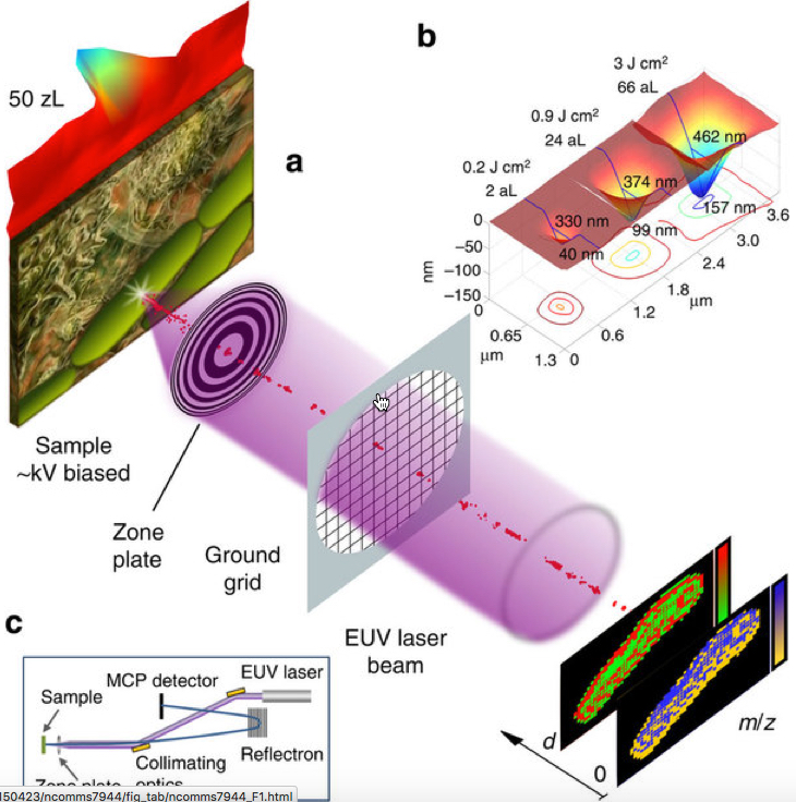
Schematics showing the focused extreme ultraviolet laser beam ablating (removal of material from the surface) a sample to produce an ion stream that is analyzed by a mass spectrometer. (b) Atomic force microscope (AFM) images of craters ablated in polymethyl methacrylate (PMMA) by a single EUV laser shot at different irradiation particle rated. The craters show smooth profiles with no signs of thermal damage. (c) Schematic of the instrument setup including the collimating extreme ultraviolet laser optics, focusing zone plate and spectrometer. (credit: Ilya Kuznetsov et al./Nature Communications)

The earlier generation of laser-based mass-spectral imaging could identify the chemical composition of a cell and could map its surface in two dimensions at microscale (about one micrometer), but could not chart cellular anatomy at more-detailed nanoscale dimensions and in 3-D, Crick said.

The research is described in an open-access paper in Nature Communications and was funded by a $1 million grant from the National Institutes of Health as part of an award to the Rocky Mountain Regional Center of Excellence for Biodefense and Emerging Infectious Disease Research. The optical equipment that focuses the laser beam was created by the Center for X-Ray Optics at the Lawrence Berkeley National Laboratory in Berkeley, Calif.

A special issue of Optics and Photonics News this month highlights the CSU research as among “the most exciting peer-reviewed optics research to have emerged over the past 12 months.”


CSU College of Veterinary Medicine and Biomedical Sciences | Nanoscale Mass-Spectral Imaging in 3-D at Colorado State University


Abstract of Three-dimensional nanoscale molecular imaging by extreme ultraviolet laser ablation mass spectrometry

Analytical probes capable of mapping molecular composition at the nanoscale are of critical importance to materials research, biology and medicine. Mass spectral imaging makes it possible to visualize the spatial organization of multiple molecular components at a sample’s surface. However, it is challenging for mass spectral imaging to map molecular composition in three dimensions (3D) with submicron resolution. Here we describe a mass spectral imaging method that exploits the high 3D localization of absorbed extreme ultraviolet laser light and its fundamentally distinct interaction with matter to determine molecular composition from a volume as small as 50 zl in a single laser shot. Molecular imaging with a lateral resolution of 75 nm and a depth resolution of 20 nm is demonstrated. These results open opportunities to visualize chemical composition and chemical changes in 3D at the nanoscale.

Periodic table of protein complexes helps predict novel protein structures

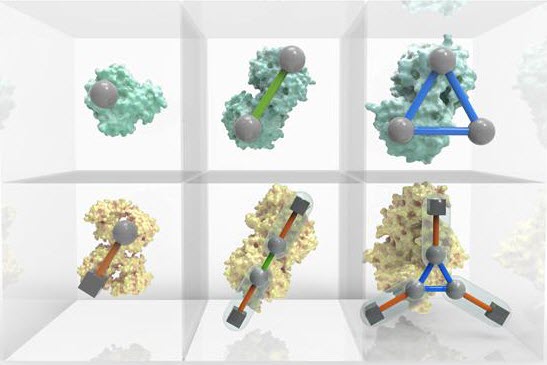
An interactive Periodic Table of Protein Complexes is available at http://sea31.user.srcf.net/periodictable/ (credit: EMBL-EBI/Spencer Phillips)

The Periodic Table of Protein Complexes, developed by researchers in the UK and to be published Dec. 11 in the journal Science, offers a new way of looking at the enormous variety of structures that proteins can build in nature. More importantly, it suggests which ones might be discovered next and how entirely novel structures could be engineered.

Created by an interdisciplinary team led by researchers at the Wellcome Genome Campus and the University of Cambridge, the Table provides a valuable tool for research into evolution and protein engineering.

Handling complexity

Almost every biological process depends on proteins interacting and assembling into complexes in a specific way, and many diseases are associated with problems in complex assembly. “Evolution has given rise to a huge variety of protein complexes, and it can seem a bit chaotic,” explains Joe Marsh of the MRC Human Genetics Unit at the University of Edinburgh. “But if you break down the steps proteins take to become complexes, there are some basic rules that can explain almost all of the assemblies people have observed so far.”

Fundamentally, protein complex assembly can be seen as endless variations on dimerization (one doubles, and becomes two), cyclisation (one forms a ring of three or more), and subunit addition (two different proteins bind to each other). Because these happen in a fairly predictable way, it’s not as hard as you might think to predict how a novel protein would form.

“By analyzing the tens of thousands of protein complexes for which three-dimensional structures have already been experimentally determined, we could see repeating patterns in the assembly transitions that occur — and with new data from mass spectrometry we could start to see the bigger picture,” says Marsh.


Abstract of Principles of assembly reveal a periodic table of protein complexes

INTRODUCTION: The assembly of proteins into complexes is crucial for most biological processes. The three-dimensional structures of many thousands of homomeric and heteromeric protein complexes have now been determined, and this has had a broad impact on our understanding of biological function and evolution. Despite this, the organizing principles that underlie the great diversity of protein quaternary structures observed in nature remain poorly understood, particularly in comparison with protein folds, which have been extensively classified in terms of their architecture and evolutionary relationships.

RATIONALE: In this work, we sought a comprehensive understanding of the general principles underlying quaternary structure organization. Our approach was to consider protein complexes in terms of their assembly. Many protein complexes assemble spontaneously via ordered pathways in vitro, and these pathways have a strong tendency to be evolutionarily conserved. Furthermore, there are strong similarities between protein complex assembly and evolutionary pathways, with assembly pathways often being reflective of evolutionary histories, and vice versa. This suggests that it may be useful to consider the types of protein complexes that have evolved from the perspective of what assembly pathways are possible.

RESULTS: We first examined the fundamental steps by which protein complexes can assemble, using electrospray mass spectrometry experiments, literature-curated assembly data, and a large-scale analysis of protein complex structures. We found that most assembly steps can be classified into three basic types: dimerization, cyclization, and heteromeric subunit addition. By systematically combining different assembly steps in different ways, we were able to enumerate a large set of possible quaternary structure topologies, or patterns of key interfaces between the proteins within a complex. The vast majority of real protein complex structures lie within these topologies. This enables a natural organization of protein complexes into a “periodic table,” because each heteromer can be related to a simpler symmetric homomer topology. Exceptions are mostly the result of quaternary structure assignment errors, or cases where sequence-identical subunits can have different interactions and thus introduce asymmetry. Many of these asymmetric complexes fit the paradigm of a periodic table when their assembly role is considered. Finally, we implemented a model based on the periodic table, which predicts the expected frequencies of each quaternary structure topology, including those not yet observed. Our model correctly predicts quaternary structure topologies of recent crystal and electron microscopy structures that are not included in our original data set.

CONCLUSION: This work explains much of the observed distribution of known protein complexes in quaternary structure space and provides a framework for understanding their evolution. In addition, it can contribute considerably to the prediction and modeling of quaternary structures by specifying which topologies are most likely to be adopted by a complex with a given stoichiometry, potentially providing constraints for multi-subunit docking and hybrid methods. Lastly, it could help in the bioengineering of protein complexes by identifying which topologies are most likely to be stable, and thus which types of essential interfaces need to be engineered.

Chemicals that make plants defend themselves could replace pesticides

Researchers used the relative induction of GUS activity as a screening tool for identifying new chemical elicitors that induce resistance in rice to the white-backed planthopper Sogatella furcifera (credit: Xingrui He et al./Bioorganic & Medicinal Chemistry Letters)

Chemical triggers that make plants defend themselves against insects could replace pesticides, causing less damage to the environment. New research published in an open-access paper in Bioorganic & Medicinal Chemistry Letters identifies five chemicals that trigger rice plants to fend off a common pest — the white-backed planthopper, Sogatella furcifera.

Pesticides have a detrimental effect on ecosystems, ravaging food chains and damaging the environment. One of the problems with many pesticides is that they kill indiscriminately.

Sogatella furcifera (credit: BIO Photography Group/CNC, Biodiversity Institute of Ontario)

For rice plants, this means pesticides kill the natural enemies of one of their biggest pests, the white-backed planthopper Sogatella furcifera. This pest attacks rice, leading to yellowing or “hopper burn,” which causes the plants to wilt and can damage the grains. It also transmits a virus disease called, southern rice black-streaked dwarf virus, which stunts the plants’ growth and stops them from “heading,” which is when pollination occurs.

Left untreated, many of the insects’ eggs would be eaten, but when pesticides are used, these hatch, leading to even more insects on the plants. What’s more, in some areas as many as a third of the planthoppers are resistant to pesticides.

“The extensive application of chemical insecticides not only causes severe environmental and farm produce pollution but also damages the ecosystem,” explained Dr. Jun Wu, one of the authors of the study and professor at Zhejiang University 
in China. “Therefore, developing safe and effective methods to control insect pests is highly desired; this is why we decided to investigate these chemicals.”

Enhancing plants’ natural defense mechanisms

Plants have natural self-defense mechanisms that kick in when they are infested with pests like the planthopper. This defense mechanism can be switched on using chemicals that do not harm the environment and are not toxic to the insects or their natural enemies.

In the new study, researchers from Zhejiang University 
in China developed a new way of identifying these chemicals. Using a specially designed screening system, they determined to what extent different chemicals switched on the plants’ defense mechanism. The team designed and synthesized 29 phenoxyalkanoic acid derivatives. Of these, they identified five that could be effective at triggering the rice plants to defend themselves.

The researchers used bioassays to show that these chemicals could trigger the plant defense mechanism and repel the white-backed planthopper, which suggests potential use in insect pest management.

“We demonstrate for the first time that some phenoxyalkanoic acid derivatives have the potential to become such plant protection agents against the rice white-backed planthopper,” said Dr. Yonggen Lou, one of the authors of the study and professor at Zhejiang University 
in China. “This new approach to pest management could help protect the ecosystem while defending important crops against attack.”

The next step for the research will be to explore how effective the chemicals are at boosting the plants’ defenses and controlling planthoppers in the field.


Abstract of Finding new elicitors that induce resistance in rice to the white-backed planthopper Sogatella furcifera

Herein we report a new way to identify chemical elicitors that induce resistance in rice to herbivores. Using this method, by quantifying the induction of chemicals for GUS activity in a specific screening system that we established previously, 5 candidate elicitors were selected from the 29 designed and synthesized phenoxyalkanoic acid derivatives. Bioassays confirmed that these candidate elicitors could induce plant defense and then repel feeding of white-backed planthopper Sogatella furcifera.

Parkinson’s disease researchers discover a way to reprogram the genome to produce dopamine neurons

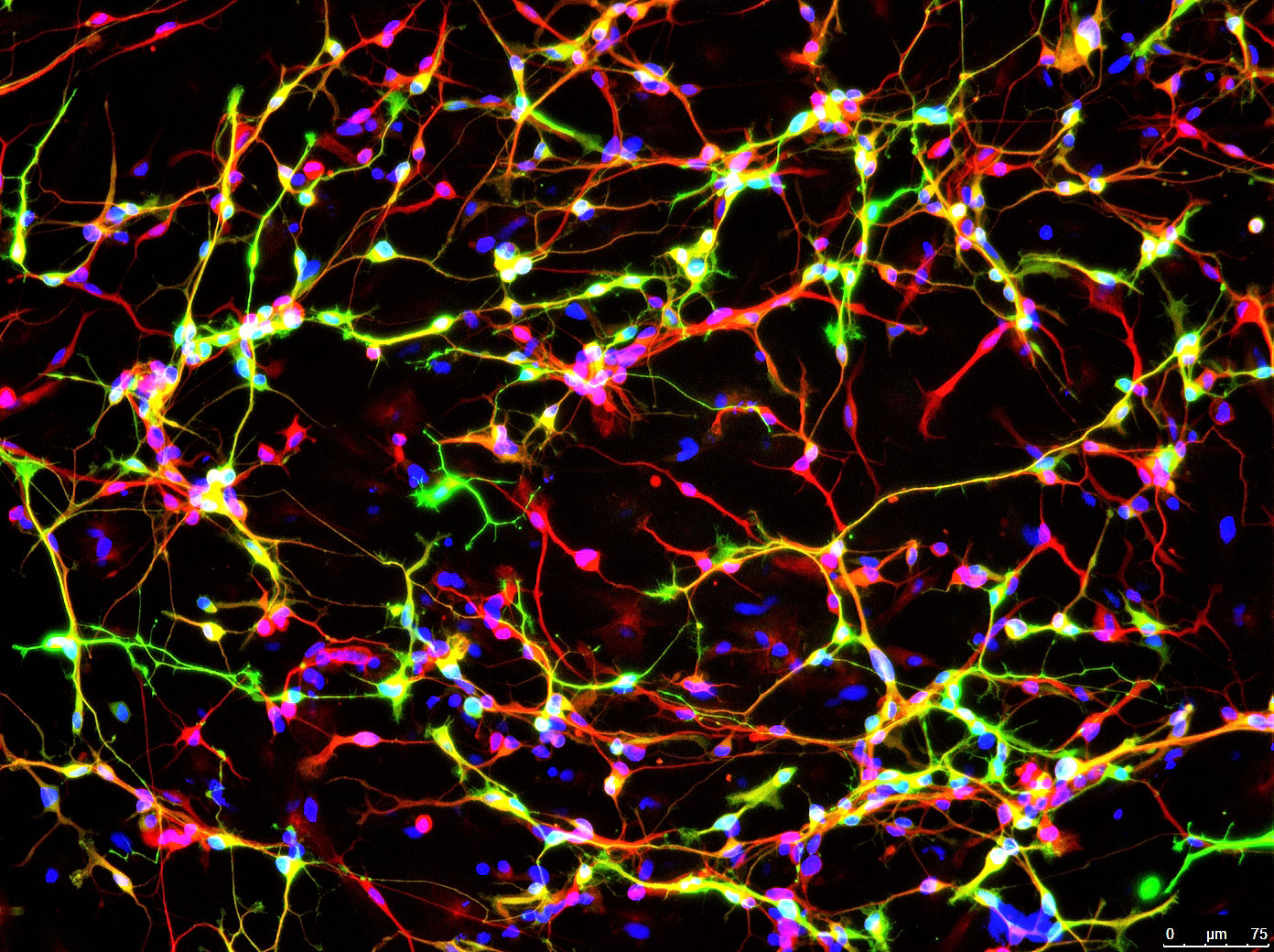
Image shows a protein found only in neurons (red) and an enzyme that synthesizes dopamine (green). Cell DNA is labeled in blue. (credit: Jian Feng, University at Buffalo)

Parkinson’s disease researchers at the Jacobs School of Medicine and Biomedical Sciences at the University at Buffalo have developed a way to ramp up the conversion of skin cells into neurons that can produce dopamine.

For decades, the elusive holy grail in Parkinson’s disease research has been finding a way to repair faulty dopamine neurons and put them back into patients, where they will start producing dopamine again. Researchers have tried fetal material, which is difficult to obtain and of variable quality, and embryonic stem cells (a long process with a low yield), and more recently, skin cells (difficult to obtain sufficient quantities of neurons).

To control movement and balance, dopamine signals travel from the substantia nigra in the midbrain up to brain regions including the corpus striatum, the globus pallidus, and the thalamus. But in Parkinson’s disease, most of the dopamine signals from the substantia nigra are lost. (credit: NIH)

Bypassing the cellular “gatekeeper”

The new UB research, published Dec. 7 in an open-access article in Nature Communications, is based on their discovery that p53, a transcription factor protein, acts as a gatekeeper protein.

“We found that p53 tries to maintain the status quo in a cell; it guards against changes from one cell type to another,” explained Jian Feng, PhD, senior author and professor in the Department of Physiology and Biophysics in the Jacobs School of Medicine and Biomedical Sciences at UB.

This is, p53 acts as a kind of gatekeeper protein to prevent conversion into another type of cell. “Once we lowered the expression of p53, then things got interesting: We were able to reprogram the [skin cell] fibroblasts into neurons much more easily.”

The advance may also be important for basic cell biology, Feng said. “This is a generic way for us to change cells from one type to another,” he said. “It proves that we can treat the cell as a software system when we remove the barriers to change. If we can identify transcription factor combinations that control which genes are turned on and off, we can change how the genome is being read. We might be able to play with the system more quickly and we might be able to generate tissues similar to those in the body, even brain tissue.

“People like to think that things proceed in a hierarchical way, that we start from a single cell and develop into an adult with about 40 trillion cells, but our results prove that there is no hierarchy,” he continued. “All our cells have the same source code as our first cell; this code is read differently to generate all types of cells that make up the body.”

Generating new dopamine neurons via cellular conversion

Timing was key to their success.  “We found that the point in the cell cycle just before the cell tries to sense its environment to ensure that all is ready for duplicating the genome is the prime time when the cell is receptive to change,” said Feng.

By lowering the genomic gatekeeper p53 at the right time of cell cycle, they could easily turn the skin cells into dopamine neurons, using transcription-factor combinations discovered in previous studies. These manipulations turn on the expression of Tet1, a DNA modification enzyme that changes how the genome is read.

“Our method is faster and much more efficient than previously developed ones,” said Feng. “The best previous method could take two weeks to produce 5 percent dopamine neurons. With ours, we got 60 percent dopamine neurons in ten days.”

The researchers have done multiple experiments to prove that these neurons are functional mid-brain dopaminergic neurons, the type lost in Parkinson’s disease.

The finding may enable researchers to generate patient-specific neurons in a dish that could then be transplanted into the brain to repair the faulty neurons, or used to efficiently screen new treatments for Parkinson’s disease.


Abstract of Cell cycle and p53 gate the direct conversion of human fibroblasts to dopaminergic neurons

The direct conversion of fibroblasts to induced dopaminergic (iDA) neurons and other cell types demonstrates the plasticity of cell fate. The low efficiency of these relatively fast conversions suggests that kinetic barriers exist to safeguard cell-type identity. Here we show that suppression of p53, in conjunction with cell cycle arrest at G1 and appropriate extracellular environment, markedly increase the efficiency in the transdifferentiation of human fibroblasts to iDA neurons by Ascl1, Nurr1, Lmx1a and miR124. The conversion is dependent on Tet1, as G1 arrest, p53 knockdown or expression of the reprogramming factors induces Tet1 synergistically. Tet1 knockdown abolishes the transdifferentiation while its overexpression enhances the conversion. The iDA neurons express markers for midbrain DA neurons and have active dopaminergic transmission. Our results suggest that overcoming these kinetic barriers may enable highly efficient epigenetic reprogramming in general and will generate patient-specific midbrain DA neurons for Parkinson’s disease research and therapy.

Hybrid solid-state chips and biological cells integrated at molecular level

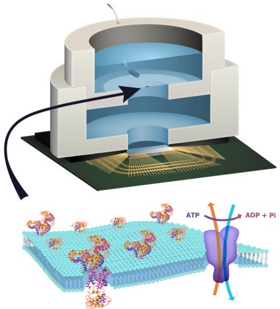
Illustration depicting a biocell attached to a CMOS integrated circuit with a membrane containing sodium-potassium pumps in pores. Energy is stored chemically in ATP molecules. When the energy is released as charged ions (which are then converted to electrons to power the chip at the bottom of the experimental device), the ATP is converted to ADP + inorganic phosphate. (credit: Trevor Finney and Jared Roseman/Columbia Engineering)

Columbia Engineering researchers have combined biological and solid-state components for the first time, opening the door to creating entirely new artificial biosystems.

In this experiment, they used a biological cell to power a conventional solid-state complementary metal-oxide-semiconductor (CMOS) integrated circuit. An artificial lipid bilayer membrane containing adenosine triphosphate (ATP)-powered ion pumps (which provide energy for cells) was used as a source of ions (which were converted to electrons to power the chip).

The study, led by Ken Shepard, Lau Family Professor of Electrical Engineering and professor of biomedical engineering at Columbia Engineering, was published online today (Dec. 7, 2015) in an open-access paper in Nature Communications.

How to build a hybrid biochip

Living systems achieve this functionality with their own version of electronics based on lipid membranes and ion channels and pumps, which act as a kind of “biological transistor.” Charge in the form of ions carry energy and information, and ion channels control the flow of ions across cell membranes.

Solid-state systems, such as those in computers and communication devices, use electrons; their electronic signaling and power are controlled by field-effect transistors.

To build a prototype of their hybrid system, Shepard’s team packaged a CMOS integrated circuit (IC) with an ATP-harvesting “biocell.” In the presence of ATP, the system pumped ions across the membrane, producing an electrical potential (voltage)* that was harvested by the integrated circuit.

“We made a macroscale version of this system, at the scale of several millimeters, to see if it worked,” Shepard notes. “Our results provide new insight into a generalized circuit model, enabling us to determine the conditions to maximize the efficiency of harnessing chemical energy through the action of these ion pumps. We will now be looking at how to scale the system down.”

While other groups have harvested energy from living systems, Shepard and his team are exploring how to do this at the molecular level, isolating just the desired function and interfacing this with electronics. “We don’t need the whole cell,” he explains. “We just grab the component of the cell that’s doing what we want. For this project, we isolated the ATPases because they were the proteins that allowed us to extract energy from ATP.”

The capability of a bomb-sniffing dog, no Alpo required

Next, the researchers plan to go much further, such as recognizing specific molecules and giving chips the potential to taste and smell.

The ability to build a system that combines the power of solid-state electronics with the capabilities of biological components has great promise, they believe. “You need a bomb-sniffing dog now, but if you can take just the part of the dog that is useful — the molecules that are doing the sensing — we wouldn’t need the whole animal,” says Shepard.

The technology could also provide a power source for implanted electronic devices in ATP-rich environments such as inside living cells, the researchers suggest.

*  “In general, integrated circuits, even when operated at the point of minimum energy in subthreshold, consume on the order of 10−2 W mm−2 (or assuming a typical silicon chip thickness of 250 μm, 4 × 10⁻² W mm−3). Typical cells, in contrast, consume on the order of 4 × 10⁻⁶ W mm−3. In the experiment, a typical active power dissipation for the IC circuit was 92.3 nW, and the active average harvesting power was 71.4 fW for the biocell (the discrepancy is managed through duty-cycled operation of the IC).” — Jared M. Roseman et al./Nature Communications


Biologists induce flatworms to grow heads and brains of other species

Tufts biologists induced one species of flatworm —- G. dorotocephala, top left — to grow heads and brains characteristic of other species of flatworm, top row, without altering genomic sequence. Examples of the outcomes can be seen in the bottom row of the image. (credit: Center for Regenerative and Developmental Biology, School of Arts and Sciences, Tufts University.)

Tufts University biologists have electrically modified flatworms to grow heads and brains characteristic of another species of flatworm — without altering their genomic sequence. This suggests bioelectrical networks as a new kind of epigenetics (information existing outside of a genomic sequence) to determine large-scale anatomy.

Besides the overall shape of the head, the changes included the shape of the brain and the distribution of the worm’s adult stem cells.

The discovery could help improve understanding of birth defects and regeneration by revealing a new pathway for controlling complex pattern formation similar to how neural networks exploit bioelectric synapses to store and re-write information in the brain.

The findings are detailed in the open-access cover story of the November 2015 edition of the International Journal of Molecular Sciences, appearing online Nov. 24.

“These findings raise significant questions about how genes and bioelectric networks interact to build complex body structures,” said the paper’s senior author Michael Levin, Ph.D., who holds the Vannevar Bush Chair in biology and directs the Center for Regenerative and Developmental Biology in the School of Arts and Sciences at Tufts. Knowing how shape is determined and how to influence it is important because biologists could use that knowledge, for example, to fix birth defects or cause new biological structures to grow after an injury, he explained.

How they did it

The researchers worked with Girardia dorotocephala — free-living planarian flatworms, which have remarkable regenerative capacity. They induced the development of different species-specific head shapes by interrupting gap junctions, which are protein channels that enable cells to communicate with each other by passing electrical signals back and forth.

A conceptual model of shape change driven by physiological network dynamics. Planaria regeneration (B) parallels classical neural network behavior (A); both can be described in terms of free energy landscapes with multiple attractor states. (credit: Maya Emmons-Bell et al./Int. J. Mol. Sci.)

The ease with which a particular shape could be coaxed from a G. dorotocephala worm was proportional to the proximity of the target worm on the evolutionary timeline. The closer the two species were related, the easier it was to effect the change. This observation strengthens the connection to evolutionary history, suggesting that modulation of physiological circuits may be one more tool exploited by evolution to alter animal body plans.

However, this shape change was only temporary. Weeks after the planaria completed regeneration to the other species’ head shapes, the worms once again began remodeling and re-acquired their original head morphology. Additional research is needed to determine how this occurs. The authors also presented a computational model that explains how changes in cell-to-cell communication can give rise to the diverse shape types.

The interdisciplinary research involved U.S.- and Canada-based biologists and European mathematicians.


Abstract of Gap Junctional Blockade Stochastically Induces Different Species-Specific Head Anatomies in Genetically Wild-Type Girardia dorotocephala Flatworms

The shape of an animal body plan is constructed from protein components encoded by the genome. However, bioelectric networks composed of many cell types have their own intrinsic dynamics, and can drive distinct morphological outcomes during embryogenesis and regeneration. Planarian flatworms are a popular system for exploring body plan patterning due to their regenerative capacity, but despite considerable molecular information regarding stem cell differentiation and basic axial patterning, very little is known about how distinct head shapes are produced. Here, we show that after decapitation in G. dorotocephala, a transient perturbation of physiological connectivity among cells (using the gap junction blocker octanol) can result in regenerated heads with quite different shapes, stochastically matching other known species of planaria (S. mediterraneaD. japonica, and P. felina). We use morphometric analysis to quantify the ability of physiological network perturbations to induce different species-specific head shapes from the same genome. Moreover, we present a computational agent-based model of cell and physical dynamics during regeneration that quantitatively reproduces the observed shape changes. Morphological alterations induced in a genomically wild-type G. dorotocephala during regeneration include not only the shape of the head but also the morphology of the brain, the characteristic distribution of adult stem cells (neoblasts), and the bioelectric gradients of resting potential within the anterior tissues. Interestingly, the shape change is not permanent; after regeneration is complete, intact animals remodel back to G. dorotocephala-appropriate head shape within several weeks in a secondary phase of remodeling following initial complete regeneration. We present a conceptual model to guide future work to delineate the molecular mechanisms by which bioelectric networks stochastically select among a small set of discrete head morphologies. Taken together, these data and analyses shed light on important physiological modifiers of morphological information in dictating species-specific shape, and reveal them to be a novel instructive input into head patterning in regenerating planaria.

A sensory illusion that makes yeast cells self-destruct

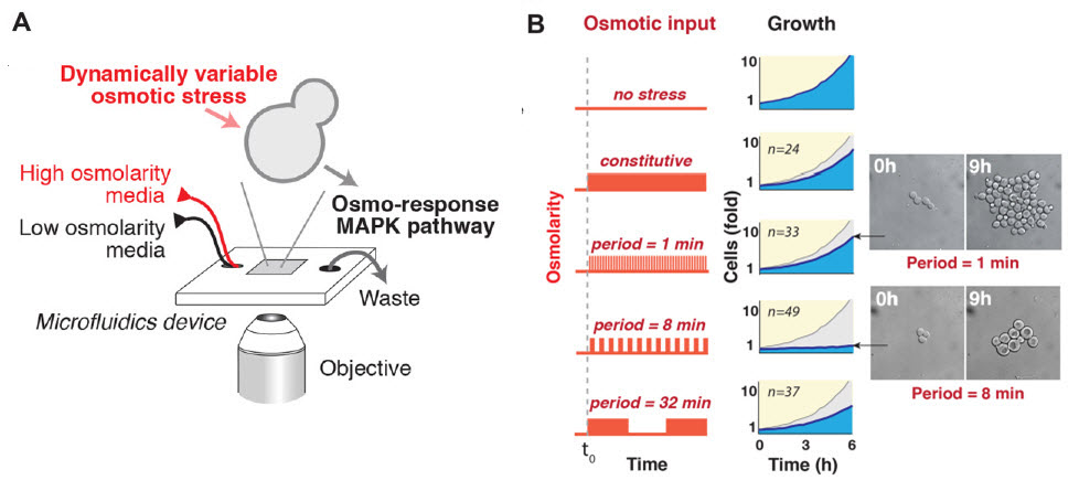
Effects of osmotic changes on yeast cell growth. (A) Schematic of the flow chamber used to create osmotic level oscillations for different periods of time. (B) Cell growth for these periods. The graphs show the average number of progeny cells (blue) before and after applying stress for different periods (gray shows orginal “no stress” line). The inset shows representative images of cells for two periods. (credit: Amir Mitchell et al./Science)

UC San Francisco researchers have discovered that even brainless single-celled yeast have “sensory biases” that can be hacked by a carefully engineered illusion — a finding that could be used to develop new approaches to fighting diseases such as cancer.

In the new study, published online Thursday November 19 in Science Express, Wendell Lim, PhD, the study’s senior author*, and his team discovered that yeast cells falsely perceive a pattern of osmotic levels (by applying potassium chloride) that alternate in eight minute intervals as massive, continuously increasing stress. In response, the microbes over-respond and kill themselves. (In their natural environment, salt stress normally gradually increases.)

The results, Lim says, suggest a whole new way of looking at the perceptual abilities of simple cells and this power of illusion could even be used to develop new approaches to fighting cancer and other diseases.

“Our results may also be relevant for cellular signaling in disease, as mutations affecting cellular signaling are common in cancer, autoimmune disease, and diabetes,” the researchers conclude in the paper. “These mutations may rewire the native network, and thus could modify its activation and adaptation dynamics. Such network rewiring in disease may lead to changes that can be most clearly revealed by simulation with oscillatory inputs or other ‘non-natural’ patterns.

“The changes in network response behaviors could be exploited for diagnosis and functional profiling of disease cells, or potentially taken advantage of as an Achilles’ heel to selectively target cells bearing the diseased network.”


UC San Francisco (UCSF) | Sensory Illusion Causes Cells to Self-Destruct

* Chair of the Department of Cellular and Molecular Pharmacology at UCSF, director of the UCSF Center for Systems and Synthetic Biology, and a Howard Hughes Medical Institute (HHMI) investigator.

** Normally, sensor molecules in a yeast cell detect changes in salt concentration and instruct the cell to respond by producing a protective chemical. The researchers found that the cells were perfectly capable of adapting when they flipped the salt stress on and off every minute or every 32 minutes. But to their surprise, when they tried an eight-minute oscillation of precisely the same salt level the cells quickly stopped growing and began to die off.


Abstract of Oscillatory stress stimulation uncovers an Achilles’ heel of the yeast MAPK signaling network

Cells must interpret environmental information that often changes over time. We systematically monitored growth of yeast cells under various frequencies of oscillating osmotic stress. Growth was severely inhibited at a particular resonance frequency, at which cells show hyperactivated transcriptional stress responses. This behavior represents a sensory misperception—the cells incorrectly interpret oscillations as a staircase of ever-increasing osmolarity. The misperception results from the capacity of the osmolarity-sensing kinase network to retrigger with sequential osmotic stresses. Although this feature is critical for coping with natural challenges—like continually increasing osmolarity—it results in a tradeoff of fragility to non-natural oscillatory inputs that match the retriggering time. These findings demonstrate the value of non-natural dynamic perturbations in exposing hidden sensitivities of cellular regulatory networks.