E-coli bacteria, found in some China farms and patients, cannot be killed with antiobiotic drug of last resort

Colistin antibiotic overused in farm animals in China apparently caused E-coli bacteria to become completely resistant to treatment; E-coli strain has already spread to Laos and Malaysia (credit: Yi-Yun Liu et al./Lancet Infect Dis)

Widespread E-coli bacteria that cannot be killed with the antiobiotic drug of last resort — colistin — have been found in samples taken from farm pigs, meat products, and a small number of patients in south China, including bacterial strains with epidemic potential, an international team of scientists revealed in a paper published Thursday Nov. 19 in the journal The Lancet Infectious Diseases.

The scientists in China, England, and the U.S. found a new gene, MCR-1, carried in E-coli bacteria strain SHP45. MCR-1 enables bacteria to be highly resistant to colistin and other polymyxins drugs.

“The emergence of the MCR-1 gene in China heralds a disturbing breach of the last group of antibiotics — polymixins — and an end to our last line of defense against infection,” said Professor Timothy Walsh, from the Cardiff University School of Medicine, who collaborated on this research with scientists from South China Agricultural University.

Walsh, an expert in antibiotic resistance, is best known for his discovery in 2011 of the NDM-1 disease-causing antibiotic-resistant superbug in New Delhi’s drinking water supply. “The rapid spread of similar antibiotic-resistant genes such as NDM-1 suggests that all antibiotics will soon be futile in the face of previously treatable gram-negative bacterial infections such as E.coli and salmonella,” he said.

Likely to spread worldwide; already found in Laos and Malaysia

The MCR-1 gene was found on plasmids — mobile DNA that can be easily copied and transferred between different bacteria, suggesting an alarming potential to spread and diversify between different bacterial populations.
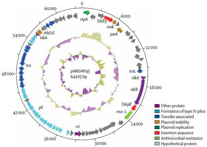
Structure of plasmid pHNSHP45 carrying MCR-1 from Escherichia coli strain SHP45 (credit: Yi-Yun Liu et al./Lancet Infect Dis)

“We now have evidence to suggest that MCR-1-positive E.coli has spread beyond China, to Laos and Malaysia, which is deeply concerning,” said Walsh.  “The potential for MCR-1 to become a global issue will depend on the continued use of polymixin antibiotics, such as colistin, on animals, both in and outside China; the ability of MCR-1 to spread through human strains of E.coli; and the movement of people across China’s borders.”

“MCR-1 is likely to spread to the rest of the world at an alarming rate unless we take a globally coordinated approach to combat it. In the absence of new antibiotics against resistant gram-negative pathogens, the effect on human health posed by this new gene cannot be underestimated.”

“Of the top ten largest producers of colistin for veterinary use, one is Indian, one is Danish, and eight are Chinese,” The Lancet Infectious Diseases notes. “Asia (including China) makes up 73·1% of colistin production with 28·7% for export including to Europe.29 In 2015, the European Union and North America imported 480 tonnes and 700 tonnes, respectively, of colistin from China.”

Urgent need for coordinated global action

“Our findings highlight the urgent need for coordinated global action in the fight against extensively resistant and pan-resistant gram-negative bacteria,” the journal paper concludes.

“The implications of this finding are enormous,” an associated editorial comment to the The Lancet Infectious Diseases paper stated. “We must all reiterate these appeals and take them to the highest levels of government or face increasing numbers of patients for whom we will need to say, ‘Sorry, there is nothing I can do to cure your infection.’”

Margaret Chan, MD, Director-General of the World Health Organization, warned in 2011 that “the world is heading towards a post-antibiotic era, in which many common infections will no longer have a cure and, once again, kill unabated.”

“Although in its 2012 World Health Organization Advisory Group on Integrated Surveillance of Antimicrobial Resistance (AGISAR) report the WHO concluded that colistin should be listed under those antibiotics of critical importance, it is regrettable that in the 2014 Global Report on Surveillance, the WHO did not to list any colistin-resistant bacteria as part of their ‘selected bacteria of international concern,’” The Lancet Infectious Diseases paper says, reflecting WHO’s inaction in Ebola-stricken African countries, as noted last September by the international medical humanitarian organization Médecins Sans Frontières.

Funding for the E-coli bacteria study was provided by the Ministry of Science and Technology of China and National Natural Science Foundation of China.


Abstract of Emergence of plasmid-mediated colistin resistance mechanism MCR-1 in animals and human beings in China: a microbiological and molecular biological study

Until now, polymyxin resistance has involved chromosomal mutations but has never been reported via
horizontal gene transfer. During a routine surveillance project on antimicrobial resistance in commensal Escherichia coli from food animals in China, a major increase of colistin resistance was observed. When an E coli strain, SHP45, possessing colistin resistance that could be transferred to another strain, was isolated from a pig, we conducted further analysis of possible plasmid-mediated polymyxin resistance. Herein, we report the emergence of the first plasmid-mediated polymyxin resistance mechanism, MCR-1, in Enterobacteriaceae.

The mcr-1 gene in E coli strain SHP45 was identified by whole plasmid sequencing and subcloning. MCR-1 mechanistic studies were done with sequence comparisons, homology modelling, and electrospray ionisation mass spectrometry. The prevalence of mcr-1 was investigated in E coli and Klebsiella pneumoniae strains collected from five provinces between April, 2011, and November, 2014. The ability of MCR-1 to confer polymyxin resistance in vivo was examined in a murine thigh model.

Polymyxin resistance was shown to be singularly due to the plasmid-mediated mcr-1 gene. The plasmid carrying mcr-1 was mobilised to an E coli recipient at a frequency of 10−1 to 10−3 cells per recipient cell by conjugation, and maintained in K pneumoniae and Pseudomonas aeruginosa. In an in-vivo model, production of MCR-1 negated the efficacy of colistin. MCR-1 is a member of the phosphoethanolamine transferase enzyme family, with expression in E coli resulting in the addition of phosphoethanolamine to lipid A. We observed mcr-1 carriage in E coli isolates collected from 78 (15%) of 523 samples of raw meat and 166 (21%) of 804 animals during 2011–14, and 16 (1%) of 1322 samples from inpatients with infection.

The emergence of MCR-1 heralds the breach of the last group of antibiotics, polymyxins, by plasmid-mediated resistance. Although currently confined to China, MCR-1 is likely to emulate other global resistance mechanisms such as NDM-1. Our findings emphasise the urgent need for coordinated global action in the fight against pan-drug-resistant Gram-negative bacteria.

Allen Institute researchers decode patterns that make our brains human

Percentage of known neuron-, astrocyte- and oligodendrocyte-enriched genes in 32 modules, ordered by proportion of neuron-enriched gene membership. (credit: Michael Hawrylycz et al./Nature Neuroscience)

Allen Institute researchers have identified a surprisingly small set of just 32 gene-expression patterns for all 20,000 genes across 132 functionally distinct human brain regions, and these patterns appear to be common to all individuals.

In research published this month in Nature Neuroscience, the researchers used data for six brains from the publicly available Allen Human Brain Atlas. They believe the study is important because it could provide a baseline from which deviations in individuals may be measured and associated with diseases, and could also provide key insights into the core of the genetic code that makes our brains distinctly human.

While many of these patterns were similar in human and mouse, many genes showed different patterns in human. Surprisingly, genes associated with neurons were most conserved (consistent) across species, while those for the supporting glial cells showed larger differences. The most highly stable genes (the genes that were most consistent across all brains) include those associated with diseases and disorders like autism and Alzheimer’s, and these genes include many existing drug targets.

These patterns provide insights into what makes the human brain distinct and raise new opportunities to target therapeutics for treating disease.

The researchers also found that the pattern of gene expression in cerebral cortex is correlated with “functional connectivity” as revealed by neuroimaging data from the Human Connectome Project.

“The human brain is phenomenally complex, so it is quite surprising that a small number of patterns can explain most of the gene variability across the brain,” says Christof Koch, Ph.D., President and Chief Scientific Officer at the Allen Institute for Brain Science. “There could easily have been thousands of patterns, or none at all. This gives us an exciting way to look further at the functional activity that underlies the uniquely human brain.”


Abstract of Canonical genetic signatures of the adult human brain

The structure and function of the human brain are highly stereotyped, implying a conserved molecular program responsible for its development, cellular structure and function. We applied a correlation-based metric called differential stability to assess reproducibility of gene expression patterning across 132 structures in six individual brains, revealing mesoscale genetic organization. The genes with the highest differential stability are highly biologically relevant, with enrichment for brain-related annotations, disease associations, drug targets and literature citations. Using genes with high differential stability, we identified 32 anatomically diverse and reproducible gene expression signatures, which represent distinct cell types, intracellular components and/or associations with neurodevelopmental and neurodegenerative disorders. Genes in neuron-associated compared to non-neuronal networks showed higher preservation between human and mouse; however, many diversely patterned genes displayed marked shifts in regulation between species. Finally, highly consistent transcriptional architecture in neocortex is correlated with resting state functional connectivity, suggesting a link between conserved gene expression and functionally relevant circuitry.

3-D printed ‘building blocks’ of life

Images of printed embryonic stem cells, or embryoid bodies (credit: Liliang Ouyang et al./Biofabrication)

Chinese and U.S. scientists have developed a 3-D printing method capable of producing embryoid bodies — highly uniform “blocks” of embryonic stem cells. These cells, which are capable of generating all cell types in the body, could be used to build tissue structures and potentially even micro-organs.

The results were published Wednesday Nov. 4 in an open-access paper in the journal Biofabrication. “The embryoid body is uniform and homogenous, and serves as a much better starting point for further tissue growth,” explains Wei Sun, a lead author on the paper.

The researchers, based at Tsinghua University, Beijing, China, and Drexel University, Philadelphia, used extrusion-based 3-D printing to produce a grid-like 3-D structure to grow an embryoid body that demonstrated cell viability and rapid self-renewal for 7 days while maintaining high pluripotentcy.

“Two other common methods of printing these cells are two-dimensional (in a petri dish) or via the ‘suspension’ method [see 'Better bioprinting with stem cells'], where a ‘stalagmite’ of cells is built up by material being dropped via gravity,” said Sun. ”However, these don’t show the same cell uniformity and homogenous proliferation. I think that we’ve produced a 3-D microenvironment that is much more like that found in vivo for growing embryoid bodies, which explains the higher levels of cell proliferation.”

The researchers hope that this technique can be developed to produce embryoid bodies at high throughput, providing the basic building blocks for other researchers to perform experiments on tissue regeneration and/or for drug screening studies.

The researchers say the next step is to find out more about how to vary the size of the embryoid body by changing the printing and structural parameters, and how varying the embryoid body size leads to “manufacture” of different cell types.

“In the longer term, we’d like to produce controlled heterogeneous embryonic bodies,” said Sun. “This would promote different cell types developing next to each other, which would lead the way for growing micro-organs from scratch within the lab.”


Abstract of Three-dimensional bioprinting of embryonic stem cells directs highly uniform embryoid body formation

With the ability to manipulate cells temporarily and spatially into three-dimensional (3D) tissue-like construct, 3D bioprinting technology was used in many studies to facilitate the recreation of complex cell niche and/or to better understand the regulation of stem cell proliferation and differentiation by cellular microenvironment factors. Embryonic stem cells (ESCs) have the capacity to differentiate into any specialized cell type of the animal body, generally via the formation of embryoid body (EB), which mimics the early stages of embryogenesis. In this study, extrusion-based 3D bioprinting technology was utilized for biofabricating ESCs into 3D cell-laden construct. The influence of 3D printing parameters on ESC viability, proliferation, maintenance of pluripotency and the rule of EB formation was systematically studied in this work. Results demonstrated that ESCs were successfully printed with hydrogel into 3D macroporous construct. Upon process optimization, about 90% ESCs remained alive after the process of bioprinting and cell-laden construct formation. ESCs continued proliferating into spheroid EBs in the hydrogel construct, while retaining the protein expression and gene expression of pluripotent markers, like octamer binding transcription factor 4, stage specific embryonic antigen 1 and Nanog. In this novel technology, EBs were formed through cell proliferation instead of aggregation, and the quantity of EBs was tuned by the initial cell density in the 3D bioprinting process. This study introduces the 3D bioprinting of ESCs into a 3D cell-laden hydrogel construct for the first time and showed the production of uniform, pluripotent, high-throughput and size-controllable EBs, which indicated strong potential in ESC large scale expansion, stem cell regulation and fabrication of tissue-like structure and drug screening studies.

Engineers design enhanced magnetic protein nanoparticles to better track cells


X-ray crystal structure of iron storage ferritin PFt displaying the internal cavity of the protein in which one of the subunits is highlighted in yellow (credit: Yuri Matsumoto/Nature Communications)

MIT engineers have designed magnetic protein nanoparticles that can be used to track cells or to monitor interactions within cells. The particles, described Monday (Nov. 2) in an open-access paper in Nature Communications, are an enhanced version of a naturally occurring, weakly magnetic protein called ferritin.

“We used the tools of protein engineering to try to boost the magnetic characteristics of this protein,”  says Alan Jasanoff, an MIT professor of biological engineering and the paper’s senior author.

The new “hypermagnetic” protein nanoparticles can be produced within cells, allowing the cells to be imaged or sorted using magnetic techniques. This eliminates the need to tag cells with synthetic particles and allows the particles to sense other molecules inside cells.

Genetically encoded magnetic particles

Previous research has yielded synthetic magnetic particles for imaging or tracking cells, but it can be difficult to deliver these particles into the target cells.

In the new study, Jasanoff and colleagues set out to create magnetic particles that are genetically encoded. With this approach, the researchers deliver a gene for a magnetic protein into the target cells, prompting the cells to start producing the protein on their own.

“Rather than actually making a nanoparticle in the lab and attaching it to cells or injecting it into cells, all we have to do is introduce a gene that encodes this protein,” says Jasanoff, who is also an associate member of MIT’s McGovern Institute for Brain Research.

As a starting point, the researchers used ferritin, which carries a supply of iron atoms that every cell needs as components of metabolic enzymes. In hopes of creating a more magnetic version of ferritin, the researchers created about 10 million variants and tested them in yeast cells.

After repeated rounds of screening, the researchers used one of the most promising candidates to create a magnetic sensor consisting of enhanced ferritin modified with a protein tag that binds with another protein called streptavidin. This allowed them to detect whether streptavidin was present in yeast cells; however, this approach could also be tailored to target other interactions.

The mutated protein appears to successfully overcome one of the key shortcomings of natural ferritin: it’s difficult to load with iron, says Alan Koretsky, a senior investigator at the National Institute of Neurological Disorders and Stroke.

“To be able to make more magnetic indicators for MRI would be fabulous, and this is an important step toward making that type of indicator more robust,” says Koretsky, who was not part of the research team.

Sensing cell signals

Because the engineered ferritins are genetically encoded, they can be manufactured within cells that are programmed to make them respond only under certain circumstances, such as when the cell receives some kind of external signal, when it divides, or when it differentiates into another type of cell. Researchers could track this activity using magnetic resonance imaging (MRI), potentially allowing them to observe communication between neurons, activation of immune cells, or stem cell differentiation, among other phenomena.

Such sensors could also be used to monitor the effectiveness of stem cell therapies, Jasanoff says.

“As stem cell therapies are developed, it’s going to be necessary to have noninvasive tools that enable you to measure them,” he says. Without this kind of monitoring, it would be difficult to determine what effect the treatment is having, or why it might not be working.

The researchers are now working on adapting the magnetic sensors to work in mammalian cells. They are also trying to make the engineered ferritin even more strongly magnetic.


Abstract of Engineering intracellular biomineralization and biosensing by a magnetic protein

Remote measurement and manipulation of biological systems can be achieved using magnetic techniques, but a missing link is the availability of highly magnetic handles on cellular or molecular function. Here we address this need by using high-throughput genetic screening in yeast to select variants of the iron storage ferritin (Ft) that display enhanced iron accumulation under physiological conditions. Expression of Ft mutants selected from a library of 107 variants induces threefold greater cellular iron loading than mammalian heavy chain Ft, over fivefold higher contrast in magnetic resonance imaging, and robust retention on magnetic separation columns. Mechanistic studies of mutant Ft proteins indicate that improved magnetism arises in part from increased iron oxide nucleation efficiency. Molecular-level iron loading in engineered Ft enables detection of individual particles inside cells and facilitates creation of Ft-based intracellular magnetic devices. We demonstrate construction of a magnetic sensor actuated by gene expression in yeast.

First complete pictures of cells’ DNA-copying machinery

These cartoons show the old “textbook” view of the replisome, left, and the new view, right, revealed by electron micrograph images in the current study. Prior to this study, scientists believed the two polymerases (green) were located at the bottom (or back end) of the helicase (tan), adding complementary DNA strands to the split DNA to produce copies side by side. The new images reveal that one of the polymerases is actually located at the front end (top) of the helicase. The scientists are conducting additional studies to explore the biological significance of this unexpected location. (credit: Brookhaven National Laboratory)

The first-ever electron microscope images of the protein complex that unwinds, splits, and copies double-stranded DNA reveal something rather different from the standard textbook view.

The images, created by scientists at the U.S. Department of Energy’s Brookhaven National Laboratory with partners from Stony Brook University and Rockefeller University, offer new insight into how this molecular machinery functions, including new possibilities about its role in DNA “quality control” and cell differentiation.

Huilin Li, a biologist with a joint appointment at Brookhaven Lab and Stony Brook University says the new images show the fully assembled and fully activated helicase protein complex — which encircles and separates the two strands of the DNA double helix as it passes through a central pore in the structure — and how the helicase coordinates with the two polymerase enzymes that duplicate each strand to copy the genome.

Three blind men and an elephant

Studying this molecular machinery, known collectively as a “replisome,” and the details of its DNA-copying process can help scientists understand what happens when DNA is miscopied — a major source of mutation that can lead to cancer. Scientists can also learn more about how a single cell can eventually develop into the many cell types that make up a multicellular organism.

“All the textbook drawings and descriptions of how a replisome should look and work are based on biochemical and genetic studies,” Li said, likening the situation to the famous parable of the three blind men trying to describe an elephant, each looking at only one part.

To test these assumptions, Li’s group turned to electron microscopy (EM). The team’s first-ever images of an intact replisome revealed that only one of the polymerases is located at the back of the helicase.

The other is on the front side of the helicase, where the helicase first encounters the double-stranded helix. This means that while one of the two split DNA strands is acted on by the polymerase at the back end, the other has to thread itself back through or around the helicase to reach the front-side polymerase before having its new complementary strand assembled.

Unforeseen functions?

The counterintuitive position of one polymerase at the front of the helicase suggests that it may have an unforeseen function. The authors suggest several possibilities, including keeping the two “daughter” strands separate to help organize them during replication and cell division. It might also be possible that, as the single strand moves over other portions of the structure, some “surveillance” protein components check for lesions or mistakes in the nucleotide sequence before it gets copied — a sort of molecular quality control.

This architecture could also potentially play an important role in developmental biology by providing a pathway for treating the two daughter strands differently. Many modifications to DNA, including how it is packaged with other proteins, control which of the many genes in the sequence are eventually expressed in cells. An asymmetric replisome may result in asymmetric treatment of the two daughter strands during cell division, an essential step for making different tissues within a multicellular organism.

“Clearly, further studies will be required to understand the functional implications of the unexpected replisome architecture reported here,” concludes the researchers’ paper published Monday (Nov. 2) online by the journal Nature Structural & Molecular Biology.


Brookhaven National Laboratory | Three-dimensional structure of the active DNA helicase bound to the front-end DNA polymerase (Pol epsilon). The DNA polymerase epsilon (green) sits on top rather than the bottom of the helicase.


Abstract of The Architecture of a Eukaryotic Replisome

At the eukaryotic DNA replication fork, it is widely believed that the Cdc45–Mcm2–7–GINS (CMG) helicase is positioned in front to unwind DNA and that DNA polymerases trail behind the helicase. Here we used single-particle EM to directly image a Saccharomyces cerevisiae replisome. Contrary to expectations, the leading strand Pol ε is positioned ahead of CMG helicase, whereas Ctf4 and the lagging-strand polymerase (Pol) α–primase are behind the helicase. This unexpected architecture indicates that the leading-strand DNA travels a long distance before reaching Pol ε, first threading through the Mcm2–7 ring and then making a U-turn at the bottom and reaching Pol ε at the top of CMG. Our work reveals an unexpected configuration of the eukaryotic replisome, suggests possible reasons for this architecture and provides a basis for further structural and biochemical replisome studies.

A powerful new ‘tool’ for assembling biomolecules

Proposed new simplified chemical reaction for assembling biomolecules in a single chemical reaction (credit: Tiffany Piou & Tomislav Rovis/Nature)

Colorado State University chemists have invented a single chemical reaction that couples two constituent chemicals into a carbon-carbon bond, while simultaneously introducing a nitrogen component. The process promises to replace a multi-step, expensive, and complex process needed when synthesizing new chemicals — for drug creation and testing, for example.

The researchers were able to control this reaction to make the nitrogen atoms go exactly where they want them to, making for precision chemistry that they believe could revolutionize pharmaceutical and biomaterials manufacturing.

The achievement is detailed in the journal Nature, published today (Oct. 21).

Achieving a critical carbon-nitrogen bond

The researchers explain in a statement that “almost every significant carbon-based biomolecule contains a nitrogen compound, or amine. Achieving this carbon-nitrogen bond in the lab, though, is tricky business. Drug companies know it well…. They must first create the carbon-carbon bonds, and then introduce the nitrogen to make a molecule that will do something useful.”

Ball-and-stick model of the ethylene (ethene) molecule, C2H4, the simplest alkene (credit: Benjah-bmm27 CC)

The chemists’ starting materials were simply oil refinery byproducts called olefins, or alkenes. They mixed in a specially engineered reagant, then used a complex based on the precious metal rhodium to reliably and specifically trigger the elusive carbon-nitrogen bonds.

Allene (left) and propyne (right) are examples of isomers containing different bond types (double and triple carbon bonds in this case) — with different functionalities. (credit: Wikipedia)

The innovation also controls molecular isomers (an isomer is a molecule with the same chemical formula as another molecule, but with a different chemical structure). Some isomers are mirror images, like right and left gloves, and although they’re chemically identical, their functionalities are strikingly different. Being able to select for a single isomer is critical to safety and efficacy — so much so that the FDA mandates that only single-isomer drugs be marketed for human use.

Take thalidomide, infamous for causing severe birth defects when taken by pregnant women in the 1950s. Chemically, thalidomide comes in two mirror-image isomeric forms. One caused the defects, one didn’t.

“For this reason, spatial display of groups in molecules is incredibly important,” said organic chemist Tomislav Rovis, professor of chemistry in the College of Natural Sciences at CSU. Rovis led the research with postdoctoral researcher Tiffany Piou, who designed all the chemical building blocks and ran the experiments.

“Tiffany’s finding gives us a leg up to do this in a carboamination reaction, by making the carbon carbon bond, and delivering the nitrogen selectively,” Rovis said.

The researchers hope their approach, which they liken to a tool in a toolbox, can be polished, perfected and used widely to make organic chemistry easier, and applied to many different fields.


Abstract of Rhodium-catalysed syn-carboamination of alkenes via a transient directing group

Alkenes are the most ubiquitous prochiral functional groups—those that can be converted from achiral to chiral in a single step—that are accessible to synthetic chemists. For this reason, difunctionalization reactions of alkenes (whereby two functional groups are added to the same double bond) are particularly important, as they can be used to produce highly complex molecular architectures12. Stereoselective oxidation reactions, including dihydroxylation, aminohydroxylation and halogenation3456, are well established methods for functionalizing alkenes. However, the intermolecular incorporation of both carbon- and nitrogen-based functionalities stereoselectively across an alkene has not been reported. Here we describe the rhodium-catalysed carboamination of alkenes at the same (syn) face of a double bond, initiated by a carbon–hydrogen activation event that uses enoxyphthalimides as the source of both the carbon and the nitrogen functionalities. The reaction methodology allows for the intermolecular, stereospecific formation of one carbon–carbon and one carbon–nitrogen bond across an alkene, which is, to our knowledge, unprecedented. The reaction design involves the in situ generation of a bidentate directing group and the use of a new cyclopentadienyl ligand to control the reactivity of rhodium. The results provide a new way of synthesizing functionalized alkenes, and should lead to the convergent and stereoselective assembly of amine-containing acyclic molecules.

How to control heartbeats more precisely, using light

Using computer-generated light patterns, researchers were able to control the direction of spiraling electrical waves in heart cells. (credit: Eana Park)

Researchers from Oxford and Stony Brook universities has found a way to precisely control the electrical waves that regulate the rhythm of our heartbeat — using light. Their results are published in the journal Nature Photonics.

Cardiac cells in the heart and neurons in the brain communicate by electrical signals, and these messages of communication travel fast from cell to cell as “excitation waves.”

For heart patients there are currently two options to keep these waves in check: electrical devices (pacemakers or defibrillators) or drugs (e.g., beta blockers). However, these methods are relatively crude: they can stop or start waves but cannot provide fine control over the wave speed and direction.

Gil Bub, from Oxford University explained: ‘When there is scar tissue in the heart or fibrosis, this can cause part of the wave to slow down. That can cause re-entrant waves which spiral back around the tissue, causing the heart to beat much too quickly, which can be fatal. If we can control these spirals, we could prevent that.

The optogenetics solution

The solution the researchers found was optogenetics, which uses genetic modification to alter cells so that they can be activated by light. Until now, it has mainly been used to activate individual cells or to trigger excitation waves in tissue, especially in neuroscience research. “We wanted to use it to very precisely control the activity of millions of cells,” said Bub.

A light-activated protein called channelrhodopsin was delivered to heart cells using gene therapy techniques so that they could be controlled by light. Then, using a computer-controlled light projector, the team was able to control the speed of the cardiac waves, their direction and even the orientation of spirals in real time — something that never been shown for waves in a living system before.

In the short term, the ability to provide fine control means that researchers are able to carry out experiments at a level of detail previously only available using computer models. They can now compare those models to experiments with real cells, potentially improving our understanding of how the heart works. The research can also be applied to the physics of such waves in other processes. In the long run, it might be possible to develop precise treatments for heart conditions.

“Precise control of the direction, speed and shape of such excitation waves would mean unprecedented direct control of organ-level function, in the heart or brain, without having to focus on manipulating each cell individually,” said Stony Brook University scientist Emilia Entcheva.

The team stresses that there are significant hurdles before this could offer new treatments; a key issue is being able to alter the heart to be light-sensitized and being able to get the light to desired locations. However, as gene therapy moves into the clinic and with miniaturization of optical devices, use of this all-optical technology may become possible.

In the meantime, the research enables scientists to look into the physics behind many biological processes, including those in our own brains and hearts.

University of Oxford | Controlling heart tissue with light


Abstract of Optical control of excitation waves in cardiac tissue

In nature, macroscopic excitation waves are found in a diverse range of settings including chemical reactions, metal rust, yeast, amoeba and the heart and brain. In the case of living biological tissue, the spatiotemporal patterns formed by these excitation waves are different in healthy and diseased states. Current electrical and pharmacological methods for wave modulation lack the spatiotemporal precision needed to control these patterns. Optical methods have the potential to overcome these limitations, but to date have only been demonstrated in simple systems, such as the Belousov–Zhabotinsky chemical reaction. Here, we combine dye-free optical imaging with optogenetic actuation to achieve dynamic control of cardiac excitation waves. Illumination with patterned light is demonstrated to optically control the direction, speed and spiral chirality of such waves in cardiac tissue. This all-optical approach offers a new experimental platform for the study and control of pattern formation in complex biological excitable systems.

A portable paper-smartphone device that analyzes trace pesticides

The prototype smartphone-based pesticide-detection system (credit: Qingsong Mei et al./Biosensors and Bioelectronics)

A new system that may allow people to detect pesticides cheaply and rapidly, combining a paper sensor and an Android program on a smartphone, has been developed by researchers in China and Singapore, according to a new study published in Biosensors and Bioelectronics.

As the potential effects of pesticides on health become clearer, it is increasingly important to be able to detect them in the environment and on foods, but existing gear that purpose is large, expensive, and slow.

Smaller detectors have been developed using paper as a sensor material, but they have not produced strong enough signals for detection. Now researchers at Hefei University of Technology in China and the National University of Singapore have developed a portable smartphone-based detection system using a paper sensor that they say produces signals stronger enough to allow for pesticide detection.

The researchers tested it on thiram, which is used to prevent fungal diseases in seed and crops and an animal repellent to protect fruit trees.

The device uses nanoparticles covered with copper ions that are coated onto paper, causing pesticide molecules to attach to the copper ions. A near-infrared mini-laser shines a light onto the paper, the smartphone detects the absorption spectrum, and an Android app then calculates pesticide concentration, down to 0.1 μM (micromolar) concentration.

The researchers are now developing kits that can multiplex (detect different molecules simultaneously), which would allow for testing food before using it in a meal, for example.

This work was supported by the National Natural Science Foundation of China and the Fundamental Research Funds for the Central Universities.


Abstract of Smartphone based visual and quantitative assays on upconversional paper sensor

The integration of smartphone with paper sensors recently has been gain increasing attentions because of the achievement of quantitative and rapid analysis. However, smartphone based upconversional paper sensors have been restricted by the lack of effective methods to acquire luminescence signals on test paper. Herein, by the virtue of 3D printing technology, we exploited an auxiliary reusable device, which orderly assembled a 980 nm mini-laser, optical filter and mini-cavity together, for digitally imaging the luminescence variations on test paper and quantitative analyzing pesticide thiram by smartphone. In detail, copper ions decorated NaYF4:Yb/Tm upconversion nanoparticles were fixed onto filter paper to form test paper, and the blue luminescence on it would be quenched after additions of thiram through luminescence resonance energy transfer mechanism. These variations could be monitored by the smartphone camera, and then the blue channel intensities of obtained colored images were calculated to quantify amounts of thiram through a self-written Android program installed on the smartphone, offering a reliable and accurate detection limit of 0.1 μM for the system. This work provides an initial demonstration of integrating upconversion nanosensors with smartphone digital imaging for point-of-care analysis on a paper-based platform.