Engineered viruses provide quantum-based enhancement of energy transport

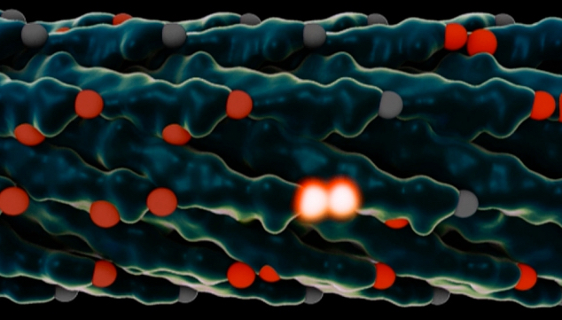
Rendering of a virus used in the MIT experiments. The light-collecting centers, called chromophores, are in red, and chromophores that just absorbed a photon of light are glowing white. After the virus is modified to adjust the spacing between the chromophores, energy can jump from one set of chromophores to the next faster and more efficiently. (credit: the researchers and Lauren Alexa Kaye)

MIT engineers have achieved a significant efficiency boost in a light-harvesting system, using genetically engineered viruses to achieve higher efficiency in transporting energy from receptors to reaction centers where it can be harnessed, making use of the exotic effects of quantum mechanics. Emulating photosynthesis in nature, it could lead to inexpensive and efficient solar cells or light-driven catalysis,

This achievement in coupling quantum research and genetic manipulation, described this week in the journal Nature Materials, was the work of MIT professors Angela Belcher, an expert on engineering viruses to carry out energy-related tasks, and Seth Lloyd, an expert on quantum theory and its potential applications, and 15 collaborators at MIT and in Italy.

The “Quantum Goldilocks Effect”

In photosynthesis, a photon hits a receptor called a chromophore, which in turn produces an exciton — a quantum particle of energy. This exciton jumps from one chromophore to another until it reaches a reaction center, where that energy is harnessed to build the molecules that support life, or photosynthesis.

But the hopping pathway of excitons is random and inefficient unless it takes advantage of quantum effects that allow it, in effect, to take multiple pathways at once and select the best ones, behaving more like a wave than a particle.

To do that, the chromophores have to be arranged just right, with exactly the right amount of space between them. This, Lloyd explains, is known as the “Quantum Goldilocks Effect.”

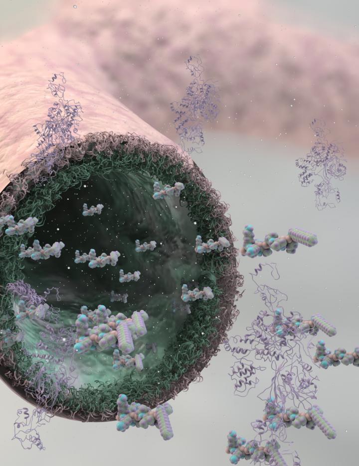
Molecular models of the genetically engineered viruses. Left virus has long inter-binding site distances of 16Å and 33Å within two proteins. Right virus has closer inter-binding site distances of approximately 10Å and 13Å, achieving faster excitation-energy transport speed. (credit: Heechul Park et al./Nature Materials)

That’s where the virus comes in. By engineering a virus that Belcher has worked with for years, the team was able to get it to bond with multiple synthetic chromophores — or, in this case, organic dyes. The researchers were then able to produce many varieties of the virus, with slightly different spacings between those synthetic chromophores, and select the ones that performed best.

In the end, they were able to more than double excitons’ speed, increasing the distance they traveled before dissipating — a significant improvement in the efficiency of the process.

The project started from a chance meeting at a conference in Italy. Lloyd and Belcher, a professor of biological engineering, were reporting on different projects they had worked on, and began discussing the possibility of a project encompassing their very different expertise. Lloyd, whose work is mostly theoretical, pointed out that the viruses Belcher works with have the right length scales to potentially support quantum effects.

In 2008, Lloyd had published a paper demonstrating that photosynthetic organisms transmit light energy efficiently because of these quantum effects. When he saw Belcher’s report on her work with engineered viruses, he wondered if that might provide a way to artificially induce a similar effect, in an effort to approach nature’s efficiency.

“I had been talking about potential systems you could use to demonstrate this effect, and Angela said, ‘We’re already making those,’” Lloyd recalls. Eventually, after much analysis, “We came up with design principles to redesign how the virus is capturing light, and get it to this quantum regime.”

Within two weeks, Belcher’s team had created their first test version of the engineered virus. Many months of work then went into perfecting the receptors and the spacings.

Once the team engineered the viruses, they were able to use laser spectroscopy and dynamical modeling to watch the light-harvesting process in action, and to demonstrate that the new viruses were indeed making use of quantum coherence to enhance the transport of excitons.

“It was really fun,” Belcher says. “A group of us who spoke different [scientific] languages worked closely together, to both make this class of organisms, and analyze the data. That’s why I’m so excited by this.”

Inexpensive and efficient solar cells or light-driven catalysis

While this initial result is essentially a proof of concept rather than a practical system, it points the way toward an approach that could lead to inexpensive and efficient solar cells or light-driven catalysis, the team says. So far, the engineered viruses collect and transport energy from incoming light, but do not yet harness it to produce power (as in solar cells) or molecules (as in photosynthesis). But this could be done by adding a reaction center, where such processing takes place, to the end of the virus where the excitons end up.

“This is exciting and high-quality research,” says Alán Aspuru-Guzik, a professor of chemistry and chemical biology at Harvard University who was not involved in this work. The research, he says, “combines the work of a leader in theory (Lloyd) and a leader in experiment (Belcher) in a truly multidisciplinary and exciting combination that spans biology to physics to potentially, future technology.”

“Access to controllable excitonic systems is a goal shared by many researchers in the field,” Aspuru-Guzik adds. “This work provides fundamental understanding that can allow for the development of devices with an increased control of exciton flow.”

The research was supported by the Italian energy company Eni through the MIT Energy Initiative. The team included researchers at the University of Florence, the University of Perugia, and Eni.


MIT | See how researchers genetically engineer viruses to more efficiently transport energy.


Abstract of Enhanced energy transport in genetically engineered excitonic networks

One of the challenges for achieving efficient exciton transport in solar energy conversion systems is precise structural control of the light-harvesting building blocks. Here, we create a tunable material consisting of a connected chromophore network on an ordered biological virus template. Using genetic engineering, we establish a link between the inter-chromophoric distances and emerging transport properties. The combination of spectroscopy measurements and dynamic modelling enables us to elucidate quantum coherent and classical incoherent energy transport at room temperature. Through genetic modifications, we obtain a significant enhancement of exciton diffusion length of about 68% in an intermediate quantum-classical regime.

Protein-folding discovery opens a window on basic life processes

Biochemists have discovered “impossible” shapes of proteins as they shift from one stable shape to a different, folded one. (credit: Oregon State University)

Biochemists at Oregon State University have made a fundamental discovery about protein structure that sheds new light on how proteins fold — one of the most basic processes of life. Even the process of thinking involves proteins at the end of one neuron passing a message to different proteins on the next neuron.

The findings, announced today (Oct. 16) in an open-access paper in Science Advances, promises to help scientists better understand some important changes that proteins undergo.

Scientists previously thought is was impossible to characterize these changes, in part because the transitions are so incredibly small and fleeting. Proteins convert from one observable shape to another in less than one trillionth of a second, and in molecules that are less than one millionth of an inch in size. These changes have been simulated by computers, but no one had ever observed how they happen.

Hiding in plain sight

“Actual evidence of these transitions was hiding in plain sight all this time,” said Andrew Brereton, an OSU doctoral student and lead author on this study. “We just didn’t know what to look for, and didn’t understand how significant it was.”

X-ray crystallography has been able to capture images of proteins in their more stable shapes. But the changes in shape needed for those transitions are fleeting and involve distortions in the molecules that are extreme and difficult to predict.

What the OSU researchers discovered is that these stable shapes actually contained some parts that were  trapped in the act of changing shape, conceptually similar to finding mosquitos trapped in amber.

“We discovered that some proteins were holding single building blocks in shapes that were supposed to be impossible to find in a stable form,” said Andrew Karplus, the corresponding author on the study and a distinguished professor of biochemistry and biophysics in the OSU College of Science.

“Apparently about one building block out of every 6,000 gets trapped in a highly unlikely shape that is like a single frame in a movie,” Karplus said. “The set of these trapped residues taken together have basically allowed us to make a movie that shows how these special protein shape changes occur. And what this movie shows has real differences from what the computer simulations have predicted.”

As with most fundamental discoveries, the researchers said, the full value of the findings may take years or decades to play out.

The movie below, created by Andrew E. Brereton and P. Andrew Karplus, is an alanine dipeptide animation generated according to the “general” model of the ψ ~ +90° conformational transition described in their paper.


Abstract of Native proteins trap high-energy transit conformations

During protein folding and as part of some conformational changes that regulate protein function, the polypeptide chain must traverse high-energy barriers that separate the commonly adopted low-energy conformations. How distortions in peptide geometry allow these barrier-crossing transitions is a fundamental open question. One such important transition involves the movement of a non-glycine residue between the left side of the Ramachandran plot (that is, ϕ < 0°) and the right side (that is, ϕ > 0°). We report that high-energy conformations with ϕ ~ 0°, normally expected to occur only as fleeting transition states, are stably trapped in certain highly resolved native protein structures and that an analysis of these residues provides a detailed, experimentally derived map of the bond angle distortions taking place along the transition path. This unanticipated information lays to rest any uncertainty about whether such transitions are possible and how they occur, and in doing so lays a firm foundation for theoretical studies to better understand the transitions between basins that have been little studied but are integrally involved in protein folding and function. Also, the context of one such residue shows that even a designed highly stable protein can harbor substantial unfavorable interactions.

Chemical transformation of human astroglial cells into neurons for brain repair

Astroglial cells before (top) and after (bottom) treatment with small-molecule cocktails (credit: Gong Chen lab, Penn State University)

Researchers have succeeded in transforming human support brain cells, called astroglial cells, into functioning neurons for brain repair.

The new technology opens the door to future development of drugs that patients could take as pills to regenerate neurons and to restore brain functions lost after traumatic injuries, stroke, or diseases such as Alzheimer’s.

Previous research, such as conventional stem-cell therapy, has required brain surgery, so it is much more invasive and prone to immune-system rejection and other problems.

The new research, led by Gong Chen, Professor of Biology and the Verne M. Willaman Chair in Life Sciences at Penn State University, was published online today (Oct. 15) in the journal Cell Stem Cell.

“We have discovered a cocktail of small molecules that can reprogram human brain astroglial cells into neuron-like cells after eight to ten days of chemical treatment,” Chen said. The reprogrammed nerves survived for more than five months in cell culture, where they formed functional synaptic networks.

The scientists also injected the reprogrammed human neurons into the brains of living mice, where they integrated into the neural circuits and survived there for at least one month.

“The small molecules are not only easy to synthesize and package into drug pills, but also much more convenient for use by patients than other methods now being developed,” Chen said.

Converting astroglial cells into neurons

Astroglial cells surround neurons and provide them with support, protection, oxygen, and nutrients. But when brain tissues are damaged by strokes or trauma, the astroglial cells react by multiplying — sometimes so much that they clog up the nervous system by forming a scar. These astroglial scars — a difficult research challenge for many decades — can cause health problems by preventing nerve regeneration and by blocking nerve-to-nerve communications between different regions of the brain.

Chen’s group previously invented a method to convert astroglial cells into neurons using viral particles. But Chen also wanted to investigate whether small chemical compounds, which could be packaged into swallowable pills, could also do the job.

Five students on Chen’s research team, led by graduate student Lei Zhang, tested hundreds of different conditions and eventually identified a cocktail of small molecules that can convert human astroglial cells into functional neurons in a cell-culture dish in the laboratory. The students found that adding small molecules in a certain sequence transformed the cultured human astroglial cells from a flat, polygon shape into a neuron-like shape with long “arms” called axons and dendrites.

“These chemically generated neurons are comparable to normal brain neurons in terms of firing electric activity and release of neurotransmitters,” Chen said. “Importantly, the human astroglial-converted neurons survived longer than five months in cell culture and longer than one month in the living mouse brain after transplantation.”

Chen acknowledges that further development, laboratory testing, and a series of clinical trials are still required, but he hopes that this new technology may have broad applications in the future treatment of stroke, Alzheimer’s disease, Parkinson’s disease, and other neurological disorders.

“Our dream is that, one day, patients with brain disorders can take drug pills at home to regenerate neurons inside their brains without any brain surgery and without any cell transplantation,” he said.

Scientists from Emory University School of Medicine were also involved in the research.


Abstract of Small Molecules Efficiently Reprogram Human Astroglial Cells into Functional Neurons

We have recently demonstrated that reactive glial cells can be directly reprogrammed into functional neurons by a single neural transcription factor, NeuroD1. Here we report that a combination of small molecules can also reprogram human astrocytes in culture into fully functional neurons. We demonstrate that sequential exposure of human astrocytes to a cocktail of nine small molecules that inhibit glial but activate neuronal signaling pathways can successfully reprogram astrocytes into neurons in 8-10 days. This chemical reprogramming is mediated through epigenetic regulation and involves transcriptional activation of NEUROD1 and NEUROGENIN2. The human astrocyte-converted neurons can survive for >5 months in culture and form functional synaptic networks with synchronous burst activities. The chemically reprogrammed human neurons can also survive for >1 month in the mouse brain in vivo and integrate into local circuits. Our study opens a new avenue using chemical compounds to reprogram reactive glial cells into functional neurons.

FDA approves the first 3D-printed drug product

How ZipDose Technology works. The result is a porous drug product that disintegrates with just a sip of liquid. (credit: Aprecia Pharmaceuticals Company)

The FDA has approved the first 3D-printed drug — Aprecia’s SPRITAM (levetiracetam) for oral use as a prescription adjunctive therapy in the treatment of seizures in adults and children with epilepsy.

SPRITAM manufacturing uses 3D printing to produce a porous formulation that rapidly disintegrates with a sip of liquid, making it easier to swallow.

SPRITAM® levetiracetam, for oral use: 750 mg (foreground) and 1000 mg (background) (credit: Aprecia Pharmaceuticals Company)

The “ZipDose Technology” allows for delivering a high drug load, up to 1,000 mg in a single dose, which is expected to help patients take their medication as prescribed. SPRITAM is expected to be available in the first quarter of 2016.

Nearly three million people in the United States have been diagnosed with active epilepsy, with an estimated 460,000 of those cases occurring in children.

How to grow old brain cells for studying age-related diseases

Salk scientists developed a new technique to grow aged brain cells from patients’ skin. Fibroblasts (cells in connective tissue) from elderly human donors are directly converted into induced neurons, as shown here. (credit: Salk Institute)

Scientists have developed a first-ever technique for using skin samples from older patients to create brain cells — without first rolling back the youthfulness clock in the cells. The new technique, which yields cells resembling those found in older people’s brains, will be a boon to scientists studying age-related diseases like Alzheimer’s and Parkinson’s.

“This lets us keep age-related signatures in the cells so that we can more easily study the effects of aging on the brain,” says Rusty Gage, a professor in the Salk Institute’s Laboratory of Genetics and senior author of the paper, published yesterday (October 8, 2015) in Cell Stem Cell.

“By using this powerful approach, we can begin to answer many questions about the physiology and molecular machinery of human nerve cells — not just around healthy aging but pathological aging as well,” says Martin Hetzer, a Salk professor also involved in the work.

Over the past few years, researchers have increasingly turned to human stem cells (instead of animals) to study various diseases in humans. For example, scientists can take patients’ skin cells and turn them into induced pluripotent stem cells, which have the ability to become any cell in the body. From there, researchers can prompt the stem cells to turn into brain cells for further study. But this process — even when taking skin cells from an older human — doesn’t guarantee stem cells with “older” properties.

Epigenetic signatures in older cells —patterns of chemical marks on DNA that dictate what genes are expressed when — were reset to match younger signatures in the process. This made studying the aging of the human brain difficult, since researchers couldn’t create “old” brain cells with the approach.

Induced neurons

The researchers decided to try another approach, turning to an even newer technique that lets them directly convert skin cells to neurons, creating what’s called an induced neuron. “A few years ago, researchers showed that it’s possible to do this, completely bypassing the stem cell precursor state,” says Jerome Mertens, a postdoctoral research fellow and first author of the new paper.

The scientists collected skin cells from 19 people, aged from birth to 89, and prompted them to turn into brain cells using both the induced pluripotent stem cell technique and the direct conversion approach. Then, they compared the patterns of gene expression in the resulting neurons with cells taken from autopsied brains.

When the induced pluripotent stem cell method was used, as expected, the patterns in the neurons were indistinguishable between young and old derived samples. But brain cells that had been created using the direct conversion technique had different patterns of gene expression depending on whether they were created from young donors or older adults.

“The neurons we derived showed differences depending on donor age,” says Mertens. “And they actually show changes in gene expression that have been previously implicated in brain aging.” For instance, levels of a nuclear pore protein called RanBP17 — whose decline is linked to nuclear transport defects that play a role in neurodegenerative diseases — were lower in the neurons derived from older patients.

Now that the direct conversion of skin cells to neurons has been shown to retain these signatures of age, Gage expects the technique to become a valuable tool for studying aging. And, while the current work only tested its effectiveness in creating brain cells, he suspects a similar method will let researchers create aged heart and liver cells as well.

Scientists at Friederich-Alexander University Erlangen-Nuremberg and Tsinghau University were also involved in the study, which was supported by grants from the G. Harold & Leila Y. Mathers Charitable Foundation, the JPB Foundation, the Leona M. and Harry B. Helmsley Charitable Trust, Annette Merle-Smith, CIRM, the German Federal Ministry of Education and Research, and the Glenn Foundation for Medical Research.


Abstract of Human induced neurons retain aging transcriptome signatures that identify compromised nucleocytoplasmic compartmentalization during aging

Aging is a major risk factor for many human diseases, and in vitro generation of human neurons is an attractive approach for modeling aging-related brain disorders. However, modeling aging in differentiated human neurons has proved challenging. We generated neurons from human donors across a broad range of ages, either by iPSC-based reprogramming and differentiation or by direct conversion into induced neurons (iNs). While iPSCs and derived neurons did not retain aging-associated gene signatures, iNs displayed age-specific transcriptional profiles and revealed age-associated decreases in the nuclear transport receptor RanBP17. We detected an age-dependent loss of nucleocytoplasmic compartmentalization (NCC) in donor fibroblasts and corresponding iNs, and found that reduced RanBP17 impaired NCC in young cells while iPSC rejuvenation restored NCC in aged cells. These results show that iNs retain important aging-related signatures, thus allowing modeling of the aging process in vitro, and identify impaired NCC as an important factor in human aging.

Detecting infectious and autoimmune antibodies with a DNA nanomachine

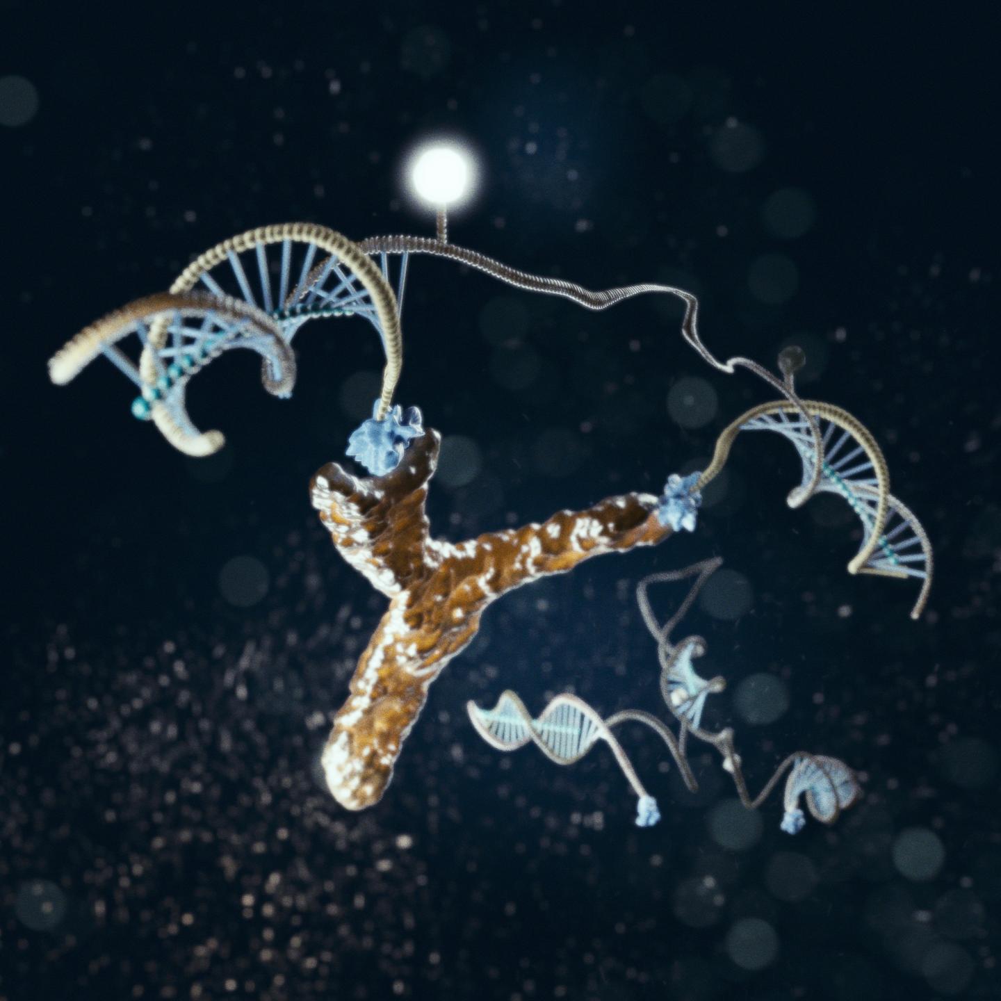
A nanoscale DNA “machine,” shown in this illustration bound to an antibody (yellow), rapidly lights up when it recognizes specific target antibodies (credit: Marco Tripodi)

An international team of scientists has developed a nanomachine using synthetic DNA for rapid, sensitive, low-cost diagnosis of infectious and auto-immune diseases, including HIV, at the point of care. It aims to replace the current slow, cumbersome, and expensive current process of detecting the protein antibodies used for diagnosis.

An antibody causes a structural change (or switch) in the device, which generates a light signal. The sensor does not need to be chemically activated and is rapid — acting within five minutes — enabling the targeted antibodies to be easily detected, even in complex clinical samples such as blood serum.

The antibody-targeting sensor is composed of a light-emitting fluorophore (F) and quencher (green circle) connected to two single-stranded DNA tails joined to the appropriate recognition element (red hexagons) for a given test. When a target antibody is detected by the two recognition elements, they open the stem, activating the fluorophore. (credit: S. Ranallo et al./Angew. Chem. Int. Ed.)

The research is described in the October issue of the journal Angewandte Chemie.

“One of the advantages of our approach is that it is highly versatile,” said Prof. Francesco Ricci, of the University of Rome, Tor Vergata, senior co-author of the study. “This DNA nanomachine can be in fact custom-modified so that it can detect a huge range of antibodies; this makes our platform adaptable for many different diseases.”

“Our modular platform provides significant advantages over existing methods for the detection of antibodies,” added Prof. Vallée-Bélisle of the University of Montreal, the other senior co-author of the paper. “It is rapid, does not require reagent chemicals, and may prove to be useful in a range of different applications such as point-of-care diagnostics and bioimaging.”

The researchers plan to allow the light-emitting signal to be detected by a mobile phone.

A University of California, Santa Barbara scientist was also involved in the research.


Abstract of A Modular, DNA-Based Beacon for Single-Step Fluorescence Detection of Antibodies and Other Proteins

A versatile platform for the one-step fluorescence detection of both monovalent and multivalent proteins has been developed. This system is based on a conformation-switching stem–loop DNA scaffold that presents a small-molecule, polypeptide, or nucleic-acid recognition element on each of its two stem strands. The steric strain associated with the binding of one (multivalent) or two (monovalent) target molecules to these elements opens the stem, enhancing the emission of an attached fluorophore/quencher pair. The sensors respond rapidly (<10 min) and selectively, enabling the facile detection of specific proteins even in complex samples, such as blood serum. The versatility of the platform was demonstrated by detecting five bivalent proteins (four antibodies and the chemokine platelet-derived growth factor) and two monovalent proteins (a Fab fragment and the transcription factor TBP) with low nanomolar detection limits and no detectable cross-reactivity.

Study of protein folds adds to evidence that viruses are alive and ancient

The diverse physical attributes, genome sizes and lifestyles of viruses make them difficult to classify. A new study uses protein folds as evidence that viruses are living entities that belong on their own branch of the tree of life. (credit: Julie McMahon)

Viruses are actually living entities that share a long evolutionary history with cells, researchers report in a study that traces viral evolution back to a time when neither viruses nor cells existed in the forms recognized today.

The new findings appear in an open-access paper in the journal Science Advances.

Some scientists have argued that viruses are nonliving entities, bits of DNA and RNA shed by cellular life. They point to the fact that viruses are not able to replicate outside of host cells, and rely on cells’ protein-building machinery to function. But much evidence supports the idea that viruses are not that different from other living entities, said University of Illinois crop sciences and Carl R. Woese Institute for Genomic Biology professor Gustavo Caetano-Anollés, who led a new analysis with graduate student Arshan Nasir.

“Many organisms require other organisms to live, including bacteria that live inside cells, and fungi that engage in obligate parasitic relationships — they rely on their hosts to complete their lifecycle,” he said. “And this is what viruses do.”

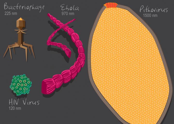
The discovery of the giant mimiviruses in the early 2000s challenged traditional ideas about the nature of viruses, Caetano-Anollés said. “These giant viruses were not the tiny Ebola virus, which has only seven genes. These are massive in size and massive in genomic repertoire,” he said. “Some are as big physically and with genomes that are as big or bigger than bacteria that are parasitic.”

Some giant viruses also have genes for proteins that are essential to translation, the process by which cells read gene sequences to build proteins, Caetano-Anollés said. The lack of translational machinery in viruses was once cited as a justification for classifying them as nonliving, he said.

“This is no more,” Caetano-Anollés said. “Viruses now merit a place in the tree of life. Obviously, there is much more to viruses than we once thought.”

A new virus taxonomy

Caetano-Anollés is the co-author of a report by the International Committee on the Taxonomy of Viruses that recognized seven orders of viruses, based on their shapes and sizes, genetic structure and means of reproducing.

“Under this classification, viral families belonging to the same order have likely diverged from a common ancestral virus,” the authors wrote. “However, only 26 (of 104) viral families have been assigned to an order, and the evolutionary relationships of most of them remain unclear.”

Part of the confusion stems from the abundance and diversity of viruses. Less than 4,900 viruses have been identified and sequenced so far, even though scientists estimate there are more than a million viral species. Many viruses are tiny — significantly smaller than bacteria or other microbes — and contain only a handful of genes. Others, like the recently discovered mimiviruses, are huge, with genomes bigger than those of some bacteria.

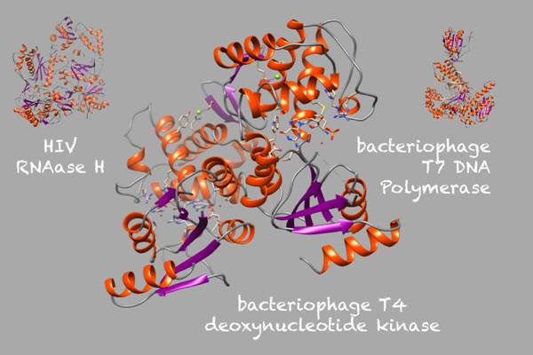
The new study focused on the vast repertoire of protein structures, called “folds,” that are encoded in the genomes of all cells and viruses. Folds are the structural building blocks of proteins, giving them their complex, three-dimensional shapes. By comparing fold structures across different branches of the tree of life, researchers can reconstruct the evolutionary histories of the folds and of the organisms whose genomes code for them.

A new study analyzes the distinct, three-dimensional “folds” structures found in proteins. Pictured here are folds found in viruses. (credit: Arshan Nasir)

The researchers chose to analyze protein folds because the sequences that encode viral genomes are subject to rapid change; their high mutation rates can obscure deep evolutionary signals, Caetano-Anollés said. Protein folds are better markers of ancient events because their three-dimensional structures can be maintained even as the sequences that code for them begin to change.

Shared protein folds between cells and viruses

The researchers analyzed all of the known folds in 5,080 organisms representing every branch of the tree of life, including 3,460 viruses. Using advanced bioinformatics methods, they identified 442 protein folds that are shared between cells and viruses, and 66 that are unique to viruses.

“This tells you that you can build a tree of life, because you’ve found a multitude of features in viruses that have all the properties that cells have,” Caetano-Anollés said. “Viruses also have unique components besides the components that are shared with cells.”

In fact, the analysis revealed genetic sequences in viruses that are unlike anything seen in cells, Caetano-Anollés said. This contradicts one hypothesis that viruses captured all of their genetic material from cells. This and other findings also support the idea that viruses are “creators of novelty,” he said.

Using the protein-fold data available in online databases, Nasir and Caetano-Anollés used computational methods to build trees of life that included viruses.

The data suggest “that viruses originated from multiple ancient cells … and co-existed with the ancestors of modern cells,” the researchers wrote. These ancient cells likely contained segmented RNA genomes, Caetano-Anollés said.

The data also suggest that at some point in their evolutionary history, not long after modern cellular life emerged, most viruses gained the ability to encapsulate themselves in protein coats that protected their genetic payloads, enabling them to spend part of their lifecycle outside of host cells and spread, Caetano-Anollés said. The protein folds that are unique to viruses include those that form these viral “capsids.”

“These capsids became more and more sophisticated with time, allowing viruses to become infectious to cells that had previously resisted them,” Nasir said. “This is the hallmark of parasitism.”


Abstract of A phylogenomic data-driven exploration of viral origins and evolution

The origin of viruses remains mysterious because of their diverse and patchy molecular and functional makeup. Although numerous hypotheses have attempted to explain viral origins, none is backed by substantive data. We take full advantage of the wealth of available protein structural and functional data to explore the evolution of the proteomic makeup of thousands of cells and viruses. Despite the extremely reduced nature of viral proteomes, we established an ancient origin of the “viral supergroup” and the existence of widespread episodes of horizontal transfer of genetic information. Viruses harboring different replicon types and infecting distantly related hosts shared many metabolic and informational protein structural domains of ancient origin that were also widespread in cellular proteomes. Phylogenomic analysis uncovered a universal tree of life and revealed that modern viruses reduced from multiple ancient cells that harbored segmented RNA genomes and coexisted with the ancestors of modern cells. The model for the origin and evolution of viruses and cells is backed by strong genomic and structural evidence and can be reconciled with existing models of viral evolution if one considers viruses to have originated from ancient cells and not from modern counterparts.

New test detects almost all viruses that infect people, animals

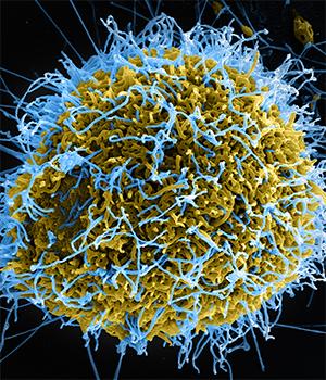
A new test developed at Washington University School of Medicine in St. Louis can detect virtually any virus that infects people and animals, including the Ebola virus (above) (credit: National Institute of Allergy and Infectious Diseases)

A new test that detects virtually any virus that infects people and animals has been developed at Washington University School of Medicine in St. Louis.

Current tests aren’t sensitive enough to detect low virus levels or are limited to detecting only those viruses suspected of being responsible for a patient’s illness.

“With this test, you don’t have to know what you’re looking for,” said the study’s senior author, Gregory Storch, MD, the Ruth L. Siteman Professor of Pediatrics. “It can efficiently detect viruses that are present at very low levels. We think the test will be especially useful in situations where a diagnosis remains elusive after standard testing or in situations in which the cause of a disease outbreak is unknown.”

Results published online in an open-access paper in the journal Genome Research demonstrate that in patient samples the new test, called ViroCap, can detect viruses not found by standard testing based on genome sequencing.

The test could be used to detect outbreaks of deadly viruses such as Ebola, Marburg, and severe acute respiratory syndrome (SARS), as well as more routine viruses, including rotavirus and norovirus, both of which cause severe gastrointestinal infections.

As sensitive as PCR assays, but also tests for related viruses and subtypes

Developed in collaboration with the university’s McDonnell Genome Institute, the test sequences and detects viruses in patient samples and is just as sensitive as the gold-standard polymerase chain reaction (PCR) assays, which are used widely in clinical laboratories. However, even the most expansive PCR assays can only screen for up to about 20 similar viruses at the same time.

“The test is so sensitive that it also detects variant strains of viruses that are closely related genetically,” said corresponding author Todd Wylie, an instructor of pediatrics. “Slight genetic variations among viruses often can’t be distinguished by currently available tests and complicate physicians’ ability to detect all variants with one test.”

In addition, because the test includes detailed genetic information about various strains of particular viruses, subtypes can be identified easily. For example, the study showed that while standard testing identified a virus as influenza A, which causes seasonal flu, the new test indicated that the virus was a particularly harsh subtype called H3N2.

Last flu season, H3N2 contributed to some 36,000 deaths in the United States. And in some patients — particularly young children, older adults and people with weakened immune systems — knowing that the H3N2 strain is present may alter treatment.

To develop the test, the researchers targeted unique stretches of DNA or RNA from every known group of viruses that infects humans and animals. In all, the research team included 2 million unique stretches of genetic material from viruses in the test. These stretches of material are used as probes to pluck out viruses in patient samples that are a genetic match. The matched viral material then is analyzed using high-throughput genetic sequencing. As completely novel viruses are discovered, their genetic material could easily be added to the test, Storch said.

The researchers plan to conduct additional research to validate the accuracy of the test, so it could be several years before it is clinically available.

In the meantime, the technology can be used by scientists to study viruses in a research setting. Kristine Wylie investigates the viruses that set up residence in and on the human body, collectively known as the virome. The new test will provide a way to capture the full breadth and depth of such viruses, and deepen understanding of how they play a role in keeping the body healthy.

“It also may be possible to modify the test so that it could be used to detect pathogens other than viruses, including bacteria, fungi and other microbes, as well as genes that would indicate the pathogen is resistant to treatment with antibiotics or other drugs,” said co-author Kristine Wylie, PhD, assistant professor of pediatrics.

* The researchers evaluated the new test in two sets of biological samples — for example, from blood, stool and nasal secretions — from patients at St. Louis Children’s Hospital. In the first, standard testing that relied on genome sequencing had detected viruses in 10 of 14 patients. But the new test found viruses in the four children that earlier testing had missed. Standard testing failed to detect common, everyday viruses: influenza B, a cause of seasonal flu; parechovirus, a mild gastrointestinal and respiratory virus; herpes virus 1, responsible for cold sores in the mouth; and varicella-zoster virus, which causes chickenpox.

In a second group of children with unexplained fevers, standard testing had detected 11 viruses in the eight children evaluated. But the new test found another seven, including a respiratory virus called human adenovirus B type 3A, which usually is harmless but can cause severe infections in some patients.

In all, the number of viruses detected in the two patient groups jumped to 32 from 21, a 52 percent increase.


Abstract of Enhanced virome sequencing through solution-based capture enrichment

Metagenomic shotgun sequencing (MSS) is an important tool for characterizing viral populations. It is culture-independent, requires no a priori knowledge of the viruses in the sample, and may provide useful genomic information. However, MSS can lack sensitivity and may yield insufficient data for detailed analysis. We have created a targeted sequence capture panel, ViroCap, designed to enrich nucleic acid from DNA and RNA viruses from 34 families that infect vertebrate hosts. A computational approach condensed nearly 1 billion base pair (bp) of viral reference sequence into less than 200 million bp of unique, representative sequence suitable for targeted sequence capture. We compared the effectiveness of detecting viruses in standard MSS versus MSS following targeted sequence capture. First, we analyzed two sets of samples, one derived from samples submitted to a diagnostic virology laboratory and one derived from samples collected in a study of fever in children. We detected 14 and 18 viruses in the two sets, comprising 19 genera from 10 families, with dramatic enhancement of genome representation following capture enrichment. The median fold-increases in percent viral reads post-capture were 674 and 296. Median breadth of coverage increased from 2.1% to 83.2% post-capture in the first set and from 2.0% to 75.6% in the second set. Next, we analyzed samples containing a set of diverse anellovirus sequences and demonstrated that ViroCap could be used to detect viral sequences with up to 58% variation from the references used to select capture probes. ViroCap substantially enhances MSS for a comprehensive set of viruses and has utility for research and clinical applications.

Self-assembling material could lead to artificial arteries

Illustration showing creation of synthetic capillaries from peptides and proteins (credit: QMUL)

Researchers at Queen Mary University of London (QMUL) have developed a new bioinspired process using self-assembling organic molecules that can develop into complex tubular tissue-like structures. The process could lead to creating synthetic tissues that emulate veins, arteries, or even the blood-brain barrier, and that exhibit dynamic behaviors found in biological tissues like growth, morphogenesis, and healing.

The process uses solutions of peptide and protein molecules that self-assemble to form a dynamic tissue that can be guided to grow into complex shapes without the use of molds or techniques like 3-D printing.

Adipose-derived stem cells (mADSCs) seeded onto the protein/peptide membrane (credit: QMUL)

According to the researchers, the finding could allow scientists to study diseases such as Alzheimer’s with a high level of similarity to the real tissue and create better implants, complex tissues, and more effective drug-screening methods.

The study appeared September 28 in the journal Nature Chemistry. It has been partly funded by the European Research Council.


QMUL | Demonstrating a dynamic self-assembling protein-peptide membrane


Abstract of Co-assembly, spatiotemporal control and morphogenesis of a hybrid protein–peptide system

Controlling molecular interactions between bioinspired molecules can enable the development of new materials with higher complexity and innovative properties. Here we report on a dynamic system that emerges from the conformational modification of an elastin-like protein by peptide amphiphiles and with the capacity to access, and be maintained in, non-equilibrium for substantial periods of time. The system enables the formation of a robust membrane that displays controlled assembly and disassembly capabilities, adhesion and sealing to surfaces, self-healing and the capability to undergo morphogenesis into tubular structures with high spatiotemporal control. We use advanced microscopy along with turbidity and spectroscopic measurements to investigate the mechanism of assembly and its relation to the distinctive membrane architecture and the resulting dynamic properties. Using cell-culture experiments with endothelial and adipose-derived stem cells, we demonstrate the potential of this system to generate complex bioactive scaffolds for applications such as tissue engineering.

‘Designless’ nanoscale logic circuits resemble Darwinian evolution and neural networks

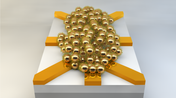
Illustration of a nanoparticle network (about 200 nanometers in diameter). By applying electrical signals at the electrodes (yellow), and using artificial evolution, this disordered network can be configured into useful logic circuits. (credit: University of Twente)

Researchers at the University of Twente in The Netherlands have designed and demonstrated working electronic logic circuits produced using methods that resemble Darwinian evolution and neural networks like the human brain.

In a radical “designless” approach, the researchers used a 200-nanometer-wide cluster of 20-nanometer gold nanoparticles. They applied a series of voltages to eight electrodes and determined the resulting set of 16 different two-input Boolean logic gates.

Artificial evolution

Instead of designing logic circuits with specified functions, as with conventional transistors, this approach works around — or can even take advantage of — any material defects.

To do this, the researchers used an artificial evolution model — one that runs in less than an hour, rather than millions of years. “Natural evolution has led to powerful ‘computers’ like the human brain, which can solve complex problems in an energy-efficient way,” the researchers note. “Nature exploits complex networks that can execute many tasks in parallel.”

“This is the first time that scientists have succeeded in realizing robust electronics with small dimensions that can compete with commercial technology.”

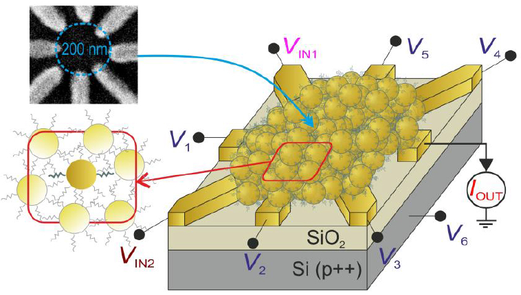
Schematic device layout of the disordered network of gold nanoparticles, separated by ~1 nm 1-octanethiols, in between eight titanium-gold electrodes, as shown in the scanning electron micrograph (top inset). The gold nanoparticles act as single-electron transistors (SETs) at low temperature (<15 K). (credit: S. K. Bose, et al./Nature Nanotechnology)

Conventional transistors are limited to a handful of atoms. It would a major challenge to produce chips in which the millions of transistors required have the same characteristics, according to the researchers from the Twente MESA+ Institute for Nanotechnology and the CTIT Institute for ICT Research. Current transistor designs are also limited by their energy consumption, which is reaching unacceptable levels.

According to University of Twente prof. Wilfred van der Wiel, the logic circuits they discovered currently have limited computing power. “But with this research we have delivered a proof of principle. By scaling up the system, real added value will be produced in the future. This type of circuitry uses much less energy, both in production and use.” The researchers anticipate a wide range of applications — for example, in portable electronics and in the medical world.

“By choosing a smaller nanoparticle diameter, and scaling down the electrode geometry accordingly, our network would not only further reduce area, but room-temperature operation would come into sight as well,” the researchers note in a paper in the journal Nature Nanotechnology.

Mimicking brain-like systems

The researchers also contrast their “designless” reconfigurable approach with massively parallel (but still design-constrained) architectures such as IBM’s TrueNorth brain-inspired chip.

“An especially interesting avenue to explore is the suitability of this system for advanced functionality that is hard (or expensive) to realize in a conventional architecture, such as pattern recognition by mimicking brain-like systems, or simulations of complex physical systems,” the researchers note in the paper. “Our evolutionary approach works around device-to-device variations at the nanoscale and the accompanying uncertainties in performance, which is becoming more and more a bottleneck for the miniaturization of conventional electronic circuits.”


Abstract of Evolution of a designless nanoparticle network into reconfigurable Boolean logic

Natural computers exploit the emergent properties and massive parallelism of interconnected networks of locally active components. Evolution has resulted in systems that compute quickly and that use energy efficiently, utilizing whatever physical properties are exploitable. Man-made computers, on the other hand, are based on circuits of functional units that follow given design rules. Hence, potentially exploitable physical processes, such as capacitive crosstalk, to solve a problem are left out. Until now, designless nanoscale networks of inanimate matter that exhibit robust computational functionality had not been realized. Here we artificially evolve the electrical properties of a disordered nanomaterials system (by optimizing the values of control voltages using a genetic algorithm) to perform computational tasks reconfigurably. We exploit the rich behaviour that emerges from interconnected metal nanoparticles, which act as strongly nonlinear single-electron transistors, and find that this nanoscale architecture can be configured in situ into any Boolean logic gate. This universal, reconfigurable gate would require about ten transistors in a conventional circuit. Our system meets the criteria for the physical realization of (cellular) neural networks: universality (arbitrary Boolean functions), compactness, robustness and evolvability, which implies scalability to perform more advanced tasks. Our evolutionary approach works around device-to-device variations and the accompanying uncertainties in performance. Moreover, it bears a great potential for more energy-efficient computation, and for solving problems that are very hard to tackle in conventional architectures.