Living implants

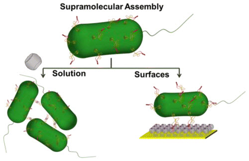
“Living implant” using a bacterial strain (credit: Shrikrishnan Sankaran et al./ACS Nano)

A method for merging bacteria in human cells as “living implants” has been developed by University of Twente researchers. The implants could include stents equipped with bacteria on which endothelial cells (cells that form the lining of blood vessels) can grow, or bacteria that can release medicines in specific parts of the body.

They achieved this by supramolecular assembly, modifying the DNA of E. coli bacteria in such a way that a small molecule called Cucurbit[8]uril attaches to a protein on the cell membrane. This substance could then attach to other building blocks, forming a sort of natural Velcro.

The research was published in the journal ACS Nano. It was sponsored by the European Union.


Abstract of Incorporating Bacteria as a Living Component in Supramolecular Self-Assembled Monolayers through Dynamic Nanoscale Interactions

Supramolecular assemblies, formed through noncovalent interactions, has become particularly attractive to develop dynamic and responsive architectures to address living systems at the nanoscale. Cucurbit[8]uril (CB[8]), a pumpkin shaped macrocylic host molecule, has been successfully used to construct various self-assembled architectures for biomedical applications since it can simultaneously bind two aromatic guest molecules within its cavity. Such architectures can also be designed to respond to external stimuli. Integrating living organisms as an active component into such supramolecular architectures would add a new dimension to the capabilities of such systems. To achieve this, we have incorporated supramolecular functionality at the bacterial surface by genetically modifying a transmembrane protein to display a CB[8]-binding motif as part of a cystine-stabilized miniprotein. We were able to confirm that this supramolecular motif on the bacterial surface specifically binds CB[8] and forms multiple intercellular ternary complexes leading to aggregation of the bacterial solution. We performed various aggregation experiments to understand how CB[8] interacts with this bacterial strain and also demonstrate that it can be chemically reversed using a competitor. To confirm that this strain can be incorporated with a CB[8] based architecture, we show that the bacterial cells were able to adhere to CB[8] self-assembled monolayers (SAMs) on gold and still retain considerable motility for several hours, indicating that the system can potentially be used to develop supramolecular bacterial biomotors. The bacterial strain also has the potential to be combined with other CB[8] based architectures like nanoparticles, vesicles and hydrogels.

Ultrafast lasers enable 3-D micropatterning of biocompatible hydrogels

Illustration of laser-based micropatterning of silk hydrogels. The transparent gels enable the laser’s photons (pink) to be absorbed more than 10 times deeper than with other materials without damaging the cells (green) surrounding the letters (credit: M.B. A)

Tufts University biomedical engineers have developed low-energy, ultrafast laser technology for micropatterning high-resolution, 3-D structures in silk-protein hydrogels.

Micropatterning is used to bring oxygen and nutrients to rapidly proliferating cells in an engineered tissue scaffold. The goal is “to controllably guide cell growth and create an artificial vasculature (blood vessel system) within an already densely seeded silk hydrogel,” said Fiorenzo G. Omenetto, Ph.D., senior author of a paper on the work in PNAS Early Edition published September 15 online before print.

Current patterning techniques allow for the production of random, micrometer-scale pores and also the creation of larger channels that are hundreds of micrometers in diameter, but there is little in between.

The Tufts researchers used the ultrafast femtosecond laser to generate scalable, high-resolution 3-D pores (voids) within silk protein hydrogel, a soft, transparent biomaterial that supports cell growth and allows cells to penetrate deep within it. The researchers were able to create pores at multiple scales as small as 10 micrometers and as large at 400 micrometers over a large volume.

The transparent silk gels also enabled the laser’s photons to be absorbed nearly 1 cm below the surface of the gel — more than ten times deeper than with other materials — without damaging adjacent material.

The laser treatment can be done while keeping the cell culture sealed and sterile. Unlike most 3-D printing, this technique does not require photoinitiators (compounds that promote photoreactivity but are typically not biocompatible).

Omenetto is associate dean for research, professor of biomedical engineering and Frank C. Doble professor at Tufts School of Engineering and also holds an appointment in physics in the School of Arts and Sciences.

The research team reported similar results in vitro and in a preliminary in vivo study in mice.


Abstract of Laser-based three-dimensional multiscale micropatterning of biocompatible hydrogels for customized tissue engineering scaffolds

Light-induced material phase transitions enable the formation of shapes and patterns from the nano- to the macroscale. From lithographic techniques that enable high-density silicon circuit integration, to laser cutting and welding, light–matter interactions are pervasive in everyday materials fabrication and transformation. These noncontact patterning techniques are ideally suited to reshape soft materials of biological relevance. We present here the use of relatively low-energy (< 2 nJ) ultrafast laser pulses to generate 2D and 3D multiscale patterns in soft silk protein hydrogels without exogenous or chemical cross-linkers. We find that high-resolution features can be generated within bulk hydrogels through nearly 1 cm of material, which is 1.5 orders of magnitude deeper than other biocompatible materials. Examples illustrating the materials, results, and the performance of the machined geometries in vitro and in vivo are presented to demonstrate the versatility of the approach.

DNA-based nanodevices for molecular medicine: an overview

Virus-protein-coated DNA origami nanostructures. With the help of protein encapsulation, DNA origamis can be transported into human cells much more efficiently. (credit: Veikko Linko and Mauri Kostiainen)

KurzweilAI has covered a wide variety of research projects that explore how DNA molecules can be assembled into complex nanostructures for molecular-scale diagnostics, smart drug-delivery, and other uses. For example, tailored DNA structures could find targeted cancer cells and release their molecular payload (drugs or antibodies) selectively.

An article written by researchers from Aalto University just published in Trends in Biotechnology journal, comparing biological DNA-nanomachine developments and their uses, should help put this varied research in perspective.

The authors explain that “the field of structural DNA nanotechnology started around 30 years ago when Ned Seeman performed pioneering research with DNA junctions and lattice. … The key player in the fast development of DNA nanotechnology was the invention of DNA origami in 2006. The DNA origami method is based on folding a long
single-stranded ‘DNA scaffold strand’ into a customized shape with a set of short synthetic strands that act as ‘staples’ to bind the overall structure together.”

“This method is the starting point for practically all other straightforward design approaches available today,” says Veikko Linko, an Academy of Finland postdoctoral researcher from Biohybrid Materials Group and first author.

The accurate shape of a DNA origami nanostructure can be used to create entirely metallic nanoparticles on silicon substrates. (credit: Veikko Linko, Boxuan Shen and Mauri Kostiainen with permission from Royal Society of Chemistry)

Versatile DNA nanostructures

The most important feature of a DNA-based nanostructure is its modularity, the authors note. DNA structures can be fabricated with nanometer-precision, and other molecules such as RNA, proteins, peptides and drugs can be anchored to them with the same resolution.

Such a high precision can be exploited in creating nanosized optical devices as well as molecular platforms and bar codes for various imaging techniques and analytics.

The author further point out that for molecular medicine, DNA-based devices could be used for detecting single molecules and modulating cell signaling. In the near future, highly sophisticated DNA robots could even be used in creating artificial immune systems, they note.

In addition, a system based on tailored DNA devices could help to avoid unnecessary drug treatments, since programmed DNA-nanorobots could detect various agents from the blood stream, and immediately start the battle against disease.


Abstract of DNA Nanostructures as Smart Drug-Delivery Vehicles and Molecular Devices

DNA molecules can be assembled into custom predesigned shapes via hybridization of sequence-complementary domains. The folded structures have high spatial addressability and a tremendous potential to serve as platforms and active components in a plethora of bionanotechnological applications. DNA is a truly programmable material, and its nanoscale engineering thus opens up numerous attractive possibilities to develop novel methods for therapeutics. The tailored molecular devices could be used in targeting cells and triggering the cellular actions in the biological environment. In this review we focus on the DNA-based assemblies – primarily DNA origami nanostructures – that could perform complex tasks in cells and serve as smart drug-delivery vehicles in, for example, cancer therapy, prodrug medication, and enzyme replacement therapy.

DNA-guided 3-D printing of human tissue

Reconstituting epithelial (skin) microtissues with programmed size, shape, composition, spatial heterogeneity, and embedding extracellular matrix. Scheme and images of fully embedded aggregates of human luminal and myoepithelial cells. (credit: Michael E Todhunter et al./Nature Methods)

A new technique developed by UCSF scientists for building organoids (tiny models of human tissues) more precisely turns human cells into the biological equivalent of LEGO bricks. Called DNA Programmed Assembly of Cells (DPAC), it allows researchers in hours to create arrays of thousands of custom-designed organoids, such as models of human mammary glands containing several hundred cells each.

These mini-tissues in a dish can be used to study how particular structural features of tissue affect normal growth or go awry in cancer. They could be used for therapeutic drug screening and to help teach researchers how to grow whole human organs.

The new technique, reported in an open-the journal Nature Methods on Aug. 31, allows for researchers to “take any cell type we want and program just where it goes,” said  Zev Gartner, PhD, the paper’s senior author and an associate professor of pharmaceutical chemistry at UCSF. “We can precisely control who’s talking to whom and who’s touching whom at the earliest stages.”

There are very few limits to the tissues this technology can mimic, he said. “One potential application would be that within the next couple of years, we could be taking samples of different components of a cancer patient’s mammary gland and building a model of their tissue to use as a personalized drug screening platform. Another is to use the rules of tissue growth we learn with these models to one day grow complete organs.”

Studying how the cells of complex tissues like the mammary gland self-organize, make decisions as groups, and break down in disease has been a challenge to researchers. The living organism is often too complex to identify the specific causes of a particular cellular behavior. On the other hand, cells in a dish lack the critical element of realistic 3-D structure.

DNA as molecular Velcro and bar code

To specify the 3-D structure of their organoids, the researchers incubate cells with tiny snippets of single-stranded DNA engineered to slip into the cells’ outer membranes, covering each cell like the hairs on a tennis ball. These DNA strands act both as a sort of molecular Velcro and as a bar code that specifies where each cell belongs within the organoid. When two cells incubated with complementary DNA strands come in contact, they stick fast. If the DNA sequences don’t match, the cells float on by. Cells can be incubated with several sets of DNA bar codes to specify multiple allowable partners.

A whole-mount image of a digitized mouse mammary fat pad (reproduced with permission of W. Muller) used to print a pattern of DNA spots, and rendered as a 1.6-cm-long pattern of single cells fully embedded in gelatinous protein mixture (credit: Michael E Todhunter et al./Nature Methods)

To turn these cellular LEGOs into arrays of organoids that can be used for research, Gartner’s team lays down the cells in layers, with multiple sets of cells designed to stick to particular partners. This lets them build up complex tissue components like the mammary gland. It also lets them experiment with specifically adding in a single cell with a known cancer mutation to different parts of the organoid to observe its effects.

To demonstrate the precision of the technique and its ability to generalize to many different human tissue types, the research team created several proof-of-principle organoid arrays mimicking human tissues such as branching vasculature and mammary glands.

In one experiment, the researchers created arrays of mammary epithelial cells and asked how adding one or more cells expressing low levels of the cancer gene RasG12V affected the cells around them. They found that normal cells grow faster when in an organoid with cells expressing RasG12V at low levels, but required more than one mutant cell to kick-start this abnormal growth. They also found that placing cells with low RasG12V expression at the end of a tube of normal cells allowed the mutant cells to branch and grow, drawing normal cells behind them like a bud at the tip of a growing tree branch.

Gartner’s group plans to use the technique to investigate what cellular or structural changes in mammary glands can lead to the breakdown of tissue architecture associated with tumors that metastasize, invading other parts of the body and threatening the life of the patient. They also hope to use what they learn from simple models of different tissue types to ultimately build functional human tissues like lung and kidney and neural circuits using larger-scale techniques.


Abstract of Programmed synthesis of three-dimensional tissues

Reconstituting tissues from their cellular building blocks facilitates the modeling of morphogenesis, homeostasis and disease in vitro. Here we describe DNA-programmed assembly of cells (DPAC), a method to reconstitute the multicellular organization of organoid-like tissues having programmed size, shape, composition and spatial heterogeneity. DPAC uses dissociated cells that are chemically functionalized with degradable oligonucleotide ‘Velcro’, allowing rapid, specific and reversible cell adhesion to other surfaces coated with complementary DNA sequences. DNA-patterned substrates function as removable and adhesive templates, and layer-by-layer DNA-programmed assembly builds arrays of tissues into the third dimension above the template. DNase releases completed arrays of organoid-like microtissues from the template concomitant with full embedding in a variety of extracellular matrix (ECM) gels. DPAC positions subpopulations of cells with single-cell spatial resolution and generates cultures several centimeters long. We used DPAC to explore the impact of ECM composition, heterotypic cell-cell interactions and patterns of signaling heterogeneity on collective cell behaviors.

First application to pursue genome editing research in human embryos

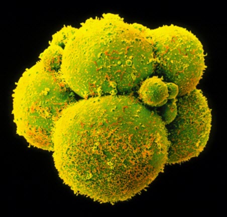
Human embryos are at the center of a debate over the ethics of gene editing (credit: Dr. Yorgos Nikas/SPL)

The first application to pursue CRISPR/Cas9 genome-editing research in viable human embryos has been submitted to the UK’s fertility regulator by a team of researchers affiliated with the Francis Crick Institute in London.

“This research proposal is a troubling and provocative move,” commented Marcy Darnovsky, PhD, Executive Director of the Center for Genetics and Society.

“Modifying the genes of human embryos is deeply controversial because it can be used for worthwhile research on the one hand, or to produce genetically modified human beings on the other. A global public conversation about preventing such misuses is just getting underway, and this proposal could short-circuit those deliberations.

“It’s illegal in the UK and dozens of other countries to use a modified embryo to initiate a pregnancy, but in others — notably the U.S. — we don’t have that legal protection,” Darnovsky added. “If scientists and the regulatory agency in the UK are serious about responsible use of powerful new gene altering technologies, they won’t be rushing ahead in ways that could open the door to a world of genetically modified humans.”

If the UK Human Fertilisation & Embryology Authority were to issue this license, this would be the first approval of genome editing research on the human germline by a national regulatory body.

The resulting experiments would be the second of their kind in this highly controversial area of research. In April, scientists working out of China published research that they had created the first genetically modified human embryos—these embryos were nonviable, and the results of the CRISPR/Cas9 engineering were highly unsuccessful: producing off target mutations and mosaicism that underlined the limitations of our current understandings of genetics and genomics.

The response from the scientific community and the public after the first human embryo gene editing experiment in April was swift. Many scientists voiced support for either a pause or a moratorium on human germline modification.

On September 14, the National Academies announced that the International Summit on Human Gene Editing scheduled for December will now be co-hosted by the Royal Society (UK) and the Chinese Academy of Sciences.

A new process for studying proteins associated with diseases

Schematic of phosphoprotein biosynthesis from E. coli bacteria. Sep-OTS: genetically encoded phosphoserine; CFPS: cell-free protein synthesis; NTPs: nucleoside triphosphates; lysis:  breaking down cell membrane. (credit: Javin P. Oza et al./Nature Communications)

Researchers from Northwestern University and Yale University have developed a new technology to help scientists understand how proteins work and fix them when they are broken. Such knowledge could pave the way for new drugs for a myriad of diseases, including cancer.

The human body turns its proteins on and off (to alter their function and activity in cells) using “phosphorylation” — the reversible attachment of phosphate groups to proteins. These “decorations” on proteins provide an enormous variety of functions and are essential to all forms of life. Little is known, however, about how this important dynamic process works in humans.

Phosphorylation: a hallmark of disease

Using a special strain of E. coli bacteria, the researchers built a cell-free protein synthesis platform technology that can manufacture large quantities of these human phosphoproteins for scientific study. The goal is to enable scientists to learn more about the function and structure of phosphoproteins and identify which ones are involved in disease.

The study was published Sept. 9 in an open-access paper by the journal Nature Communications.

Trouble in the phosphorylation process can be a hallmark of disease, such as cancer, inflammation and Alzheimer’s disease. The human proteome (the entire set of expressed proteins) is estimated to be phosphorylated at more than 100,000 unique sites, making study of phosphorylated proteins and their role in disease a daunting task.

“Our technology begins to make this a tractable problem,”  said  Michael C. Jewett, an associate professor of chemical and biological engineering who led the Northwestern team. “We now can make these special proteins at unprecedented yields, with a freedom of design that is not possible in living organisms. The consequence of this innovative strategy is enormous.”

A “plug-and-play” protein expression platform

Jewett and his colleagues combined state-of-the-art genome engineering tools and engineered biological “parts” into a “plug-and-play” protein expression platform that is cell-free. Cell-free systems activate complex biological systems without using living intact cells. Crude cell lysates, or extracts, are employed instead.

The researchers prepared cell lysates of genomically recoded bacteria that incorporate amino acids not found in nature. This allowed them to harness the cell’s engineered machinery and turn it into a factory, capable of on-demand biomanufacturing new classes of proteins.

To demonstrate their cell-free platform technology, the researchers produced a human kinase that is involved in tumor cell proliferation and showed that it was functional and active. Kinase is an enzyme (a protein acting as a catalyst) that transfers a phosphate group onto a protein. Through this process, kinases activate the function of proteins within the cell. Kinases are implicated in many diseases and, therefore, of particular interest.

“The ability to produce kinases for study should be useful in learning how these proteins function and in developing new types of drugs,” Jewett said.


Abstract of Robust production of recombinant phosphoproteins using cell-free protein synthesis

Understanding the functional and structural consequences of site-specific protein phosphorylation has remained limited by our inability to produce phosphoproteins at high yields. Here we address this limitation by developing a cell-free protein synthesis (CFPS) platform that employs crude extracts from a genomically recoded strain of Escherichia coli for site-specific, co-translational incorporation of phosphoserine into proteins. We apply this system to the robust production of up to milligram quantities of human MEK1 kinase. Then, we recapitulate a physiological signalling cascade in vitro to evaluate the contributions of site-specific phosphorylation of mono- and doubly phosphorylated forms on MEK1 activity. We discover that only one phosphorylation event is necessary and sufficient for MEK1 activity. Our work sets the stage for using CFPS as a rapid high-throughput technology platform for direct expression of programmable phosphoproteins containing multiple phosphorylated residues. This work will facilitate study of phosphorylation-dependent structure–function relationships, kinase signalling networks and kinase inhibitor drugs.

New optogenetics process could lead to neurological enhancements and treatments

Artist’s representation of a calcium ion channel affected by OptoSTIM1 (credit: Institute for Basic Science)

An advanced process for precision control of cellular calcium ion (Ca2+) channels in living organisms has been engineered by a research team at the Korea Advanced Institute of Science and Technology (KAIST) and the IBS Center for Cognition and Sociality.

Calcium ions are a crucial part of diverse cellular functions such as contraction, excitation, growth, differentiation and death. Severe Ca2+ deficiency is linked to cardiac arrhythmia, cognitive impairment, and ataxia.

The new process uses optogenetics, or control of cells by light. The researchers added a new light-sensitive, plant-human hybrid protein to cells to efficiently modulate calcium ion channels in cells by shining blue light on them.

The hybrid protein combines a photoreceptor protein called cryptochrome 2 (Cry2) from a small, flowering plant Arabidopsis thaliana with the STromal Interaction Molecule 1 (STIM1), a protein found in almost all animals that opens cellular Ca2+ channels.

They named the resultant hybrid molecule OptoSTIM1.

When they shined blue light on the OptoSTIM1-expressing cells, they were able to coax them to open their Ca2+ channels and allow an influx of 5 to 10 times more Ca2+ than in previous studies.

Increasing learning capacity in mice

Mouse with blue light apparatus attached (credit: Institute For Basic Science)

To test the functional effect of the Ca2+ influx, the IBS team introduced OptoSTIM1 to the hippocampus of a living mouse. They compared sets of light-illuminated mice to non-illuminated mice expressing OptoSTIM1 in an environment in which they introduced a conditioning cue followed by a fear stimulus.

In subsequent tests they observed that light-illuminated mice with the OptoSTIM1 expression had a nearly twofold increase in fear response when placed in the testing environment without the conditioning cue than the non-light-stimulated mice. That indicated that the OptoSTIM1 expression (and resultant Ca2+ uptake) was an effective method for memory enhancement.

Neurological enhancements and treatments

The researchers say this work opens the door for future research into optogenetically enhanced memory and learning studies and into treating neurological diseases that are a result of a dysfunction in Ca2+ regulation.

This may also be a step towards discovering applications for drugs as well as therapeutic Ca2+ modulation. According to Kyung, “There are diseases that result from dysfunction in cellular Ca2+ regulation, such as Alzheimer’s disease, so we can apply our system to those areas and hopefully in the near future help people to recover from those diseases.”

This may also allow for future non-invasive and non-drug treatments or may help to mitigate and eventually cure some neurological diseases.

Team is led by Won Do Heo, associate professor together with Professor Yong-Mahn Han and Professor Daesoo Kim.


Abstract of Optogenetic control of endogenous Ca2+ channels in vivo

Calcium (Ca2+) signals that are precisely modulated in space and time mediate a myriad of cellular processes, including contraction, excitation, growth, differentiation and apoptosis. However, study of Ca2+ responses has been hampered by technological limitations of existing Ca2+-modulating tools. Here we present OptoSTIM1, an optogenetic tool for manipulating intracellular Ca2+ levels through activation of Ca2+-selective endogenous Ca2+ release−activated Ca2+ (CRAC) channels. Using OptoSTIM1, which combines a plant photoreceptor and the CRAC channel regulator STIM1 (ref. 4), we quantitatively and qualitatively controlled intracellular Ca2+ levels in various biological systems, including zebrafish embryos and human embryonic stem cells. We demonstrate that activating OptoSTIM1 in the CA1 hippocampal region of mice selectively reinforced contextual memory formation. The broad utility of OptoSTIM1 will expand our mechanistic understanding of numerous Ca2+-associated processes and facilitate screening for drug candidates that antagonize Ca2+ signals.

The CRISPR controversy: faster, cheaper gene editing vs. bioethicists

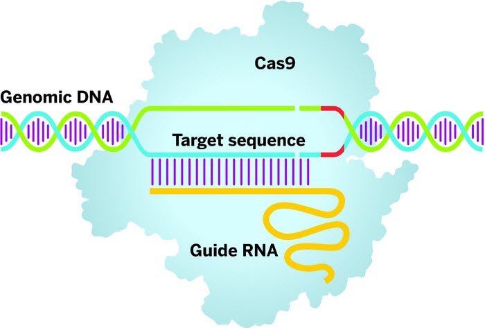
Clustered regularly interspaced short palindromic repeats (CRISPRs) technology employs a guide RNA to direct the Cas9 enzyme (light blue) to a target DNA sequence. Once there, Cas9 will bind when it finds a protospacer-adjacent motif sequence (red) in the DNA and cut both strands, priming the gene sequence for editing. (credit: Adapted from OriGene Technologies)

Within the past few years, a new technology has made altering genes in plants and animals much easier than before. The tool, called CRISPR/Cas9 or just CRISPR, has spurred a flurry of research that could one day lead to hardier crops and livestock, as well as innovative biomedicines.

But along with potential benefits, it raises red flags, according to an open-access article in Chemical & Engineering News (C&EN), the weekly newsmagazine of the American Chemical Society.

Ann M. Thayer, a senior correspondent at C&EN, notes that scientists have long had the ability to remove, repair or insert genetic material in cells. But the process was time consuming and expensive. CRISPR (“clustered regularly interspaced short palinodromic repeats”) streamlines gene editing dramatically. Its simplicity has enabled far more scientists to get involved in such work. In a short time, they have now used CRISPR to edit genes in insects, plants, fish, rodents and monkeys.

The potential agricultural and medical applications that could result from the tool in the future have attracted the interest of venture capitalists and pharmaceutical companies, the article says. While it seems CRISPR work is moving full-steam ahead, a couple of recent developments could check its growth.

In April, Chinese scientists reported that they had attempted to alter a gene in nonviable human embryos. The announcement sparked bioethicists to call for a more cautious approach to gene editing. The other wrench in the system is an ongoing dispute over who should be awarded the patent for inventing CRISPR. Until these issues are resolved, some investors and researchers will opt to wait on the sidelines.


McGovern Institute for Brain Research at MIT | Genome Editing with CRISPR-Cas9

Why human genome editing research is essential

(credit: NIH)

Research involving editing the human genome, including research with human embryos, is essential to gain basic understanding of biology and germ cells and should be permitted, according to one of the first global meetings to debate the controversial new techniques.

The bold statement was published today (Thursday, Sept. 10) by the Hinxton Group, a global network of stem cell researchers, bioethicists, and experts on policy and scientific publishing, who met in Manchester, England, September 3–4.

Not ready for clinical applications

“We believe that while this technology has tremendous value to basic research and enormous potential for somatic clinical uses, it is not sufficiently developed to consider human genome editing for clinical reproductive purposes at this time,” the consensus statement reads.

Discussions at the meeting included the most contentious aspects of these new technologies — the implications for any children born with engineered genetic modifications, and also successive generations who would inherit those genetic changes, according to Debra Mathews, a member of the Hinxton Group steering committee.

“While there is controversy and deep moral disagreement about human germline genetic modification, what is needed is not to stop all discussion, debate and research, but rather to engage with the public, policymakers and the broader scientific community, and to weigh together the potential benefits and harms of human genome editing for research and human health,” says Mathews, the Assistant Director for Science Programs at the Johns Hopkins Berman Institute of Bioethics.

The consensus statement addresses these ethical concerns, with the group agreeing that, “given all safety, efficacy and governance needs are met, there may be morally acceptable uses of this technology in human reproduction, though further substantial discussion and debate will be required.”

Basic research with human embryos

In the meantime, knowledge gained through basic science research is essential to human understanding of both ourselves and other life, the group says. “Much of our knowledge of early development comes from studies of mouse embryos, yet it is becoming clear that gene activity and even some cell types are very different in human embryos.”

Genome editing techniques could be used to ask how cell types are specified in the early embryo and the nature and importance of the genes involved,” says Robin Lovell-Badge, a member of the Hinxton Group steering committee and Group Leader, and head of the Laboratory of Stem Cell Biology and Developmental Genetics, The Francis Crick Institute.

The statement emphasizes the importance of “meaningful and substantial public engagement” to decision-making about genome editing, stating that policy restraints on science should have justification that “that reaches beyond disagreements based solely on divergent moral convictions.”

“The relevant regulatory distinction should be not between using genome editing in somatic cells and using it in embryos, but between research and reproduction: whether those embryos are ever destined to be implanted, says Sarah Chan, another steering committee member and a Chancellor’s Fellow at the Usher Institute for Population Health Sciences and Informatics, University of Edinburgh.

“Restricting research because of concerns that reproductive application is premature and dangerous will ensure that it remains forever premature and dangerous, for want of better knowledge,” Chan says.

Carbon dioxide capture by a novel material that mimics a plant enzyme

Atomic structure of the adsorbed* carbon dioxide (gray sphere bonded to two red spheres) inserted between the manganese (green sphere) and amine (blue sphere) groups within the novel metal-organic framework, forming a linear chain of ammonium carbamate (top). Some hydrogen atoms (white sphere) are omitted for clarity. (credit: Image courtesy of Thomas McDonald, Jarad Mason, and Jeffrey Long)

A novel porous material that achieves carbon dioxide (CO2) capture-and-release with only small shifts in temperature has been developed by a team of researchers at the Center for Gas Separations Relevant to Clean Energy Technologies, led by the University of California, Berkeley (a DOE Energy Frontier Research Center), and associates.

This metal-organic framework (MOF) structure, which adsorbs* CO2, closely resembles an enzyme found in plants known as RuBisCO, which captures CO2 from the atmosphere for conversion into nutrients.

The discovery* paves the way for designing more efficient materials that dramatically reduce overall energy cost of carbon capture. Such materials could be used for carbon capture from fossil-fuel-based power plants as well as from the atmosphere, mitigating the greenhouse effect.

The enhanced carbon capture efficiency of the new class of materials could allow for dramatic reductions in the overall energy cost of carbon capture in power plants or even from the atmosphere, according to the researchers.

* Adsorbed CO2 is captured on the surface of a material; absorbed CO2 is captured inside the material.

** The cooperative mechanism for carbon dioxide (CO2) adsorption in porous MOF materials:

First, a CO2 molecule gets inserted between a metal ion and an amine group within the cylindrical pore of the MOF. Interestingly, the chemical environment of the MOF with the adsorbed CO2  is very similar to that of plant enzyme RuBisCO with a bound CO2.

RuBisCO plays an essential role in biological carbon fixation by plants and conversion into nutrients. In the case of the newly synthesized diamine-appended MOFs, however, the inserted CO2 reorganizes the chemical environment at the adjacent metal ion site to be just right for the insertion of the next CO2.

As more CO2 enters the pore, a cooperative domino effect ensues that leads to the formation of linear chains of ammonium carbamate along the cylindrical pore surfaces of the MOF.

Gas adsorption measurements show the high selectivity of the material for CO2 from the typical composition of flue gas from fossil-fuel-based power plants that contains nitrogen, water, and CO2.

Furthermore, the material has large working capacities — the amount of CO2 adsorbed and desorbed for a given amount of material — that are enabled by only moderate temperature shifts for the adsorption and desorption processes.

Finally, the research points out that changing the strength of the metal-diamine bond through metal substitution allows for rational tuning of the adsorption and desorption properties.


Abstract of Cooperative insertion of CO2 in diamine-appended metal-organic frameworks

The process of carbon capture and sequestration has been proposed as a method of mitigating the build-up of greenhouse gases in the atmosphere. If implemented, the cost of electricity generated by a fossil fuel-burning power plant would rise substantially, owing to the expense of removing CO2 from the effluent stream. There is therefore an urgent need for more efficient gas separation technologies, such as those potentially offered by advanced solid adsorbents. Here we show that diamine-appended metal-organic frameworks can behave as ‘phase-change’ adsorbents, with unusual step-shaped CO2 adsorption isotherms that shift markedly with temperature. Results from spectroscopic, diffraction and computational studies show that the origin of the sharp adsorption step is an unprecedented cooperative process in which, above a metal-dependent threshold pressure, CO2 molecules insert into metal-amine bonds, inducing a reorganization of the amines into well-ordered chains of ammonium carbamate. As a consequence, large CO2 separation capacities can be achieved with small temperature swings, and regeneration energies appreciably lower than achievable with state-of-the-art aqueous amine solutions become feasible. The results provide a mechanistic framework for designing highly efficient adsorbents for removing CO2 from various gas mixtures, and yield insights into the conservation of Mg2+ within the ribulose-1,5-bisphosphate carboxylase/oxygenase family of enzymes.