Silk bio-ink could help advance tissue engineering using 3-D printers

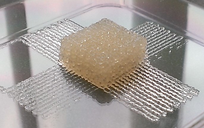
Scientists have developed a silk-based, 3-D printer ink for use in biomedical implants or tissue engineering (credit: American Chemical Society )

Tufts University scientists have developed a silk-based bio-ink that could allow for printing tissues that could be loaded with pharmaceuticals, cytokines (for directing stem cell functions), and antibiotics (for controlling infections), for example, or used in biomedical implants and tissue engineering.

Current 3-D printing processes are limited to simple body parts such as bone. And most inks currently being developed for 3-D printing are made of thermoplastics, silicones, collagen, gelatin, or alginate, which have limits. For example, the temperatures, pH changes and crosslinking methods that may be required to toughen some of these materials can damage cells or other biological components that researchers would want to add to the inks.

To address these bio-ink limitations, Tufts Stern Family Professor of Engineering David L. Kaplan and associates combined silk proteins, which are biocompatible, and glycerol, a non-toxic sugar alcohol commonly found in food and pharmaceutical products. The resulting ink was clear, flexible, stable in water, and didn’t require any processing methods, such as high temperatures, that would limit its versatility.

The researchers reported their research findings in the journal ACS Biomaterials Science & Engineering.

(a−e) Printing of 10% silk, 2% glycerol bioink into regular or irregular constructs; (a) 5 μm thick prints; (b) optically clear 250 μm thick printed mesh with regular geometry; (c) flexible, clear, 250 μm thick printed tubes; (d) irregular CAD geometry printed onto an aluminum surface; (e) Silk Lab logo printed onto a silk film substrate;
(f) printing of a 5% silk, 10% gelatin biogel mesh with simple geometry onto planar print surface; (g) a printed biogel mesh structure measuring 17 mm × 17 mm × 6 mm, microchannels are 250 μm wide; (h) SEM of a cryogel mesh created from the biogel structures via lyophilization; (i−k) printing of regular and irregular shaped constructs was performed using programming generated from CAD geometry; biogel was composed of 5% agar, 5% silk, 1% glycerol. (credit: Rod R. Jose/ACS Biomater. Sci. Eng)


Abstract of Polyol-Silk Bioink Formulations as Two-Part Room-Temperature Curable Materials for 3D Printing

Silk-based bioinks were developed for 2D and 3D printing. By incorporating nontoxic polyols into silk solutions, two-part formulations with self-curing features at room temperature were generated. By varying the formulations the crystallinity of the silk polymer matrix could be controlled to support printing in 2D and 3D formats interfaced with CAD geometry and with good feature resolution. The self-curing phenomenon was tuned and exploited in order to demonstrate the formation of both structural and support materials. Biocompatible aqueous protein inks for printing that avoid the need for chemical or photo initiators and that form aqueous-stable structures with good resolution at ambient temperatures provide useful options for biofunctionalization and a broad range of applications.

Engineered bacteria form multicellular circuit to control protein expression

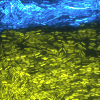
Two strains of synthetically engineered bacteria cooperate to create multicellular phenomena. Their fluorescence indicates the engineered capabilities have been activated. (credit: Bennett Lab/Rice University)

Rice University scientists and associates have created a biological equivalent to a computer circuit using multiple types of bacteria that change protein expression. The goal is to modify biological systems by controlling how bacteria influence each other. This could lead to bacteria that, for instance, beneficially alter the gut microbiome (collection of microorganisms) in humans.

The research is published in the journal Science.

Humans’ stomachs have a lot of different kinds of bacteria contained in the microbiome. “They naturally form a large consortium,” said Rice synthetic biologist Matthew Bennett. The idea is to engineer bacteria to be part of a consortium. “Working together allows them to effect more change than if they worked in isolation.”

In the proof-of-concept study, Bennett and his team created two strains of genetically engineered bacteria that regulate the production of proteins essential to intercellular signaling pathways, which allow cells to coordinate their efforts, generally in beneficial ways.

The synthetic microbial consortium oscillator yo-yo

The activator strain up-regulates genes in both strains; the repressor strain down-regulates genes in both strains, generating an oscillation of gene transcription in the bacterial population (credit: Ye Chen et al.)

“The main push in synthetic biology has been to engineer single cells,” Bennett said. “But now we’re moving toward multicellular systems. We want cells to coordinate their behaviors in order to elicit a populational response, just the way our bodies do.”

Bennett and his colleagues achieved their goal by engineering common Escherichia coli bacteria. By creating and mixing two genetically distinct populations, they prompted the bacteria to form a consortium.

The bacteria worked together by doing opposite tasks: One was an activator that up-regulated the expression of targeted genes; the other was a repressor that down-regulated specific genes. Together, they created oscillations of gene transcription in the bacterial population.

The two novel strains of bacteria sent out intercellular signaling molecules and created linked positive and negative feedback loops that affected gene production in the entire population. Both strains were engineered to make fluorescent reporter genes so their activities could be monitored. The bacteria were confined to microfluidic devices in the lab, where they could be monitored easily during each hours-long experiment.

When the bacteria were cultured in isolation, the protein oscillations did not appear, the researchers wrote.

Programmed yogurt, anyone?

Bennett said his lab’s work will help researchers understand how cells communicate, an important factor in fighting disease. “We have many different types of cells in our bodies, from skin cells to liver cells to pancreatic cells, and they all coordinate their behaviors to make us work properly,” he said. “To do this, they often send out small signaling molecules that are produced in one cell type and effect change in another cell type.

“We take that principle and engineer it into these very simple organisms to see if we can understand and build multicellular systems from the ground up.”

Ultimately, people might ingest the equivalent of biological computers that can be programmed through one’s diet, Bennett said. “One idea is to create a yogurt using engineered bacteria,” he said. “The patient eats it and the physician controls the bacteria through the patient’s diet. Certain combinations of molecules in your food can turn systems within the synthetic bacteria on and off, and then these systems can communicate with each other to effect change within your gut.”

KAIST and University of Houston scientists were also involved in the research. The National Institutes of Health, the Robert A. Welch Foundation, the Hamill Foundation, the National Science Foundation, and the China Scholarship Council supported the research.


Abstract of Emergent genetic oscillations in a synthetic microbial consortium

A challenge of synthetic biology is the creation of cooperative microbial systems that exhibit population-level behaviors. Such systems use cellular signaling mechanisms to regulate gene expression across multiple cell types. We describe the construction of a synthetic microbial consortium consisting of two distinct cell types—an “activator” strain and a “repressor” strain. These strains produced two orthogonal cell-signaling molecules that regulate gene expression within a synthetic circuit spanning both strains. The two strains generated emergent, population-level oscillations only when cultured together. Certain network topologies of the two-strain circuit were better at maintaining robust oscillations than others. The ability to program population-level dynamics through the genetic engineering of multiple cooperative strains points the way toward engineering complex synthetic tissues and organs with multiple cell types.

Anti-cancer vaccine uses patient’s own cancer cells to trigger immune responses

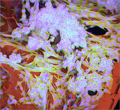
Cancerous melanoma cells, shown with their cell bodies (green) and nuclei (blue), are nestled in tiny hollow lumens (tubes) within the cryogel (red) structure. (credits: Thomas Ferrante, Sidi A. Bencherif / Wyss Institute at Harvard University)

A new biologically inspired “injectable cryogel whole-cell cancer vaccine” combines patient-specific harvested cancer cells and immune-stimulating chemicals or biological molecules to help the body attack cancer. It has been developed by scientists at Harvard’s Wyss Institute and Dana-Farber Cancer Institute.

This new approach is simpler and more economical than other cancer cell transplantation therapies, which harvest tumor cells and then genetically engineer them to trigger immune responses once they are transplanted back into the patient’s body, the researchers say.

The research, headed by Wyss Core Faculty member David Mooney, Ph.D., was reported online in an open-access paper in Nature Communications on August 12.

Minimally invasive cryogels

The new anti-cancer vaccine uses the patient’s own cancer cells to trigger immune responses. The cryogels are a type of hydrogel made up of hydrophilic (water-compatible) polymer chains that are cross-linked and can hold up to 99 percent water.

They are created by freezing a solution of the polymer that is in the process of gelling. When thawed back again to room temperature, the substance turns into a highly interconnected pore-containing hydrogel, which is similar in composition to bodily soft tissues in terms of their water content, structure, and mechanics.

This scanning electron microscopy image shows the thawed cryogel with its well-organized interconnected porous architecture ready to be infused with cancer cells and immune factors. (credits: Ellen Roche, James Weaver, Sidi A. Bencherif / Wyss Institute at Harvard University)

By adjusting their shape, physical properties, and chemical composition, Mooney’s team generated sponge-like, porous cryogels that can be infused with living cells, biological molecules, or drugs for a variety of potential therapeutic applications, including cancer immunotherapy.

The cryogels are minimally invasive because of their extreme flexibility and resilience, enabling them to be compressed to a fraction of their size and injected underneath the skin via a surgical needle. Once injected, they quickly bounce back to their original dimensions to do their job.

“After injection into the body, the cryogels can release their immune-enhancing factors in a highly controlled fashion to recruit specialized immune cells, which then make contact and read unique signatures off the patient’s tumor cells, also contained in the cryogels,” said Sidi Bencherif, the study’s co-first author and a Research Associate in Mooney’s research group.

This has two consequences, he said: “Immune cells become primed to mount a robust and destructive response against patient-specific tumor tissue and the immune tolerance developing within the tumor microenvironment is broken.”

Shrinking tumors

In experimental animal models on melanoma (skin) tumors, results show that using the cryogel to deliver whole cells and drugs triggers a dramatic immune response that can shrink tumors and even prophylactically (in advance) protect animals from tumor growth. With the pre-clinical success of the new cancer cell vaccination technology, Mooney and his team are going to explore how this cryogel-based method could be more broadly useful to treat a number of different cancer types.

“This new injectable form of this biomaterials-based cancer vaccine will help to expand the cancer immunotherapy arsenal, and it’s a great example of how engineering and materials science can be used to mimic the body’s own natural responses in a truly powerful way,” said Don Ingber, the Wyss Institute’s Founding Director, who also is the Judah Folkman Professor of Vascular Biology at Harvard Medical School and Boston Children’s Hospital, and Professor of Bioengineering at SEAS.

Mooney is also the Robert P. Pinkas Family Professor of Bioengineering at the Harvard John A. Paulson School of Engineering and Applied Sciences.


Abstract of Automated adaptive inference of phenomenological dynamical models

Dynamics of complex systems is often driven by large and intricate networks of microscopic interactions, whose sheer size obfuscates understanding. With limited experimental data, many parameters of such dynamics are unknown, and thus detailed, mechanistic models risk overfitting and making faulty predictions. At the other extreme, simple ad hoc models often miss defining features of the underlying systems. Here we develop an approach that instead constructs phenomenological, coarse-grained models of network dynamics that automatically adapt their complexity to the available data. Such adaptive models produce accurate predictions even when microscopic details are unknown. The approach is computationally tractable, even for a relatively large number of dynamical variables. Using simulated data, it correctly infers the phase space structure for planetary motion, avoids overfitting in a biological signalling system and produces accurate predictions for yeast glycolysis with tens of data points and over half of the interacting species unobserved.

How to reprogram cancer cells back to normal

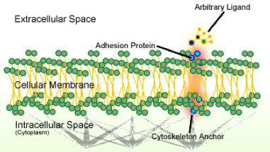
Schematic of cell adhesion (credit: Wikipedia)

A way to potentially reprogram cancer cells back to normalcy has been discovered by researchers on Mayo Clinic’s Florida campus.

The finding, published in Nature Cell Biology, represents “an unexpected new biology that provides the code, the software for turning off cancer,” says the study’s senior investigator, Panos Anastasiadis, Ph.D., chair of the Department of Cancer Biology on Mayo Clinic’s Florida campus.

MicroRNAs — short, non-coding RNAs present in all living organisms that were formerly considered “junk DNA” — have been shown to regulate the expression of at least half of all human genes. These single-stranded RNAs exert their regulatory action by binding messenger RNAs and preventing their translation into proteins. (credit: Firefly BioWorks)

That code was unraveled by the discovery that adhesion proteins — the glue that keeps cells together — interact with a key player in the production of molecules called microRNAs (miRNAs).

The miRNAs orchestrate whole cellular programs by simultaneously regulating expression of a group of genes. The investigators found that when normal cells come in contact with each other, a specific subset of miRNAs suppresses (or blocks) genes that promote cell growth.

However, when adhesion is disrupted in cancer cells, these miRNAs are misregulated and cells grow out of control. The investigators showed, in laboratory experiments, that restoring normal miRNA levels in cancer cells can reverse that aberrant cell growth.

“The study brings together two so-far unrelated research fields — cell-to-cell adhesion and miRNA biology — to resolve a long-standing problem* about the role of adhesion proteins in cell behavior that was baffling scientists,” says the study’s lead author Antonis Kourtidis, Ph.D., a research associate in Anastasiadis’ lab. “Most significantly, it uncovers a new strategy for cancer therapy,” he adds.

“By administering the affected miRNAs in cancer cells to restore their normal levels, we should be able to re-establish the brakes and restore normal cell function,” Anastasiadis says. “Initial experiments in some aggressive types of cancer are indeed very promising.”


Mayo Clinic | Mayo Clinic Researchers Find New Code That Makes Reprogramming of Cancer Cells Possible

* That problem arose from conflicting reports about E-cadherin and p120 catenin — adhesion proteins that are essential for normal epithelial tissues to form, and which have long been considered to be tumor suppressors.

“However, we and other researchers had found that this hypothesis didn’t seem to be true, since both E-cadherin and p120 are still present in tumor cells and required for their progression,” Anastasiadis says. “That led us to believe that these molecules have two faces — a good one, maintaining the normal behavior of the cells, and a bad one that drives tumorigenesis.”

Their theory turned out to be true, but what was regulating this behavior was still unknown. To answer this, the researchers studied a new protein called PLEKHA7, which associates with E-cadherin and p120 only at the top, or the “apical” part of normal polarized epithelial cells. The investigators discovered that PLEKHA7 maintains the normal state of the cells, via a set of miRNAs, by tethering the microprocessor to E-cadherin and p120. In this state, E-cadherin and p120 exert their good tumor suppressor sides.

However, “when this apical adhesion complex was disrupted after loss of PLEKHA7, this set of miRNAs was misregulated, and the E-cadherin and p120 switched sides to become oncogenic,” Dr. Anastasiadis says.

“We believe that loss of the apical PLEKHA7-microprocessor complex is an early and somewhat universal event in cancer,” he adds. “In the vast majority of human tumor samples we examined, this apical structure is absent, although E-cadherin and p120 are still present. This produces the equivalent of a speeding car that has a lot of gas (the bad p120) and no brakes (the PLEKHA7-microprocessor complex).


Abstract of Distinct E-cadherin-based complexes regulate cell behaviour through miRNA processing or Src and p120 catenin activity

E-cadherin and p120 catenin (p120) are essential for epithelial homeostasis, but can also exert pro-tumorigenic activities. Here, we resolve this apparent paradox by identifying two spatially and functionally distinct junctional complexes in non-transformed polarized epithelial cells: one growth suppressing at the apical zonula adherens (ZA), defined by the p120 partner PLEKHA7 and a non-nuclear subset of the core microprocessor components DROSHA and DGCR8, and one growth promoting at basolateral areas of cell–cell contact containing tyrosine-phosphorylated p120 and active Src. Recruitment of DROSHA and DGCR8 to the ZA is PLEKHA7 dependent. The PLEKHA7–microprocessor complex co-precipitates with primary microRNAs (pri-miRNAs) and possesses pri-miRNA processing activity. PLEKHA7 regulates the levels of select miRNAs, in particular processing of miR-30b, to suppress expression of cell transforming markers promoted by the basolateral complex, including SNAI1, MYC and CCND1. Our work identifies a mechanism through which adhesion complexes regulate cellular behaviour and reveals their surprising association with the microprocessor.

Paper-based test can quickly diagnose Ebola in remote areas


American Chemical Society | A simple, cheap test for Ebola, dengue and yellow fever

MIT researchers have developed a low-cost, paper-based device that changes color, depending on whether the patient has Ebola, dengue, or yellow fever. The test is designed to facilitate diagnosis in remote, low-resource settings, takes minutes, and does not need electricity to read out results.

The team described their approach Tuesday (Aug. 18) at the 250th National Meeting & Exposition of the American Chemical Society (ACS), updating the MIT announcement in February.

Standard approaches for diagnosing viral infections require technical expertise and expensive equipment, says MIT researcher Kimberly Hamad-Schifferli, Ph.D. “Typically, people perform PCR and ELISA, which are highly accurate, but they need a controlled lab environment.” Polymerase chain reaction (PCR) and enzyme-linked immunosorbent assay (ELISA) are bioassays that detect pathogens directly or indirectly, respectively.

Color-changing paper devices that work like over-the-counter pregnancy tests offer a possible solution. “These are not meant to replace PCR and ELISA [lab tests], because we can’t match their accuracy,” Hamad-Schifferli says. “This is a complementary technique for places with no running water or electricity.”

Hamad-Schifferli and her team at the Massachusetts Institute of Technology, Harvard Medical School and the U.S. FDA use silver nanoparticles in a rainbow of colors. The sizes of the nanoparticles determine their colors.

When a fever strikes in a developing area, the immediate concern may be: Is it the common flu or something much worse that requires quarantine? A paper-based diagnostic test that distinguishes between yellow fever virus, Ebola, and dengue, using different colored nanoparticles tagged with virus-specific antibodies (credit: Chunwan Yen)

The researchers attached red, green, or orange nanoparticles to antibodies that specifically bind to proteins from the organisms that cause yellow fever, Ebola, or dengue, respectively. They introduced the antibody-tagged nanoparticles onto the end of a small strip of paper. In the paper’s middle, the researchers affixed “capture” antibodies to three test lines at different locations, one for each disease.

To test the device, the researchers spiked blood samples with the viral proteins and then dropped small volumes onto the end of the paper device. If a sample contained dengue proteins, for example, the dengue antibody, which was attached to a green nanoparticle, latched onto one of those proteins. This complex then migrated through the paper, until reaching the dengue fever test line, where a second dengue-specific antibody captured it. That stopped the complex from going farther down the strip, and the test line turned green. When the researchers tested samples with proteins from Ebola or yellow fever, the antibody complexes migrated to different places on the strip and turned red or orange.

“Using other laboratory tests, we know the typical concentrations of yellow fever or dengue virus in patient blood. We know that the paper-based test is sensitive enough to detect concentrations well below that range,” says Hamad-Schifferli. “It’s hard to get that information for Ebola, but we can detect down to tens of nanograms per milliliter — that’s pretty sensitive and might work with patient samples.”

Next, the researchers plan to produce kits for free distribution. “We’re giving people the components so they can build the devices themselves,” says Hamad-Schifferli. The kits will provide a flexible platform for making paper devices that can detect any disease of interest, given the right antibody. “We are trying to move this into the field and put it in the hands of the people who need it,” she says.


American Chemical Society | Paper-based test can quickly diagnose Ebola in remote areas (press conference)


Abstract of Multicolored silver nanoparticles for multiplexed disease diagnostics: Distinguishing dengue, Yellow Fever, and Ebola viruses

Rapid point-of-care (POC) diagnostic devices are needed for field-forward screening of severe acute systemic febrile illnesses. Multiplexed rapid lateral flow diagnostics have the potential to distinguish among multiple pathogens, thereby facilitating diagnosis and improving patient care. Here, we present a platform for multiplexed pathogen detection using multi-colored prism-shaped silver nanoparticles (AgNPs). We exploit the size-dependent optical properties of Ag NPs to construct a multiplexed paperfluidic lateral flow POC sensor. AgNPs of different sizes were conjugated to antibodies that bind to specific biomarkers. Red AgNPs were conjugated to antibodies that could recognize the glycoprotein for Ebola virus, green AgNPs to those that could recognize nonstructural protein 1 for dengue virus, and orange AgNPs for non structural protein 1 for yellow fever virus. Presence of each of the biomarkers resulted in a different colored band on the test line in the lateral flow test. Thus, we were able to use NP color to distinguish among three pathogens that cause a febrile illness. Because positive test lines can be imaged by eye or a mobile phone camera, the approach is adaptable to low-resource, widely deployable settings. This design requires no external excitation source and permits multiplexed analysis in a single channel, facilitating integration and manufacturing.

Most complete functioning human-brain model to date, according to researchers

This image of the lab-grown brain is labeled to show identifiable structures: the cerebral hemisphere, the optic stalk, and the cephalic flexure, a bend in the mid-brain region, all characteristic of the human fetal brain (credit: The Ohio State University)

Scientists at The Ohio State University have developed a miniature human brain in a dish with the equivalent brain maturity of a five-week-old fetus.

The brain organoid, engineered from adult human skin cells, is the most complete human brain model yet developed, said Rene Anand, a professor of biological chemistry and pharmacology at Ohio State.

The lab-grown brain, about the size of a pencil eraser, has an identifiable structure and contains 99 percent of the genes present in the human fetal brain. Such a system will enable ethical and more rapid, accurate testing of experimental drugs before the clinical trial stage. It is intended to advance studies of genetic and environmental causes of central nervous system disorders.

“It not only looks like the developing brain, its diverse cell types express nearly all genes like a brain,” Anand said. “The power of this brain model bodes very well for human health because it gives us better and more relevant options to test and develop therapeutics other than [using] rodents.”

Anand reported on his lab-grown brain today (August 18) at the 2015 Military Health System Research Symposium in Ft. Lauderdale, Florida.

The main thing missing in this model is a vascular system. But what is there — a spinal cord, all major regions of the brain, multiple cell types, signaling circuitry and even a retina — has the potential to dramatically accelerate the pace of neuroscience research, said Anand, who is also a professor of neuroscience.

Organoid derivation and development (credit: Rene Anand and Susan McKay)

Created from pluripotent stem cells

“In central nervous system diseases, this will enable studies of either underlying genetic susceptibility or purely environmental influences, or a combination,” he said. According to genomic science, “there are up to 600 genes that give rise to autism, but we are stuck there. Mathematical correlations and statistical methods are insufficient in themselves to identify causation. You need an experimental system — you need a human brain.”

Anand’s method is proprietary and he has filed an invention disclosure with the university. He said he used techniques to differentiate pluripotent stem cells into cells that are designed to become neural tissue, components of the central nervous system or other brain regions..

High-resolution imaging of the organoid identifies functioning neurons and their signal-carrying extensions — axons and dendrites — as well as astrocytes, oligodendrocytes and microglia. The model also activates markers for cells that have the classic excitatory and inhibitory functions in the brain, and that enable chemical signals to travel throughout the structure.

It takes about 15 weeks to build a model system developed to match the 5-week-old fetal human brain. Anand and colleague Susan McKay, a research associate in biological chemistry and pharmacology, let the model continue to grow to the 12-week point, observing expected maturation changes along the way.

“If we let it go to 16 or 20 weeks, that might complete it, filling in that 1 percent of missing genes. We don’t know yet,” he said.

Models of brain disorders and injury with civilian and military uses

He and McKay have already used the platform to create brain organoid models of Alzheimer’s and Parkinson’s diseases and autism in a dish. They hope that with further development and the addition of a pumping blood supply, the model could be used for stroke therapy studies. For military purposes, the system offers a new platform for the study of Gulf War illness, traumatic brain injury, and post-traumatic stress disorder.

Anand hopes his brain model could be incorporated into the Microphysiological Systems program, a platform the Defense Advanced Research Projects Agency is developing by using engineered human tissue to mimic human physiological systems.

Support for the work came from the Marci and Bill Ingram Research Fund for Autism Spectrum Disorders and the Ohio State University Wexner Medical Center Research Fund.

Anand and McKay are co-founders of a Columbus-based start-up company, NeurXstem, to commercialize the brain organoid platform, and have applied for funding from the federal Small Business Technology Transfer program to accelerate its drug discovery applications.

How to create a genius mouse

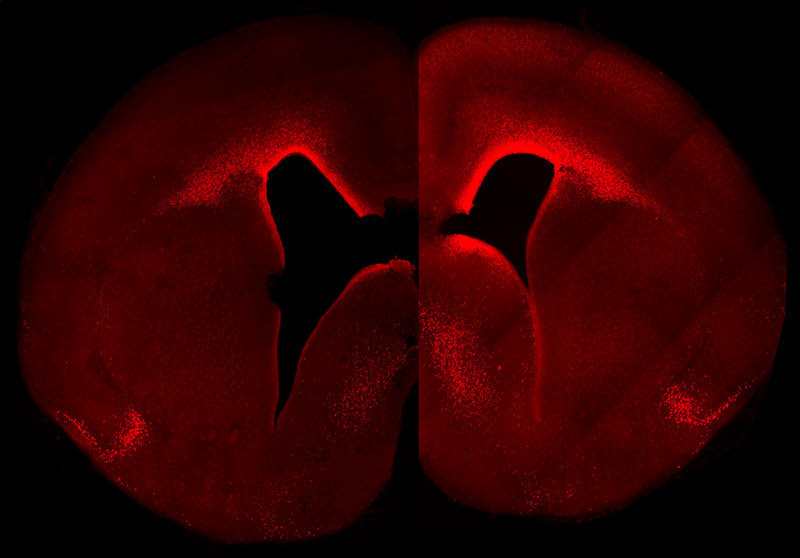
The left-brain hemisphere of a normal mouse shows the normal level and cellular distribution of the Pax6 gene expression in the developing neocortex. The right-brain hemisphere shows a sustained, primate-like Pax6 expression pattern in the neocortex of a double transgenic mouse embryo. These animals have more Pax6-positive progenitor cells and a higher Pax6 expression level in the germinal layer close to the ventricle in the right hemisphere. (credit: © MPI of Molecular Cell Biology & Genetics)

Researchers at the Max Planck Institute of Molecular Cell Biology and Genetics have created a transgenic mouse in which a gene called Pax6, during embryonic development, is highly expressed in a specific group of brain cortical cells called neural progenitor stem cells (the cells that generate all cells that make up the brain).

The resulting mouse brain generated more neurons than normal and exhibited primate-like features — notably those in the top layer, a characteristic feature of an expanded neocortex.

Mouse basal progenitors, in contrast to human, do not express Pax6. In humans, basal progenitors can undergo multiple rounds of cell division, thereby substantially increasing neuron number and ultimately the size of the neocortex.

“The evolutionary expansion of the neocortex is a hallmark of species with higher cognitive functions,” explains Wieland Huttner, the research group leader and director at the MPI-CBG. “Our findings contribute to our understanding of the molecular mechanisms underlying this expansion. While the findings demonstrate how altering the expression of a single key gene can make a big difference to brain development, a future challenge will be to obtain a comprehensive, integrated view of all the molecular changes that made our brains big.”

The study was published in an open-access paper in the journal PLOS Biology.

The paper, reassuringly, did not mention plans to create a transgenic genius cat in case the transgenic mouse gene escaped the laboratory.


Abstract of Sustained Pax6 Expression Generates Primate-like Basal Radial Glia in Developing Mouse Neocortex

The evolutionary expansion of the neocortex in mammals has been linked to enlargement of the subventricular zone (SVZ) and increased proliferative capacity of basal progenitors (BPs), notably basal radial glia (bRG). The transcription factor Pax6 is known to be highly expressed in primate, but not mouse, BPs. Here, we demonstrate that sustaining Pax6 expression selectively in BP-genic apical radial glia (aRG) and their BP progeny of embryonic mouse neocortex suffices to induce primate-like progenitor behaviour. Specifically, we conditionally expressed Pax6 by in utero electroporation using a novel, Tis21–CreERT2 mouse line. This expression altered aRG cleavage plane orientation to promote bRG generation, increased cell-cycle re-entry of BPs, and ultimately increased upper-layer neuron production. Upper-layer neuron production was also increased in double-transgenic mouse embryos with sustained Pax6 expression in the neurogenic lineage. Strikingly, increased BPs existed not only in the SVZ but also in the intermediate zone of the neocortex of these double-transgenic mouse embryos. In mutant mouse embryos lacking functional Pax6, the proportion of bRG among BPs was reduced. Our data identify specific Pax6 effects in BPs and imply that sustaining this Pax6 function in BPs could be a key aspect of SVZ enlargement and, consequently, the evolutionary expansion of the neocortex.

How aging cripples the immune system

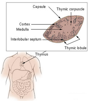
Thymus (credit: Wikimedia Commons)

Aging cripples the production of new immune cells, decreasing the immune system’s response to vaccines and putting the elderly at risk of infection, but antioxidants in the diet may slow this damaging process.

That’s a new finding by scientists from the Florida campus of The Scripps Research Institute (TSRI), published in an open-access paper in the journal Cell Reports.

The problem is focused on an organ called the thymus, which produces T lymphocytes (a type of white blood cell) — critical immune cells that must be continuously replenished so they can respond to new infections.

“The thymus begins to atrophy rapidly in very early adulthood, simultaneously losing its function,” said TSRI Professor Howard Petrie.

“This new study shows for the first time a mechanism for the long-suspected connection between normal immune function and antioxidants.”

How antioxidant enzyme deficiency leads to metabolic damage

Scientists have been hampered in their efforts to develop specific immune therapies for the elderly by a lack of knowledge of the underlying mechanisms of this process.

To explore these mechanisms, Petrie and his team developed a computational approach for analyzing the activity of genes in two major cell types in the thymus — stromal cells and lymphoid cells — in mouse tissues, which are similar to human tissues in terms of function and age-related atrophy.

The team found that stromal cells were specifically deficient in an antioxidant enzyme called catalase. That resulted in elevated levels of the reactive oxygen byproducts of metabolism, which cause accelerated metabolic damage.*

New support for the “free-radical theory” of aging

(Credit: Bethany Christmann)

Taken together, the findings provide support for the “free-radical theory” of aging, which proposes that reactive oxygen species (such as hydrogen peroxide), produced during normal metabolism (and from other sources) cause cellular damage that contributes to aging and age-related diseases.

Free radicals are especially reactive atoms or groups of atoms that have one or more unpaired electrons.  Besides those produced in the body as a by-product of normal metabolism, they can also be introduced from an outside source, such as tobacco smoke or other toxins.

Other studies have suggested that sex hormones, particularly androgens such as testosterone, play a major role in the aging process. But according to the researchers, those studies have failed to answer the key question: why does the thymus atrophy so much more rapidly than other body tissues?

“There’s no question that the thymus is remarkably responsive to androgens,” Petrie noted, “but our study shows that the fundamental mechanism of aging in the thymus, namely accumulated metabolic damage, is the same as in other body tissues. However, the process is accelerated in the thymus by a deficiency in the essential protective effects of catalase, which is found at higher levels in almost all other body tissues.”

It’s complicated

However, lowering free radicals with antioxidants has not always conferred the expected benefits, according to Senior scientist and Buck Institute professor Judith Campisi, PhD.

In a study published August 3 online in Proceedings of the National Academy of Sciences, scientists in her lab bred mice that produced excess free radicals that damaged the mitochondria in their skin. Based on the free-radical theory, the scientists expected to see accelerated aging across the mouse lifespan.

Instead, they saw a surprising benefit in young animals: accelerated wound healing due to increased epidermal (skin) differentiation and re-epithelialization.

However, the mice paid a price over time. Campisi said mitochondrial damage from excess free radicals caused some of the skin cells to go into senescence — they stopped dividing and started accumulating.  Campisi said that over time, the energy available to the epidermal stems cells was depleted — the stem cells simply became too scarce and the mice showed expected signs of aging: thin skin and poor wound healing.

“It may be that nature used free radicals to optimize skin health, but because this process is not deleterious to the organism until later in life, past its reproductive age, there was no [evolutionary benefit from evolving] ways to alter this mechanism,” suggested Michael Velarde, PhD, a postdoctoral fellow in the Campisi lab.

* To confirm the central role of catalase, the scientists increased levels of this enzyme in genetically altered animal models, resulting in preservation of thymus size for a much longer period. In addition, animals that were given two common dietary antioxidants, including vitamin C, were also protected from the effects of aging on the thymus.


Abstract of Metabolic Damage and Premature Thymus Aging Caused by Stromal Catalase Deficiency

T lymphocytes are essential mediators of immunity that are produced by the thymus in proportion to its size. The thymus atrophies rapidly with age, resulting in progressive diminution of new T cell production. This decreased output is compensated by duplication of existing T cells, but it results in gradual dominance by memory T cells and decreased ability to respond to new pathogens or vaccines. Here, we show that accelerated and irreversible thymic atrophy results from stromal deficiency in the reducing enzyme catalase, leading to increased damage by hydrogen peroxide generated by aerobic metabolism. Genetic complementation of catalase in stromal cells diminished atrophy, as did chemical antioxidants, thus providing a mechanistic link between antioxidants, metabolism, and normal immune function. We propose that irreversible thymic atrophy represents a conventional aging process that is accelerated by stromal catalase deficiency in the context of an intensely anabolic (lymphoid) environment.

Abstract of Pleiotropic age-dependent effects of mitochondrial dysfunction on epidermal stem cells

Tissue homeostasis declines with age partly because stem/progenitor cells fail to self-renew or differentiate. Because mitochondrial damage can accelerate aging, we tested the hypothesis that mitochondrial dysfunction impairs stem cell renewal or function. We developed a mouse model, Tg(KRT14-cre/Esr1)20Efu/J × Sod2tm1Smel, that generates mitochondrial oxidative stress in keratin 14-expressing epidermal stem/progenitor cells in a temporally controlled manner owing to deletion of Sod2, a nuclear gene that encodes the mitochondrial antioxidant enzyme superoxide dismutase 2 (Sod2). Epidermal Sod2 loss induced cellular senescence, which irreversibly arrested proliferation in a fraction of keratinocytes. Surprisingly, in young mice, Sod2 deficiency accelerated wound closure, increasing epidermal differentiation and reepithelialization, despite the reduced proliferation. In contrast, at older ages, Sod2 deficiency delayed wound closure and reduced epidermal thickness, accompanied by epidermal stem cell exhaustion. In young mice, Sod2 deficiency accelerated epidermal thinning in response to the tumor promoter 12-O-tetradecanoylphorbol-13-acetate, phenocopying the reduced regeneration of older Sod2-deficient skin. Our results show a surprising beneficial effect of mitochondrial dysfunction at young ages, provide a potential mechanism for the decline in epidermal regeneration at older ages, and identify a previously unidentified age-dependent role for mitochondria in skin quality and wound closure.

Move over, autonomous AI weapons, there’s a new risk in town: ‘gene drives’

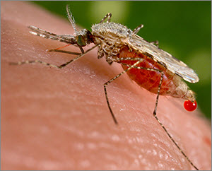
Wyss Institute scientists believe that synthetic gene drives, if researched responsibly, might be used in the future to render mosquito populations unable to transmit malaria (credit: CDC)

An international group of 26 experts, including prominent genetic engineers and fruit fly geneticists, has unanimously recommended a series of preemptive measures to safeguard gene drive research from accidental (or intentional) release from laboratories.

RNA-guided gene drives are genetic elements — found naturally in the genomes of most of the world’s organisms — that increase the chance of the gene they carry being passed on to all offspring. So  they can quickly spread through populations if not controlled.

Looking to these natural systems, researchers around the world, including some  scientists, are developing synthetic gene drives that could one day be leveraged by humans to purposefully alter the traits of wild populations of organisms to prevent disease transmission and eradicate invasive species.

What could possibly go wrong?

These synthetic gene drives, designed using an RNA-guided gene editing system called CRISPR, could one day improve human health and the environment by preventing mosquitoes and ticks from spreading diseases such as malaria and Lyme; by promoting sustainable agriculture through control of crop pests without the use of toxic pesticides and herbicides; and by protecting at-risk ecosystems from the spread of destructive, invasive species such as rats or cane toads.

Most genome alterations don’t persist in nature. Only 50 percent of transgenic mosquito offspring (left) will carry the altered gene, so it may persist at low frequency or go instinct. With gene drive (right), using CRISPR/Cas9, all of the offspring will carry the altered gene and will be inherited through the population. (credit: adapted from Wyss Institute video)

However, the development of RNA-guided gene drive technology calls for enhanced safety measures. That’s because its capability to also affect shared ecosystems if organisms containing synthetic gene drives are accidentally or deliberately released from a laboratory. This potential risk is especially relevant with highly mobile species such as fruit flies or mosquitoes.

Guidelines available

“One of the great successes of engineering is the development of safety features, such as the rounding of sharp corners on objects and the invention of airbags for cars, and in biological engineering we want to emulate the process of designing safety features in ways relevant to the technologies we develop,” said Wyss Core Faculty member George Church, Ph.D., who leads the Synthetic Biology Platform at the Wyss Institute. Church is also the Robert Winthrop Professor of Genetics at Harvard Medical School and Professor of Health Sciences and Technology at Harvard and MIT.

At the Wyss Institute, enhanced protocols for safely and securely researching emerging biotechnologies, including RNA–guided gene drives, have already been formally implemented. The safeguards were put in place proactively, step–by–step, in direct parallel with the development of the first RNA-guided gene drives at the Wyss Institute.

The working documents have been made publicly available by the Institute to encourage widespread adoption of multi-tier confinement and risk assessment procedures. Church was instrumental in the design of the enhanced biosafety and biosecurity protocols.

Now, research teams from the Wyss Institute and University of California, San Diego — the only two groups to have published work on RNA-guided CRISPR gene drives — have proactively assembled an international group of 26 experts, including prominent genetic engineers and fruit fly geneticists, to unanimously recommend a series of preemptive measures to safeguard gene drive research.

Open-access research recommended

Led by Wyss Institute Technology Development Fellow, Kevin Esvelt, Ph.D., and UC San Diego Professor of Cell and Developmental Biology Ethan Bier, Ph.D., the 26 authors of this consensus recommendation, which is published online in Science Express journal and includes representatives from every major group known to be working on gene drives, calls for all researchers to use multiple confinement strategies in order to prevent the accidental alteration of wild populations.

The group also provides explicit recommended guidelines for regulatory authorities evaluating proposed new work. And Esvelt and others are hopeful that the field of gene drive research is so nascent that it may be possible to build a community of scientists that share their research with the public throughout the development process.

“This would promote collaboration and avoid needless duplication of efforts among different research groups while allowing diverse voices to help guide the development of a technology that could improve our shared world,” said Esvelt. “And eventually, it might inspire a similar shift towards full transparency in other scientific fields of collective public importance.”

“The scientific community has a responsibility to the public and to the environment to constantly assess how new biotechnologies could potentially impact our world,” said Wyss Institute Founding Director Donald E. Ingber, M.D., Ph.D.

“This proactive consensus recommendation — reached in an extraordinary demonstration of the power of scientific collaboration over competition — provides concrete, useful guidelines for safeguarding our shared ecosystem while ensuring that remarkable breakthroughs, such as synthetic gene drives, can be applied to their full potential for the greater good.”


Wyss Institute at Harvard University | CRISPER-Cas9: Gene Drive

This animation explains how gene drives could one day be used to spread gene alterations through targeted wild populations over many generations, for purposes such as preventing spread of insect-borne disease and controlling invasive plant species. To ensure gene drives have the potential to be used for the greater good in the future, Wyss Institute Technology Development Fellow Kevin Esvelt, Ph.D., has co-led an international consensus of 26 scientists to recommend safeguards to prevent synthetic gene drive research from having any accidental impacts on the world’s shared ecosystems.


Abstract of A mucosal vaccine against Chlamydia trachomatis generates two waves of protective memory T cells

INTRODUCTION: Administering vaccines through nonmucosal routes often leads to poor protection against mucosal pathogens, presumably because such vaccines do not generate memory lymphocytes that migrate to mucosal surfaces. Although mucosal vaccination induces mucosa-tropic memory lymphocytes, few mucosal vaccines are used clinically; live vaccine vectors pose safety risks, whereas killed pathogens or molecular antigens are usually weak immunogens when applied to intact mucosa. Adjuvants can boost immunogenicity; however, most conventional mucosal adjuvants have unfavorable safety profiles. Moreover, the immune mechanisms of protection against many mucosal infections are poorly understood.

RATIONALE: One case in point is Chlamydia trachomatis (Ct), a sexually transmitted intracellular bacterium that infects >100 million people annually. Mucosal Ct infections can cause female infertility and ectopic pregnancies. Ct is also the leading cause of preventable blindness in developing countries and induces pneumonia in infants. No approved vaccines exist to date. Here, we describe a Ct vaccine composed of ultraviolet light–inactivated Ct (UV-Ct) conjugated to charge-switching synthetic adjuvant nanoparticles (cSAPs). After immunizing mice with live Ct, UV-Ct, or UV-Ct–cSAP conjugates, we characterized mucosal immune responses to uterine Ct rechallenge and dissected the underlying cellular mechanisms.

RESULTS: In previously uninfected mice, Ct infection induced protective immunity that depended on CD4 T cells producing the cytokine interferon-γ, whereas uterine exposure to UV-Ct generated tolerogenic Ct-specific regulatory T cells, resulting in exacerbated bacterial burden upon Ct rechallenge. In contrast, mucosal immunization with UV-Ct–cSAP elicited long-lived protection. This differential effect of UV-Ct–cSAP versus UV-Ct was because the former was presented by immunogenic CD11b+CD103 dendritic cells (DCs), whereas the latter was presented by tolerogenic CD11bCD103+ DCs. Intrauterine or intranasal vaccination, but not subcutaneous vaccination, induced genital protection in both conventional and humanized mice. Regardless of vaccination route, UV-Ct–cSAP always evoked a robust systemic memory T cell response. However, only mucosal vaccination induced a wave of effector T cells that seeded the uterine mucosa during the first week after vaccination and established resident memory T cells (TRM cells). Without TRM cells, mice were suboptimally protected, even when circulating memory cells were abundant. Optimal Ct clearance required both early uterine seeding by TRM cells and infection-induced recruitment of a second wave of circulating memory cells.

CONCLUSIONS: Mucosal exposure to both live Ct and inactivated UV-Ct induces antigen-specific CD4 T cell responses. While immunogenic DCs present the former to promote immunity, the latter is instead targeted to tolerogenic DCs that exacerbate host susceptibility to Ct infection. By combining UV-Ct with cSAP nanocarriers, we have redirected noninfectious UV-Ct to immunogenic DCs and achieved long-lived protection. This protective vaccine effect depended on the synergistic action of two memory T cell subsets with distinct differentiation kinetics and migratory properties. The cSAP technology offers a platform for efficient mucosal immunization that may also be applicable to other mucosal pathogens.
A fast, accurate, nanoscale ‘biochemical nose’ sensor

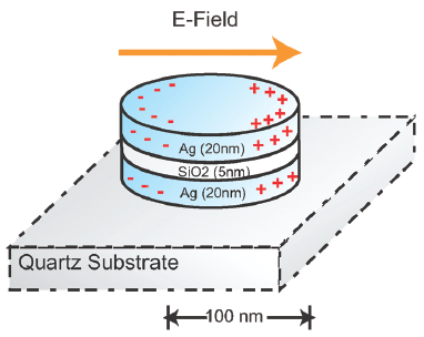
A nanoplasmonic resonator (NPR) consists of a thin silicon dioxide layer sandwiched between metallic nanodisks. NPRs can enhance surface-enhanced Raman spectroscopic (SERS) signals by a factor of 60 billion to detect target molecules with high sensitivity. (credit: Cheng Sun et al./ ACS Nano)

Imagine being able to test your food in your kitchen to quickly determine if it carried any deadly microbes. Technology now being commercialized by Optokey may soon make that possible.

Optokey, a startup based in Hayward, California, has developed a miniaturized sensor using surface-enhanced Raman spectroscopy (SERS) that can quickly and accurately detect or diagnose substances at a molecular level. The technology is based on research conducted at Lawrence Berkeley National Laboratory (Berkeley Lab) and published in 2010.

Molecular fingerprinting

“Our system can do chemistry, biology, biochemistry, molecular biology, clinical diagnosis, and chemical analysis,” said Optokey president and co-founder Fanqing Frank Chen, a scientist at Berkeley Lab who was co-author of an ACS Nano paper on the research. The system can be implemented “very cheaply, without much human intervention,” he said.

SERS is a highly sensitive analytical tool used for “molecular fingerprinting,” but the results have not easily reproducible. Chen and colleagues developed a solution to this problem using what they called “nanoplasmonic resonators,” which measures the interaction of photons with an activated surface using nanostructures to do chemical and biological sensing. The method produces measurements much more reliably.

“At Optokey we’re able to mass produce this nanoplasmonic resonator on a wafer scale,” Chen said. “We took something from the R&D realm and turned it into something industrial-strength.”

The miniaturized sensors use a microfluidic control system for “lab on a chip” automated liquid sampling. “We’re leveraging knowledge acquired from high-tech semiconductor manufacturing methods to get the cost, the volume, and the accuracy in the chip,” said VP of Manufacturing Robert Chebi, a veteran of the microelectronic industry who previously worked at Lam Research and Applied Materials. “We’re also leveraging all the knowledge in lasers and optics for this specific Raman-based method.”

A biochemical nose

Chebi calls Optokey’s product a “biochemical nose,” or an advanced nanophotonic automated system, with sensitivity to the level of a single molecule, far superior to sensors on the market today, he claims. “Today’s detection and diagnosis methods are far from perfect … Also, our system can provide information in minutes, or even on a continuous basis, versus other methods where it could take hours or even days, if samples have to be sent to another lab.”

The potential applications include food safety, environmental monitoring (of both liquids and gases), medical diagnosis, and chemical analysis. Optokey’s customers include a major European company interested in food safety, a Chinese petrochemical company interested in detecting impurities in its products, and a German company interested in point-of-care diagnosis.

“The product we’re envisioning is something that is compact and automated but also connected, and it can go into schools, restaurants, factories, hospitals, ambulances, airports, and even battlefields,” Chen said. Next, they plan to introduce it in the smart home, where a nanophotonic sensor could be built to scan for pollutants not just in food but also in air and water.

Key discovery: nanoplasmonic resonators

Ultimately, Chen and his Berkeley Lab group developed about 20 patents involving hybrid bionanomaterials. The key discovery that led to the formation of Optokey was the development of nanoplasmonic resonators to dramatically improve the signal and reliability of Raman spectroscopy. The method was initially used in the research lab to quickly and accurately detect a biomarker for prostate cancer, which has a high rate of false positives using conventional diagnostic tools.

“There was 10 years of research that went into this, funded by NIH, DARPA, the federal government, private foundations,” said Chen. “Berkeley Lab has a really good culture of multidisciplinary research, excellent engineering, and very strong basic science. Plus it has strong support for startups.”


Abstract of Time-Resolved Single-Step Protease Activity Quantification Using Nanoplasmonic Resonator Sensors

Protease activity measurement has broad application in drug screening, diagnosis and disease staging, and molecular profiling. However, conventional immunopeptidemetric assays (IMPA) exhibit low fluorescence signal-to-noise ratios, preventing reliable measurements at lower concentrations in the clinically important picomolar to nanomolar range. Here, we demonstrated a highly sensitive measurement of protease activity using a nanoplasmonic resonator (NPR). NPRs enhance Raman signals by 6.1 × 10¹⁰ times in a highly reproducible manner, enabling fast detection of proteolytically active prostate-specific antigen (paPSA) activities in real-time, at a sensitivity level of 6 pM (0.2 ng/mL) with a dynamic range of 3 orders of magnitude. Experiments on extracellular fluid (ECF) from the paPSA-positive cells demonstrate specific detection in a complex biofluid background. This method offers a fast, sensitive, accurate, and one-step approach to detect the proteases’ activities in very small sample volumes.