Looking at living cells down to individual molecules

3D rendered correlative AFM/PALM image of a fixed mammalian cell (mouse embryonic fibroblast (MEF) cell) expressing the fusion protein paxillin-mEOS2 (credit: Pascal D. Odermatt et al./Nano Letters)

EPFL scientists have captured images of living cells with unprecedented nanoscale resolution — even the evolution of their structure and molecular characteristics.

They did that by combining two cutting edge microscopy techniques — high-speed atomic force microscopy and a single-molecule-localization, super-resolution optical imaging system — into one instrument.

Their work was published in the journal ACS Nano Letters.

The “correlated single molecule localization microscope” combines two methods:

  • An atomic force microscope (AFM) “feels” the surface being observed using a tiny force sensitive needle, capturing the 3D structure. It is installed above the sample.
  • Meanwhile, the microscope technique, known at PALM (photo-activated localization microscopy), observes the sample from below. It selectively stains the sample with fluorescent molecules to label certain selected molecules by making them blink, and then follows their path in the interior of a cell. Its inventors were awarded the Nobel Prize last year.

The scientists also developed special software that assembles the images from the two instruments, providing a precise 3D visualization of the observed sample.

Correlative AFM-SMLM: instrument setup. (a) Schematic of the aligned optical path with the AFM cantilever. By laterally translating the incoming laser beam using a micrometer screw, the TIRF illumination condition is enabled. The AFM cantilever is centered in the field of view by adjusting the position of the inverted optical microscope mounted on an x/y-translation stage (as shown in b and c). (b) Mechanical integration of an inverted optical microscope and the AFM. The inverted optical microscope is mounted on an x/y-translation stage. Around it a mechanical support structure is built to hold the AFM in place without mechanically contacting the microscope body. The whole instrument is placed on a vibration isolation platform inside an acoustic isolation box. (c) Photograph of the instrument and (d) zoom in to the AFM cantilever aligned to the optical axis. (credit: Pascal D. Odermatt et al./Nano Letters)

By taking successive images of the same living cell, the scientists were, for the first time ever, able to follow the behavior of protein clusters in relation to the 3D structure of the cell. “That could, for example, allow us to observe the inner workings of cell division, or unravel how stem cells react to mechanical forces” says Henrik Deschout, post doctoral researcher in EPFL’s Laboratory of Nanometer-Scale Biology, which is directed by Aleksandra Radenovic.

The prototype stage has already attracted the interest of many other researchers as well as leading microscope manufacturers. The microscope could be of great interest to researchers in cellular-, micro- and mechanobiology, allowing scientists to shed new light on the intricate mechanisms occurring in living cells, the researchers say.


Abstract of High-Resolution Correlative Microscopy: Bridging the Gap between Single Molecule Localization Microscopy and Atomic Force Microscopy

Nanoscale characterization of living samples has become essential for modern biology. Atomic force microscopy (AFM) creates topological images of fragile biological structures from biomolecules to living cells in aqueous environments. However, correlating nanoscale structure to biological function of specific proteins can be challenging. To this end we have built and characterized a correlated single molecule localization microscope (SMLM)/AFM that allows localizing specific, labeled proteins within high-resolution AFM images in a biologically relevant context. Using direct stochastic optical reconstruction microscopy (dSTORM)/AFM, we directly correlate and quantify the density of localizations with the 3D topography using both imaging modalities along (F-)actin cytoskeletal filaments. In addition, using photo activated light microscopy (PALM)/AFM, we provide correlative images of bacterial cells in aqueous conditions. Moreover, we report the first correlated AFM/PALM imaging of live mammalian cells. The complementary information provided by the two techniques opens a new dimension for structural and functional nanoscale biology.

Heating and cooling genetic samples with light leads to ultrafast DNA diagnostics

An artist’s rendering of photonic PCR on a chip using light to rapidly heat and cool electrons at the surface of a thin film of gold. This method yields gene amplification results in mere minutes, and promises to transform point-of-care diagnostics in fields as diverse as medicine, food security and evolutionary biology. (credit: Luke Lee’s BioPOETS lab)

New technology developed by bioengineers at the University of California, Berkeley, promises to dramatically speed up the polymerase chain reaction (PCR) DNA test and make it cheaper and more portable by simply accelerating the heating and cooling of genetic samples with the switch of a light.

This turbocharged thermal cycling, described in an open-access paper published Friday July 31 in the journal Light: Science & Application, greatly expands the clinical and research applications of the PCR test, with results in minutes instead of an hour or more.

The PCR test, which amplifies a single copy of a DNA sequence to produce thousands to millions of copies, has become vital in genomics applications, ranging from cloning research to forensic analysis to paternity tests. PCR is used in the early diagnosis of hereditary and infectious diseases, and even for analysis of ancient DNA samples of mummies and mammoths.

The huge impact of the PCR test in modern science was recognized in 1993 with a Nobel Prize in Chemistry for its inventors, Kary Mullis and Michael Smith.

Using light-emitting diodes (LEDs), the UC Berkeley researchers were able to heat electrons at the interface of thin films of gold and a DNA solution. They clocked the speed of heating the solution at around 55 degrees Fahrenheit per second. The rate of cooling was equally impressive, coming in at about 43.9 degrees per second.

The heating-time bottleneck

“PCR is powerful, and it is widely used in many fields, but existing PCR systems are relatively slow,” said study senior author Luke Lee, a professor of bioengineering. “It is usually done in a lab because the conventional heater used for this test requires a lot of power and is expensive. Because it takes an hour or longer to complete each test, it is not practical for use for point-of-care diagnostics. Our system can generate results within minutes.”

Schematic showing the ultrafast photonic PCR using LED lights under a thin gold film to amplify genetic samples. The repeated heating and cooling process, called thermal cycling, is needed to separate the double-stranded DNA (1-Denaturation). Complementary bases from a primer then bind to the single strand (2-Annealing and extension), resulting in two copies of the gene. The process is repeated for at least 30 cycles. (credit: Jun Ho Son, UC Berkeley)

The slowdown in conventional PCR tests comes from the time it takes to heat and cool the DNA solution. The PCR test requires repeated temperature changes — an average of 30 thermal cycles at three different temperatures — to amplify the genetic sequence. This process involves breaking up the double-stranded DNA and binding the single strand with a matching primer. With each heating-cooling cycle, the amount of the DNA sample is doubled.

To speed up this thermal cycling, Lee and his team of researchers took advantage of plasmonics, or the interaction between light and free electrons on a metal’s surface. When exposed to light, the free electrons get excited and begin to oscillate, generating heat. Once the light is off, the oscillations and the heating stop.

Gold, it turns out, is a popular metal for this plasmonic photothermal heating because it is so efficient at absorbing light. It has the added benefit of being inert to biological systems, so it can be used in biomedical applications.

For their experiments, the researchers used thin films of gold that were 120 nanometers thick, or about the width of a rabies virus. The gold was deposited onto a plastic chip with microfluidic wells to hold the PCR mixture with the DNA sample.

The light source was an array of off-the-shelf LEDs positioned beneath the PCR wells. The peak wavelength of the blue LED light was 450 nanometers, tuned to get the most efficient light-to-heat conversion.

The researchers were able to cycle from 131 degrees to 203 degrees Fahrenheit 30 times in less than five minutes.

They tested the ability of the photonic PCR system to amplify a sample of DNA, and found that the results compared well with conventional PCR tests.

“This photonic PCR system is fast, sensitive and low-cost,” said Lee, who is also co-director of the Berkeley Sensor and Actuator Center. “It can be integrated into an ultrafast genomic diagnostic chip, which we are developing for practical use in the field. Because this technology yields point-of-care results, we can use this in a wide range of settings, from rural Africa to a hospital ER.”


Abstract of Ultrafast photonic PCR

Nucleic acid amplification and quantification via polymerase chain reaction (PCR) is one of the most sensitive and powerful tools for clinical laboratories, precision medicine, personalized medicine, agricultural science, forensic science and environmental science. Ultrafast multiplex PCR, characterized by low power consumption, compact size and simple operation, is ideal for timely diagnosis at the point-of-care (POC). Although several fast/ultrafast PCR methods have been proposed, the use of a simple and robust PCR thermal cycler remains challenging for POC testing. Here, we present an ultrafast photonic PCR method using plasmonic photothermal light-to-heat conversion via photon–electron–phonon coupling. We demonstrate an efficient photonic heat converter using a thin gold (Au) film due to its plasmon-assisted high optical absorption (approximately 65% at 450 nm, the peak wavelength of heat source light-emitting diodes (LEDs)). The plasmon-excited Au film is capable of rapidly heating the surrounding solution to over 150 °C within 3 min. Using this method, ultrafast thermal cycling (30 cycles; heating and cooling rate of 12.79±0.93 °C s−1 and 6.6±0.29 °C s−1, respectively) from 55 °C (temperature of annealing) to 95 °C (temperature of denaturation) is accomplished within 5 min. Using photonic PCR thermal cycles, we demonstrate here successful nucleic acid (λ-DNA) amplification. Our simple, robust and low cost approach to ultrafast PCR using an efficient photonic-based heating procedure could be generally integrated into a variety of devices or procedures, including on-chip thermal lysis and heating for isothermal amplifications.

Intracellular microlasers for precise labeling of a trillion individual cells

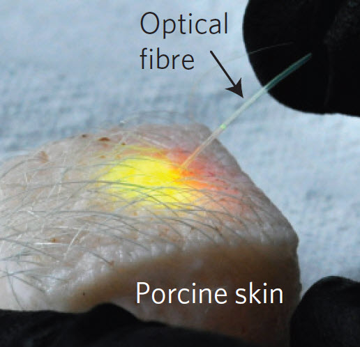
Massachusetts General Hospital investigators have induced subcutaneous fat cells in a piece of skin from a pig to emit laser light in response to energy delivered through an optical fiber (credit: Matjaž Humar and Seok Hyun Yun/Nature Photonics)

Imagine being able to label a trillion cells in the body to detect what’s going on in each individual cell.

That’s the eventual goal of a Massachusetts General Hospital (MGH) study to allow individual cells to produce laser light. The wavelengths of light emitted by these intracellular microlasers differ based on factors such as the size, shape, and composition of each microlaser, allowing precise labeling of individual cells.

“The fluorescent dyes currently used for research and for medical diagnosis are limited because they emit a very broad spectrum of light,” explains Seok Hyun Yun, PhD, of the Wellman Center for Photomedicine at MGH, corresponding author of the report. “As a result, only a handful of dyes can be used at a time, since their spectral signatures would overlap.”

(Left) Bright-field image of a HeLa cell containing a polystyrene fluorescent bead. (Right) False-color image of the cell. (scale bars: 10 micrometers) (credit: Matjaž Humar and Seok Hyun Yun/Nature Photonics)

Lead author Matjaž Humar, PhD, also of the Wellman Center, adds, “The narrow-band spectrum of light emitted by these intracellular lasers would allow us to label thousands — in principle, up to a trillion — of cells individually [the estimated number of cells in the human body], and the very specific wavelengths emitted by these microlasers also would allow us to measure small changes happening within a cell with much greater sensitivity than is possible with broadband fluorescence.”

The trick is to use solid plastic fluorescent microbeads, which are readily taken up into cells, each with a unique signature spectrum based on the size and number of beads within a cell and the fluorescent dye used.

“One immediate application of these intracellular lasers could be basic studies, such as understanding how cells move and respond to external forces,” says Yun, an associate professor of Dermatology at Harvard Medical School.

“Another challenging step will be figuring out how to use biologically generated energy from mechanical movement or a biochemical reaction to pump a cellular laser in a living body. Cells are smart machines, and we are interested in exploiting their amazing capabilities by developing smart-cell lasers that might be able to find diseases and fire light at them on their own.

“We can envision lasers completely made out of materials that are safe for use within the human body, which could enable remote sensing within the body or be used in laser-light therapies.”

The researchers’ report has received Advance Online Publication in Nature Photonics.


Abstract of Intracellular microlasers

Optical microresonators, which confine light within a small cavity, are widely exploited for various applications ranging from the realization of lasers and nonlinear devices to biochemical and optomechanical sensing. Here we use microresonators and suitable optical gain materials inside biological cells to demonstrate various optical functions in vitro including lasing. We explore two distinct types of microresonator—soft and hard—that support whispering-gallery modes. Soft droplets formed by injecting oil or using natural lipid droplets support intracellular laser action. The laser spectra from oil-droplet microlasers can chart cytoplasmic internal stress (∼500 pN μm–2) and its dynamic fluctuations at a sensitivity of 20 pN μm–2 (20 Pa). In a second form, whispering-gallery modes within phagocytized polystyrene beads of different sizes enable individual tagging of thousands of cells easily and, in principle, a much larger number by multiplexing with different dyes.

Ebola vaccine found 100% effective in initial trial

The Ebola vaccine being prepared for injection (credit: WHO/S. Hawkey)

An Ebola vaccine known as VSV-EBOV, provided by Merck, Sharp & Dohme, has shown 100% efficacy in individuals, according to results from an interim analysis published (open access) today (July 31) in the British journal The Lancet.

“This is an extremely promising development,” said Margaret Chan, M.D., Director-General of the World Health Organization. “The credit goes to the Guinean Government, the people living in the communities and our partners in this project. An effective vaccine will be another very important tool for both current and future Ebola outbreaks.”

An independent body of international experts — the Data and Safety Monitoring Board — conducted the review.

Based on the results, the Guinean national regulatory authority and ethics review committee have approved continuation of the trial to acquire conclusive evidence for the vaccine’s capacity to protect populations through what is called “herd immunity.”

“The ‘ring’ vaccination method adopted for the vaccine trial is based on the smallpox eradication strategy,” said John-Arne Røttingen, Director of the Division of Infectious Disease Control at the Norwegian Institute of Public Health and Chair of the Study Steering Group.

“The premise is that by vaccinating all people who have come into contact with an infected person you create a protective ‘ring’ and stop the virus from spreading further. This strategy has helped us to follow the dispersed epidemic in Guinea, and will provide a way to continue this as a public health intervention in trial mode.”

“This record-breaking work marks a turning point in the history of health R&D,” said Assistant Director-General Marie-Paule Kieny, who leads the Ebola Research and Development effort at WHO. “We now know that the urgency of saving lives can accelerate R&D. We will harness this positive experience to develop a global R&D preparedness framework so that if another major disease outbreak ever happens again, for any disease, the world can act quickly and efficiently to develop and use medical tools and prevent a large-scale tragedy.”

VSV-EBOV was developed by the Public Health Agency of Canada.

Scientists successfully edit human immune-system T cells

Efficient editing (cutting out) of CXCR4, a protein receptor that HIV can use to infect T cells (credit: Kathrin Schumann et al./PNAS)

In a project led by investigators at UC San Francisco , scientists have devised a new strategy to precisely modify human immune-system T cells, using the popular genome-editing system known as CRISPR/Cas9. T cells play important roles in a wide range of diseases, from diabetes to AIDS to cancer, so this achievement provides a path toward CRISPR/Cas9-based therapies for many serious health problems, the scientists say. It also provides a versatile new tool for research on T cell function.

Specifically, the researchers disabled a protein on the T-cell surface called CXCR4, which can be exploited by HIV when the virus infects T cells and causes AIDS. The group also successfully shut down PD-1. Scientists have shown that using drugs to block PD-1 coaxes T cells to attack tumors.

The CRISPR/Cas9 system makes it possible to easily and inexpensively edit genetic information in virtually any organism. T cells, which circulate in the blood, are an obvious candidate for medical applications of the technology, as these cells are at the center of many disease processes, and could be easily gathered from patients, edited with CRISPR/Cas9, then returned to the body to exert therapeutic effects.

A CRISPR/Cas9 breakthrough

Cas9, an enzyme in the CRISPR system that makes cuts in DNA and allows new genetic sequences to be inserted, is generally introduced into cells by using viruses or circular bits of DNA called plasmids. Then, in a separate step, a genetic construct known as single-guide RNA, which steers Cas9 to the specific spots in DNA where cuts are desired, is also placed into the cells.

Until recently, however, editing human T cells with CRISPR/Cas9 has been inefficient, with only a relatively small percentage of cells being successfully modified. And while scientists have had some success in switching off genes by inserting or deleting random sequences, they have not yet been able to use CRISPR/Cas9 to paste in (or “knock in”) specific new sequences to correct mutations in T cells.

Now, as reported in an open-access paper online in Proceedings of the National Academy of Sciences on July 27, 2015, the team has cracked these problems by streamlining the delivery of Cas9 and single-guide RNA to cells.

In lab dishes, the group assembled Cas9 ribonucleoproteins (RNPs), which combine the Cas9 protein with single-guide RNA. They then used a method known as electroporation, in which electrical field essentially punches tiny holes in membranes to make them more permeable so that these RNPs can we quickly delivered to the interior of the cells.

With these innovations, the researchers successfully edited CXCR4 and PD-1, even knocking in new sequences to replace specific genetic “letters” in these proteins. The group was then able to sort the cells, using markers expressed on the cell surface, to help pull out successfully edited cells for research, and eventually for therapeutic use.

The new work was done under the auspices of the Innovative Genomics Initiative (IGI), a joint UC Berkeley-UCSF program co-directed by Berkeley’s Jennifer Doudna, PhD, who is world-renowned for her pioneering research on CRISPR/Cas9, and Jonathan Weissman, PhD, professor of cellular and molecular pharmacology at UCSF and a Howard Hughes Medical Institute (HHMI) investigator.

Doudna, professor of chemistry and of cell and molecular biology at Berkeley, an HHMI investigator and co-corresponding author of the new paper, said that the research is a significant step forward in bringing the power of CRISPR/Cas9 editing to human biology and medicine.”

‘Designer babies’ concern

Recent reports of CRISPR/Cas9 editing of human embryos for possible heritable germline modification have stirred up an ethical controversy. But with this new protocol, T cells are created anew in each individual, so modifications would not be passed on to future generations, explained Alexander Marson, PhD, a UCSF Sandler Fellow and an affiliate member of the IGI, and senior and co-corresponding author of the new study.

“There’s actually well-trodden ground putting modified T cells into patients. There are companies out there already doing it and figuring out the safety profile, so there’s increasing clinical infrastructure that we could potentially piggyback on as we work out more details of genome editing,” Marson said. “I think CRISPR-edited T cells will eventually go into patients, and it would be wrong not to think about the steps we need to take to get there safely and effectively.”

He hopes that Cas9-based therapies for T cell-related disorders, which include autoimmune diseases as well as immunodeficiencies such as “bubble boy disease,” will enter the clinic in the future.

The research was supported by a gift from Jake Aronov, and by the UCSF Sandler Fellows Program, the National Institutes of Health, the National MS Society, and the Howard Hughes Medical Institute.


Abstract of Generation of knock-in primary human T cells using Cas9 ribonucleoproteins

T-cell genome engineering holds great promise for cell-based therapies for cancer, HIV, primary immune deficiencies, and autoimmune diseases, but genetic manipulation of human T cells has been challenging. Improved tools are needed to efficiently “knock out” genes and “knock in” targeted genome modifications to modulate T-cell function and correct disease-associated mutations. CRISPR/Cas9 technology is facilitating genome engineering in many cell types, but in human T cells its efficiency has been limited and it has not yet proven useful for targeted nucleotide replacements. Here we report efficient genome engineering in human CD4+ T cells using Cas9:single-guide RNA ribonucleoproteins (Cas9 RNPs). Cas9 RNPs allowed ablation of CXCR4, a coreceptor for HIV entry. Cas9 RNP electroporation caused up to ∼40% of cells to lose high-level cell-surface expression of CXCR4, and edited cells could be enriched by sorting based on low CXCR4 expression. Importantly, Cas9 RNPs paired with homology-directed repair template oligonucleotides generated a high frequency of targeted genome modifications in primary T cells. Targeted nucleotide replacement was achieved in CXCR4 and PD-1 (PDCD1), a regulator of T-cell exhaustion that is a validated target for tumor immunotherapy. Deep sequencing of a target site confirmed that Cas9 RNPs generated knock-in genome modifications with up to ∼20% efficiency, which accounted for up to approximately one-third of total editing events. These results establish Cas9 RNP technology for diverse experimental and therapeutic genome engineering applications in primary human T cells.

A simulated robot with bacterial brain

Computational Simulation of microbiome-host interactions. (A) A basic gene circuit forms the core of all simulated gene network behavior. (B) Green fluorescent protein (GFP, shown as a green dot) from this circuit is conceptualized to be detected by an onboard miniature, epifluorescent microscope (EFM). (C) A computational simulation of microbiome GFP production based upon an analytical model for the circuit in (A). In a built system, this protein fluorescence signal would be the light detected by the EFM. (D) The conceptualized robot uses onboard electronics to convert the measured light signals into electrical (voltage) signals. (E) Voltage signals meeting specific criteria activate pre-programmed robot motion subroutines. (F) The resulting emergent behavior potentially leads a robot to a carbon fuel depot. Here, robot behavior resulting from a simulation of the circuit in (A) is shown. The robot was programmed with motion subroutines that activate to seek arabinose (synthesized from glucose, orange square) depots following receipt of lactose (cyan triangles). (credit: Keith C. Heyde & Warren C. Ruder/Scientific Reports)

Virginia Tech scientist Warren Ruder, an assistant professor of biological systems engineering, has created an in silico (computer-simulated) model of a biomimetic robot controlled by a bacterial brain.

The study was inspired by real-world experiments where the mating behavior of fruit flies was manipulated using bacteria, and in which mice exhibited signs of lower stress when implanted with probiotics (“healthy” bacteria).

A math model of microbiome-controlled behavior

The deeper motivation for the study was to understand how the microbiome (the bacteria in the human body, thought to number ten times more than human cells) might influence human behavior. For example, some studies show that the gut microbiome influences human eating behavior and dietary choices to favor the survival of the bacteria. (See Do gut bacteria control your mind? for example.)

As explained in an open-access paper published recently in Scientific Reports, Ruder’s study revealed unique decision-making behavior by a bacteria-robot system by coupling and computationally simulating equations that describe three distinct elements: engineered gene circuits in E. coli, microfluid bioreactors, and robot movement.

In the mathematical model, the theoretical robot was equipped with sensors and a miniature microscope to measure the color. The hypothetical robot was designed to read E. coli bacterial gene expression levels (how much protein is created by specific genes), using light sensors in miniature microscopes. The bacteria turned green or red, depending on what they ate.

Bacteria that act like tigers?

Interestingly, the bacteria in the model began to approach a fuel source with “stalk-pause-strike” behavior, characteristic of predators.

Ruder’s modeling study also demonstrates that these sorts of biosynthetic experiments could be done in the future with a minimal amount of funds, opening up the field to a much larger pool of researchers.

Understanding the biochemical sensing between organisms could have far reaching implications in ecology, biology, and robotics, Ruder suggests.

In agriculture, bacteria-robot model systems could enable robust studies that explore the interactions between soil bacteria and livestock. In healthcare, further understanding of bacteria’s role in controlling gut physiology could lead to bacteria-based prescriptions (probiotics) to treat mental and physical illnesses. Ruder also envisions droids that could execute tasks such as deploying bacteria to remediate oil spills.

Bacteria effects on behavior

The findings also add to the ever-growing body of research about bacteria in the human body that are thought to regulate health and mood, and especially the theory that bacteria also affect behavior.

“We hope to help democratize the field of synthetic biology for students and researchers all over the world with this model,” said Ruder. “In the future, rudimentary robots and E. coli that are already commonly used separately in classrooms could be linked with this model to teach students from elementary school through the Ph.D.-level about bacterial relationships with other organisms.”

Ruder plans next to create a real-world version of the experiment, creating mobile robots with bioreactors on board that harbor living colonies of bacteria that direct the robot’s behavior.

The Air Force Office of Scientific Research funded the mathematical modeling of gene circuitry in E. coli, and the Virginia Tech Student Engineers’ Council has provided funding to move these models and resulting mobile robots into the classroom as teaching tools.


Virginia Tech | Scientist shows bacteria could control robots


Abstract of Exploring Host-Microbiome Interactions using an in Silico Model of Biomimetic Robots and Engineered Living Cells

The microbiome’s underlying dynamics play an important role in regulating the behavior and health of its host. In order to explore the details of these interactions, we created anin silico model of a living microbiome, engineered with synthetic biology, that interfaces with a biomimetic, robotic host. By analytically modeling and computationally simulating engineered gene networks in these commensal communities, we reproduced complex behaviors in the host. We observed that robot movements depended upon programmed biochemical network dynamics within the microbiome. These results illustrate the model’s potential utility as a tool for exploring inter-kingdom ecological relationships. These systems could impact fields ranging from synthetic biology and ecology to biophysics and medicine.

Novel DNA origami structures

The versatility of the DNA origami 3D wireframe design technique created by Arizona State University Biodesign Institute researcher Hao Yan is demonstrated with the construction of this snub cube model, an Archimedean solid with 60 edges, 24 vertices and 38 faces including 6 squares and 32 equilateral triangles. (credit: TED-43/Wikimedia Commons)

Hao Yan, a researcher at Arizona State University’s Biodesign Institute, has extended DNA origami — which uses combinations of DNA base pairs to create new 2-D and 3-D nanoforms — into imaginative new forms that may one day lead to microelectronics and biomedical innovations.

“Earlier design methods [for DNA origami] used strategies including parallel arrangement of DNA helices to approximate arbitrary shapes, but precise fine-tuning of DNA wireframe architectures that connect vertices in 3D space has required a new approach,” says Yan, the Milton D. Glick Distinguished Chair of Chemistry and Biochemistry at ASU and directs Biodesign’s Center for Molecular Design and Biomimetics.

The new study describes wireframe structures of high complexity and programmability that are fabricated by precise control of branching and curvature, using novel organizational principles for the designs. (Wireframes are skeletal three-dimensional models represented purely through lines and vertices.)

The resulting nanoforms include symmetrical lattice arrays, quasicrystalline structures, curvilinear arrays, and a simple wire art sketch in the 100-nm scale, as well as 3-D objects including a snub cube with 60 edges and 24 vertices and a reconfigurable Archimedean solid that can be controlled to make the unfolding and refolding transitions between 3D and 2D.

The research appears in the advanced online edition of the journal Nature Nanotechnology.


DNA origami: how it works

In previous investigations, the Yan group created subtle architectural forms at an astonishingly minute scale, some measuring only tens of nanometers across — roughly the diameter of a virus particle. These nano-objects include spheres, spirals, flasksMöbius forms, and even an autonomous spider-like robot capable of following a prepared DNA track.

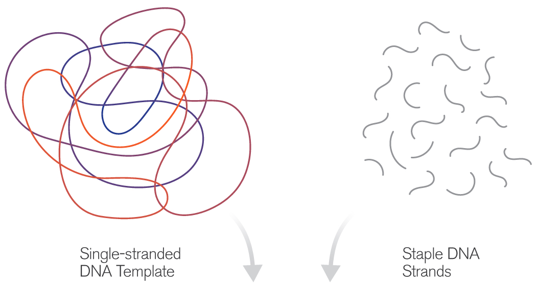
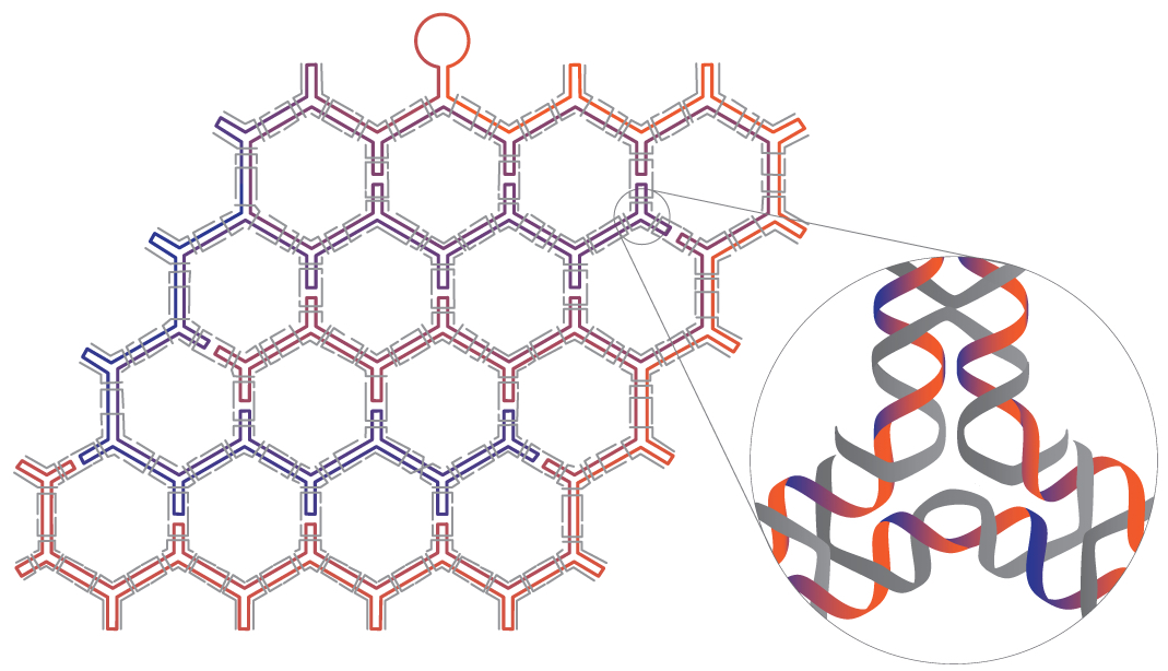
(credit: Fei Zhang et al./Nature Nanotechnology)

The technique of DNA origami capitalizes on the simple base-pairing properties of DNA, a molecule built from the four nucleotides Adenine (A), Thymine (T), Cytosine (C) and Guanine (G). The rules of the game are simple: A’s always pair with T’s and C’s with G’s. Using this abbreviated vocabulary, the myriad body plans of all living organisms are constructed; though duplicating even Nature’s simpler designs has required great ingenuity.

The basic idea of DNA origami is to use a length of single-stranded DNA as a scaffold for the desired shape. Base-pairing of complementary nucleotides causes the form to fold and self-assemble. The process is guided by the addition of shorter “staple strands,” which act to help fold the scaffold and to hold the resulting structure together. Various imaging technologies are used to observe the tiny structures, including fluorescence-, electron- and atomic force microscopy.


DNA origami originally produced nanoarchitectures of purely aesthetic interest, but refinements of the technique have opened the door to a new range of exciting applications including molecular cages for the encapsulation of molecules, enzyme immobilization and catalysis, chemical and biological sensing tools, drug delivery mechanisms, and molecular computing devices.

The technique described in the new study takes this approach a step further, allowing researchers to overcome local symmetry restrictions, creating wireframe architectures with higher order arbitrariness and complexity. Here, each line segment and vertex is individually designed and controlled. The number of arms emanating from each vertex may be varied from 2 to 10 and the precise angles between adjacent arms can be modified.

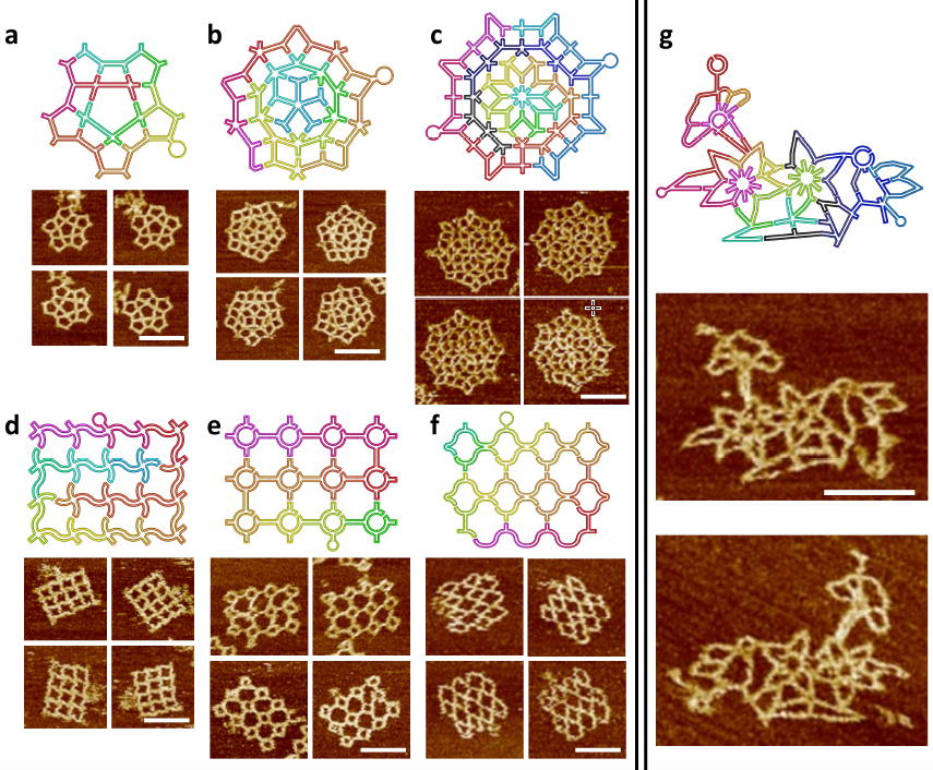
The scaffold folding path and representative AFM images for intricate 2D patterns. (a) A star-shape pattern without translational symmetry. (a) A Penrose tiling. (c) An 8-fold quasicrystalline pattern. (d-f) Three curved structures. (d) A waving grid. (e) A sphere array. (f) A fishnet. (g) A flower-and-bird pattern. All scale bars are 100 nm. Two scaffolds (one colorful and one black-blue) are used in c and g. (credit: Fei Zhang et al./Nature Nanotechnology)

In the current study, the method was first applied to symmetrical, regularly repeating polygonal designs, including hexagonal, square and triangular tiling geometries. Such common designs are known as tessellation patterns. A clever strategy involving a series of bridges and loops was used to properly route the scaffold strand, allowing it to pass through the entire structure, touching all lines of the wireframe once and only once. Staple strands were then applied to complete the designs.

In subsequent stages, the researchers created more complex wireframe structures, without the local translational symmetry found in the tessellation patterns. Three such patterns were made, including a star shape, a 5-fold Penrose tile and an 8-fold quasicrystalline pattern. (Quasicrystals are structures that are highly ordered but non-periodic. Such patterns can continuously fill available space, but are not translationally symmetric.)

Loop structures inserted into staple strands and unpaired nucleotides at the vertex points of the scaffold strands were also used, allowing researchers to perform precision modifications to the angles of junction arms.

The new design rules were next tested with the assembly of increasingly complex nanostructures, involving vertices ranging from 2 to 10 arms, with many different angles and curvatures involved, including a complex pattern of birds and flowers. The accuracy of the design was subsequently confirmed by AFM imaging, proving that the method could successfully yield highly sophisticated wireframe DNA nanostructures.

3D wire-frame Archimedean solid structures. (a) A 3D model of an Archimedean solid cuboctahedron with 12 vertices and 24 edges. Each vertex is a 4 Å~ 4 junction, and each edge is a 14-turn long double DNA duplex. (b) Left: Models showing possible conformations of the structure when deposited on mica surface; Right: the corresponding AFM images. (c) The reconfiguration between 3D and 2D can be realized by strand displacement by adding fuel and set strands. Top: reconfiguration schematics, Bottom: AFM images showing the transition. All scale bars in AFM images are 100 nm. (d) A 3D model of snub cube with 24 vertices and 60 edges. Each vertex is 5 Å~ 4 junction, and each edge is a 5-turn double DNA duplex. (e) Three views of the DNA snub cube from the design model (top of page). (credit: Fei Zhang et al./Nature Nanotechnology)

The method was then adapted to produce a number of 3D structures as well, including a cuboctahedron, and another Archimedian solid known as a snub cube — a structure with 60 edges, 24 vertices and 38 faces, including 6 squares and 32 equilateral triangles.

The authors stress that the new design innovations described can be used to compose and construct any imaginable wireframe nanostructure— a significant advancement for the burgeoning field.

On the horizon, nanoscale structures may one day be marshaled to hunt cancer cells in the body or act as robot assembly lines for the design of new drugs.


Abstract of Complex wireframe DNA origami nanostructures with multi-arm junction vertices

Structural DNA nanotechnology and the DNA origami technique, in particular, have provided a range of spatially addressable two- and three-dimensional nanostructures. These structures are, however, typically formed of tightly packed parallel helices. The development of wireframe structures should allow the creation of novel designs with unique functionalities, but engineering complex wireframe architectures with arbitrarily designed connections between selected vertices in three-dimensional space remains a challenge. Here, we report a design strategy for fabricating finite-size wireframe DNA nanostructures with high complexity and programmability. In our approach, the vertices are represented by n × 4 multi-arm junctions (n = 2–10) with controlled angles, and the lines are represented by antiparallel DNA crossover tiles of variable lengths. Scaffold strands are used to integrate the vertices and lines into fully assembled structures displaying intricate architectures. To demonstrate the versatility of the technique, a series of two-dimensional designs including quasi-crystalline patterns and curvilinear arrays or variable curvatures, and three-dimensional designs including a complex snub cube and a reconfigurable Archimedean solid were constructed.

An anti-inflammatory ‘smart drug’ that activates only in high-inflammation areas

Ben-Gurion University of the Negev (BGU) in Israel and University of Colorado researchers have developed a dynamic anti-inflammatory “smart” drug that can target specific sites in the body and could enhance the body’s natural ability to fight infection while reducing side effects.

This protein molecule, reported in the current issue of Journal of Immunology, has an exceptional property: when injected, it’s non-active. But upon reaching a local site with excessive inflammation, it becomes activated. Most other anti-inflammatory agents have broad effects in the body.

“This development is important because inhibition of inflammation in a non-specific manner reduces the natural ability to fight infections and is a common side effect of anti-inflammatory biologic therapeutics,” says Dr. Peleg Rider of BGU’s Department of Clinical Biochemistry and Pharmacology.

Using such a non-specific agent means any patient who suffers from local inflammation could be exposed to opportunistic infections at distant sites, such as lungs, which could risk tuberculosis, for example. This is especially a concern for immunosuppressed patients, as well as older patients and patients undergoing chemotherapy as part of an anti-cancer treatment course.

Mimicking a natural inflammatory process

The “chimeric IL-1Ra” protein combines an anti-inflammatory protein (right) with a peptide (left) that inactivates the IL-1Ra protein — except when the IL-1Ra portion encounters inflammatory enzymes, resulting in cleaving and releasing anti-inflammatory molecules (credit: Peleg Rider et al./Journal of Immunology)

“The beauty of this invention lies in the use of a known natural biological code,” Rider explains. “We mimicked a natural process that occurs during inflammation.”

Here’s how it works. The protein molecule (known as Chimeric IL-1Ra) is actually a chimera, combining two protein domains — both originating from the potent inflammatory IL-1 (interleukin family) protein (a group of 11 cytokines). (IL-1 normally plays a central role in regulating both immune and inflammatory responses to infections.)

The first part of this chimeric protein (IL-1beta) keeps a potent IL-1 natural infllammatory inhibitor (known as IL-1Ra) inactive. But when the IL-1Ra protein molecule encounters inflammatory enzymes, it springs into action, overriding IL-1beta, and is cleaved (split open), releasing powerful active molecules to reduce inflammation.

Rider, along with BGU’s Dr. Eli Lewis and Prof. Charles Dinarello of the University of Colorado, demonstrated their findings in a mouse model of local inflammation. They showed that leukocytes, which infiltrate inflammatory sites, indeed activate the chimeric protein, which in turn reduces local inflammation. The extent of activation of the protein correlated with the amount of inflammatory stimuli.

“Thus, a point that is highly relevant to clinical practice arises. Upon resolution of inflammation, the activation of the protein is also reduced and side effects are avoided,” Rider explains.

The new chimeric molecule was patented by BGN Technologies, BGU’s technology transfer company, and by the University of Colorado.

The research was supported by the Kamin program of Israel’s Ministry of Economy’s Chief Scientist’s Office.


Abstract of  IL-1 Receptor Antagonist Chimeric Protein: Context-Specific and Inflammation-Restricted Activation

Both IL-1α and IL-1β are highly inflammatory cytokines mediating a wide spectrum of diseases. A recombinant form of the naturally occurring IL-1R antagonist (IL-1Ra), which blocks IL-1R1, is broadly used to treat autoimmune and autoinflammatory diseases; however, blocking IL-1 increases the risk of infection. In this study, we describe the development of a novel form of recombinant IL-1Ra, termed chimeric IL-1Ra. This molecule is a fusion of the N-terminal peptide of IL-1β and IL-1Ra, resulting in inactive IL-1Ra. Because the IL-1β N-terminal peptide contains several protease sites clustered around the caspase-1 site, local proteases at sites of inflammation can cleave chimeric IL-1Ra and turn IL-1Ra active. We demonstrate that chimeric IL-1Ra reduces IL-1–mediated inflammation in vitro and in vivo. This unique approach limits IL-1 receptor blockade to sites of inflammation, while sparing a multitude of desired IL-1–related activities, including host defense against infections and IL-1–mediated repair.

The CRISPR craze: genome editing technologies poised to revolutionize medicine and industry

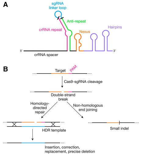
Genome editing by engineered Cas9 systems (credit: Mary Ann Liebert, Inc., publishers)

CRISPR/Cas systems for genome editing have revolutionized biological research over the past three years, and their ability to make targeted changes in DNA sequences in living cells with relative ease and affordability is now being applied to clinical medicine and will have a significant impact on advances in drug and other therapies, agriculture, and food products.

The power and promise of this innovation are presented in the Review article “The Bacterial Origins of the CRISPR Genome-Editing Revolution,” published in a special issue of Human Gene Therapy, a peer-reviewed journal from Mary Ann Liebert, Inc., publishers. The article is available open access until October 15, 2015 on the Human Gene Therapy website.

Erik Sontheimer, University of Massachusetts Medical School, Worcester, and Rodolphe Barrangou, North Carolina State University, Raleigh, describe the origins of this technology, which were derived from DNA sequences found in many bacteria known as clustered, regularly interspaced, short palindromic repeats (CRISPR) regions. These are part of bacteria’s protective immune system.

These regions have been developed into genome editing tools comprised of a “hardware” component (an RNA-guided DNA-targeting system that breaks a DNA strand at a specific site, with the help of the Cas protein), and a “software” component that can be programmed, and re-programmed, to repair or replace a faulty gene.

Impacts beyond academic research

“Although the CRISPR craze has yielded tremendous scientific progress and critical technological advances, it is important to keep in mind that the sgRNA–Cas9 technology is only 3 years old, and that notwithstanding current progress and momentum, we are yet to fully unleash the potential of these tools,” the authors note.

“Beyond academic research and the media, the most significant impact of CRISPR may well turn out to be in industry, with unprecedented levels of interest and investment from multiple distinct business segments, including pharmaceuticals and biotech, as well as covering the food supply chain from agriculture to livestock to other food products.”


Abstract of The Bacterial Origins of the CRISPR Genome-Editing Revolution

Like most of the tools that enable modern life science research, the recent genome-editing revolution has its biological roots in the world of bacteria and archaea. Clustered, regularly interspaced, short palindromic repeats (CRISPR) loci are found in the genomes of many bacteria and most archaea, and underlie an adaptive immune system that protects the host cell against invasive nucleic acids such as viral genomes. In recent years, engineered versions of these systems have enabled efficient DNA targeting in living cells from dozens of species (including humans and other eukaryotes), and the exploitation of the resulting endogenous DNA repair pathways has provided a route to fast, easy, and affordable genome editing. In only three years after RNA-guided DNA cleavage was first harnessed, the ability to edit genomes via simple, user-defined RNA sequences has already revolutionized nearly all areas of biological science. CRISPR-based technologies are now poised to similarly revolutionize many facets of clinical medicine, and even promise to advance the long-term goal of directly editing genomic sequences of patients with inherited disease. In this review, we describe the biological and mechanistic basis for these remarkable immune systems, and how their engineered derivatives are revolutionizing basic and clinical research.

Deep Genomics launches, uniting deep learning and genome biology

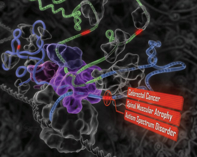
“Deep learning” reveals the genetic origins of disease. A computational system mimics the biology of RNA splicing by correlating DNA elements with splicing levels in healthy human tissues. The system can scan DNA and identify damaging genetic variants, including those deep within introns.This procedure has led to insights into the genetics of autism, cancers, and spinal muscular atrophy. (credit: Hui Y. Xiong et al./Science)

Deep Genomics, a University of Toronto spinoff, launched today (July 22), combining deep learning and artificial intelligence with the study of the human genome. The company is building on more than a decade of research and expertise in both fields.

Using deep learning allows Deep Genomics to predict the consequences of genomic alteration on various cell mechanisms to make life-changing decisions, potentially via personalized medicine treatment, the researchers say.

“Our vision is to change the course of genomic medicine and help save lives by determining smarter treatment options,” says Brendan Frey, the company’s president and CEO, a Fellow of the Canadian Institute for Advanced Research, and a Professor at the University of Toronto.

SPIDEX, a Google for DNA mutation effects

Professor Brendan Frey (center-right) and colleagues at the University of Toronto Faculty of Applied Science & Engineering (credit: Roberta Baker/ U of T Engineering)

The scientific community has spent decades searching for mutations within specific genes that can be connected to disease, such as the BRCA-1 and BRCA-2 genes for breast cancer. But there is a vast amount of mutations and combinations of mutations that have neither been observed nor studied, posing a challenge for diagnostics and therapeutics today.

“We envision a future where computers are trusted to predict the outcome of laboratory experiments and treatments, long before anyone picks up a test tube. Our first step will be to open up a genome-wide database of over 300 million potentially disease-causing variants, most of which are in regions of the genome that can’t be examined using other methods.”

Deep Genomics’ first product, called SPIDEX, provides information about how these DNA mutations may alter splicing in the cell, a process that is crucial for normal development. It also connects the dots between a variant or mutation of unknown significance and a variant that has been linked to a disease to determine its level of danger.

Because errant splicing is behind many diseases and disorders, including cancers and autism spectrum disorder, SPIDEX has immediate and practical importance for genetic testing and pharmaceutical development. The science validating the SPIDEX tool was described in the January 9, 2015 issue of the journal Science.

Labs will send the mutations they’ve collected to Deep Genomics, and the company will use their proprietary deep learning system, which includes SPIDEX, to “read” the genome and assess how likely the mutation is to cause a problem. SPIDEX can also connect the dots between a variant of unknown significance and a variant that has been linked to disease.

The company plans to further grow its team of machine learning, genome biology, and computational biology experts, and continue to invent new deep learning technologies and work with diagnosticians and biologists to understand the many complex ways that cells interpret DNA.

The company’s scientific advisory board includes Yann LeCun, Director, Facebook AI Research; Stephen Scherer, Director, The Center for Applied Genomics; and Jordan Lerner-Ellis, Director, Molecular Diagnostics at Mount Sinai Hospital.

More information: www.deepgenomics.com.


Abstract of The human splicing code reveals new insights into the genetic determinants of disease

To facilitate precision medicine and whole-genome annotation, we developed a machine-learning technique that scores how strongly genetic variants affect RNA splicing, whose alteration contributes to many diseases. Analysis of more than 650,000 intronic and exonic variants revealed widespread patterns of mutation-driven aberrant splicing. Intronic disease mutations that are more than 30 nucleotides from any splice site alter splicing nine times as often as common variants, and missense exonic disease mutations that have the least impact on protein function are five times as likely as others to alter splicing. We detected tens of thousands of disease-causing mutations, including those involved in cancers and spinal muscular atrophy. Examination of intronic and exonic variants found using whole-genome sequencing of individuals with autism revealed misspliced genes with neurodevelopmental phenotypes. Our approach provides evidence for causal variants and should enable new discoveries in precision medicine.