Saturn moon Titan has chemical that could form bio-like ‘membranes’ says NASA

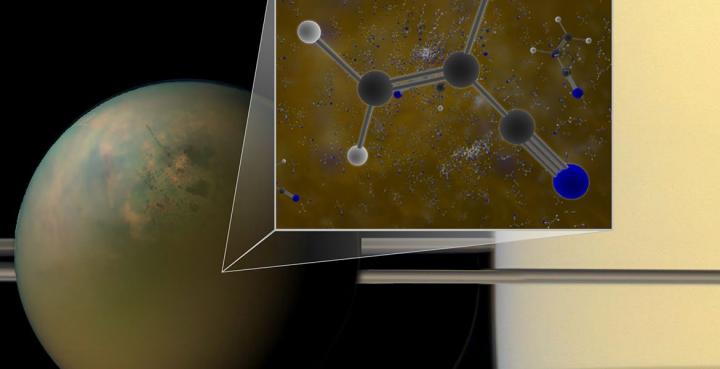
Molecules of vinyl cyanide reside in the atmosphere of Titan, Saturn’s largest moon, says NASA. Titan is shown here in an optical (atmosphere) infrared (surface) composite from NASA’s Cassini spacecraft. Titan’s atmosphere is a veritable chemical factory, harnessing the light of the sun and the energy from fast-moving particles that orbit around Saturn to convert simple organic molecules into larger, more complex chemicals. (credit: B. Saxton (NRAO/AUI/NSF); NASA)

NASA researchers have found large quantities (2.8 parts per billion) of acrylonitrile* (vinyl cyanide, C2H3CN) in Titan’s atmosphere that could self-assemble as a sheet of material similar to a cell membrane.

Acrylonitrile (credit: NASA Goddard)

Consider these findings, presented July 28, 2017 in the open-access journal Science Advances, based on data from the ALMA telescope in Chile (and confirming earlier observations by NASA’s Cassini spacecraft):

Azotozome illustration (credit: James Stevenson/Cornell)

1. Researchers have proposed that acrylonitrile molecules could come together as a sheet of material similar to a cell membrane. The sheet could form a hollow, microscopic sphere that they dubbed an “azotosome.”

A bilayer, made of two layers of lipid molecules (credit: Mariana Ruiz Villarreal/CC)

2. The azotosome sphere could serve as a tiny storage and transport container, much like the spheres that biological lipid bilayers can form. The thin, flexible lipid bilayer is the main component of the cell membrane, which separates the inside of a cell from the outside world.

“The ability to form a stable membrane to separate the internal environment from the external one is important because it provides a means to contain chemicals long enough to allow them to interact,” said Michael Mumma, director of the Goddard Center for Astrobiology, which is funded by the NASA Astrobiology Institute.

Organic rain falling on a methane sea on Titan (artist’s impression) (credit: NASA Goddard)

3. Acrylonitrile condenses in the cold lower atmosphere and rains onto its solid icy surface, ending up in seas of methane liquids on its surface.

Illustration showing organic compounds in Titan’s seas and lakes (ESA)

4. A lake on Titan named Ligeia Mare that could have accumulated enough acrylonitrile to form about 10 million azotosomes in every milliliter (quarter-teaspoon) of liquid. Compare that to roughly a million bacteria per milliliter of coastal ocean water on Earth.

Chemistry in Titan’s atmosphere. Nearly as large as Mars, Titan has a hazy atmosphere made up mostly of nitrogen with a smattering of organic, carbon-based molecules, including methane (CH4) and ethane (C2H6). Planetary scientists theorize that this chemical make-up is similar to Earth’s primordial atmosphere. The conditions on Titan, however, are not conducive to the formation of life as we know it; it’s simply too cold (95 kelvins or -290 degrees Fahrenheit). (credit: ESA)

6. A related open-access study published July 26, 2017 in The Astrophysical Journal Letters notes that Cassini has also made the surprising detection of negatively charged molecules known as “carbon chain anions” in Titan’s upper atmosphere. These molecules are understood to be building blocks towards more complex molecules, and may have acted as the basis for the earliest forms of life on Earth.

“This is a known process in the interstellar medium, but now we’ve seen it in a completely different environment, meaning it could represent a universal process for producing complex organic molecules,” says Ravi Desai of University College London and lead author of the study.

* On Earth, acrylonitrile  is used in manufacturing of plastics.


NASA Goddard | A Titan Discovery


Abstract of ALMA detection and astrobiological potential of vinyl cyanide on Titan

Recent simulations have indicated that vinyl cyanide is the best candidate molecule for the formation of cell membranes/vesicle structures in Titan’s hydrocarbon-rich lakes and seas. Although the existence of vinyl cyanide (C2H3CN) on Titan was previously inferred using Cassini mass spectrometry, a definitive detection has been lacking until now. We report the first spectroscopic detection of vinyl cyanide in Titan’s atmosphere, obtained using archival data from the Atacama Large Millimeter/submillimeter Array (ALMA), collected from February to May 2014. We detect the three strongest rotational lines of C2H3CN in the frequency range of 230 to 232 GHz, each with >4σ confidence. Radiative transfer modeling suggests that most of the C2H3CN emission originates at altitudes of ≳200 km, in agreement with recent photochemical models. The vertical column densities implied by our best-fitting models lie in the range of 3.7 × 10¹³ to 1.4 × 1014 cm−2. The corresponding production rate of vinyl cyanide and its saturation mole fraction imply the availability of sufficient dissolved material to form ~107 cell membranes/cm3 in Titan’s sea Ligeia Mare.

A living programmable biocomputing device based on RNA

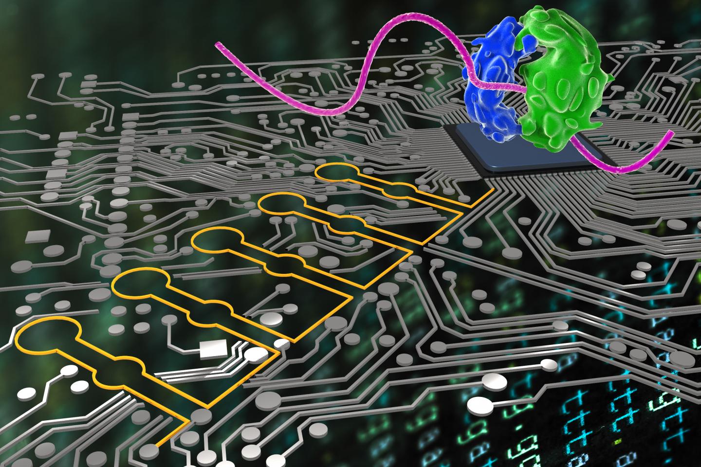
“Ribocomputing devices” ( yellow) developed by a team at the Wyss Institute can now be used by synthetic biologists to sense and interpret multiple signals in cells and logically instruct their ribosomes (blue and green) to produce different proteins. (credit: Wyss Institute at Harvard University)

Synthetic biologists at Harvard’s Wyss Institute for Biologically Inspired Engineering and associates have developed a living programmable “ribocomputing” device based on networks of precisely designed, self-assembling synthetic RNAs (ribonucleic acid). The RNAs can sense multiple biosignals and make logical decisions to control protein production with high precision.

As reported in Nature, the synthetic biological circuits could be used to produce drugs, fine chemicals, and biofuels or detect disease-causing agents and release therapeutic molecules inside the body. The low-cost diagnostic technologies may even lead to nanomachines capable of hunting down cancer cells or switching off aberrant genes.

Biological logic gates

Similar to a digital circuit, these synthetic biological circuits can process information and make logic-guided decisions, using basic logic operations — AND, OR, and NOT. But instead of detecting voltages, the decisions are based on specific chemicals or proteins, such as toxins in the environment, metabolite levels, or inflammatory signals. The specific ribocomputing parts can be readily designed on a computer.

E. coli bacteria engineered to be ribocomputing devices output a green-glowing protein when they detect a specific set of programmed RNA molecules as input signals (credit: Harvard University)

The research was performed with E. coli bacteria, which regulate the expression of a fluorescent (glowing) reporter protein when the bacteria encounter a specific complex set of intra-cellular stimuli. But the researchers believe ribocomputing devices can work with other host organisms or in extracellular settings.

Previous synthetic biological circuits have only been able to sense a handful of signals, giving them an incomplete picture of conditions in the host cell. They are also built out of different types of molecules, such as DNAs, RNAs, and proteins, that must find, bind, and work together to sense and process signals. Identifying molecules that cooperate well with one another is difficult and makes development of new biological circuits a time-consuming and often unpredictable process.

Brain-like neural networks next

Ribocomputing devices could also be freeze-dried on paper, leading to paper-based biological circuits, including diagnostics that can sense and integrate several disease-relevant signals in a clinical sample, the researchers say.

The next stage of research will focus on the use of RNA “toehold” technology* to produce neural networks within living cells — circuits capable of analyzing a range of excitatory and inhibitory inputs, averaging them, and producing an output once a particular threshold of activity is reached. (Similar to how a neuron averages incoming signals from other neurons.)

Ultimately, researchers hope to induce cells to communicate with one another via programmable molecular signals, forming a truly interactive, brain-like network, according to lead author Alex Green, an assistant professor at Arizona State University’s Biodesign Institute.

Wyss Institute Core Faculty member Peng Yin, Ph.D., who led the study, is also Professor of Systems Biology at Harvard Medical School.

The study was funded by the Wyss Institute’s Molecular Robotics Initiative, a Defense Advanced Research Projects Agency (DARPA) Living Foundries grant, and grants from the National Institute of Health (NIH), the Office of Naval Research (ONR), the National Science Foundation (NSF) and the Defense Threat Reduction Agency (DTRA).

* The team’s approach evolved from its previous development of “toehold switches” in 2014 — programmable hairpin-like nano-structures made of RNA. In principle, RNA toehold wwitches can control the production of a specific protein: when a desired complementary “trigger” RNA, which can be part of the cell’s natural RNA repertoire, is present and binds to the toehold switch, the hairpin structure breaks open. Only then will the cell’s ribosomes get access to the RNA and produce the desired protein.


Wyss Institute | Mechanism of the Toehold Switch


Abstract of Complex cellular logic computation using ribocomputing devices

Synthetic biology aims to develop engineering-driven approaches to the programming of cellular functions that could yield transformative technologies. Synthetic gene circuits that combine DNA, protein, and RNA components have demonstrated a range of functions such as bistability, oscillation, feedback, and logic capabilities. However, it remains challenging to scale up these circuits owing to the limited number of designable, orthogonal, high-performance parts, the empirical and often tedious composition rules, and the requirements for substantial resources for encoding and operation. Here, we report a strategy for constructing RNA-only nanodevices to evaluate complex logic in living cells. Our ‘ribocomputing’ systems are composed of de-novo-designed parts and operate through predictable and designable base-pairing rules, allowing the effective in silico design of computing devices with prescribed configurations and functions in complex cellular environments. These devices operate at the post-transcriptional level and use an extended RNA transcript to co-localize all circuit sensing, computation, signal transduction, and output elements in the same self-assembled molecular complex, which reduces diffusion-mediated signal losses, lowers metabolic cost, and improves circuit reliability. We demonstrate that ribocomputing devices in Escherichia coli can evaluate two-input logic with a dynamic range up to 900-fold and scale them to four-input AND, six-input OR, and a complex 12-input expression (A1 AND A2 AND NOT A1*) OR (B1 AND B2 AND NOT B2*) OR (C1 AND C2) OR (D1 AND D2) OR (E1 AND E2). Successful operation of ribocomputing devices based on programmable RNA interactions suggests that systems employing the same design principles could be implemented in other host organisms or in extracellular settings.

33 blood-cancer patients have dramatic clinical remission with new T-cell therapy

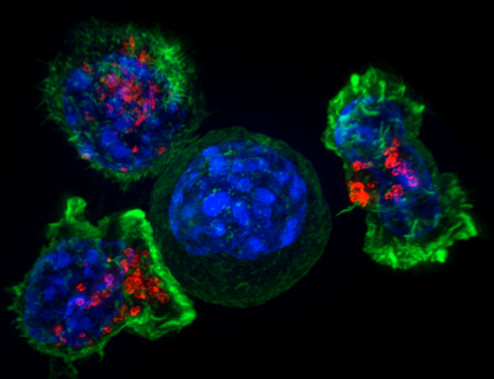
Image of a group of killer T cells (green and red) surrounding a cancer cell (blue, center)  (credit: NIH)

Chinese doctors have reported success with a new type of immunotherapy for multiple myeloma*, a blood cancer: 33 out of 35 patients in a clinical trial had clinical remission within two months.

The researchers used a type of T cell called “chimeric antigen receptor (CAR) T.”** In a phase I clinical trial in China, the patient’s own T cells were collected, genetically reprogrammed in a lab, and injected back into the patient. The reprogramming involved inserting an artificially designed gene into the T-cell genome, which helped the genetically reprogrammed cells find and destroy cancer cells throughout the body.

The study was presented Monday (June 5, 2017) at the American Society of Clinical Oncology (ASCO) conference in Chicago.

“Although recent advances in chemotherapy have prolonged life expectancy in multiple myeloma, this cancer remains incurable,” said study author Wanhong Zhao, MD, PhD, an associate director of hematology at The Second Affiliated Hospital of Xi’an Jiaotong University in Xi’an, China. “It appears that with this novel immunotherapy there may be a chance for cure in multiple myeloma, but we will need to follow patients much longer to confirm that.”***

U.S. clinical trial planned

“While it’s still early, these data are a strong sign that CAR T-cell therapy can send multiple myeloma into remission,” said ASCO expert Michael S. Sabel, MD, FACS. “It’s rare to see such high response rates, especially for a hard-to-treat cancer. This serves as proof that immunotherapy and precision medicine research pays off. We hope that future research builds on this success in multiple myeloma and other cancers.”

The researchers plan to enroll a total of 100 patients in this continuing clinical trial at four participating hospitals in China. “In early 2018 we also plan to launch a similar clinical trial in the United States. Looking ahead, we would also like to explore whether BCMA CAR T-cell therapy benefits patients who are newly diagnosed with multiple myeloma,” said Zhao.

This study was funded by Legend Biotech Co.

* Multiple myeloma is a cancer of plasma cells, which make antibodies to fight infections. Abnormal plasma cells can crowd out or suppress the growth of other cells in the bone marrow. This suppression may result in anemia, excessive bleeding, and a decreased ability to fight infection. Multiple myeloma is a relatively uncommon cancer. This year, an estimated 30,300 people [Ref. 2] in the United States will be diagnosed with multiple myeloma, and 114,250 [Ref. 3] were diagnosed with this cancer worldwide in 2012. In the United States, only about half of patients survive five years after being diagnosed with multiple myeloma. — American Society of Clinical Oncology

** Over the past few years, CAR T-cell therapy targeting a B-cell biomarker called CD19 proved very effective in initial trials for acute lymphoblastic leukemia (ALL) and some types of lymphoma, but until now, there has been little success with CAR T-cell therapies targeting other biomarkers in other types of cancer. This is one of the first clinical trials of CAR T cells targeting BCMA, which was discovered to play a role in progression of multiple myeloma in 2004. — American Society of Clinical Oncology

*** To date, 19 patients have been followed for more than four months, a pre-set time for full efficacy assessment by the International Myeloma Working Group (IMWG) consensus. Of the 19 patients, 14 have reached stringent complete response (sCR) criteria, one patient has reached partial response, and four patients have achieved very good partial remission (VgPR) criteria in efficacy.  There has been only a single case of disease progression from VgPR; an extramedullary lesion of the VgPR patient reappeared three months after disappearing on CT scans. There has not been a single case of relapse among patients who reached sCR criteria. The five patients who have been followed for over a year (12–14 months) all remain in sCR status and are free of minimal residual disease as well (have no detectable cancer cells in the bone marrow). Cytokine release syndrome or CRS, a common and potentially dangerous side effect of CAR T-cell therapy, occurred in 85% of patients, but it was only transient. In the majority of patients symptoms were mild and manageable. CRS is associated with symptoms such as fever, low blood pressure, difficulty breathing, and problems with multiple organs. Only two patients on this study experienced severe CRS (grade 3) but recovered upon receiving tocilizumab (Actemra, an inflammation-reducing treatment commonly used to manage CRS in clinical trials of CAR T-cell therapy). No patients experienced neurologic side effects, another common and serious complication from CAR T-cell therapy. American Society of Clinical Oncology


Abstract of Durable remissions with BCMA-specific chimeric antigen receptor (CAR)-modified T cells in patients with refractory/relapsed multiple myeloma.

Background: Chimeric antigen receptor engineered T cell (CAR-T) is a novel immunotherapeutic approach for cancer treatment and has been clinically validated in the treatment of acute lymphoblastic leukemia (ALL). Here we report an encouraging breakthrough of treating multiple myeloma (MM) using a CAR-T designated LCAR-B38M CAR-T, which targets principally BCMA. Methods: A single arm clinical trial was conducted to assess safety and efficacy of this approach. A total of 19 patients with refractory/relapsed multiple myeloma were included in the trial. The median number of infused cells was 4.7 (0.6 ~ 7.0) × 10e6/ kg. The median follow-up times was 208 (62 ~ 321) days. Results: Among the 19 patients who completed the infusion, 7 patients were monitored for a period of more than 6 months. Six out of the 7 achieved complete remission (CR) and minimal residual disease (MRD)-negative status. The 12 patients who were followed up for less than 6 months met near CR criteria of modified EBMT criteria for various degrees of positive immunofixation. All these effects were observed with a progressive decrease of M-protein and thus expected to eventually meet CR criteria. In the most recent follow-up examination, all 18 survived patients were determined to be free of myeloma-related biochemical and hematologic abnormalities. One of the most common adverse event of CAR-T therapy is acute cytokine release syndrome (CRS). This was observed in 14 (74%) patients who received treatment. Among these 14 patients there were 9 cases of grade 1, 2 cases of grade 2, 1 case of grade 3, and 1 case of grade 4 patient who recovered after treatments. Conclusions: A 100% objective response rate (ORR) to LCAR-B38M CAR-T cells was observed in refractory/relapsed myeloma patients. 18 out of 19 (95%) patients reached CR or near CR status without a single event of relapse in a median follow-up of 6 months. The majority (14) of the patients experienced mild or manageable CRS, and the rest (5) were even free of diagnosable CRS. Based on the encouraging safety and efficacy outcomes, we believe that our LCAR-B38M CAR-T cell therapy is an innovative and highly effective treatment for multiple myeloma.

New antibiotic could eliminate the global threat of antibiotic-resistant infections

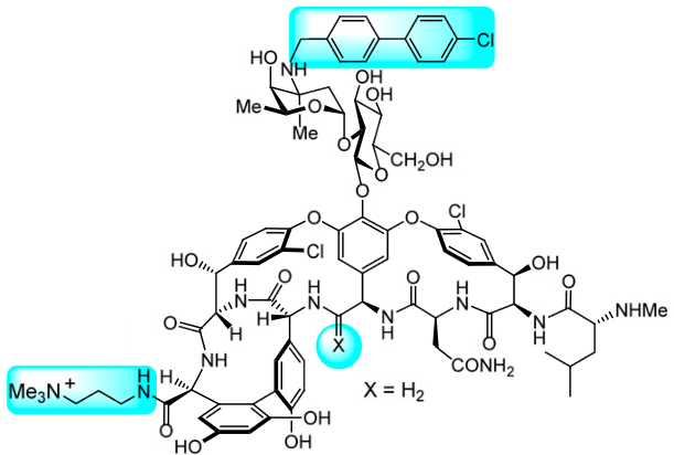
Modified vancomycin antibiotic (credit: Akinori Okano et al./PNAS)

Scientists at The Scripps Research Institute (TSRI) have discovered a way to structurally modify the antibiotic called vancomycin to make an already-powerful version of the antibiotic even more potent — an advance that could eliminate the threat of antibiotic-resistant infections for years to come.

“Doctors could use this modified form of vancomycin without fear of resistance emerging,” said Dale Boger, co-chair of TSRI’s Department of Chemistry, whose team announced the finding Monday (May 29, 2016) in the journal Proceedings of the National Academy of Sciences.

“The death of a hospitalized patient in Reno Nevada for whom no available antibiotics worked highlights what World Health Organization and other public-health experts have been warning: antibiotic resistance is a serious threat and has gone global,” KurzweilAI reported in January 2017. The new finding promises to lead to a solution.

First antibiotic to have three independent mechanisms of action

Vancomycin  has been prescribed by doctors for 60 years, and bacteria are only now developing resistance to it, according to Boger, who called vancomycin “magical” for its proven strength against infections. Previous studies by Boger and his colleagues at TSRI had shown that it is possible to add two modifications to vancomycin to make it even more potent. “With these modifications, you need less of the drug to have the same effect,” Boger said.

The new study shows that scientists can now make a third modification that interferes with a bacterium’s cell wall in a new way, with promising results. Combined with the previous modifications, this alteration gives vancomycin a 1,000-fold increase in activity, meaning doctors would need to use less of the antibiotic to fight infection.

The discovery makes this version of vancomycin the first antibiotic to have three independent mechanisms of action. “This increases the durability of this antibiotic,” said Boger. “Organisms just can’t simultaneously work to find a way around three independent mechanisms of action. Even if they found a solution to one of those, the organisms would still be killed by the other two.”

Tested against Enterococci bacteria, the new version of vancomycin killed both vancomycin-resistant Enterococci and the original forms of Enterococci. The next step in this research is to design a way to synthesize the modified vancomycin using fewer steps in the lab; the current method takes 30 steps.

The study was supported by the National Institutes of Health.

Could there be life below Saturn’s moon Enceladus and Jupiter’s moon Europa?

Illustration (not to scale) of the plume (white ejections) of Saturnian moon Enceladus, based on analysis of data from NASA’s Cassini spacecraft, which dived through the Enceladus plume in 2015. Scientists have now discovered hydrogen gas in the erupting material in the plume — providing further evidence for hydrothermal activity and making it more likely that the underground ocean of Enceladus could have conditions suitable for microbial life. (credit: NASA/JPL-Caltech)

Two NASA missions — Cassini and Hubble — have provided new evidence for life on icy, ocean-bearing moons of Saturn and Jupiter, NASA announced Friday, April 14, 2017.

Scientists from Southwest Research Institute (SwRI) have discovered hydrogen gas in the plume of material erupting from Saturn’s moon Enceladus — suggesting conditions suitable for microbial life in an underground ocean. The finding, published April 14, 2017 in the journal Science, was based on analysis of data from NASA’s Cassini spacecraft.

The researchers suggest that the hydrogen was most likely formed in chemical reactions between the moon’s rocky core and warm water from vents in the moon’s subsurface ocean floor. These vents could have features similar to hydrothermal vents on Earth, which emit hot, mineral-laden fluid containing hydrogen (in the form of hydrogen sulfide) and are thought to power microbe life on the seafloor.*

This infographic illustrates how scientists on NASA’s Cassini mission think water interacts with rock at the bottom of the ocean of Saturn’s icy moon Enceladus, producing hydrogen gas (H2). The graphic shows water from the ocean circulating through the seafloor, where it is heated and interacts chemically with the rock. This warm water, laden with minerals and dissolved gases (including hydrogen and possibly methane) then pours into the ocean, creating chimney-like vents through the ice. The scientists have determined that nearly 98 percent of the gas in the plume is water vapor, about 1 percent is hydrogen, and the rest is a mixture of other molecules including carbon dioxide, methane (CH4), and ammonia (NH3). (credit: NASA/JPL-Caltech/Southwest Research Institute)

“The amount of molecular hydrogen we detected is high enough to support microbes similar to those that live near hydrothermal vents on Earth,” said SwRI’s Christopher Glein, PhD, a co-author on the paper and a pioneer of extraterrestrial chemical oceanography. “If similar organisms are present in Enceladus, they could ‘burn’ the hydrogen to obtain energy for chemosynthesis, which could conceivably serve as a foundation for a larger ecosystem.”

New Hubble observations suggest where to look for signs of life on Europa

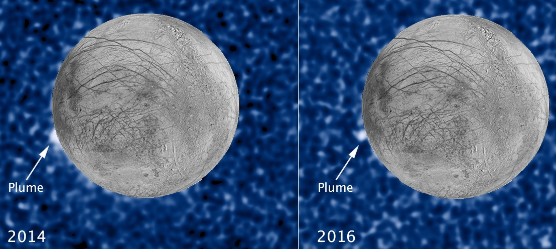
Best evidence yet for reoccurring water vapor plumes erupting from Jupiter’s Europa moon (credit: NASA, ESA W. Sparks (STScI), USGS Astrogeology Science Center)

NASA also announced new Hubble Space Telescope observations of Jupiter’s moon Europa, reported in a paper published in The Astrophysical Journal Letters. A newly discovered plume seen towering 62 miles above the surface in 2016 is at precisely the same location as a similar plume seen on the moon two years earlier by Hubble. The scientists suggest this offers a promising location for study of Europa’s internal water and ice — and for seeking evidence of Europa’s habitability.

Scientists hope to learn more with NASA’s Europa Clipper mission, planned for launch in the 2020s. It will feature a powerful ultraviolet camera that will make similar measurements to Hubble’s (but from thousands of times closer) and a next-generation version of the Cassini instrument.

* NASA astrobiologists suggest that bacteria living in and around the dark hydrothermal vents extract their energy from hydrogen sulfide (HS) and other molecules that billow out of the seafloor. Just like plants, the bacteria use their energy to build sugars out of carbon dioxide and water; sugars then provide fuel and raw material for the rest of the microbe’s activities. But  instead of photosynthesis, the microbes derive their energy from chemicals in a process called “chemosynthesis”: Hydrothermal vents extract their energy from hydrogen sulfide (HS) and other molecules that billow out of the seafloor. Recently, researchers have determined that fossilized evidence of bacteria from ancient seafloor hydrothermal vent-precipitates (found in the Nuvvuagittuq belt in Quebec, Canada) is at least 3.77 billion years old (or possibly as much as 4.28 billion years old). The minimum age of the fossils would make them the oldest indication of life on Earth so far.


ScienceAtNASA | ScienceCasts: Ocean Worlds


NASA Goddard | Europa Water Vapor Plumes — More Hubble Evidence


UCLTV | World’s oldest fossils unearthed (UCL)


Abstract of Cassini finds molecular hydrogen in the Enceladus plume: Evidence for hydrothermal processes

Saturn’s moon Enceladus has an ice-covered ocean; a plume of material erupts from cracks in the ice. The plume contains chemical signatures of water-rock interaction between the ocean and a rocky core. We used the Ion Neutral Mass Spectrometer onboard the Cassini spacecraft to detect molecular hydrogen in the plume. By using the instrument’s open-source mode, background processes of hydrogen production in the instrument were minimized and quantified, enabling the identification of a statistically significant signal of hydrogen native to Enceladus. We find that the most plausible source of this hydrogen is ongoing hydrothermal reactions of rock containing reduced minerals and organic materials. The relatively high hydrogen abundance in the plume signals thermodynamic disequilibrium that favors the formation of methane from CO2 in Enceladus’ ocean.

Abstract of Active Cryovolcanism on Europa?

Evidence for plumes of water on Europa has previously been found using the Hubble Space Telescope using two different observing techniques. Roth et al. found line emission from the dissociation products of water. Sparks et al. found evidence for off-limb continuum absorption as Europa transited Jupiter. Here, we present a new transit observation of Europa that shows a second event at the same location as a previous plume candidate from Sparks et al., raising the possibility of a consistently active source of erupting material on Europa. This conclusion is bolstered by comparison with a nighttime thermal image from the Galileo Photopolarimeter-Radiometer that shows a thermal anomaly at the same location, within the uncertainties. The anomaly has the highest observed brightness temperature on the Europa nightside. If heat flow from a subsurface liquid water reservoir causes the thermal anomaly, its depth is ≈1.8–2 km, under simple modeling assumptions, consistent with scenarios in which a liquid water reservoir has formed within a thick ice shell. Models that favor thin regions within the ice shell that connect directly to the ocean, however, cannot be excluded, nor modifications to surface thermal inertia by subsurface activity. Alternatively, vapor deposition surrounding an active vent could increase the thermal inertia of the surface and cause the thermal anomaly. This candidate plume region may offer a promising location for an initial characterization of Europa’s internal water and ice and for seeking evidence of Europa’s habitability.

Nanopores map small changes in DNA for early cancer detection

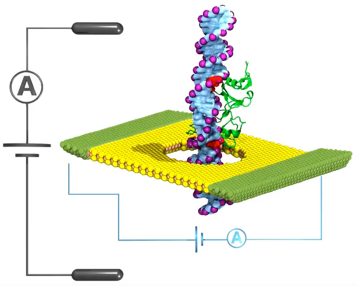
To detect DNA methylation changes (for cancer early warning), researchers punched a tiny hole (pore) in a flat sheet of graphene (or other  2D material). They then submerged the material in a salt solution and applied an electrical voltage to force the DNA molecule through the pore. A dip in the ionic current (black A) identified a methyl group (green) is passing through, but a dip in the electrical current (blue A) could detect smaller DNA changes. (credit: Beckman Institute Nanoelectronics and Nanomaterials Group)

University of Illinois researchers have designed a high-resolution method to detect, count, and map tiny additions to DNA called methylations*, which can be a early-warning sign of cancer.

The method threads DNA strands through a tiny hole, called a nanopore, in an atomically thin sheet of graphene or other 2D material** with an electrical current running through it.

Many methylations packed close together suggest an early stage of cancer, explained study leader Jean-Pierre Leburton, a professor of electrical and computer engineering at Illinois.

There have been previous attempts to use nanopores to detect methylation (by measuring ionic changes), which have been limited in resolution (how precise the measurement is). The Illinois group instead applied a current directly to the conductive sheet surrounding the pore. Working with Klaus Schulten, a professor of physics at Illinois, Leburton’s group at Illinois’ Beckman Institute for Advanced Science and Technology, they used advanced computer simulations to test applying current to different flat materials, such as graphene and molybdenum disulfide, while methylated DNA was threaded through.

“Our simulations indicate that measuring the current through the membrane instead of just the solution around it is much more precise,” Leburton said. “If you have two methylations close together, even only 10 base pairs away, you continue to see two dips and no overlapping. We also can map where they are on the strand, so we can see how many there are and where they are.”

Leburton’s group is now working with collaborators to improve DNA threading, to cut down on noise in the electrical signal, and to perform experiments to verify their simulations.

The study was published in 2D Materials and Applications, a new open-access journal from Nature Press. Grants from Oxford Nanopore Technology, the Beckman Institute, the National Institutes of Health, and the National Science Foundation supported this work.

* Methylation refers to the addition of a methyl group, which contains one carbon atom bonded to three hydrogen atoms, with the formula CH3.

** Such as graphene and molybdenum disulfide (MoS2).


NewsIllinois | Nanopore detection of DNA methylation

This contact lens could someday measure blood glucose and other signs of disease

Transparent biosensors in contact lenses (made visible in this artist’s rendition) could soon help track our health. (credit: Jack Forkey/Oregon State University)

Transparent biosensors embedded into contact lenses could soon allow doctors and patients to monitor blood glucose levels and many other telltale signs of disease from teardops without invasive tests, according to Oregon State University chemical engineering professor Gregory S. Herman, Ph.D. who presented his work Tuesday April 4, 2017 at the American Chemical Society (ACS) National Meeting & Exposition.

Herman and two colleagues previously invented a compound composed of indium gallium zinc oxide (IGZO). This semiconductor is the same one that has revolutionized electronics, providing higher resolution displays on televisions, smartphones and tablets while saving power and improving touch-screen sensitivity.

In his research, Herman’s goal was to find a way to help people with diabetes continuously monitor their blood glucose levels more efficiently using bio-sensing contact lenses. Continuous glucose monitoring — instead of the prick-and-test approach — helps reduce the risk of diabetes-related health problems. But most continuous glucose monitoring systems require inserting electrodes in various locations under the skin. This can be painful, and the electrodes can cause skin irritation or infections.

Herman says bio-sensing contact lenses could eliminate many of these problems and improve compliance since users can easily replace them on a daily basis. And, unlike electrodes on the skin, they are invisible, which could help users feel less self-conscious about using them.

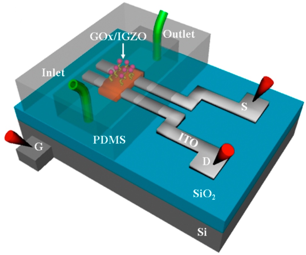
A schematic illustration of an experimental device (credit: Du X et al./ ACS Applied Materials & Interfaces)

To test this idea, Herman and his colleagues first developed an inexpensive method to make IGZO electronics. Then, they used the approach to fabricate a biosensor containing a transparent sheet of IGZO field-effect transistors and glucose oxidase, an enzyme that breaks down glucose. When they added glucose to the mixture, the enzyme oxidized the blood sugar. As a result, the pH level in the mixture shifted and, in turn, triggered changes in the electrical current flowing through the IGZO transistor.

In conventional biosensors, these electrical changes would be used to measure the glucose concentrations in the interstitial fluid under a patient’s skin. But glucose concentrations are much lower in the eye. So any biosensors embedded into contact lenses will need to be far more sensitive. To address this problem, the researchers created nanostructures within the IGZO biosensor that were able to detect glucose concentrations much lower than found in tears.*

In theory, Herman says, more than 2,000 transparent biosensors — each measuring a different bodily function — could be embedded in a 1-millimeter square patch of an IGZO contact lens. Once developed, the biosensors could transmit vital health information to smartphones and other Wi-Fi or Bluetooth-enabled devices.

Herman’s team has already used the IGZO system in catheters to measure uric acid, a key indicator of kidney function, and is exploring the possibility of using it for early detection of cancer and other serious conditions. However, Herman says it could be a year or more before a prototype bio-sensing contact lens is ready for animal testing.

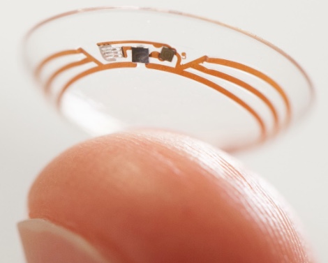
(credit: Google)

The concept appears similar to Goggle’s smart contact lens project, using a tiny wireless chip and miniaturized glucose sensor that are embedded between two layers of soft contact lens material, announced in 2014, but Herman says the Google design is more limited and that the research has stalled.

Herman acknowledges funding from the Juvenile Diabetes Research Foundation and the Northwest Nanotechnology Infrastructure, a member of the National Nanotechnology Coordinated Infrastructure, which is supported by the National Science Foundation.

* “We have functionalized the back-channel of IGZO-FETs with aminosilane groups that are cross-linked to glucose oxidase and have demonstrated that these devices have high sensitivity to changes in glucose concentrations. Glucose sensing occurs through the decrease in pH during glucose oxidation, which modulates the positive charge of the aminosilane groups attached to the IGZO surface. The change in charge affects the number of acceptor-like surface states which can deplete electron density in the n-type IGZO semiconductor. Increasing glucose concentrations leads to an increase in acceptor states and a decrease in drain-source conductance due to a positive shift in the turn-on voltage. The functionalized IGZO-FET devices are effective in minimizing detection of interfering compounds including acetaminophen and ascorbic acid.” — Du XLi YMotley JRStickle WFHerman GS, Glucose Sensing Using Functionalized Amorphous In-Ga-Zn-O Field-Effect Transistors. ACS Applied Materials & Interfaces. 2016 03 30.


Abstract of Implantable indium gallium zinc oxide field effect biosensors

Amorphous indium gallium zinc oxide (IGZO) field effect transistors (FETs) are a promising technology for a wide range of electronic applications including implantable and wearable biosensors. We have recently developed novel, low-cost methods to fabricate IGZO-FETs, with a wide range of form factors. Attaching self-assembled monolayers (SAM) to the IGZO backchannel allows us to precisely control surface chemistry and improve stability of the sensors. Functionalizing the SAMs with enzymes provides excellent selectivity for the sensors, and effectively minimizes interference from acetaminophen/ascorbic acid. We have recently demonstrated that a nanostructured IGZO network can significantly improve sensitivity as a sensing transducer, compared to blanket IGZO films. In Figure (a) we show a scanning electron microscopy image of a nanostructured IGZO transducer located between two indium tin oxide source/drain electrodes. In Figure (b) we show an atomic force microscope image of the close packed hexagonal IGZO nanostructured network (3×3 mm2), and Figure (c) shows the corresponding height profile along the arrow shown in (b). We will discuss reasons for improved sensitivity for the nanostructured IGZO, and demonstrate high sensitivity for glucose sensing. Finally, fully transparent glucose sensors have been fabricated directly on catheters, and have been characterized by a range of techniques. These results suggest that IGZO-FETs may provide a means to integrate fully transparent, highly-sensitive sensors into contact lenses.

The next agricultural revolution: a ‘bionic leaf’ that could help feed the world

The radishes on the right were grown with the help of a bionic leaf that produces fertilizer with bacteria, sunlight, water, and air. (credit: Nocera lab, Harvard University)

Harvard University chemists have invented a new kind of “bionic” leaf that uses bacteria, sunlight, water, and air to make fertilizer right in the soil where crops are grown. It could make possible a future low-cost commercial fertilizer for poorer countries in the emerging world.

The invention deals with the renewed challenge of feeding the world as the population continues to balloon.* “When you have a large centralized process and a massive infrastructure, you can easily make and deliver fertilizer,” Daniel Nocera, Ph.D., says. “But if I said that now you’ve got to do it in a village in India onsite with dirty water — forget it. Poorer countries in the emerging world don’t always have the resources to do this. We should be thinking of a distributed system because that’s where it’s really needed.”

The research was presented at the national meeting of the American Chemical Society (ACS) today (April 3, 2017). The new bionic leaf builds on a previous Nocera-team invention: the “artificial leaf” — a device that mimics photosynthesis: When exposed to sunlight, it mimics a natural leaf by splitting water into hydrogen and oxygen. These two gases would be stored in a fuel cell, which can use those two materials to produce electricity from inexpensive materials.

That was followed by “bionic leaf 2.0,” a water-splitting system that carbon dioxide out of the air and uses solar energy plus hydrogen-eating Ralstonia eutropha bacteria to produce liquid fuel with 10 percent efficiency, compared to the 1 percent seen in the fastest-growing plants. It provided biomass and liquid fuel yields that greatly exceeded those from natural photosynthesis.

Fertilizer created from sunlight + water + carbon dioxide and nitrogen from the air

For the new “bionic leaf,” Nocera’s team has designed a system in which bacteria use hydrogen from the water split by the artificial leaf plus carbon dioxide from the atmosphere to make a bioplastic that the bacteria store inside themselves as fuel. “I can then put the bug [bacteria] in the soil because it has already used the sunlight to make the bioplastic,” Nocera says. “Then the bug pulls nitrogen from the air and uses the bioplastic, which is basically stored hydrogen, to drive the fixation cycle to make ammonia for fertilizing crops.”

The researchers have used their approach to grow five crop cycles of radishes. The vegetables receiving the bionic-leaf-derived fertilizer weigh 150 percent more than the control crops. The next step, Nocera says, is to boost throughput so that one day, farmers in India or sub-Saharan Africa can produce their own fertilizer with this method.

Nocera said a paper describing the new system will be submitted for publication in about six weeks.

* The first “green revolution” in the 1960s saw the increased use of fertilizer on new varieties of rice and wheat, which helped double agricultural production. Although the transformation resulted in some serious environmental damage, it potentially saved millions of lives, particularly in Asia, according to the United Nations (U.N.) Food and Agriculture Organization. But the world’s population continues to grow and is expected to swell by more than 2 billion people by 2050, with much of this growth occurring in some of the poorest countries, according to the U.N. Providing food for everyone will require a multi-pronged approach, but experts generally agree that one of the tactics will have to involve boosting crop yields to avoid clearing even more land for farming.


American Chemical Society | A ‘bionic leaf’ could help feed the world

Scientists grow beating heart tissue on spinach leaves

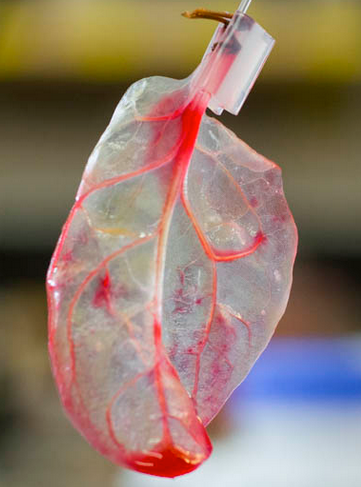
(credit: Worcester Polytechnic Institute)

A research team headed by Worcester Polytechnic Institute (WPI) scientists* has solved a major tissue engineering problem holding back the regeneration of damaged human tissues and organs: how to grow small, delicate blood vessels, which are beyond the capabilities of 3D printing.**

The researchers used plant leaves as scaffolds (structures) in an attempt to create the branching network of blood vessels — down to the capillary scale — required to deliver the oxygen, nutrients, and essential molecules required for proper tissue growth.

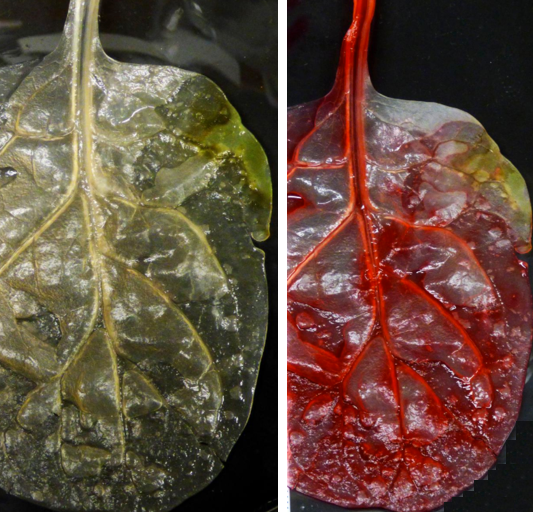
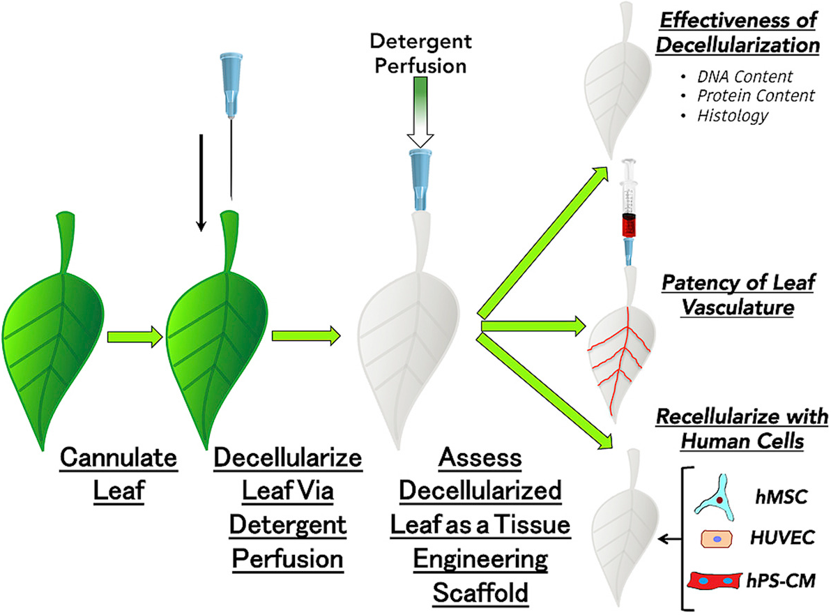
In a series of unconventional experiments, the team cultured beating human heart cells on spinach leaves that were stripped of plant cells.*** The researchers first decellularized spinach leaves (removed cells, leaving only the veins) by perfusing (flowing) a detergent solution through the leaves’ veins. What remained was a framework made up primarily of biocompatible cellulose, which is already used in a wide variety of regenerative medicine applications, such as cartilage tissue engineering, bone tissue engineering, and wound healing.

A spinach leaf (left) was decellularized in 7 days, leaving only the scaffold (right), which served as an intact vascular network. As a test, red dye was pumped through its veins, simulating blood, oxygen, and nutrients. Cardiomyocytes (cardiac muscle cells) derived from human pluripotent stem cells were then seeded onto the surface of the leaf scaffold, forming cell clusters that demonstrated cardiac contractile function and calcium-handling capabilities for 21 days. (credit: Worcester Polytechnic Institute)

After testing the spinach vascular (leaf vessel structure) system mechanically by flowing fluids and microbeads similar in size to human blood cells through it, the researchers seeded the vasculature with human umbilical vein endothelial cells (HUVECs) to grow endothelial cells (which line blood vessels).

Human mesenchymal stem cells (hMSC) and human pluripotent stem-cell-derived cardiomyocytes (cardiac muscle cells) (hPS-CM) were then seeded to the outer surfaces of  the plant scaffolds. The cardiomyocytes spontaneously demonstrated cardiac contractile function (beating) and calcium-handling capabilities over the course of 21 days.

The decellurize-recellurize process (credit: Joshua R. Gershlak et al./Biomaterials)

The future of ”crossing kingdoms”

These proof-of-concept studies may open the door to using multiple spinach leaves to grow layers of healthy heart muscle, and a potential tissue engineered graft based upon the plant scaffolds could use multiple leaves, where some act as arterial support and some act as venous return of blood and fluids from human tissue, say the researchers.

“Our goal is always to develop new therapies that can treat myocardial infarction, or heart attacks,” said Glenn Gaudette, PhD, professor of biomedical engineering at WPI and corresponding author of an open-access paper in the journal Biomaterials, published online in advance of the May 2017 issue.

“Unfortunately, we are not doing a very good job of treating them today. We need to improve that. We have a lot more work to do, but so far this is very promising.”

Currently, it’s not clear how the plant vasculature would be integrated into the native human vasculature and whether there would be an immune response, the authors advise.

The researchers are also now optimizing the decellularization process and seeing how well various human cell types grow while they are attached to (and potentially nourished by) various plant-based scaffolds that could be adapted for specialized tissue regeneration studies. “The cylindrical hollow structure of the stem of Impatiens capensis might better suit an arterial graft,” the authors note. “Conversely, the vascular columns of wood might be useful in bone engineering due to their relative strength and geometries.”

Other types of plants could also provide the framework for a wide range of other tissue engineering technologies, the authors suggest.****

The authors conclude that “development of decellularized plants for scaffolding opens up the potential for a new branch of science that investigates the mimicry between kingdoms, e.g., between plant and animal. Although further investigation is needed to understand future applications of this new technology, we believe it has the potential to develop into a ‘green’ solution pertinent to a myriad of regenerative medicine applications.”

* The research team also includes human stem cell and plant biology researchers at the University of Wisconsin-Madison, and Arkansas State University-Jonesboro.

** The research is driven by the pressing need for organs and tissues available for transplantation, which far exceeds their availability. More than 100,000 patients are on the donor waiting list at any given time and an average of 22 people die each day while waiting for a donor organ or tissue to become available, according to a 2016 paper in the American Journal of Transplantation

*** In addition to spinach leaves, the team successfully removed cells from parsley, Artemesia annua (sweet wormwood), and peanut hairy roots.

**** “Tissue engineered scaffolds are typically produced either from animal-derived or synthetic biomaterials, both of which have a large cost and large environmental impact. Animal-derived biomaterials used extensively as scaffold materials for tissue engineering include native [extracellular matrix]  proteins such as collagen I or fibronectin and whole animal tissues and organs. Annually, 115 million animals are estimated to be used in research. Due to this large number, a lot of energy is necessary for the upkeep and feeding of such animals as well as to dispose of the large amount of waste that is generated. Along with this environmental impact, animal research also has a plethora of ethical considerations, which could be alleviated by forgoing animal models in favor of more biologically relevant in vitro human tissue models,” the authors advise.

Worcester Polytechnic Institute | Spinach leaves can carry blood to grow human tissues