Travelers to Mars risk leukemia cancer, weakened immune function from radiation, NASA-funded study finds

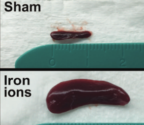
The spleen from a mouse exposed to a mission-relevant dose (20 cGy, 1 GeV/n) of iron ions (bottom) was ~ 30 times the normal volume compared with the spleen from a control mouse (top). (credit: C Rodman et al./Leukemia)

Radiation encountered in deep space travel may increase the risk of leukemia cancer in humans traveling to Mars, NASA-funded researchers at the Wake Forest Institute for Regenerative Medicine and colleagues have found, using mice transplanted with human stem cells.

“Our results are troubling because they show radiation exposure could potentially increase the risk of leukemia,” said Christopher Porada, Ph.D., associate professor of regenerative medicine and senior researcher on the project.

Radiation exposure is believed to be one of the most dangerous aspects of traveling to Mars, according to NASA. The average distance to Mars is 140 million miles, and a round trip could take three years.

The goal of the study, published in the journal Leukemia, was to assess the direct effects of simulated solar energetic particles (SEP) and galactic cosmic ray (GCR) radiation on human hematopoietic stem cells (HSCs). These stem cells comprise less than 0.1% of the bone marrow of adults, but produce the many types of blood cells that circulate through the body and work to transport oxygen, fight infection, and eliminate any malignant cells that arise.

For the study, human HSCs from healthy donors of typical astronaut age (30–55 years) were exposed to Mars mission-relevant doses of protons and iron ions — the same types of radiation that astronauts would be exposed to in deep space, followed by laboratory and animal studies to define the impact of the exposure.

“Radiation exposure at these levels was highly deleterious to HSC function, reducing their ability to produce almost all types of blood cells, often by 60–80 percent,” said Porada. “This could translate into a severely weakened immune system and anemia during prolonged missions in deep space.”

The radiation also caused mutations in genes involved in the hematopoietic process and dramatically reduced the ability of HSCs to give rise to mature blood cells.

Previous studies had already demonstrated that exposure to high doses of radiation, such as from X-rays, can have harmful (even life-threatening) effects on the body’s ability to make blood cells, and can significantly increase the likelihood of cancers, especially leukemias. However, the current study was the first to show a damaging effect of lower, mission-relevant doses of space radiation.

Mice develop T-cell acute lymphoblastic leukemia, weakened immune function

The next step was to assess how the cells would function in the human body. For that purpose, mice were transplanted with GCR-irradiated human HSCs, essentially “humanizing” the animals. The mice developed what appeared to be T-cell acute lymphoblastic leukemia — the first demonstration that exposure to space radiation may increase the risk of leukemia in humans.

“Our results show radiation exposure could potentially increase the risk of leukemia in two ways,” said Porada. “We found that genetic damage to HSCs directly led to leukemia. Secondly, radiation also altered the ability of HSCs to generate T and B cells, types of white blood cells involved in fighting foreign ‘invaders’ like infections or tumor cells. This may reduce the ability of the astronaut’s immune system to eliminate malignant cells that arise as a result of radiation-induced mutations.”

Porada said the findings are particularly troubling given previous work showing that conditions of weightlessness/microgravity present during spaceflight can also cause marked alterations in astronaut’s immune function, even after short duration missions in low-earth orbit, where they are largely protected from cosmic radiation.

Taken together, the results indicate that the combined exposure to microgravity and SEP/GCR radiation that would occur during extended deep space missions, such as to Mars, could potentially exacerbate the risk of immune-dysfunction and cancer,

NASA’s Human Research Program is also exploring conditions of microgravity, isolation and confinement, hostile and closed environments, and distance from Earth. The ultimate goal of the research is to make space missions as safe as possible.

Researchers at Wake Forest Baptist Medical Center, Brookhaven National Laboratory, and the University of California Davis Comprehensive Cancer Center were also involved in the study.


Abstract of In vitro and in vivo assessment of direct effects of simulated solar and galactic cosmic radiation on human hematopoietic stem/progenitor cells

Future deep space missions to Mars and near-Earth asteroids will expose astronauts to chronic solar energetic particles (SEP) and galactic cosmic ray (GCR) radiation, and likely one or more solar particle events (SPEs). Given the inherent radiosensitivity of hematopoietic cells and short latency period of leukemias, space radiation-induced hematopoietic damage poses a particular threat to astronauts on extended missions. We show that exposing human hematopoietic stem/progenitor cells (HSC) to extended mission-relevant doses of accelerated high-energy protons and iron ions leads to the following: (1) introduces mutations that are frequently located within genes involved in hematopoiesis and are distinct from those induced by γ-radiation; (2) markedly reduces in vitro colony formation; (3) markedly alters engraftment and lineage commitment in vivo; and (4) leads to the development, in vivo, of what appears to be T-ALL. Sequential exposure to protons and iron ions (as typically occurs in deep space) proved far more deleterious to HSC genome integrity and function than either particle species alone. Our results represent a critical step for more accurately estimating risks to the human hematopoietic system from space radiation, identifying and better defining molecular mechanisms by which space radiation impairs hematopoiesis and induces leukemogenesis, as well as for developing appropriately targeted countermeasures.

Scientists reverse aging in mice by repairing damaged DNA

A research team led by Harvard Medical School professor of genetics David Sinclair, PhD, has made a discovery that could lead to a revolutionary new drug that allows cells to repair DNA damaged by aging, cancer, and radiation.

In a paper published in the journal Science on Friday (March 24), the scientists identified a critical step in the molecular process related to DNA damage.

The researchers found that a compound known as NAD (nicotinamide adenine dinucleotide), which is naturally present in every cell of our body, has a key role as a regulator in protein-to-protein interactions that control DNA repair. In an experiment, they found that treating mice with a NAD+ precursor called NMN (nicotinamide mononucleotide) improved their cells’ ability to repair DNA damage.

“The cells of the old mice were indistinguishable from the young mice, after just one week of treatment,” said senior author Sinclair.

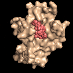
Disarming a rogue agent: When the NAD molecule (red) binds to the DBC1 protein (beige), it prevents DBC1 from attaching to and incapacitating a protein (PARP1) that is critical for DNA repair. (credit: David Sinclair)

Human trials of NMN therapy will begin within the next few months to “see if these results translate to people,” he said. A safe and effective anti-aging drug is “perhaps only three to five years away from being on the market if the trials go well.”

What it means for astronauts, childhood cancer survivors, and the rest of us

The researchers say that in addition to reversing aging, the DNA-repair research has attracted the attention of NASA. The treatment could help deal with radiation damage to astronauts in its Mars mission, which could cause muscle weakness, memory loss, and other symptoms (see “Mars-bound astronauts face brain damage from galactic cosmic ray exposure, says NASA-funded study“), and more seriously, leukemia cancer and weakened immune function (see “Travelers to Mars risk leukemia cancer, weakend immune function from radiation, NASA-funded study finds“).

The treatment could also help travelers aboard aircraft flying across the poles. A 2011 NASA study showed that passengers on polar flights receive about 12 percent of the annual radiation limit recommended by the International Committee on Radiological Protection.

The other group that could benefit from this work is survivors of childhood cancers, who are likely to suffer a chronic illness by age 45, leading to accelerated aging, including cardiovascular disease, Type 2 diabetes, Alzheimer’s disease, and cancers unrelated to the original cancer, the researchers noted.

For the past four years, Sinclair’s team has been working with spinoff MetroBiotech on developing NMN as a drug. Sinclair previously made a link between the anti-aging enzyme SIRT1 and resveratrol. “While resveratrol activates SIRT1 alone, NAD boosters [like NMN] activate all seven sirtuins, SIRT1-7, and should have an even greater impact on health and longevity,” he says.

Sinclair is also a professor at the University of New South Wales School of Medicine in Sydney, Australia.


Abstract of A conserved NAD+ binding pocket that regulates protein-protein interactions during aging

DNA repair is essential for life, yet its efficiency declines with age for reasons that are unclear. Numerous proteins possess Nudix homology domains (NHDs) that have no known function. We show that NHDs are NAD+ (oxidized form of nicotinamide adenine dinucleotide) binding domains that regulate protein-protein interactions. The binding of NAD+ to the NHD domain of DBC1 (deleted in breast cancer 1) prevents it from inhibiting PARP1 [poly(adenosine diphosphate–ribose) polymerase], a critical DNA repair protein. As mice age and NAD+ concentrations decline, DBC1 is increasingly bound to PARP1, causing DNA damage to accumulate, a process rapidly reversed by restoring the abundance of NAD+. Thus, NAD+ directly regulates protein-protein interactions, the modulation of which may protect against cancer, radiation, and aging.

Do-it-yourself robotics kit gives science, tech, engineering, math students tools to automate biology and chemistry experiments

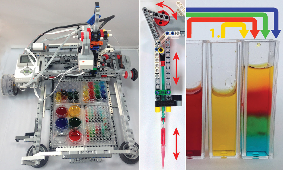
Bioengineers combined a Lego Mindstorms system (left) with a motorized pipette (center) for dropping fluids, allowing for simple experiments like showing how liquids of different salt densities can be layered. (credit: Riedel-Kruse Lab)

Stanford bioengineers have developed liquid-handling robots to allow students to modify and create their own robotic systems that can transfer precise amounts of fluids between flasks, test tubes, and experimental dishes.

The bioengineers combined a Lego Mindstorms robotics kit with a cheap and easy-to-find plastic syringe to create robots that approach the performance of the far more costly automation systems found at universities and biotech labs.

Step-by-step DIY plans

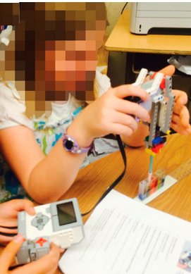
Children 10–13 years old built and explored the functionality of these robots by performing experiments (credit: Lukas C. Gerber et al./PloS Biology)

The idea is to enable students to learn the basics of robotics and the wet sciences in an integrated way. Students learn STEM skills like mechanical engineering, computer programming, and collaboration while gaining a deeper appreciation of the value of robots in life-sciences experiments.

“We really want kids to learn by doing,” said Ingmar Riedel-Kruse, assistant professor of bioengineering and a member of Stanford Bio-X, who led the team. “We show that with a few relatively inexpensive parts, a little training and some imagination, students can create their own liquid-handling robots and then run experiments on it — so they learn about engineering, coding, and the wet sciences at the same time.”

In an open-access paper in the journal PLoS Biology and on Riedel-Kruse’s lab website, the team offers step-by-step building plans and several fundamental experiments targeted to elementary, middle and high school students. They also offer experiments that students can conduct using common household consumables like food coloring, yeast or sugar.

In one experiment, colored liquids with distinct salt concentrations are layered atop one another to teach about liquid density. Other tests measure whether liquids are acids like vinegar or bases like baking soda, or which sugar concentration is best for yeast.

Funding was provided by grants from the National Science Foundation (Cyberlearning and National Robotics Initiative).


Stanford University School of Engineering | SFENG Robots Riedel Kruse v4


Abstract of Liquid-handling Lego robots and experiments for STEM education and research

Liquid-handling robots have many applications for biotechnology and the life sciences, with increasing impact on everyday life. While playful robotics such as Lego Mindstorms significantly support education initiatives in mechatronics and programming, equivalent connections to the life sciences do not currently exist. To close this gap, we developed Lego-based pipetting robots that reliably handle liquid volumes from 1 ml down to the sub-μl range and that operate on standard laboratory plasticware, such as cuvettes and multiwell plates. These robots can support a range of science and chemistry experiments for education and even research. Using standard, low-cost household consumables, programming pipetting routines, and modifying robot designs, we enabled a rich activity space. We successfully tested these activities in afterschool settings with elementary, middle, and high school students. The simplest robot can be directly built from the widely used Lego Education EV3 core set alone, and this publication includes building and experiment instructions to set the stage for dissemination and further development in education and research.

Graphene sheets allow for very-low-cost diagnostic devices

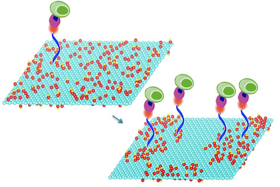
A new, very-low-cost diagnostic method. Mild heating of graphene oxide sheets makes it possible to bond particular compounds (blue, orange, purple) to the sheets’ surface, a new study shows. These compounds in turn select and bond with specific molecules of interest, including DNA and proteins, or even whole cells. In this image, the treated graphene oxide on the right has oxygen molecules (red) clustered together, making it nearly twice as efficient at capturing cells (green) as the material on the left. (credit:  the researchers)

A new method developed at MIT and National Chiao Tung University, based on specially treated sheets of graphene oxide, could make it possible to capture and analyze individual cells from a small sample of blood. It could potentially lead to very-low-cost diagnostic devices (less than $5 a piece) that are mass-producible and could be used almost anywhere for point-of-care testing, especially in resource-constrained settings.

A single cell can contain a wealth of information about the health of an individual. The new system could ultimately lead to a variety of simple devices that could perform a variety of sensitive diagnostic tests, even in places far from typical medical facilities, for cancer screening or treatment follow-up, for example.

How to capture DNA, proteins, or even whole cells for analysis

The material (graphene oxide, or GO) used in this research is an oxidized version of the two-dimensional form of pure carbon known as graphene. The key to the new process is heating the graphene oxide at relatively mild temperatures.

This low-temperature annealing, as it is known, makes it possible to bond particular compounds to the material’s surface that can be used to capture molecules of diagnostic interest.

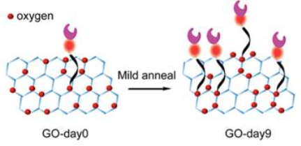
Schematic showing oxygen clustering, resulting in improved ability to recognize foreign molecules (credit: Neelkanth M. Bardhan et al./ACS Nano)

The heating process changes the material’s surface properties, causing oxygen atoms to cluster together, leaving spaces of bare graphene between them. This leaves room to attach other chemicals to the surface, which can be used to select and bond with specific molecules of interest, including DNA and proteins, or even whole cells. Once captured, those molecules or cells can then be subjected to a variety of tests.*

Nanobodies

The new research demonstrates how that basic process could potentially enable a suite of low-cost diagnostic systems.

For this proof-of-concept test, the team used molecules that can quickly and efficiently capture specific immune cells that are markers for certain cancers. They were able to demonstrate that their treated graphene oxide surfaces were almost twice as effective at capturing such cells from whole blood, compared to devices fabricated using ordinary, untreated graphene oxide.

They did this by enzymatically coating the treated graphene oxide surface with peptides called “nanobodies” — subunits of antibodies, which can be cheaply and easily produced in large quantities in bioreactors and are highly selective for particular biomolecules.**

The new process allows for rapid capture and assessment of cells or biomolecules within about 10 minutes and without the need for refrigeration of samples or incubators for precise temperature control. And the whole system is compatible with existing large-scale manufacturing methods.

The researchers believe many different tests could be incorporated on a single device, all of which could be placed on a small glass slide like those used for microscopy. The basic processing method could also make possible a wide variety of other applications, including solar cells and light-emitting devices.

The findings are reported in the journal ACS Nano. Authors include Angela Belcher, the James Mason Crafts Professor in biological engineering and materials science and engineering at MIT and a member of the Koch Institute for Integrative Cancer Research; Jeffrey Grossman, the Morton and Claire Goulder and Family Professor in Environmental Systems at MIT; Hidde L. Ploegh, a professor of biology and member of the Whitehead Institute for Biomedical Research; Guan-Yu Chen, an assistant professor in biomedical engineering at National Chiao Tung University in Taiwan; and Zeyang Li, a doctoral student at the Whitehead Institute.

“Efficiency is especially important if you’re trying to detect a rare event,” Belcher says. “The goal of this was to show a high efficiency of capture.” The next step after this basic proof of concept, she says, is to try to make a working detector for a specific disease model.

The work was supported by the Army Research Office Institute for Collaborative Biotechnologies and MIT’s Tata Center and Solar Frontiers Center.

* Other researchers have been trying to develop diagnostic systems using a graphene oxide substrate to capture specific cells or molecules, but these approaches used just the raw, untreated material. Despite a decade of research, other attempts to improve such devices’ efficiency have relied on external modifications, such as surface patterning through lithographic fabrication techniques, or adding microfluidic channels, which add to the cost and complexity. Those methods for treating graphene oxide for this purpose require high-temperature treatments or the use of harsh chemicals; the new system, which the group has patented, requires no chemical pretreatment and an annealing temperature of just 50 to 80 degrees Celsius (122 to 176 F).

** The researchers found that increasing the annealing time steadily increased the efficiency of cell capture: After nine days of annealing, the efficiency of capturing cells from whole blood went from 54 percent, for untreated graphene oxide, to 92 percent for the treated material. The team then performed molecular dynamics simulations to understand the fundamental changes in the reactivity of the graphene oxide base material. The simulation results, which the team also verified experimentally, suggested that upon annealing, the relative fraction of one type of oxygen (carbonyl) increases at the expense of the other types of oxygen functional groups (epoxy and hydroxyl) as a result of the oxygen clustering. This change makes the material more reactive, which explains the higher density of cell capture agents and increased efficiency of cell capture.


Abstract of Enhanced Cell Capture on Functionalized Graphene Oxide Nanosheets through Oxygen Clustering

With the global rise in incidence of cancer and infectious diseases, there is a need for the development of techniques to diagnose, treat, and monitor these conditions. The ability to efficiently capture and isolate cells and other biomolecules from peripheral whole blood for downstream analyses is a necessary requirement. Graphene oxide (GO) is an attractive template nanomaterial for such biosensing applications. Favorable properties include its two-dimensional architecture and wide range of functionalization chemistries, offering significant potential to tailor affinity toward aromatic functional groups expressed in biomolecules of interest. However, a limitation of current techniques is that as-synthesized GO nanosheets are used directly in sensing applications, and the benefits of their structural modification on the device performance have remained unexplored. Here, we report a microfluidic-free, sensitive, planar device on treated GO substrates to enable quick and efficient capture of Class-II MHC-positive cells from murine whole blood. We achieve this by using a mild thermal annealing treatment on the GO substrates, which drives a phase transformation through oxygen clustering. Using a combination of experimental observations and MD simulations, we demonstrate that this process leads to improved reactivity and density of functionalization of cell capture agents, resulting in an enhanced cell capture efficiency of 92 ± 7% at room temperature, almost double the efficiency afforded by devices made using as-synthesized GO (54 ± 3%). Our work highlights a scalable, cost-effective, general approach to improve the functionalization of GO, which creates diverse opportunities for various next-generation device applications.

Programmable shape-shifting molecular robots respond to DNA signals

Japanese researchers have developed an amoeba-like shape-changing molecular robot — assembled from biomolecules such as DNA, proteins, and lipids — that could act as a programmable and controllable robot for treating live culturing cells or monitoring environmental pollution, for example.

This the first time a molecular robotic system can recognize signals and control its shape-changing function, and their molecular robots could in the near future function in a way similar to living organisms, according to the researchers.

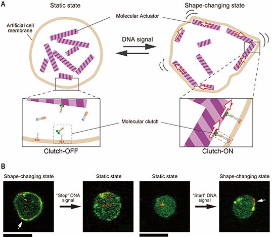
Developed by a research group at Tohoku University and Japan Advanced Institute of Science and Technology, the molecular robot integrates molecular machines within an artificial cell membrane and is about one micrometer in diameter — similar in size to human cells. It can start and stop its shape-changing function in response to a specific DNA signal.

Schematic diagram of the molecular robot. (A) In response to a start-stop DNA signal, molecular actuators (microtubules) inside the robot change the shape of the artificial cell membrane (liposome), controlled by a “molecular clutch” that transmits the force from the actuator (kinesin proteins, shown in green, assemble DNA to the cell membrane when activated). (B) Microscopy images of molecular robots. When the input DNA signal is “stop,” the clutch is turned “OFF,” deactivating the shape-changing behavior. The shape-changing is activated when the the clutch is turned “ON.” Scale bar: 20 μm. The white arrow indicates the molecular actuator part that transforms the shape of the membrane. (credit: Yusuke Sato)

The movement force is generated by molecular actuators (microtubules) controlled by a molecular clutch (composed of DNA and kinesin — a “walker” that carries molecules along microtubules in the body). The shape of the robot’s body (artificial cell membrane, or liposome — a vesicle made from a lipid bilayer) is changed (from static to active) by the actuator, triggered by specific DNA signals activated by UV irradiation.

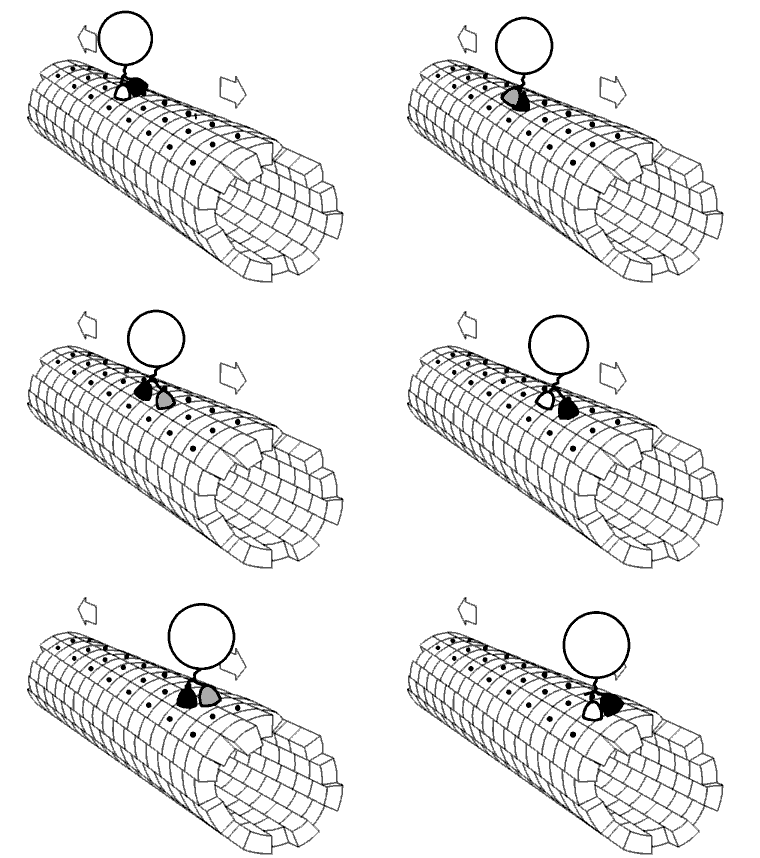
Kinesin motor protein “walking” along microtubule filament (credit: Jzp706/CC)

The realization of a molecular robot whose components are designed at a molecular level and that can function in a small and complicated environment, such as the human body, is expected to significantly expand the possibilities of robotics engineering, according to the researchers.*

“With more than 20 chemicals at varying concentrations, it took us a year and a half to establish good conditions for working our molecular robots,” says Associate Professor Shin-ichiro Nomura at Tohoku University’s Graduate School of Engineering, who led the study. “It was exciting to see the robot shape-changing motion through the microscope. It meant our designed DNA clutch worked perfectly, despite the complex conditions inside the robot.”

Programmable by DNA computing devices

The research results were published in an open-access paper in Science Robotics on March 1, 2017.

The authors say that “combining other molecular devices would lead to the realization of a molecular robot with advanced functions. For example, artificial nanopores, such as an artificial channel composed of DNA, could be used to sense signal molecules in the surrounding environments through the channel.

“In addition, the behavior of a molecular robot could be programmed by DNA computing devices, such as judging the condition of environments. These implementations could allow for the development of molecular robots capable of chemotaxis [movement in a direction corresponding to a gradient of increasing or decreasing concentration of a particular substance], [similar to] white blood cells, and beyond.”

The research was supported by the JSPS KAKENHI, AMED-CREST and Tohoku University-DIARE.

* In the current design, “there are still limitations in the functions of the robot. For example, the switching of robot behavior is not reversible. The shape change is not directional and as yet not possible for complex tasks, for example, locomotion. However, to the best of our knowledge, this is the first implementation of a molecular robot that can control its shape-changing behavior in response to specific signal molecules.” — Yusuke Sato et al./Science Robotics


Abstract of Micrometer-sized molecular robot changes its shape in response to signal molecules

Rapid progress in nanoscale bioengineering has allowed for the design of biomolecular devices that act as sensors, actuators, and even logic circuits. Realization of micrometer-sized robots assembled from these components is one of the ultimate goals of bioinspired robotics. We constructed an amoeba-like molecular robot that can express continuous shape change in response to specific signal molecules. The robot is composed of a body, an actuator, and an actuator-controlling device (clutch). The body is a vesicle made from a lipid bilayer, and the actuator consists of proteins, kinesin, and microtubules. We made the clutch using designed DNA molecules. It transmits the force generated by the motor to the membrane, in response to a signal molecule composed of another sequence-designed DNA with chemical modifications. When the clutch was engaged, the robot exhibited continuous shape change. After the robot was illuminated with light to trigger the release of the signal molecule, the clutch was disengaged, and consequently, the shape-changing behavior was successfully terminated. In addition, the reverse process—that is, initiation of shape change by input of a signal—was also demonstrated. These results show that the components of the robot were consistently integrated into a functional system. We expect that this study can provide a platform to build increasingly complex and functional molecular systems with controllable motility.

New machine-learning algorithms may revolutionize drug discovery — and our understanding of life

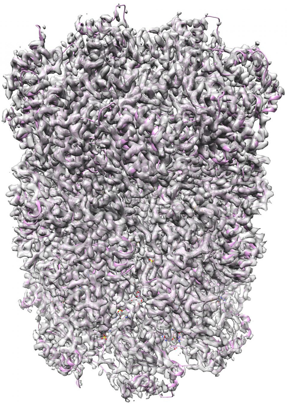
A new set of machine-learning algorithms can generate 3D structures of complex nanoscale protein molecules like this complex proteasome map refined to 2.8 Angstroms (.28 nanometer) in 70 min with 49,954 particle images (credit: Structura Biotechnology Inc.)

A new set of machine-learning algorithms developed by researchers at the University of Toronto Scarborough can generate 3D structures of nanoscale protein molecules that could not be achieved in the past. The algorithms may revolutionize the development of new drug therapies for a range of diseases and may even lead to better understand how life works at the atomic level, the researchers say.

Drugs work by binding to a specific protein molecule and changing the protein’s 3D shape, which alters the way the drug works once inside the body. The ideal drug is designed in a shape that will only bind to a specific protein or group of proteins that are involved in a disease, while eliminating side effects that occur when drugs bind to other proteins in the body.

A significant computational problem

Since proteins are tiny — about 1 to 100 nanometers — even smaller than the shortest wavelength of visible light, they can’t be seen directly without using sophisticated techniques like electron cryomicroscopy (cryo-EM). Cryo-EM uses high-power microscopes to take tens of thousands of low-resolution images of a frozen protein sample from different positions.

The computational problem is to then piece together the correct high-resolution 3D structure from these 2D images.

Existing techniques take several days or even weeks to generate a 3D structure on a cluster of computers, requiring as much as 500,000 CPU hours, according to the researchers. Also, existing techniques often generate incorrect structures unless an expert user provides an accurate guess of the molecule being studied.

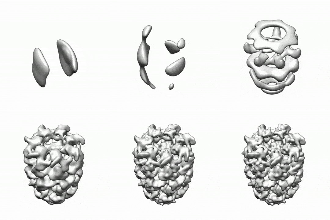
CryoSPARC machine learning algorithms can generate 3-D structures of nanoscale protein molecules (credit: Structura Biotechnology Inc)

New high-speed, deep-learning algorithms

That’s where the new set of algorithms* comes in. It reconstructs 3D structures of protein molecules using these images. “Our approach solves some of the major problems in terms of speed and number of structures you can determine,” says Professor David Fleet, chair of the Computer and Mathematical Sciences Department at U of Toronto Scarborough.

The algorithms could significantly aid in the development of new drugs because they provide a faster, more efficient means at arriving at the correct protein structure.

The new approach, called cryoSPARC, developed by the team’s startup, Structura Biotechnology Inc., eliminates the need for that prior knowledge and can make the computations possible in minutes on a single computer, using a standalone graphics processing unit (GPU) accelerated software package, according to the researchers.

The research was published in the current edition of the journal Nature Methods. It received funding from the Natural Sciences and Engineering Research Council of Canada (NSERC). The new cryo-EM platform is already being used in labs across North America, the researchers note.

* “We use an SGD [stochastic gradient descent] optimization scheme to quickly identify one or several low-resolution 3D structures that are consistent with a set of observed images. This algorithm allows for ab initio heterogeneous structure determination with no prior model of the molecule’s structure. Once approximate structures are determined, a branch-and-bound algorithm for image alignment helps rapidly refine structures to high resolution. The speed and robustness of these approaches allow structure determination in a matter of minutes or hours on a single inexpensive desktop workstation. … SGD was popularized as a key tool in deep learning for the optimization of nonconvex functions, and it results in near human-level performance in tasks like image and speech recognition.” — Ali Punjani et al./Nature Methods

University of Toronto Scarborough | New algorithms may revolutionize drug discoveries and our understanding of life


Abstract of cryoSPARC: algorithms for rapid unsupervised cryo-EM structure determination

Single-particle electron cryomicroscopy (cryo-EM) is a powerful method for determining the structures of biological macromolecules. With automated microscopes, cryo-EM data can often be obtained in a few days. However, processing cryo-EM image data to reveal heterogeneity in the protein structure and to refine 3D maps to high resolution frequently becomes a severe bottleneck, requiring expert intervention, prior structural knowledge, and weeks of calculations on expensive computer clusters. Here we show that stochastic gradient descent (SGD) and branch-and-bound maximum likelihood optimization algorithms permit the major steps in cryo-EM structure determination to be performed in hours or minutes on an inexpensive desktop computer. Furthermore, SGD with Bayesian marginalization allows ab initio 3D classification, enabling automated analysis and discovery of unexpected structures without bias from a reference map. These algorithms are combined in a user-friendly computer program named cryoSPARC

Scientists create first 3-D synchronized-beating heart tissue

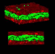
3-D fluorescent image of 3-D tissue with many cells laid down sequentially to create attached layers of alternating cell types, like membranes in the human body (credit: York University)

York University scientists have created the first in vitro (lab) 3D heart tissue made from three different types of cardiac cells that beat in synchronized harmony. It may lead to better understanding of cardiac health and improved treatments.*

The researchers constructed the heart tissue from three free-beating rat cell types: contractile cardiac muscle cells, connective tissue cells, and vascular cells. No external scaffold was used and the cells were the only building blocks of the generated cardiac tissue. The researchers believe this is the first 3D in vitro cardiac tissue with three cell types that can beat together as one entity, rather than at different intervals, with high cell density and efficient cell contacts, and without the requirement of external electrical stimulation.

The substance used to stick the cells together (ViaGlue) may also provide researchers with tools to create and test 3D in vitro cardiac tissue in their own labs to study heart disease and issues with transplantation.

“This breakthrough will allow better and earlier drug testing, and potentially eliminate harmful or toxic medications sooner,” said York U chemistry Professor Muhammad Yousaf.

For 2D or 3D cardiac tissue to be functional it needs the same high cellular density and the cells must be in contact to facilitate synchronized beating, according to the researchers. The 3D cardiac tissue was created at a millimeter scale, but larger versions could be made, said Yousaf, who has created a start-up company, OrganoLinX, to commercialize the ViaGlue reagent and to provide custom 3D tissues on demand.

“Production of 3-dimensional artificial cardiac tissues for fundamental studies of heart disease, transplantation, and evaluation of drug toxicity is an important and intense area of research,” the researchers note in a paper in open-access Nature Scientific Reports.

* Cardiovascular-associated diseases are the leading cause of death globally and are responsible for 40 per cent of deaths in North America, according to a 2011 report from the American Heart Association.


York University | York U makes a 3D heart beat as one


Abstract of Scaffold Free Bio-orthogonal Assembly of 3-Dimensional Cardiac Tissue via Cell Surface Engineering

There has been tremendous interest in constructing in vitro cardiac tissue for a range of fundamental studies of cardiac development and disease and as a commercial system to evaluate therapeutic drug discovery prioritization and toxicity. Although there has been progress towards studying 2-dimensional cardiac function in vitro, there remain challenging obstacles to generate rapid and efficient scaffold-free 3-dimensional multiple cell type co-culture cardiac tissue models. Herein, we develop a programmed rapid self-assembly strategy to induce specific and stable cell-cell contacts among multiple cell types found in heart tissue to generate 3D tissues through cell-surface engineering based on liposome delivery and fusion to display bio-orthogonal functional groups from cell membranes. We generate, for the first time, a scaffold free and stable self assembled 3 cell line co-culture 3D cardiac tissue model by assembling cardiomyocytes, endothelial cells and cardiac fibroblast cells via a rapid inter-cell click ligation process. We compare and analyze the function of the 3D cardiac tissue chips with 2D co-culture monolayers by assessing cardiac specific markers, electromechanical cell coupling, beating rates and evaluating drug toxicity.

First stable semisynthetic organism created

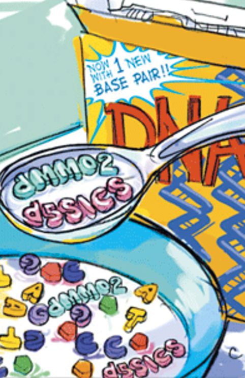
DNA — now with a new base pair! (credit: Romesberg Lab)

Scientists at The Scripps Research Institute (TSRI) have developed the first stable semisynthetic organism — a bacterium with two new synthetic bases (called X and Y) added to the four natural bases (A, T, C, and G) that every living organism possesses. Adding two more letters to expand the genetic alphabet can be used to make novel proteins for new therapeutics, according to the researchers.

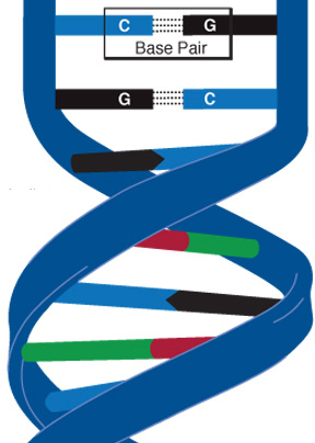
All life as we currently know it contains just four bases that pair up to form two “base pairs” — the rungs of the DNA ladder — which are simply rearranged to create different organisms.

DNA double helix with examples of base pairs (credit: National Human Genome Research Institute)

Until now. TSRI Professor Floyd Romesberg and his colleagues have now shown that their new “more life-like” single-celled organism can hold on indefinitely to the new synthetic base pair as it divides. Their research was published January 23, 2017, online ahead of print in the journal Proceedings of the National Academy of Sciences.

Snipping together a new semisynthetic organism

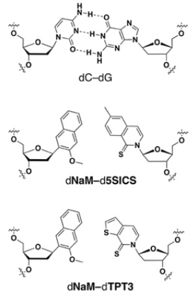
Top: chemical structure of a normal DNA base pair, with C and G bases. Below: structures of the two new X and Y synthetic bases. (credit: Yorke Zhang et al./PNAS)

To ensure the new semisynthetic organism retains the new synthetic base pair as it divides, the researchers used CRISPR-Cas9 (a sort of scissors for modifying DNA). They engineered the organism to react to a genetic sequence that doesn’t have X and Y as a foreign invader (an immune response).

So any new cell that dropped X and Y would be marked for destruction — leaving the scientists with an organism that could hold on to the new bases.

In their experiments, that enabled their semisynthetic organism to keep X and Y in its genome after dividing 60 times, leading the researchers to believe it can hold on to the new base pair indefinitely.

“We can now get the light of life to stay on,” said Romesberg, senior author of the new study. “That suggests that all of life’s processes can be subject to manipulation.”

Romesberg emphasized that this work is only in single cells and is not meant to be used in more complex organisms. So far, scientists can only get the organism to store genetic information.

While applications for this kind of organism are still far in the future, the researchers say the work could be used to create new functions for single-celled organisms that play important roles in drug discovery and “much more.” (Romesberg has created created a biotech company named Synthorx for discovering and developing novel protein therapeutics.)


“All of life’s processes can be subject to manipulation”


Next, the researchers plan to study how their new genetic code can be transcribed into RNA, the molecule in cells needed to translate DNA into useful proteins.

Scientists at the University of Grenoble and Henan Normal University were also involved in the study, which was supported by grants from the National Institutes of Health, theNational Science Foundation, the National Natural Science Foundation of China, Labex ARCANE, NanoBio-ICMG platforms, and a postdoctoral fellowship from the American Cancer Society.


Abstract of A semisynthetic organism engineered for the stable expansion of the genetic alphabet

All natural organisms store genetic information in a four-letter, two-base-pair genetic alphabet. The expansion of the genetic alphabet with two synthetic unnatural nucleotides that selectively pair to form an unnatural base pair (UBP) would increase the information storage potential of DNA, and semisynthetic organisms (SSOs) that stably harbor this expanded alphabet would thereby have the potential to store and retrieve increased information. Toward this goal, we previously reported that Escherichia coli grown in the presence of the unnatural nucleoside triphosphates dNaMTP and d5SICSTP, and provided with the means to import them via expression of a plasmid-borne nucleoside triphosphate transporter, replicates DNA containing a single dNaM-d5SICS UBP. Although this represented an important proof-of-concept, the nascent SSO grew poorly and, more problematically, required growth under controlled conditions and even then was unable to indefinitely store the unnatural information, which is clearly a prerequisite for true semisynthetic life. Here, to fortify and vivify the nascent SSO, we engineered the transporter, used a more chemically optimized UBP, and harnessed the power of the bacterial immune response by using Cas9 to eliminate DNA that had lost the UBP. The optimized SSO grows robustly, constitutively imports the unnatural triphosphates, and is able to indefinitely retain multiple UBPs in virtually any sequence context. This SSO is thus a form of life that can stably store genetic information using a six-letter, three-base-pair alphabet.

Scientists use stem cells to create human/pig chimera embryos

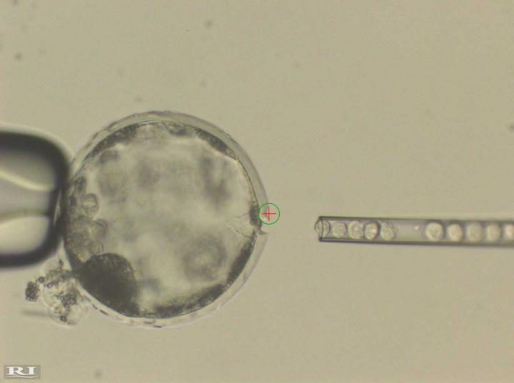
This photograph shows injection of human induced pluripotent stem* (iPS) cells into a pig blastocyst (pre-embryo). A laser beam (green circle with a red cross inside) was used to perforate an opening in the outer membrane (Zona Pellucida) of the pig blastocyst to allow easy access of an injection needle delivering human iPS cells. (credit: Courtesy of Juan Carlos Izpisua Belmonte)

In an open-access paper published online January 26, 2017 in the journal CellSalk Institute researchers report breakthroughs on multiple fronts in the race to integrate stem cells from one species into the early-stage development of another species (or chimeras**).

Scientists are still struggling to coax stem cells growing in Petri dishes to become fully functional specialized adult cells, the researchers report. “The ultimate goal is to grow functional and transplantable tissue or organs, but we are far away from that,” says lead investigator Juan Carlos Izpisua Belmonte, a professor in the Salk Institute of Biological Studies’ Gene Expression Laboratory. But their efforts to grow the first embryos containing cells from humans and pigs may someday provide a means of growing human cells, tissues, and organs for regenerative medicine. “This is an important first step.”

Meanwhile, the experiments are helping scientists understand how human stem cells grow and specialize, which may offer insights into disease onset or help establish new drug-testing platforms.

How to grow a chimera

In previous research, Izpisua Belmonte and Salk Institute staff scientist Jun Wu created a rat/mouse chimera by introducing rat cells into mouse embryos and letting the cells mature. The purpose was to find out whether rat-derived cells could rescue severe developmental defects in mouse pancreas, heart or lung. To do that, they used CRISPR genome editing tools to delete critical developmental genes in mouse egg cells. Remarkably, rat cells populated those mouse organs, filling in for the defective mouse cells.

Here, cells derived from rat pluripotent stem cells were enriched in the developing heart of a genetically modified mouse embryo. (credit: Salk Institute)

For instance, in a given cell, the researchers would delete a single gene critical for the development of an organ, such as the heart, pancreas, or eye. Then they introduced rat stem cells into the embryos to see if they would fill the open niche. “The rat cells have a functional copy of the missing mouse gene, so they can outcompete mouse cells in occupying the emptied developmental organ niches,” says Wu. As the organism matured, the rat cells filled in where mouse cells could not, forming the functional tissues of the organism’s heart, eye, or pancreas.

Amazingly, in one experiment, rat cells also grew to form a gall bladder in the mouse, even though rats themselves stopped developing this organ over the 18 million years since rats and mice separated evolutionarily. “This suggests that the reason a rat does not generate a gall bladder is not because it cannot, but because the potential has been hidden by a rat-specific developmental program,” says Wu. “The microenvironment has evolved through millions of years to choose a program that defines a rat.”

Introducing human cells in pig embryos

The team’s next step was to introduce human cells into an organism. Experiments with cow embryos were difficult and costly. They decided to use pig embryos as hosts because the size of these animals’ organs more closely resembles those of humans than those of mice. The effort required completing studies with 1,500 pig embryos and involved the contributions of more than 40 people, including pig farmers, over a four-year period. “We underestimated the effort involved,” says Izpisua Belmonte. “This required a tour de force.”

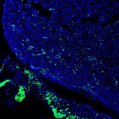
Salk scientists advance stem-cell and genome-editing technologies to help researchers study evolution and disease, test therapeutic drugs and possibly grow transplantable organs. Here, human iPS cells (green) contributed to a developing heart of 4-week-old pig embryo. (credit: Salk Institute)

The human cells survived and formed a human/pig chimera embryo. Embryos were implanted in sows and allowed to develop for between three and four weeks. “This is long enough for us to try to understand how the human and pig cells mix together early on without raising ethical concerns about mature chimeric animals,” says Izpisua Belmonte.

To ensure that, the human cells did not become precursors of brain cells that can grow into the central nervous system. Rather, they were developing into muscle cells and precursors of other organs. “At this point, we wanted to know whether human cells can contribute at all to address the ‘yes or no’ question,” he says. “Now that we know the answer is yes, our next challenge is to improve efficiency and guide the human cells into forming a particular organ in pigs.”

To do this, the researchers are using CRISPR to perform genome editing on the pig genome, as they did with mice, to open gaps that human cells can fill in. That work is in progress.

“Of course, the ultimate goal of chimeric research is to learn whether we can use stem-cell and gene-editing technologies to generate genetically matched human tissues and organs, and we are very optimistic that continued work will lead to eventual success,” says Izpisua Belmonte. “But in the process we are gaining a better understanding of species evolution as well as human embryogenesis and disease that is difficult to get in other ways.”

Other authors included scientists at the University of Murcia Campus de Espinardo, the University of California, Davis; the Universidad Católica San Antonio de Murcia; Clinica Centro Fundación Pedro Guillén; and the Hospital Clinic of Barcelona. The work was funded by The Fundación Séneca; a University of California, Davis, Academic Senate New Research Grant; the Universidad Católica San Antonio de Murcia; Fundacion Dr. Pedro Guillen; the G. Harold and Leila Y. Mathers Charitable Foundation; and The Moxie Foundation.

* Induced pluripotent stem cells (also known as iPS cells or iPSCs) are a type of pluripotent stem cell that can be generated directly from adult cells. They have the capacity to mature into many different types of cells and therefore can contribute to the formation of multiple organs or distinct cell lineages, but not all possible lineages. This developmental capacity differs from naïve stem cells, which can develop into any given cell type.

** An interspecies chimera is an organism containing cells from different species. The word “chimera” originally described mythological creatures or deities in polytheistic religions. In science, interspecies chimeras have emerged as valuable basic research tools with potential for future clinical applications.


Salk Institute | New findings highlight promise of chimeric organisms for science and medicine


Abstract of Interspecies Chimerism with Mammalian Pluripotent Stem Cells

Interspecies blastocyst complementation enables organ-specific enrichment of xenogenic pluripotent stem cell (PSC) derivatives. Here, we establish a versatile blastocyst complementation platform based on CRISPR-Cas9-mediated zygote genome editing and show enrichment of rat PSC-derivatives in several tissues of gene-edited organogenesis-disabled mice. Besides gaining insights into species evolution, embryogenesis, and human disease, interspecies blastocyst complementation might allow human organ generation in animals whose organ size, anatomy, and physiology are closer to humans. To date, however, whether human PSCs (hPSCs) can contribute to chimera formation in non-rodent species remains unknown. We systematically evaluate the chimeric competency of several types of hPSCs using a more diversified clade of mammals, the ungulates. We find that naïve hPSCs robustly engraft in both pig and cattle pre-implantation blastocysts but show limited contribution to post-implantation pig embryos. Instead, an intermediate hPSC type exhibits higher degree of chimerism and is able to generate differentiated progenies in post-implantation pig embryos.