Scientists discover precise DNA sequence code critical for turning genes on

DNA sequence signal for the activation of human genes. Each tiny human cell contains about six feet of DNA, a double-helical molecular chain containing four types of several billion chemical nucleotides — adenine (A), cytosine (C), guanine (G) and thymine (T) — arranged in a specific sequence, or code, that when transcribed guide the cell into producing specific proteins. (credit: University of California — San Diego)

Molecular biologists at the University of California, San Diego (UC San Diego) have discovered a short sequence of DNA that is essential for turning on (expressing proteins) more than half of all human genes — an achievement that should provide scientists with a better understanding of how human genes are regulated.

Knowing what turns on genes is important. Each human cell contains about six feet of DNA, a double-helical molecular chain containing several billion chemical nucleotides — adenine (A), cytosine (C), guanine (G) and thymine (T) — arranged in a specific sequence, or code. Active genes undergo a process called transcription, in which the nucleotide sequence in DNA is read and converted into a sister language called RNA for processing. The processed RNA sequence then guides the cell to produce specific proteins that are essential for normal cellular functions.
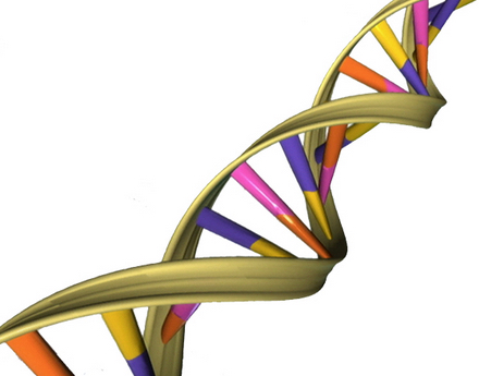
A depiction of the double helical structure of DNA. Its four nucleotide coding units (A, T, C, G) are color-coded in pink, orange, purple and yellow. (credit: NHGRI)

“In these six feet of DNA, there are tens of thousands of genes, which are segments of DNA that direct specific functions, such as the production of a hormone or an enzyme,” explains James T. Kadonaga, PhD, a molecular biology professor at UC San Diego who headed the team of researchers. “It is essential for the cell to control the activity of each of its tens of thousands of genes, because the improper control of gene activity can lead to adverse outcomes such as cell death or the formation of a cancer cell.”

The “human Initiator”

Enter the “Initiator.” The initiation of gene expression often occurs at a critical DNA sequence code called the “human Initiator.” This small piece of DNA helps gene expression machinery locate exactly where to begin transcribing. Although the concept of the Initiator has been known since the 1980s, the precise DNA sequence comprising the Initiator had eluded scientists.*

“There are many sequence signals that control gene activity in human cells and the Initiator is the most commonly occurring sequence at the start sites of genes,” Kadonaga said. Kadonaga and his team employed emerging genomic techniques and devised novel computational strategies to unlock the exact DNA sequence code for the human Initiator.

They also discovered that this sequence is located precisely at the start site of more than half of all human genes, underlining the importance of the human Initiator in the human genome. “The solution of the human Initiator code will enable us to explore new frontiers in gene regulation,” said Kadonaga. “In the future, it will be possible to use the code to identify other regulatory signals and in this way gain a more complete understanding of how human genes are turned on and off.”

“The authors verified the Initiator sequence in multiple cell lines, which is an impressive finding,” a scientist not involved in the studies told KurzweilAI. “However, none of these cell lines reflect normal human biology — they are essentially cancer cells proliferating in a dish. I would have liked to see this Initiator sequence verified in normal human cells from healthy patients.”

The research, now online and to be detailed in the February 10 print issue of the journal Genes & Development, was supported by grants from the National Institutes of Health.

* First observed by Pierre Chambon and his colleagues in Strasbourg, France in 1980, the human Initiator and its role in gene activation were articulated in 1989 by two MIT biologists, Stephen Smale and David Baltimore at MIT, who later revealed the approximate sequence code of the Initiator. Since then, however, other scientists had proposed a number of different sequences for the human Initiator, but none of them were found to be consistently associated with the start sites of human genes. As a result, the true Initiator sequence code remained a mystery — until now.


Abstract of The human initiator is a distinct and abundant element that is precisely positioned in focused core promoters

DNA sequence signals in the core promoter, such as the initiator (Inr), direct transcription initiation by RNA polymerase II. Here we show that the human Inr has the consensus of BBCA+1BW at focused promoters in which transcription initiates at a single site or a narrow cluster of sites. The analysis of 7678 focused transcription start sites revealed 40% with a perfect match to the Inr and 16% with a single mismatch outside of the CA+1 core. TATA-like sequences are underrepresented in Inr promoters. This consensus is a key component of the DNA sequence rules that specify transcription initiation in humans.

Microbiologists make big leap in developing ‘green’ electronics


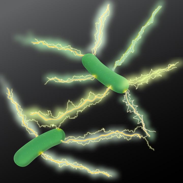
An artist’s rendition of Geobacter expressing electrically conductive nanowires. Microbiologists at UMass Amherst have discovered a new type of natural wire produced by bacteria that could greatly accelerate the development of sustainable “green” conducting materials for the electronics industry. (credit: UMass Amherst)

UMass Amherst research finds microbe yields better electronic material

Microbiologists at the University of Massachusetts Amherst report that they have discovered a new type of microbial nanowire produced by bacteria that could greatly accelerate the development of sustainable “green” conducting materials for the electronics industry.

The study by Derek Lovley and colleagues appears this week in an open-access paper in mBio, the American Society of Microbiology’s premier journal.

A bacterium known as Geobacter sulfurreducens uses the protein filaments naturally to make electrical connections with other microbes or minerals.

As Lovley explains, “Microbial nanowires are a revolutionary electronic material with substantial advantages over man-made materials. Chemically synthesizing nanowires in the lab requires toxic chemicals, high temperatures and/or expensive metals. The energy requirements are enormous. By contrast, natural microbial nanowires can be mass-produced at room temperature from inexpensive renewable feedstocks in bioreactors with much lower energy inputs. And the final product is free of toxic components.”

Confocal scanning laser micrographs of G. sulfurreducens anode biofilms harvested on day 10. Bar, 25 µm. (credit: Yang Tan et al./mBio)

The Microbial nanowires offer an unprecedented potential for developing novel electronic devices and sensors for diverse applications with a new environmentally friendly technology, Lovely says. “This is an important advance in microbial nanowire technology. The approach we outline in this paper demonstrates a rapid method for prospecting in nature to find better electronic materials.”

When his lab began looking at the protein filaments of other Geobacter species, they were surprised to find a wide range in conductivities. For example, one species recovered from uranium-contaminated soil produced poorly conductive filaments. However, another species, Geobacter metallireducens produced nanowires 5,000 times more conductive than the G. sulfurreducens wires. Lovley recalls, “I isolated metallireducens from mud in the Potomac River 30 years ago, and every couple of years it gives us a new surprise.”

In their new study supported by the U.S. Office of Naval Research, they did not study the G. metallireducens strain directly. Instead, they took the gene for the protein that assembles into microbial nanowires from it and inserted this into G. sulfurreducens. The result is a genetically modified G. sulfurreducens that expresses the G. metallireducens protein, making nanowires much more conductive than G. sulfurreducens would naturally produce.

Further, Lovley says, “We have found that G. sulfurreducens will express filament genes from many different types of bacteria. This makes it simple to produce a diversity of filaments in the same microorganism and to study their properties under similar conditions.”

The high conductivity of the G. metallireducens nanowires suggests that they may be an attractive material for the construction of conductive materials, electronic devices ,and sensors for medical or environmental applications. The authors say discovering more about the mechanisms of nanowire conductivity “provides important insight into how we might make even better wires with genes that we design ourselves.”


Abstract of Expressing the Geobacter metallireducens PilA in Geobacter sulfurreducens Yields Pili with Exceptional Conductivity

The electrically conductive pili (e-pili) of Geobacter sulfurreducens serve as a model for a novel strategy for long-range extracellular electron transfer. e-pili are also a new class of bioelectronic materials. However, the only other Geobacter pili previously studied, which were from G. uraniireducens, were poorly conductive. In order to obtain more information on the range of pili conductivities in Geobacter species, the pili of G. metallireducens were investigated. Heterologously expressing the PilA gene of G. metallireducens in G. sulfurreducens yielded a G. sulfurreducens strain, designated strain MP, that produced abundant pili. Strain MP exhibited phenotypes consistent with the presence of e-pili, such as high rates of Fe(III) oxide reduction and high current densities on graphite anodes. Individual pili prepared at physiologically relevant pH 7 had conductivities of 277 ± 18.9 S/cm (mean ± standard deviation), which is 5,000-fold higher than the conductivity of G. sulfurreducens pili at pH 7 and nearly 1 million-fold higher than the conductivity of G. uraniireducens pili at the same pH. A potential explanation for the higher conductivity of the G. metallireducens pili is their greater density of aromatic amino acids, which are known to be important components in electron transport along the length of the pilus. The G. metallireducens pili represent the most highly conductive pili found to date and suggest strategies for designing synthetic pili with even higher conductivities.

Woman dies from antibiotic-resistant bacteria when no antibiotics worked

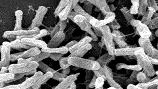
Carbapenem-resistant Enterobacteriaceae (CRE) bacteria (credit: Melissa Brower/CDC)

The death of a hospitalized patient in Reno Nevada for whom no available antibiotics worked highlights what World Health Organization and other public-health experts have been warning: antibiotic resistance is a serious threat and has gone global.

The patient — a female in her 70s — was admitted in for an infection and died in September 2016 from septic shock the CDC announced on Jan. 13. The patient had been treated for multiple infections in India before traveling to the United States. The infection that led to her hospitalization in Reno was caused by a strain of carbapenem-resistant Enterobacteriaceae (CRE)* bacteria known as Klebsiella pneumoniae. Although not all strains of Klebsiella pneumonia are CRE, the strain that infected this patient was resistant to all available antibiotics, according to the CDC. (Carbapeneum is a “drug of last resort.”)

In a paper in The Lancet in October, researchers reported that more than a third of blood infections in newborn babies involving Klebsiella pneumoniae and similar bacteria were resistant to multiple drugs to the point they were virtually untreatable and “threaten the return of a pre-antibiotic era in Indian neonatal intensive care units,” the study’s authors warned.

Spreading more widely and stealthily that thought

Highlighting the importance of this report, a new NIH-funded study published online January 16, 2017 in PNAS (Proceedings of the National Academy of Sciences) from Harvard T.H. Chan School of Public Health and the Broad Institute of MIT and Harvard found that the CRE family of highly drug-resistant and potentially deadly bacteria may be spreading more widely — and more stealthily — than previously thought.

The researchers examined CRE bacteria that were isolated from patients with infections at four U.S. hospitals. They found a wide variety of CRE . They also found a diverse array of genetic traits enabling CRE to resist antibiotics, and found that these traits are transferring easily among various CRE species.

The findings suggest that CRE may well be transmitting from person to person asymptomatically, and that genomic surveillance of this dangerous bacteria should be increased.

CRE are a class of bacteria that is resistant to multiple antibiotics, including carbapenems, which are considered last-resort drugs when other antibiotics have failed. CRE, which tend to spread in hospitals and long-term care facilities, cause an estimated 9,300 infections and 600 deaths in the U.S. each year, according to the CDC — and incidence is on the rise. CDC director Tom Frieden has called these “nightmare bacteria” because they are resistant to some of the last-ditch treatments available to doctors battling resistant infections.

A type of ultraviolet light called UVC could aid hospitals in the ongoing battle to keep drug-resistant bacteria from lingering in patient rooms and causing new infections. A CDC-NIH-funded study led by Duke Health and published in The Lancet finds use of UVC machines can cut transmission of four major superbugs by a cumulative 30 percent. (credit: Shawn Rocco/Duke Health)

Unobserved transmissions

“While the typical focus has been on treating sick patients with CRE-related infections, our new findings suggest that CRE is spreading beyond the obvious cases of disease,” said William Hanage, associate professor of epidemiology at Harvard Chan School and senior author of the study. “We need to look harder for this unobserved transmission within our communities and healthcare facilities if we want to stamp it out.”

The researchers looked at about 250 samples of CRE from hospitalized patients from three Boston-area hospitals and from one California hospital.

The researchers found what Hanage termed a “riot of diversity,” both among CRE species and among carbapenem resistance genes. They also found that resistance genes are moving easily from species to species, contributing to a continually evolving threat from CRE.

In addition, the researchers found that some CRE bacteria employ uncharacterized resistance mechanisms—implying that there are more to be discovered. The finding highlights the need for vigilance in searching for as yet unknown forms of resistance as they evolve and emerge.

What if there were no new antibacterial drugs?

But what’s also needed is new antibiotics. “Meaningful incentives for pharmaceutical companies have been suggested to encourage them to re-enter this area of drug development; these incentives include models to push companies to develop these drugs or by making it economically viable so as to pull companies back to this area as well as alternative routes of funding,” notes Prof Laura J V Piddock, Antimicrobial Agents Research Group, School of Immunity and Infection, College of Medical and Dental Sciences, University of Birmingham in “The crisis of no new antibiotics—what is the way forward?” in Lancet Infect Dis. in 2012.

“Also, few data exist on the human and economic costs of antibacterial resistance or the potential costs of inaction (i.e., what would the social effects be if no new antibacterial drugs were available and fatal infections became the norm?). To assess such costs, involvement of governmental agencies that recommend use of drugs is needed (i.e., FDA, European Medicines Agency, and in individual countries such as UK, The National Institute for Health and Clinical Excellence).”

* “Carbapenems are one of few antimicrobials that have been effective against multidrug-resistant bacteria, but their utility is threatened by the emergence of carbapenem-resistant Enterobacteriaceae (CRE),” according to an open-access PLoS ONE paper published in 2014. “Klebsiella pneumoniae is the most common CRE species in the United States, typically encountered as a hospital-acquired infection with high morbidity and mortality, and resistant to nearly all available antibiotics.” In this case, metallo-β-lactamase NDM-1 (New Delhi metallo-beta-lactamase or NDM), a version of Klebsiella pneumoniae, was found.

Synthetic stem cells offer benefits of natural stem cells without the risks

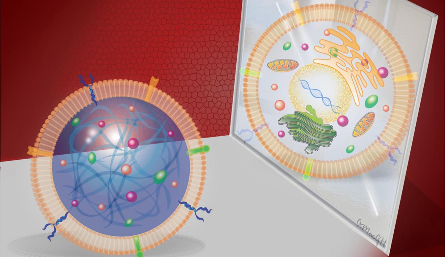
A synthetic cardiac stem cell (left) mirroring a real cardiac stem cell (right), offering therapeutic benefits without the associated risks (credit: Alice Harvey/NC State University)

Scientists have created the first synthetic version of a cardiac stem cell, offering therapeutic benefits comparable to those from natural stem cells — but without the risks and limitations, according to researchers from North Carolina State University, the University of North Carolina at Chapel Hill, and First Affiliated Hospital of Zhengzhou University in China.

The newly created “synthetic stem cells” (not actual stem cells — not even cells — just clever mimics) cannot replicate. That means they could reduce some of the risks associated with natural stem-cell therapies — including tumor growth and immune rejection. The synthetic stem calls would also avoid the fragility of natural stem cells, which require careful storage and a multi-step process of typing and characterization before they can be used.

Simulating cardiac stem cells with synthetic stem cells: a cargo-shell strategy

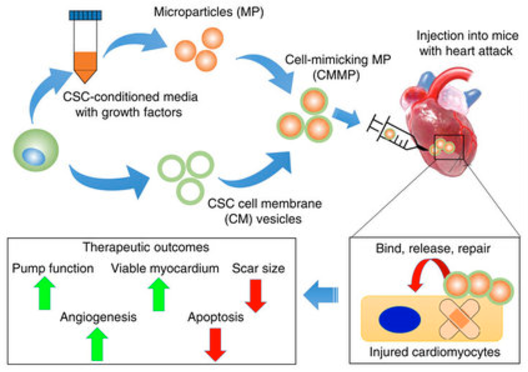
In an in vitro (test tube) experiment described in an open-access paper in Nature Communications, the scientists created synthetic “cell-mimicking microparticles” (CMMPs) and tested them on a mouse model with myocardial infarction.

The scientists found that the CMMPs’ ability to bind to cardiac tissue and promote growth after a heart attack was comparable to that of cardiac stem cells. But unlike cardiac (and other) natural stem cells, the synthetic stem cells have better preservation stability, and the technology is also generalizable to other types of stem cells, according to the researchers.

A cargo-and-shell strategy. The therapeutic potential of CMMPs was successfully tested in tissue repair of a mouse model of myocardial infarction. To create the synthetic stem cell, a microparticle (MP) was first fabricated from PLGA, or poly(lactic-co-glycolic acid) — a biodegradable, biocompatible polymer that serves as a dissolvable carrier. The MP contained conditioned media and growth factors* (the “cargo”) from cardiac stem cells (CSC) in its polymeric core — forming a synthetic cell-mimicking microparticle (CMMP). To prevent an immune reaction, the researchers also cloaked the CMMP with a coating from the cardiac stem-cell membrane (forming the “shell”). (credit: Junnan Tang et al./Nature Communications)

“The synthetic cells operate much the same way a deactivated vaccine works,” explains research-team leader Ke Cheng, associate professor of molecular biomedical sciences at NC State, associate professor in the joint biomedical engineering program at NC State and UNC, and adjunct associate professor at the UNC Eshelman School of Pharmacy. “[The synthetic cells'] membranes allow them to bypass the immune response, bind to cardiac tissue, release the growth factors, and generate repair, but they cannot amplify [and thus risk out-of-control growth, leading to tumors] by themselves. So you get the benefits of stem cell therapy without risks.”

Multiple stem-cell types and organs

The synthetic stem cells are also much more durable than human stem cells, they can tolerate harsh freezing and thawing, and they don’t have to be derived from the patient’s own cells, the researchers note.

“Although our first application targeted the heart, the CMMP strategy represents a platform technology that can be applied to multiple stem cell types and the repair of various organ systems,” the authors state in the paper.

The work was funded in part by the National Institutes of Health, NC State Chancellor’s Innovation Fund, and University of North Carolina General Assembly Research Opportunities Initiative grant.

* Stem-cell therapies work by secreting “paracrine factors,” including proteins and genetic materials, that aid damaged tissue in repairing itself.


Abstract of Therapeutic microparticles functionalized with biomimetic cardiac stem cell membranes and secretome

Stem cell therapy represents a promising strategy in regenerative medicine. However, cells need to be carefully preserved and processed before usage. In addition, cell transplantation carries immunogenicity and/or tumorigenicity risks. Mounting lines of evidences indicate that stem cells exert their beneficial effects mainly through secretion (of regenerative factors) and membrane-based cell-cell interaction with the injured cells. Herein, we fabricated a synthetic cell-mimicking microparticle (CMMP) that recapitulates stem cell functions in tissue repair. CMMPs carries similar secreted proteins and membranes as genuine cardiac stem cells do. In a mouse model of myocardial infarction, injection of CMMPs leads to preservation of viable myocardium and augmentation of cardiac functions similar to cardiac stem cell therapy. CMMPs (derived from human cells) do not stimulate T cells infiltration in immuno-competent mice. In conclusion, CMMPs act as “synthetic stem cells” which mimic the paracrine and biointerfacing activities of natural stem cells in therapeutic cardiac regeneration.

Nanoarray sniffs out and distinguishes ‘breathprints’ of multiple diseases

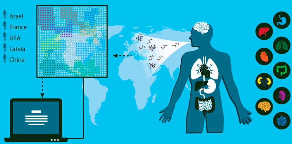
Schematic representation of the concept and design of the study, which involved collection of breath samples from 1404 patients diagnosed with one of 17 different diseases. One breath sample obtained from each subject was analyzed with the artificially intelligent nanoarray for disease diagnosis and classification (represented by patterns in the illustration), and a second sample was analyzed with gas chromatography–mass spectrometry to explore its chemical composition. (credit: Morad K. Nakhleh et al./ACS Nano)

An international team of 63 scientists in 14 clinical departments have identified a unique “breathprint” for 17 diseases with 86% accuracy and have designed a noninvasive, inexpensive, and miniaturized portable device that screens breath samples to classify and diagnose several types of diseases, they report in an open-access paper in the journal ACS Nano.

As far back as around 400 B.C., doctors diagnosed some diseases by smelling a patient’s exhaled breath, which contains nitrogen, carbon dioxide, oxygen, and a small amount of more than 100 other volatile chemical components. Relative amounts of these substances vary depending on the state of a person’s health. For example, diabetes creates a sweet breath smell. More recently, several teams of scientists have developed experimental breath analyzers, but most of these instruments focus on one disease, such as diabetes and melanoma, or a few diseases.

Detecting 17 diseases

The researchers developed an array of nanoscale sensors to detect the individual components in thousands of breath samples collected from 1404 patients who were either healthy or had one of 17 different diseases*, such as kidney cancer or Parkinson’s disease.

The team used mass spectrometry to identify the breath components associated with each disease. By analyzing the results with artificial intelligence techniques (binary classifiers), the team found that each disease produces a unique breathprint, based on differing amounts of 13 volatile organic chemical (VOC) components. They also showed that the presence of one disease would not prevent the detection of others — a prerequisite for developing a practical device to screen and diagnose various diseases.

Based on the research, the team designed an organic layer that functions as a sensing layer (recognition element) for adsorbed VOCs and an electrically conductive nanoarray based on resistive layers of molecularly modified gold nanoparticles and a random network of single-wall carbon nanotubes. The nanoparticles and nanotubes have different electrical conductivity patterns associated with different diseases.**

The authors received funding from the ERC and LCAOS of the European Union’s Seventh Framework Programme for Research and Technological Development, the EuroNanoMed Program under VOLGACORE, and the Latvian Council of Science.

* Lung cancer, colorectal cancer, head and neck cancer, ovarian cancer, bladder cancer, prostate cancer, kidney cancer, gastric cancer, Crohn’s disease, ulcerative colitis, irritable bowel syndrome, idiopathic Parkinson’s, atypical Parkinsonism, multiple sclerosis, pulmonary arterial hypertension, pre-eclampsia, and chronic kidney disease.

** During exposure to breath samples, interaction between the VOC components and the organic sensing layer changes the electrical resistance of the sensors. The relative change of sensor’s resistance at the peak (beginning), middle, and end of the exposure, as well as the area under the curve of the whole measurement were measured. All breath samples identified by the AI nanoarray were also examined using an independent lab-based analytical technique: gas chromatography linked with mass spectrometry.


Abstract of Diagnosis and Classification of 17 Diseases from 1404 Subjects via Pattern Analysis of Exhaled Molecules

We report on an artificially intelligent nanoarray based on molecularly modified gold nanoparticles and a random network of single-walled carbon nanotubes for noninvasive diagnosis and classification of a number of diseases from exhaled breath. The performance of this artificially intelligent nanoarray was clinically assessed on breath samples collected from 1404 subjects having one of 17 different disease conditions included in the study or having no evidence of any disease (healthy controls). Blind experiments showed that 86% accuracy could be achieved with the artificially intelligent nanoarray, allowing both detection and discrimination between the different disease conditions examined. Analysis of the artificially intelligent nanoarray also showed that each disease has its own unique breathprint, and that the presence of one disease would not screen out others. Cluster analysis showed a reasonable classification power of diseases from the same categories. The effect of confounding clinical and environmental factors on the performance of the nanoarray did not significantly alter the obtained results. The diagnosis and classification power of the nanoarray was also validated by an independent analytical technique, i.e., gas chromatography linked with mass spectrometry. This analysis found that 13 exhaled chemical species, called volatile organic compounds, are associated with certain diseases, and the composition of this assembly of volatile organic compounds differs from one disease to another. Overall, these findings could contribute to one of the most important criteria for successful health intervention in the modern era, viz. easy-to-use, inexpensive (affordable), and miniaturized tools that could also be used for personalized screening, diagnosis, and follow-up of a number of diseases, which can clearly be extended by further development.

Cellular reprogramming turns back the aging clock in mice

This cartoon depicts turning back the aging clock through cellular regeneration of progeria mice (credit: Juan Carlos Izpisua Belmonte Lab/Salk Institute)

Salk Institute scientists have extended the average lifespan of live mice by 30 percent, according to a study published December 15 in Cell. They did that by rolling back the “aging clock” to younger years, using cellular reprogramming.

The finding suggests that aging is reversible by winding back an animal’s biological clock to a more youthful state and that lifespan can be extended. While the research does not yet apply directly to humans, it promises to lead to improved understanding of human aging and the possibility of rejuvenating human tissues.

To achieve this, the scientists worked with “progeria” mouse models — mice that had been genetically modified to carry a mutation that leads to premature aging (allowing the aging effects to be isolated and studied).*

Rather than attempting to correct the genetic mutations that cause premature aging (a difficult challenge), the Salk team instead focused on restoring the epigenome (a system of chemical marks on the genome that control which genes are or are not expressed as proteins)**.

Partial reprogramming avoids tumors and aging effects

To wind back the clock, the scientists first reprogrammed progeria mouse adult cells (such as skin cells) into an induced pluripotent stem (iPS) cells*** — an early embryonic state in which the cell is not yet specialized (“differentiated”) and can be programmed to perform specific functions, such as becoming part of the skin or heart.

To generate iPS cells, fibroblasts (or another type of adult somatic cell) are transduced with retroviruses encoding four pluripotency factors (SOX2, KLF4, c-MYC and OCT4). These differentiated cells can be used in disease models (such as progeria) for studying the molecular basis of a broad range of human diseases that are otherwise difficult to study. (credit: Yamanaka S. et al./Nature)

The scientists did that by inducing (modifying) the expression of four specific “epigenetic marks” called “Yamanaka factors” in the iPS cells, with the goal of allowing the cells to grow into adult cells without defects.

Researchers have previously done that successfully with cells in vitro (in a test tube), allowing these factors to be expressed for the needed 2 to 3 weeks for cells. But when they tried that in vivo (in live mice), it resulted in mice that later either died immediately or developed extensive tumors.

Partial cellular reprogramming with the four Yamanaka factors (Oct4, Sox2, Klf4, c-Myc) erases cellular markers of aging in progeria mouse cells (credit: Alejandro Ocampo et al./Cell)

So instead, the Salk team used partial in vivo cellular reprogramming. They induced expression of Yamanaka factors for just 2 to 4 days instead of 2 to 3 weeks. Instead of resulting in tumors or death, that prolonged the mice lifespan. An in vitro experiment also showed reduced DNA damage accumulation and restored nuclear structure.

This image depicts the discovery by Salk Institute researchers that partial cellular reprogramming reversed cellular signs of aging such as accumulation of DNA damage. (Left) Progeria mouse fibroblast cells; (Right) Progeria mouse fibroblast cells rejuvinated by partial reprogramming. (credit: Juan Carlos Izpisua Belmonte Lab/Salk Institute)

“These changes are the result of epigenetic remodeling in the cell,” notes lead investigator Juan Carlos Izpisua Belmonte, a professor in the Salk Institute of Biological Science’s Gene Expression Laboratory.

Several organs improved. For instance, tissue from skin, spleen, kidney and stomach all had improved appearance when inspected under the microscope. The cardiovascular system, which often fails and causes early death in these prematurely aging mice, also showed improvements in structure and function.

This image shows the discovery by researchers that induction of partial cellular reprogramming improved muscle regeneration in aged mice. (Left) Impaired muscle repair in aged mice; (Right) Improved muscle regeneration in aged mice subjected to reprogramming. (credit: Juan Carlos Izpisua Belmonte Lab/Salk Institute)

The team also tested partial reprogramming in mouse models that had injury, resulting in enhanced regeneration of muscle tissue and pancreas beta cells (which store and release insulin).

Next steps will involve learning more about how the epigenome changes during partial reprogramming. “We need to go back and explore which marks are changing and driving the aging process,” says Belmonte.

This study was supported by the G. Harold and Leila Y. Mathers Charitable Foundation, The Leona M. and Harry B. Helmsley Charitable Trust, The Glenn Foundation, Universidad Católica San Antonio de Murcia (UCAM), and Fundación Dr. Pedro Guillen.

* Mice do not perfectly mimic human aging. Researchers generate and use mouse models of premature aging by introducing mutations responsible for these human diseases in mouse.

** Epigenetic marks in the DNA regulate and protect the genome. Some marks turn on specialized functions, such as skin cell machinery in a skin cell; others turn off mechanisms that aren’t needed, such as liver cell machinery in the skin. They are modified over a lifetime in response to environmental changes, and have been proposed as drivers of aging.

*** Induced pluripotent stem cells (iPS cells or iPSCs) are a type of pluripotent stem cell that can be generated directly from adult cells. The iPSC technology was pioneered by Shinya Yamanaka’s lab in Kyoto, Japan, who showed in 2006 that the introduction of four specific genes encoding transcription factors could convert adult cells into pluripotent stem cells. He was awarded the 2012 Nobel Prize in Physiology or Medicine with Sir John Gurdon.


Salk Institute | Salk scientists reverse signs of aging in mice


Abstract of In Vivo Amelioration of Age-Associated Hallmarks by Partial Reprogramming

Aging is the major risk factor for many human diseases. In vitro studies have demonstrated that cellular reprogramming to pluripotency reverses cellular age, but alteration of the aging process through reprogramming has not been directly demonstrated in vivo. Here, we report that partial reprogramming by short-term cyclic expression of Oct4Sox2Klf4, and c-Myc (OSKM) ameliorates cellular and physiological hallmarks of aging and prolongs lifespan in a mouse model of premature aging. Similarly, expression of OSKM in vivo improves recovery from metabolic disease and muscle injury in older wild-type mice. The amelioration of age-associated phenotypes by epigenetic remodeling during cellular reprogramming highlights the role of epigenetic dysregulation as a driver of mammalian aging. Establishing in vivo platforms to modulate age-associated epigenetic marks may provide further insights into the biology of aging.

Electric current and antiobiotic kill multidrug-resistant bacteria in biofilms

Washington State University researchers have successfully used a mild electric current to kill drug-resistant bacterial infections, a technology that may eventually be used to treat chronic wound infections. Red arrow indicates cell showing a stressed membrane. (credit: Washington State University)

A Washington State University research team has successfully used a mild electric current combined with an antibiotic to kill multidrug-resistant pseudomonas aeruginosa PAO1 bacteria in a lab-cultured biofilm.

These bacteria are responsible for chronic and serious infections in people with lung diseases, such as cystic fibrosis, and in chronic wounds. They also often cause pneumonia for people who are on ventilators and infections in burn victims.*

Schematic of experimental setup for the treatment of biofilm exposed to an e-scaffold, with an illustration of electrochemical production of hydrogen peroxide. The electrodes are connected to a potentiostat (600 mV source; not shown in figure). (credit: Sujala T. Sultana et al./Scientific Reports)

The researchers used an “e-scaffold” (electrochemical structure), a sort of electronic band-aid made out of conductive carbon fabric, which uses an electrical current to produce a low, constant concentration of hydrogen peroxide, an effective disinfectant, at the e-scaffold surface.

The hydrogen peroxide disrupts the biofilm (slime layer) matrix and damages the bacteria cell walls and DNA, which then allows better antibiotic penetration and efficacy against the bacteria. Bacteria that form biofilms are more difficult to kill because antibiotics only partially penetrate this protective layer. Subpopulations of “persister” cells survive treatment and are able to grow and multiply, resulting in chronic infections.

Tuning electrical stimulation to kill bacteria

Researchers have previously tried electrical stimulation, a method used to kill bacteria, for more than a century, but with limited results. In this study, the researchers determined the conditions necessary for the electrochemical reaction to produce hydrogen peroxide. The current has to be carefully controlled to assure the correct reaction at an exact rate to stop the bacteria from developing resistance, while not damaging surrounding tissue.**

The research to develop the e-scaffold actually came out of a failed attempt to improve fuel cells, according to research team leader Haluk Beyenal, the Paul Hohenschuh Distinguished Professor in WSU’s Gene and Linda Voiland School of Chemical Engineering and Bioengineering. When the researchers figured out they could only produce a small amount of electric current for their fuel cell cathode, they decided to see if they could use the process for a different purpose.

The technology may eventually be used to treat chronic wound infections, the researchers say. The researchers have filed a patent application and are working to commercialize the process. They hope to begin conducting clinical tests.

The work, reported in the online edition of npj Biofilms and Microbiomes, was supported by Beyenal’s National Science Foundation CAREER award.

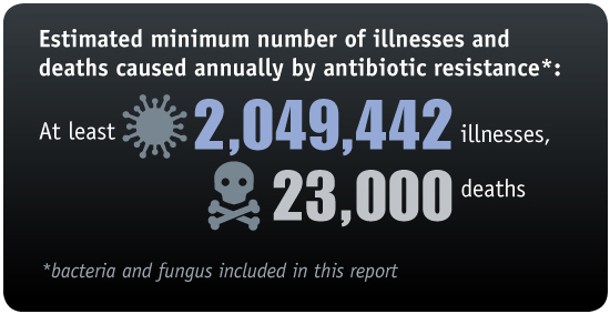
* Bacterial resistance is a growing problem around the world. While antibiotics were a miracle drug of the 20th century, their widespread use has led to drug-resistant strains of bacteria. In the U.S., at least 2 million infections and 23,000 deaths are attributable to antibiotic-resistant bacteria each year, according to the U.S. Centers for Disease Control.

** As the researchers note in the paper, alternative antimicrobial treatments such as silver or mannitol, or in combination with conventional antibiotics, have toxic side effects at adequately high concentrations, and at low concentrations they often decompose before completely eliminating biofilm communities. In addition, persister cells can regrow and form biofilms with potentially enhanced tolerance to antibiotics.


Abstract of Eradication of Pseudomonas aeruginosa biofilms and persister cells using an electrochemical scaffold and enhanced antibiotic susceptibility

Biofilms in chronic wounds are known to contain a persister subpopulation that exhibits enhanced multidrug tolerance and can quickly rebound after therapeutic treatment. The presence of these “persister cells” is partly responsible for the failure of antibiotic therapies and incomplete elimination of biofilms. Electrochemical methods combined with antibiotics have been suggested as an effective alternative for biofilm and persister cell elimination, yet the mechanism of action for improved antibiotic efficacy remains unclear. In this work, an electrochemical scaffold (e-scaffold) that electrochemically generates a constant concentration of H2O2 was investigated as a means of enhancing tobramycin susceptibility in pre-grown Pseudomonas aeruginosa PAO1 biofilms and attacking persister cells. Results showed that the e-scaffold enhanced tobramycin susceptibility in P. aeruginosa PAO1 biofilms, which reached a maximum susceptibility at 40 µg/ml tobramycin, with complete elimination (7.8-log reduction vs control biofilm cells, P ≤ 0.001). Moreover, the e-scaffold eradicated persister cells in biofilms, leaving no viable cells (5-log reduction vs control persister cells, P ≤ 0.001). It was observed that the e-scaffold induced the intracellular formation of hydroxyl free radicals and improved membrane permeability in e-scaffold treated biofilm cells, which possibly enhanced antibiotic susceptibility and eradicated persister cells. These results demonstrate a promising advantage of the e-scaffold in the treatment of persistent biofilm infections.

Caltech scientists use bacterial protein to merge silicon and carbon and create new organosilicon compounds

Artist rendering of organosilicon-based life (credit: Lei Chen and Yan Liang (BeautyOfScience.com) for Caltech)

Scientists at Caltech have “bred” a bacterial protein with the ability to make silicon-carbon bonds, with applications in several industries — something only chemists could do before. The research was published in the Nov. 24 issue of the journal Science.

Molecules with silicon-carbon (organosilicon) compounds are found in pharmaceuticals and many other products, including agricultural chemicals, paints, semiconductors, and computer and TV screens. Currently, these products are made synthetically, since silicon-carbon bonds are not found in nature.

The new research demonstrates that biology can be used to manufacture these bonds in ways that are more environmentally friendly and potentially much less expensive, according to the researchers.


Caltech | Bringing Silicon to Life: Scientists Persuade Nature to Make Silicon-Carbon Bonds

Directed evolution

The key to this research involves deliberate messing with nature: a method called directed evolution* pioneered in the early 1990s by Frances Arnold, Caltech’s Dick and Barbara Dickinson Professor of Chemical Engineering, Bioengineering and Biochemistry, and principal investigator of this project.

An example of directed evolution with comparison to natural evolution. The inner cycle indicates the 3 stages of the directed evolution cycle with the natural process being mimicked in parentheses. The outer circle demonstrates steps a typical experiment. The red symbols indicate functional variants, the pale symbols indicate variants with reduced function. (credit: Thomas Shafee CC)

Directed evolution has been used for years to make enzymes for household products, like detergents; and for “green” sustainable routes to making pharmaceuticals, agricultural chemicals, and fuels.

In directed evolution, new and better enzymes are created in labs by artificial selection, similar to the way that breeders modify corn, cows, or cats. Enzymes are a class of proteins that catalyze, or facilitate, chemical reactions. The directed evolution process begins with an enzyme that scientists want to enhance. The DNA coding for the enzyme is mutated in more-or-less random ways, and the resulting enzymes are tested for a desired trait. The top-performing enzyme is then mutated again, and the process is repeated until an enzyme that performs much better than the original is created.

Going where no enzyme has gone before

In the new study, the goal was not just to improve an enzyme’s biological function but to actually persuade it to do something that it had not done before. The researchers’ first step was to find a suitable candidate, an enzyme showing potential for making the silicon-carbon bonds.

“It’s like breeding a racehorse,” says Arnold, who is also the director of the Donna and Benjamin M. Rosen Bioengineering Center at Caltech. “A good breeder recognizes the inherent ability of a horse to become a racer and has to bring that out in successive generations. We just do it with proteins.”

An Icelandic hot spring (credit: Nordic Visitor)

The ideal candidate turned out to be a protein from a bacterium, Rhodothermus marinus, that grows in hot springs in Iceland. That protein, called cytochrome c, normally shuttles electrons to other proteins, but the researchers found that it also happens to act like an enzyme to create silicon-carbon bonds at low levels. The scientists then mutated the DNA coding for that protein within a region that specifies an iron-containing portion of the protein thought to be responsible for its silicon-carbon bond-forming activity. Next, they tested these mutant enzymes for their ability to make organosilicon compounds better than the original.

cytochrome c (credit: Caltech)

After only three rounds, they had created an enzyme that can selectively make silicon-carbon bonds 15 times more efficiently than the best catalyst invented by chemists. Furthermore, the enzyme is highly selective, which means that it makes fewer unwanted byproducts that have to be chemically separated out.

“This iron-based, genetically encoded catalyst is nontoxic, cheaper, and easier to modify compared to other catalysts used in chemical synthesis,” says Jennifer Kan, a postdoctoral scholar in Arnold’s lab and lead author of the new study. “The new reaction can also be done at room temperature and in water.”

The synthetic process for making silicon-carbon bonds often uses precious metals and toxic solvents, and requires extra processing to remove unwanted byproducts, all of which add to the cost of making these compounds.

Could life on Earth (or elsewhere) have evolved based on silicon-carbon?

The study is the first to show that nature can adapt to incorporate silicon into carbon-based molecules, the building blocks of life.

Carbon and silicon are chemically very similar, and silicon is the second most abundant element in Earth’s crust. They can both form bonds to four atoms simultaneously, making them well suited to form the long chains of molecules found in life, such as proteins and DNA. Science-fiction authors have imagined alien worlds with silicon-based life, like the lumpy Horta creatures portrayed in an episode of the 1960s TV series Star Trek.

“This study shows how quickly nature can adapt to new challenges,” says Arnold. “The DNA-encoded catalytic machinery of the cell can rapidly learn to promote new chemical reactions when we provide new reagents and the appropriate incentive in the form of artificial selection.”

However, no living organism is known [yet] to put silicon-carbon bonds together, even though silicon is so abundant, all around us, in rocks and all over the beach,” says Kan.

What about other planets (Mars has both silicon and carbon, for example) and asteroids? And could alien life have evolved silicon-carbon semiconductor brains? It would also be interesting to see if such a lifeform could be invented on Earth.

This research is funded by the National Science Foundation, the Caltech Innovation Initiative program, and the Jacobs Institute for Molecular Engineering for Medicine at Caltech.

* Not to be confused with a transhumanist concept for controlling human evolution.


Abstract of Directed evolution of cytochrome c for carbon–silicon bond formation: Bringing silicon to life

Enzymes that catalyze carbon–silicon bond formation are unknown in nature, despite the natural abundance of both elements. Such enzymes would expand the catalytic repertoire of biology, enabling living systems to access chemical space previously only open to synthetic chemistry. We have discovered that heme proteins catalyze the formation of organosilicon compounds under physiological conditions via carbene insertion into silicon–hydrogen bonds. The reaction proceeds both in vitro and in vivo, accommodating a broad range of substrates with high chemo- and enantioselectivity. Using directed evolution, we enhanced the catalytic function of cytochrome c from Rhodothermus marinus to achieve more than 15-fold higher turnover than state-of-the-art synthetic catalysts. This carbon–silicon bond-forming biocatalyst offers an environmentally friendly and highly efficient route to producing enantiopure organosilicon molecules.

Overuse and misuse of antibiotics leading to rise of resistant strains of pathogenic bacteria

Prolonged preventive antibiotics after surgical incision closure do not prevent infections, but they do change the composition of bacteria in the host at other anatomic locations. The result is resistant colonization of the patient and potentially an infection with Clostridium difficile bacteria, shown here. (credit: CDC)

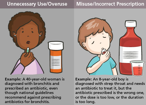
Overuse and misuse of antimicrobial agents in hospitals is an urgent problem. Surgeons around the world, who often prescribe antibiotics for surgical prophylaxis, need to take a leadership role in promoting “antimicrobial stewardship programs” (ASPs) that can optimize antimicrobial agent use in the hospital.

That’s the message from the Surgical Infection Society and the World Society of Emergency Surgery in an open-access paper entitled “Antimicrobial Stewardship: A  Call to Action for Surgeons,” published in Surgical Infections, a peer-reviewed journal published today (Tuesday, Nov. 15) online ahead of print by Mary Ann Liebert, Inc.*

The authors emphasize the urgent need to standardize the use of antimicrobial agents. Approximately 15% of antibiotic prescribing in hospitals takes place prior to surgery to prevent infection, they note.

(credit: CDC)

The paper is timed to coincide with the CDC’s “Get Smart About Antibiotics Week,” November 14–20, which aims to improve the antibiotic prescribing habits of clinicians to help combat the continuing rise of resistant strains of pathogenic bacteria.

“In addition to emerging resistance, the adverse events of decades of antibiotic use may have led to a number of other adverse consequences,” warns Surgical Infections Editor-in-Chief Donald E. Fry, MD, Northwestern University Feinberg School of Medicine, in an associated open-access editorial, “The Unintended Consequences of Antibiotic Use.”

(credit: CDC)

“The lesson of giving the preventive antibiotics before incision and subsequent contamination has been learned, but the lesson of discontinuation of the antibiotic after the incision has been closed continues to be problematic,” he notes.

“Prolonged preventive antibiotics after incision closure do not prevent infections, but they do change the composition of bacteria in the host at other anatomic locations. The result is resistant colonization of the patient and potentially Clostridium difficile infection. The aggregate effect of prolonged and unnecessary preventive antibiotics is more resistant pathogens in our hospitals.”


Risks of developing antibiotic resistance vs. prevention

A recent KurzweilAI news article reports that an international research team has developed a new antimicrobial peptide, clavanin-MO, that kills strains resistant to existing antibiotics. The researchers advised that the peptide could also be embedded in surfaces such as tabletops to make them resistant to microbial growth, as antimicrobial coatings for catheters, and in ointments to treat skin infections. Two KurzweilAI readers (“tedhowardnz” and “DevilDocNowCiv”) questioned this advice, suggesting that we need to minimize exposure to prevent antibiotic resistance.

One of the senior authors of the associated paper, César de la Fuente Nunez, has replied: “I agree exposure should be limited and regulated. Over-exposure and misuse of conventional antibiotics is one of the main reason bacteria have developed resistance to these drugs. However, it has recently been estimated that if we don’t develop novel therapies, drug-resistant infections will kill 10 million people annually, which corresponds to 1 death every 3 seconds. [We] need to come up with alternative strategies to treat these infections [that have] driven much of our research, including this study.

“Interestingly, work by others has shown that peptides, similar to the ones we have described here, do not readily select for resistance, particularly when compared to conventional antibiotics. We believe this is due to their multifunctional mechanism of action (i.e., they have multiple targets). We envision these peptides being used in adjuvant therapies with antibiotics, as they can potentiate antibiotic action, therefore reducing levels of both peptide and antibiotics required for effective treatment. This approach will help further reduce selective pressure towards the development of resistance.

“Hospital procedures currently sterilize surfaces using ethanol, etc. We think these peptides could be useful, for instance, to coat catheters to prevent microbial colonization. Catheters are often infected, meaning the patient has to undergo surgery to replace the catheter, with all the costs that are associated with this procedure. We think peptides may help prevent some of these infections.”


In related news …

  • A multi-drug resistant mycobacteria lung infection (Mycobacterium abscessus) that can cause life-threatening illness in people with cystic fibrosis (CF) and can spread from patient to patient has now spread globally and is becoming increasingly virulent, according to new research published November 11 in the journal Science. The mystery: how does the pathogen manage to spread globally? The researchers speculate that healthy individuals may be unwittingly carrying the mycobacteria between countries, possibly via fomites (carriers, such as a dish or an article of clothing) and aerosols (particles dispersed in air or a gas).
  • A previously unnoticed global outbreak of Mycobacterium chimaera, an invasive, slow-growing bacterium, is linked to heater-cooler devices (HCD) used in cardiac surgery, according to a study published Nov. 14 in Infection Control & Hospital Epidemiology. HCDs are stand-alone devices needed for heat exchange in heart-lung machines used in some 250,000 surgeries annually in the U.S., according to the CDC.
  • Symptom-free Ebola infections have been found by researchers in 14 individuals in Sierra Leone, West Africa, who showed no signs of Ebola but have evidence of prior Ebola infection in their immune systems. (This open-access link to a PLOS Neglected Tropical Diseases paper goes live upon publication.)

* Coauthored by Senior author John E. Mazuski, MD; Massimo Sartelli, Macerata Hospital (Italy); and colleagues from John Peter Smith Health Network (Fort Worth, TX), Maggiore Hospital (Parma, Italy), Papa XXIII (Bergamo, Italy), University of Texas Health Science Center (Houston), UNIVPM (Ancona, Italy), Vanderbilt University Medical Center (Nashville, TN), and Washington University School of Medicine (St. Louis, MO). Mazuski has received research support from Astra-Zeneca, Bayer, and Merck, and honoraria as an advisory board member, consultant, or speaker from Allergan, Astra-Zeneca, Bayer, and Merck. Mazuski also receives grant support from the National Institutes of Health for collaborative work on an infection control research project. He serves as the President of the Surgical Infection Society without compensation. For the remaining authors, no competing financial interests exist.


Abstract of Antimicrobial Stewardship: A Call to Action for Surgeons

Despite current antimicrobial stewardship programs (ASPs) being advocated by infectious disease specialists
and discussed by national and international policy makers, ASPs coverage remains limited to only certain hospitals
as well as specific service lines within hospitals. The ASPs incorporate a variety of strategies to optimize
antimicrobial agent use in the hospital, yet the exact set of interventions essential to ASP success remains
unknown. Promotion of ASPs across clinical practice is crucial to their success to ensure standardization of
antimicrobial agent use within an institution. To effectively accomplish this standardization, providers who
actively engage in antimicrobial agent prescribing should participate in the establishment and support of these programs. Hence, surgeons need to play a major role in these collaborations. Surgeons must be aware that
judicious antibiotic utilization is an integral part of any stewardship program and necessary to maximize clinical
cure and minimize emergence of antimicrobial resistance. The battle against antibiotic resistance should be fought
by all healthcare professionals. If surgeons around the world participate in this global fight and demonstrate
awareness of the major problem of antimicrobial resistance, they will be pivotal leaders. If surgeons fail to actively engage and use antibiotics judiciously, they will find themselves deprived of the autonomy to treat their patients.

New antimicrobial peptide kills strains resistant to existing antibiotics

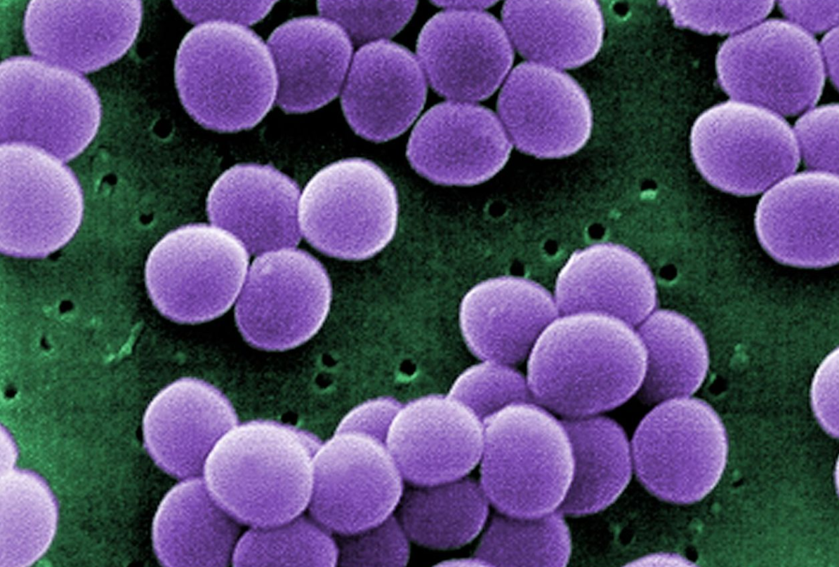
Scanning electron micrograph (SEM) showing a strain of Staphylococcus aureus bacteria taken from a vancomycin intermediate resistant culture (credit: CDC)

A team of researchers at MIT, the University of Brasilia, and the University of British Columbia has engineered an antimicrobial peptide to wipe out many types of bacteria, including some that are resistant to most antibiotics.

A recent study from a U.K. commission on antimicrobial resistance estimated that by 2050, antibiotic-resistant bacterial infections will kill 10 million people per year if no new drugs are developed.

Learning from nature

Living organisms naturally make antimicrobial peptides, as part of their immune system, to kill bacteria, as well as other persistent microbes such as viruses and fungi. It’s done by poking holes in the invaders’ cell membranes. Once inside, they can disrupt several cellular targets, including DNA, RNA, and proteins.

What also sets antimicrobial peptides apart from traditional antibiotics is their ability to recruit the host’s immune system, summoning cells called leukocytes that secrete chemicals that help kill the invading microbes.

In this new study, which appears in the Nov. 2 issue of Nature’s (open-access) Scientific Reports, the researchers began with a naturally occurring antimicrobial peptide called clavanin-A, which has proven deadly to many bacterial strains. The researchers decided to try to engineer it to make it even more effective.*

Killing resistant strains of E. coli and Staph

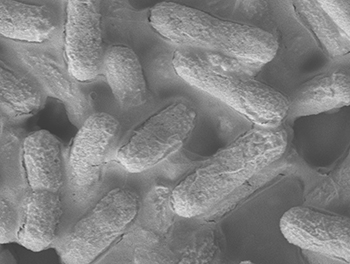
A microscopic image of E. coli bacteria (credit: A*STAR Institute of Bioengineering and Nanotechnology)

The researchers found in tests with mice that this new peptide, which they called clavanin-MO, could kill strains of Escherichia coli and Staphylococcus aureus that are resistant to most antibiotics.

The researchers also found that the clavanin-MO peptide suppresses the overactive inflammatory response that can cause sepsis, a life threatening condition, and can destroy certain biofilms (persistent thin layers of bacterial cells that form on surfaces). That raises the possibility of using them to treat infections caused by biofilms.**

The clavanin-MO peptide could also be embedded in surfaces such as tabletops to make them resistant to microbial growth, as antimicrobial coatings for catheters, and in ointments to treat skin infections caused by Staphylococcus aureus or other bacteria.

The researchers are now investigating what makes these engineered peptides more effective than the naturally occurring ones, with hopes of further improving them.

* Antimicrobial peptides have a positively charged region that allows them to poke through bacterial cell membranes, and a hydrophobic stretch that enables interaction with and translocation into membranes. The researchers decided to add a sequence of five amino acids that would make the peptides even more hydrophobic, in hopes that it would improve their killing ability.

** Such as the Pseudomonas aeruginosa infections that often affect the lungs of cystic fibrosis patients.


Abstract of An anti-infective synthetic peptide with dual antimicrobial and immunomodulatory activities

Antibiotic-resistant infections are predicted to kill 10 million people per year by 2050, costing the global economy $100 trillion. Therefore, there is an urgent need to develop alternative technologies. We have engineered a synthetic peptide called clavanin-MO, derived from a marine tunicate antimicrobial peptide, which exhibits potent antimicrobial and immunomodulatory properties both in vitro and in vivo. The peptide effectively killed a panel of representative bacterial strains, including multidrug-resistant hospital isolates. Antimicrobial activity of the peptide was demonstrated in animal models, reducing bacterial counts by six orders of magnitude, and contributing to infection clearance. In addition, clavanin-MO was capable of modulating innate immunity by stimulating leukocyte recruitment to the site of infection, and production of immune mediators GM-CSF, IFN-γ and MCP-1, while suppressing an excessive and potentially harmful inflammatory response by increasing synthesis of anti-inflammatory cytokines such as IL-10 and repressing the levels of pro-inflammatory cytokines IL-12 and TNF-α. Finally, treatment with the peptide protected mice against otherwise lethal infections caused by both Gram-negative and -positive drug-resistant strains. The peptide presented here directly kills bacteria and further helps resolve infections through its immune modulatory properties. Peptide anti-infective therapeutics with combined antimicrobial and immunomodulatory properties represent a new approach to treat antibiotic-resistant infections.