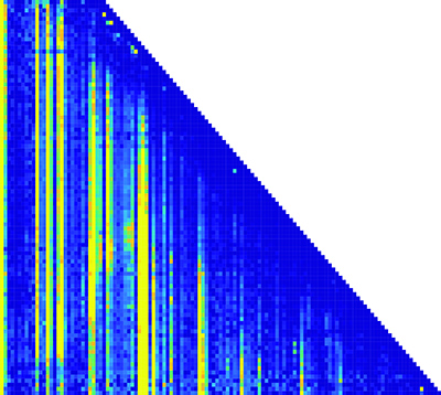
A super-high-resolution snapshot of RNA folding

This matrix of the RNA folding pathway shows how the transcription length and nucleotide positions change over time. (Nucleotide position is along the x-axis; transcription length is along the y-axis.) Each pixel in the matrix is a piece of information about the structure of the RNA molecules. (credit: Northwestern University)

Northwestern University engineers have invented a tool to make a super-high-resolution representation of RNA folding as it is being synthesized. It could potentially lead to future discoveries in basic biology, gene expression, RNA viruses, and disease.

Made up of long chains of nucleotides, RNA is responsible for many tasks in the cellular environment, including making proteins, transporting amino acids, gene expression, and carrying messages between DNA and ribosomes. To accomplish all these tasks, RNA folds into complex structures — “one of the biggest, most essential pieces of biology that we know comparatively nothing about,” said Julius B. Lucks, Ph.D, an associate professor of chemical and biological engineering at Northwestern’s McCormick School of Engineering.

RNA folding is an essential requirement to life, but is difficult to investigate because the process occurs rapidly and is extremely hard to measure. Existing technology to image RNA folding is very-low-resolution and can’t image RNA’s individual components rapidly enough to capture these processes.

A molecular microscope

Instead, Lucks’s technology combines two existing components: a next-generation sequencing technique, which is typically used for sequencing human genomes, and a chemistry technique to turn RNA structure measurements into big data.

“Instead of treating it like a genome sequencer, we’re treating it like a molecular microscope to get a massive snapshot,” Lucks said. The technique captures the RNA-folding pathway in a massive dataset. Lucks’s group then uses computational tools to mine and organize the data, which reveals points where the RNA folds and what happens after it folds.

From the structural information that they gather, the researchers can reconstruct a movie of the RNA folding process. The team plans to make the data-analysis component open source, so researchers anywhere can download and run the program.*

Lucks and his team have already used the technology to view the folding of a riboswitch, a segment of RNA that acts as a genetic “light switch” to turn protein expression on or off in response to a molecular signal, in this case fluoride.

Supported by the National Institutes of Health, the research was published online on October 31 in Nature Structural and Molecular Biology.

* The researchers say the technology can be applied to all questions pertaining to RNA, such as how it plays a role in disease. Several notable human diseases, including Ebola, hepatitis C, and measles, are caused by RNA viruses, which are viruses that have RNA as their genetic material. And just as misfolded proteins can cause diseases, such as Alzheimer’s and Parkinson’s, RNA misfolding could also play a role in human illnesses.


Abstract of Cotranscriptional folding of a riboswitch at nucleotide resolution

RNAs can begin to fold immediately as they emerge from RNA polymerase. During cotranscriptional folding, interactions between nascent RNAs and ligands are able to direct the formation of alternative RNA structures, a feature exploited by noncoding RNAs called riboswitches to make gene-regulatory decisions. Despite their importance, cotranscriptional folding pathways have yet to be uncovered with sufficient resolution to reveal how cotranscriptional folding governs RNA structure and function. To access cotranscriptional folding at nucleotide resolution, we extended selective 2′-hydroxyl acylation analyzed by primer-extension sequencing (SHAPE-seq) to measure structural information of nascent RNAs during transcription. Using cotranscriptional SHAPE-seq, we determined how the cotranscriptional folding pathway of the Bacillus cereus crcB fluoride riboswitch undergoes a ligand-dependent bifurcation that delays or promotes terminator formation via a series of coordinated structural transitions. Our results directly link cotranscriptional RNA folding to a genetic decision and establish a framework for cotranscriptional analysis of RNA structure at nucleotide resolution.

‘Nanobionic’ spinach plants detect explosives, pollution, drought

By embedding spinach leaves with carbon nanotubes, MIT engineers have transformed spinach plants into sensors that can detect explosives and wirelessly relay that information to a handheld device similar to a smartphone. (credit: Christine Daniloff/MIT)

MIT engineers have implanted spinach leaves with carbon nanotubes, resulting in a hybrid electronic system that they call “plant nanobionics” for detecting dangerous (and other) chemicals.

Two years ago, in the first demonstration of plant nanobionics, MIT engineer Michael Strano, PhD, used nanoparticles to enhance plants’ photosynthesis ability and turn them into sensors for nitric oxide, a pollutant produced by combustion.

Detecting trace molecules

In the new study, the researchers embedded the carbon-nanotube sensors for nitroaromatic compounds into the leaves of spinach plants. The plant can detect minute samples of explosives that leech into the groundwater. Carbon nanotubes can also be used as sensors to detect a wide range of molecules, including hydrogen peroxide, the explosive TNT, and the nerve gas sarin.

To read the signal, the researchers shine a laser onto the leaf, prompting the carbon nanotubes in the leaf to emit near-infrared fluorescent light. This can be detected with a small infrared camera connected to a Raspberry Pi, a $35 credit-card-sized computer, which then alerts the user with an email. The fluorescent signal could also be detected with a smartphone by removing the near-infrared filter that most camera phones have, the researchers say.

“You can apply these techniques with any living plant,” says Strano, leader of the MIT research team and the senior author of a paper describing the nanobionic plants in the Oct. 31 issue of Nature Materials. That opens the door to novel ways for plants to pick up signals that tell of environmental pollution, and even drought.

Plants “know that there is going to be a drought long before we do,” he says. “They can detect small changes in the properties of soil and water potential. If we tap into those chemical signaling pathways, there is a wealth of information to access.”


MIT | Plant-to-human communication


Abstract of Nitroaromatic detection and infrared communication from wild-type plants using plant nanobionics

Plant nanobionics aims to embed non-native functions to plants by interfacing them with specifically designed nanoparticles. Here, we demonstrate that living spinach plants (Spinacia oleracea) can be engineered to serve as self-powered pre-concentrators and autosamplers of analytes in ambient groundwater and as infrared communication platforms that can send information to a smartphone. The plants employ a pair of near-infrared fluorescent nanosensors—single-walled carbon nanotubes (SWCNTs) conjugated to the peptide Bombolitin II to recognize nitroaromatics via infrared fluorescent emission, and polyvinyl-alcohol functionalized SWCNTs that act as an invariant reference signal—embedded within the plant leaf mesophyll. As contaminant nitroaromatics are transported up the roots and stem into leaf tissues, they accumulate in the mesophyll, resulting in relative changes in emission intensity. The real-time monitoring of embedded SWCNT sensors also allows residence times in the roots, stems and leaves to be estimated, calculated to be 8.3 min (combined residence times of root and stem) and 1.9 min mm−1leaf, respectively. These results demonstrate the ability of living, wild-type plants to function as chemical monitors of groundwater and communication devices to external electronics at standoff distances.

Will we kill (or contaminate) microbial life on Mars?

These dark, narrow, 100 meter-long streaks called recurring slope lineae flowing downhill on Mars are inferred to have been formed by contemporary flowing water. Planetary scientists detected hydrated salts on these slopes at Hale crater, corroborating their original hypothesis that the streaks are indeed formed by liquid water. The blue color seen upslope of the dark streaks are thought not to be related to their formation, but instead are from the presence of the mineral pyroxene. (credits: NASA/JPL/University of Arizona)

Recent evidence of water, complex organic molecules, and methane in the Martian environment, combined with findings from the 1976 Viking mission, have led to the conclusion that existing microbial life on Mars is a possibility that must be considered, according to the authors of a paper in the journal Astrobiology (open-access until November 15, 2016).

Coauthors Gilbert V. Levin, Arizona State University, Tempe, and Patricia Ann Straat, National Institutes of Health, Bethesda, MD (retired), outline the evidence to support the “biological hypothesis,” which argues that the results of the 1976 Viking Labeled Release experiment were positive for extant microbial life on the surface of Mars.

The authors also evaluate the “non-biological hypotheses” to explain the Viking results, which many scientists support. But the authors conclude that the experimental evidence supports a biological explanation and the likelihood that microorganisms were able to evolve and adapt to be able to survive in the harsh conditions of the Martian environment.

“Even if one is not convinced that the Viking LR results give strong evidence for life on Mars, this paper clearly shows that the possibility must be considered,” says Chris McKay, PhD, Senior Editor of Astrobiology and an astrobiologist with NASA Ames Research Center.

“We cannot rule out the biological explanation,” McKay said. “This has implications for plans for sample return from Mars and for future human missions.”

Probing the Mars atmosphere for signs of life

Artist’s impression of ExoMars 2016 Trace Gas Orbiter (TGO)and separation of Schiaparelli, the entry, descent and landing demonstrator module. (credit: ESA)

ExoMars, a joint mission between the European Space Agency and Roscosmos, the Russian space agency, with the goal of exploring “whether life has ever existed on Mars,” entered Mars orbit Oct. 19. On Oct. 20, after separation, Schiaparelli, the entry, descent and landing demonstrator module, landed on Mars, but the signal was lost, according to ESA.

Loss of the Schiaparelli lander is a major disappointment. It was designed to test a new Entry Descent and Landing Module to be used by ExoMars 2020, which will be deployed in four years to land a rover designed to search for life below the planet’s surface at a depth of two meters. That’s below the zone in which UV radiation from the sun is able to destroy organic matter.

Meanwhile, with a science payload of four instruments, the Trace Gas Orbiter (TGO) will investigate trace gases — those gases that are present in small concentrations in the atmosphere — with particular focus on hydrocarbons and sulphur species, some of which could be signatures of active biological or geological processes, at present or in the past.


National Geographic Channel | MARS Trailer

The year is 2033, and humankind’s first Mars crew is about to launch. This is the story of how we make Mars home, told by the pioneers making it possible. MARS, a global miniseries event, premieres this November on National Geographic.


Abstract of The Case for Extant Life on Mars and Its Possible Detection by the Viking Labeled Release Experiment

The 1976 Viking Labeled Release (LR) experiment was positive for extant microbial life on the surface of Mars. Experiments on both Viking landers, 4000 miles apart, yielded similar, repeatable, positive responses. While the authors eventually concluded that the experiment detected martian life, this was and remains a highly controversial conclusion. Many believe that the martian environment is inimical to life and the LR responses were nonbiological, attributed to an as-yet-unidentified oxidant (or oxidants) in the martian soil. Unfortunately, no further metabolic experiments have been conducted on Mars. Instead, follow-on missions have sought to define the martian environment, mostly searching for signs of water. These missions have collected considerable data regarding Mars as a habitat, both past and present. The purpose of this article is to consider recent findings about martian water, methane, and organics that impact the case for extant life on Mars. Further, the biological explanation of the LR and recent nonbiological hypotheses are evaluated. It is concluded that extant life is a strong possibility, that abiotic interpretations of the LR data are not conclusive, and that, even setting our conclusion aside, biology should still be considered as an explanation for the LR experiment. Because of possible contamination of Mars by terrestrial microbes after Viking, we note that the LR data are the only data we will ever have on biologically pristine martian samples. Key Words: Extant life on Mars—Viking Labeled Release experiment—Astrobiology—Extraterrestrial life—Mars. Astrobiology 16, 798–810.

Zapping undifferentiated stem cells with light to prevent tumors

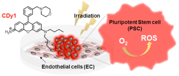
A light-activated dye turns on reactive oxygen species-mediated cell death in undifferentiated pluripotent stem cells, which could make stem cell therapies safer by preventing tumors. (credit: American Chemical Society)

Pluripotent stem cells (PSC) could be the key to a host of regeneration therapies because they can differentiate (develop) into basically any tissue type. But some PSCs in a culture dish can remain undifferentiated, and those could form teratomas — a type of tumor — if transplanted into patients.

Now a new light-based technology could remove this risk, Korean researchers report in an open-access paper in ACS Central Science.

Zapping undifferentiated PSCs

The researchers created a special dye (CDy1) that can selectively stain undifferentiated PSCs, but not differentiated ones. When the dye is exposed to light, it turns on the production of reactive oxygen species (ROS), which then kill these cells.

The researchers used undifferentiated PSCs transplanted into mice to demonstrate the method. None of the mice that received light-treated PSCs with the dye developed teratomas, whereas all of those in the control group (receiving PSCs that were not treated with light) did. The CDy1 and/or light irradiation did not negatively affect differentiated endothelial cells.

The researchers believe this dye-light combination could greatly improve the safety of a wide array of stem-cell therapies.

The researchers are affiliated with Sogang University, Republic of Korea Department of Medicine, Konkuk University, National University of Singapore, and Singapore Bioimaging Consortium (SBIC) Agency for Science, Technology and Research (A-STAR).

The work was supported by grants from the National Research Foundation of Korea and the Korea Health Industry Development Institute, and funded by the Korea government and A*STAR (Agency for Science, Technology and Research, Singapore) Biomedical Research Council and the Joint Council Office (JCO) Development Programme, A*STAR.


Abstract of Photodynamic Approach for Teratoma-Free Pluripotent Stem Cell Therapy Using CDy1 and Visible Light

Pluripotent stem cells (PSC) are promising resources for regeneration therapy, but teratoma formation is one of the critical problems for safe clinical application. After differentiation, the precise detection and subsequent elimination of undifferentiated PSC is essential for teratoma-free stem cell therapy, but a practical procedure is yet to be developed. CDy1, a PSC specific fluorescent probe, was investigated for the generation of reactive oxygen species (ROS) and demonstrated to induce selective death of PSC upon visible light irradiation. Importantly, the CDy1 and/or light irradiation did not negatively affect differentiated endothelial cells. The photodynamic treatment of PSC with CDy1 and visible light irradiation confirmed the inhibition of teratoma formation in mice, and suggests a promising new approach to safe PSC-based cell therapy.

A carbon-nanotube trap for ultra-sensitive virus detection and identification

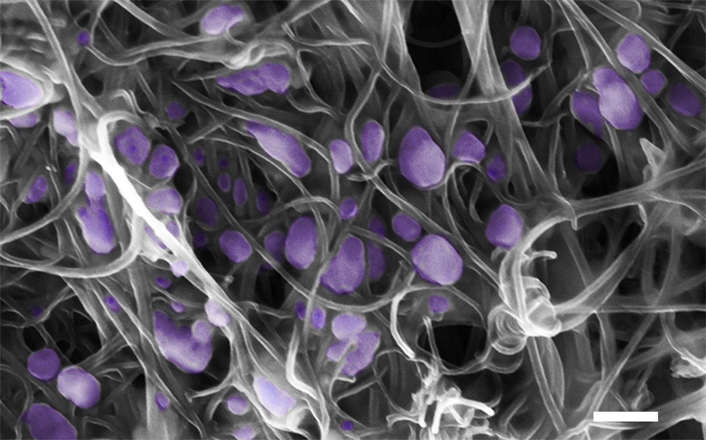
Scanning electron microscope image (scale bar, 200 nm) of the H5N2 avian influenza virus (purple) trapped inside the aligned carbon nanotubes. (credit: Penn State University)

Penn State researchers have developed a new portable microdevice that uses a forest-like array of vertically aligned carbon nanotubes to selectively trap and concentrate viruses by their size. It could improve detection of viruses and speed the process of identifying newly emerging viruses.

The research, by an interdisciplinary team of scientists at Penn State, was published in an open-access paper in the October 7, 2016 edition of the journal Science Advances.

“Detecting viruses early in an infection before symptoms appear, or from field samples, is difficult because the concentration of the viruses could be very low — often below the threshold of current detection methods,” said Mauricio Terrones, professor of physics, chemistry, and materials science and engineering.

“Early detection is important because a virus can begin to spread before we have the ability to detect it. The device we have developed allows us to selectively trap and concentrate viruses by their size — smaller than human cells and bacteria, but larger than most proteins and other macromolecules — in incredibly dilute samples.”

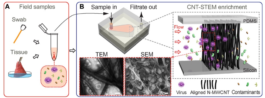
A small, portable device increases the sensitivity of virus detection by trapping and concentrating viruses in an array of carbon nanotubes. (A) Dilute samples collected from patients or the environment are passed through a filter to remove large particles such as bacteria and human cells, then (B) passed through the array of carbon nanotubes in the device. Viruses get trapped and build up to usable concentrations within the forest of nanotubes, while other smaller particles pass through and are eliminated. The concentrated virus captured in the device can then be put through a panel of tests to identify it, including molecular diagnosis by polymerase chain reaction (PCR), immunological methods, virus isolation, and genome sequencing.  The intertube distance can range from about 17 nanometers to over 300 nanometers to selectively capture viruses. (credit: Yin-Ting Yeh et al./Science Advances)

The device isolates and concentrates viruses by size, so the researchers can capture unknown viruses, said Terrones. “Once we capture and concentrate the virus, we can then use other techniques such as whole-genome sequencing to characterize it.”

Unpredictable outbreaks

Viruses such as influenza, HIV/AIDS, Ebola, and Zika can cause sudden, unpredictable outbreaks that lead to severe public-health crises. Currently available techniques for isolating and identifying the viruses that cause these outbreaks are slow, expensive, and use equipment and reagents that can be expensive, bulky, and require specialized storage.

Additionally, many recent outbreaks have been caused by newly emerging viruses for which there are no established ways to selectively isolate them for identification and characterization.

The research was funded by the U.S. National Center for Research Resources, the National Center for Advancing Translation Science, the U.S. National Institutes of Health, the U.S. Air Force Office of Scientific Research, and the Penn State Eberly College of Science, and it received support from the Penn State Huck Institutes of the Life Sciences.


Abstract of Tunable and label-free virus enrichment for ultrasensitive virus detection using carbon nanotube arrays

Viral infectious diseases can erupt unpredictably, spread rapidly, and ravage mass populations. Although established methods, such as polymerase chain reaction, virus isolation, and next-generation sequencing have been used to detect viruses, field samples with low virus count pose major challenges in virus surveillance and discovery. We report a unique carbon nanotube size-tunable enrichment microdevice (CNT-STEM) that efficiently enriches and concentrates viruses collected from field samples. The channel sidewall in the microdevice was made by growing arrays of vertically aligned nitrogen-doped multiwalled CNTs, where the intertubular distance between CNTs could be engineered in the range of 17 to 325 nm to accurately match the size of different viruses. The CNT-STEM significantly improves detection limits and virus isolation rates by at least 100 times. Using this device, we successfully identified an emerging avian influenza virus strain [A/duck/PA/02099/2012(H11N9)] and a novel virus strain (IBDV/turkey/PA/00924/14). Our unique method demonstrates the early detection of emerging viruses and the discovery of new viruses directly from field samples, thus creating a universal platform for effectively remediating viral infectious diseases.

Coming soon: a 3-D computer model of a cell

A preliminary model of a mycoplasma mycoides, a parasitic bacterium found in human urogenital and respiratory tracts. This pathogen has one of the smallest genomes of any free-living organism (525 genes). (credit: The Scripps Research Institute. Modeling by Ludovic Autin and David Goodsell, rendering by Adam Gardner.)

Advances in molecular biology and computer science may soon lead to a three-dimensional computer model of a cell, heralding a new era for biological research, medical science, and human and animal health, according to the authors of a paper recently published in the Journal of Molecular Biology.

“Cells are the foundation of life,” said Ilya Vakser, professor of computational biology and molecular biosciences and director of the Center for Computational Biology at the University of Kansas, one of the paper’s co-authors. “Recently, there has been tremendous progress in biomolecular modeling and advances at understanding life at the molecular level.

“Now, the focus is shifting to larger systems — up to the level of the entire cell. We’re trying to capture this emerging milestone development in computational structural biology, which is the tectonic shift from modeling individual biomolecular processes to modeling the entire cell.”

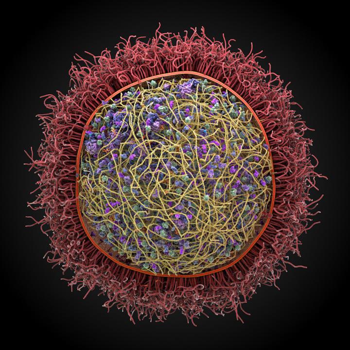
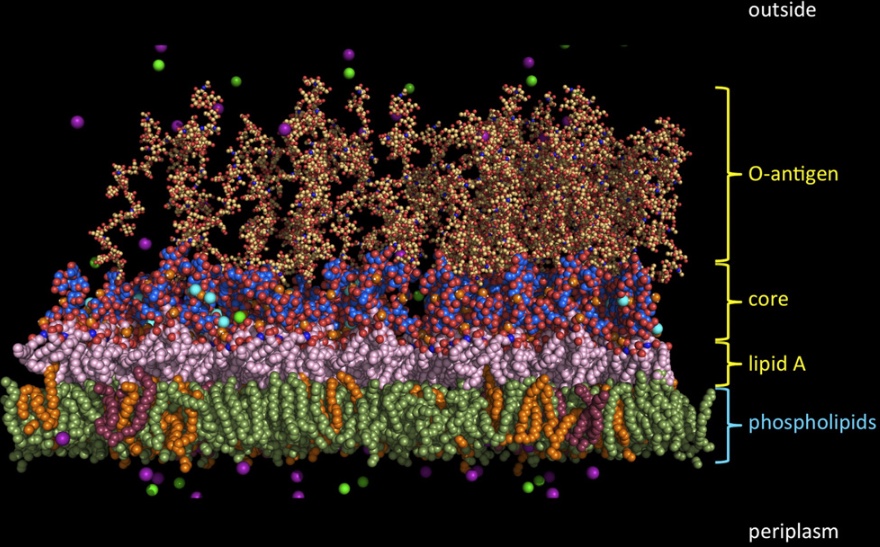
Gram-negative bacterial outer membrane molecular complexity. The image illustrates a typical E. coli outer membrane and the molecular system used to represent the complexity in molecular dynamics simulations. (credit: Wonpil Im et al./Journal of Molecular Biology)

The study surveys a range of methodologies for simulating a whole 3-D cell, including studies of biological networks, automated construction of 3-D cell models with experimental data, modeling of protein complexes, prediction of protein interactions, thermodynamic and kinetic effects of crowding cellular membrane modeling, and modeling of chromosomes.

“There are two major benefits,” Vakser said. “One is our fundamental understanding of how a cell works. You can’t claim you understand a phenomenon if you can’t model it. So this gives us insight into basic fundamentals of life at the scale of an entire cell.

“On the practical side, it will give us an improved grasp of the underlying mechanisms of diseases and also the ability to understand mechanisms of drug action, which will be a tremendous boost to our efforts at drug design. It will help us create better drug candidates, which will potentially shorten the path to new drugs.”

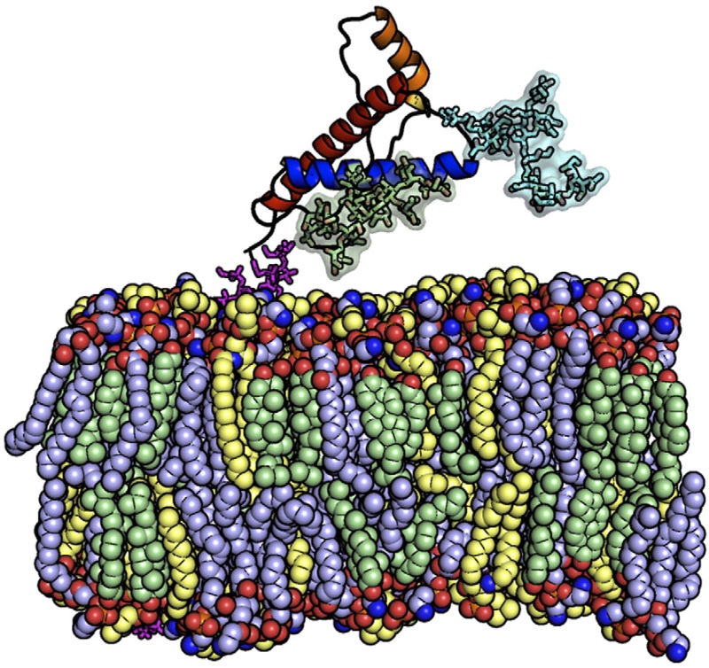
Prion protein model. Wonpil Im et al./Journal of Molecular Biology

As an example, he said a working 3-D molecular cell model could help to replace or augment phases of time-consuming and expensive drug development protocols required today to bring drug therapies from the scientist’s bench to the marketplace.

The paper’s other co-authors are Wonpil Im of Lehigh University, Jie Liang of the University of Illinois at Chicago, Sandor Vajda of Boston University, Arthur Olson of The Scripps Research Institute, and Huan-Xiang Zhou of Florida State University.


Abstract of Challenges in structural approaches to cell modeling

Computational modeling is essential for structural characterization of biomolecular mechanisms across the broad spectrum of scales. Adequate understanding of biomolecular mechanisms inherently involves our ability to model them. Structural modeling of individual biomolecules and their interactions has been rapidly progressing. However, in terms of the broader picture, the focus is shifting toward larger systems, up to the level of a cell. Such modeling involves a more dynamic and realistic representation of the interactomes in vivo, in a crowded cellular environment, as well as membranes and membrane proteins, and other cellular components. Structural modeling of a cell complements computational approaches to cellular mechanisms based on differential equations, graph models, and other techniques to model biological networks, imaging data, etc. Structural modeling along with other computational and experimental approaches will provide a fundamental understanding of life at the molecular level and lead to important applications to biology and medicine. A cross section of diverse approaches presented in this review illustrates the developing shift from the structural modeling of individual molecules to that of cell biology. Studies in several related areas are covered: biological networks; automated construction of three-dimensional cell models using experimental data; modeling of protein complexes; prediction of non-specific and transient protein interactions; thermodynamic and kinetic effects of crowding; cellular membrane modeling; and modeling of chromosomes. The review presents an expert opinion on the current state-of-the-art in these various aspects of structural modeling in cellular biology, and the prospects of future developments in this emerging field.

Genetically engineered peptides on 2D nanosheets form bio-nano interfaces

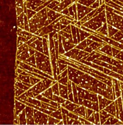
A top view of GrBP5 nanowires on a 2-D surface of graphene (credit: Mehmet Sarikaya/Scientific Reports)

Engineers at the University of Washington have created genetically engineered peptides that self-assemble into arrays of nanowires on two-dimensional nanosheets (single-layer graphene and molybdenum disulfide) to relay information across a bio-nano interface — a first step towards fully self-assembled future biomedical and electro-optical bionanoelectronic devices.

Arrays of peptides could provide organized scaffolds for functional biomolecules, enabling nanoscale bioelectronics interfaces. And designed peptides could be incorporated with metal ions or nanoparticles with specific physical characteristics, thus fine-tuning 2D device performance for chemical and biological sensors.

A bridge between biology and technology

“Bridging this divide would be the key to building the genetically engineered biomolecular solid-state devices of the future,” said UW professor Mehmet Sarikaya in the Departments of Materials Science & Engineering, senior author of an open-access paper published Sept. 22 in Scientific Reports.

The UW team is also planning to develop genetically engineered peptides with specific chemical and structural properties. Their ideal peptide would change the physical properties of synthetic materials and respond to that change. That way, it would transmit “information” from the synthetic material to other biomolecules — bridging the chemical divide between biology and technology.

The peptides function through molecular recognition — the same principles that underlie biochemical interactions such as an antibody binding to its specific antigen or protein binding to DNA.

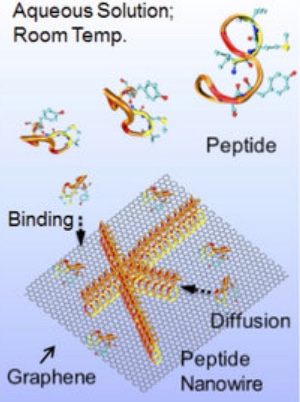
A schematic showing GrBP5 peptide self-organization with a series of surface processes on graphene: binding, diffusion and self-organization (credit: Yuhei Hayamizu et al./Scientific Reports)

In exploring the properties of 80 genetically selected peptides — which are not found in nature but have the same chemical components as peptides in all proteins — the researchers discovered that one peptide, GrBP5, showed promising interactions with the semimetal graphene. They tested GrBP5’s interactions with several other 2-D nanomaterials that “could serve as the metals or semiconductors of the future,” Sarikaya said.

Their experiments revealed that GrBP5 spontaneously organized into ordered nanowire patterns on graphene. With a few mutations, GrBP5 also altered the electrical conductivity of a graphene-based device, the first step toward transmitting electrical information from graphene to cells via peptides.

New bio-optoelectronic devices

Sarikaya’s team also modified GrBP5 to produce similar results on semiconductor material molybdenum disulfide (MoS2) and other materials* by converting a chemical signal to an optical signal. And they computationally predicted how different arrangements of GrBP5 nanowires would affect the electrical conduction or optical signal properties of each material.

A top view image of GrBP5 nanowires on a 2-D surface of molybdenum disulfide (credit: Mehmet Sarikaya/Scientific Reports)

The researchers are also seeking a peptide that could interact with materials such as gold, titanium, and even a mineral in bone and teeth.

Funded by the National Science Foundation, the UW, the National Institutes of Health, and the Japan Science and Technology Agency, the research is the focus of a new endeavor funded by the National Science Foundation’s Materials Genome Initiative. UW’s CoMotion is also working with Amazon to develop nano-sensors to detect early stages of pancreatic cancer.

* Other semiconducting 2D transition metal dichalcogenides (WSe2, WS2, MoSe2) along with insulating hBN, all with unique electronic and optical properties, were also tested.


Abstract of Bioelectronic interfaces by spontaneously organized peptides on 2D atomic single layer materials

Self-assembly of biological molecules on solid materials is central to the “bottom-up” approach to directly integrate biology with electronics. Inspired by biology, exquisite biomolecular nanoarchitectures have been formed on solid surfaces. We demonstrate that a combinatorially-selected dodecapeptide and its variants self-assemble into peptide nanowires on two-dimensional nanosheets, single-layer graphene and MoS2. The abrupt boundaries of nanowires create electronic junctions via spatial biomolecular doping of graphene and manifest themselves as a self-assembled electronic network. Furthermore, designed peptides form nanowires on single-layer MoS2 modifying both its electric conductivity and photoluminescence. The biomolecular doping of nanosheets defined by peptide nanostructures may represent the crucial first step in integrating biology with nano-electronics towards realizing fully self-assembled bionanoelectronic devices.

Graphene crowd-surfs on a lipid monolayer

Model of graphene on a lipid monolayer (credit: Universiteit Leiden)

“Crowd-surfing” on a smooth, supportive lipid monolayer, graphene could provide a versatile new platform for biosensors and drug delivery systems, researchers at Leiden University in The Netherlands have discovered.

Graphene is typically supported or sandwiched with other two-dimensional materials to promote higher mobility, ensure consistent electrical performance, and prevent environmental contamination. But combining graphene with soft, dynamic, molecular self-assembled lipid monolayers could provide a versatile platform for applications such as biosensors and drug delivery systems.

In research results published (open access) in a cover story in the journal Nanoscale on September 28, the authors note that the lipids (surprisingly) also improve graphene’s electrical conductivity. That could allow for measuring the electrical signals of graphene in the body for detecting acidity or the presence of certain proteins, for example.
This research was funded by the European Research Council, the Netherlands Organization for Scientific Research, and the Swiss National Science Foundation.

Abstract of Graphene-stabilized lipid monolayer heterostructures: a novel biomembrane superstructure

Chemically defined and electronically benign interfaces are attractive substrates for graphene and other two-dimensional materials. Here, we introduce lipid monolayers as an alternative, structurally ordered, and chemically versatile support for graphene. Deposition of graphene on the lipids resulted in a more ordered monolayer than regions without graphene. The lipids also offered graphene a more uniform and smoother support, reducing graphene hysteresis loop and the average value of the charge neutrality point under applied voltages. Our approach promises to be effective towards measuring experimentally biochemical phenomena within lipid monolayers and bilayers.

Smoking leaves ‘footprint’ in DNA

(credit: American Heart Association)

Smoking leaves its “footprint” on the human genome in the form of DNA methylation, a process that affects what genes are turned on, according to new research in Circulation: Cardiovascular Genetics, an American Heart Association journal.

The new findings could provide researchers with potential targets for new therapies.

“These results are important because methylation, as one of the mechanisms of the regulation of gene expression, affects what genes are turned on, which has implications for the development of smoking-related diseases, said Stephanie J. London, M.D., Dr.P.H., last author and deputy chief of the Epidemiology Branch at the National Institute of Environmental Health Sciences, National Institutes of Health, Research Triangle Park, North Carolina. “Even after someone stops smoking, we still see the effects of smoking on their DNA.”

The good news (for some): most DNA methylation sites returned to levels seen in never-smokers within five years of quitting smoking

Smoking remains the leading preventable cause of death worldwide. Even decades after stopping, former smokers are at long-term risk of developing diseases including some cancers, chronic obstructive pulmonary disease, and stroke. While the molecular mechanisms responsible for these long-term effects remain poorly understood, previous studies linking DNA methylation sites to genes involved with coronary heart disease and pulmonary disease suggest it may play an important role.

To find out more, researchers conducted a meta-analysis of DNA methylation sites across the human genome using blood samples taken from nearly 16,000 participants from 16 groups of the Cohorts for Heart and Aging Research in Genetic Epidemiology (CHARGE) Consortium, including a group of the Framingham Heart Study that has been followed by researchers since 1971.

The researchers compared DNA methylation sites in current and former smokers to those who never smoked. They found:

  • Smoking-associated DNA methylation sites were associated with more than 7,000 genes, or one-third of known human genes.
  • For people who stopped smoking, the majority of DNA methylation sites returned to levels seen in never-smokers within five years of quitting smoking. However, some DNA methylation sites persisted even after 30 years of quitting.
  • The most statistically significant methylation sites were linked to genes enriched for association with numerous diseases caused by cigarette smoking, such as cardiovascular diseases and certain cancers, suggesting that some of these long-lasting methylation sites may be marking genes potentially important for former smokers who are still at increased risk of developing certain diseases.

The discovery of smoking-related DNA methylation sites raises the possibility of developing biomarkers to evaluate a patient’s smoking history, as well as potentially developing new treatments targeted toward these methylation sites. However, the main analysis was not designed to examine effects over long periods of time.


Abstract of Epigenetic Signatures of Cigarette Smoking

Background — DNA methylation leaves a long-term signature of smoking exposure and is one potential mechanism by which tobacco exposure predisposes to adverse health outcomes, such as cancers, osteoporosis, lung, and cardiovascular disorders.

Methods and Results — To comprehensively determine the association between cigarette smoking and DNA methylation, we conducted a meta-analysis of genome-wide DNA methylation assessed using the Illumina BeadChip 450K array on 15,907 blood derived DNA samples from participants in 16 cohorts (including 2,433 current, 6,518 former, and 6,956 never smokers). Comparing current versus never smokers, 2,623 CpG sites (CpGs), annotated to 1,405 genes, were statistically significantly differentially methylated at Bonferroni threshold of p

Conclusions — Cigarette smoking has a broad impact on genome-wide methylation that, at many loci, persists many years after smoking cessation. Many of the differentially methylated genes were novel genes with respect to biologic effects of smoking, and might represent therapeutic targets for prevention or treatment of tobacco-related diseases. Methylation at these sites could also serve as sensitive and stable biomarkers of lifetime exposure to tobacco smoke.