Engineering ‘backup’ mitochondrial genes to restore power to cells

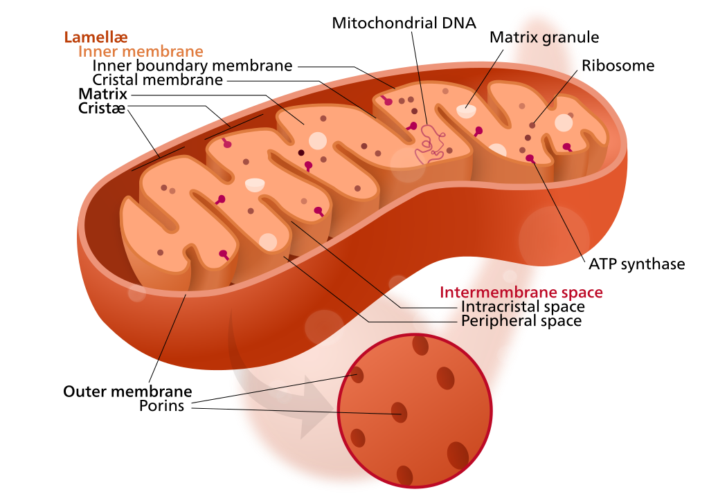
Mitochondrion structure (credit: Kelvinsong; modified by Sowlos/CC)

A new study by SENS Research Foundation, published in an open-access paper in the journal Nucleic Acids Research, explores the possibility of re-engineering mutated mitochondrial genes, which can otherwise lead to incurable disorders* and contribute to aging.

Mitochondria have their own DNA, allowing them to create proteins to supply nutrients and energy to cells. But sometimes, the DNA becomes mutated by “reactive oxygen species” generated by the mitochrondia themselves. This causes diseases in nervous, cardiovascular, and skeletal muscle tissues. More generally, “the mutations are also believed to contribute substantially to aging,” said Aubrey de Grey, PhD, a biomedical gerontologist and Chief Science Officer of SENS Research Foundation.

In the new study, the researchers developed “backup” copies of the modified mitochondrial genes, based on a patient cell line, to express the necessary proteins to produce cellular energy. The new genes would be delivered to the cell nucleii, where genes are protected from the more violent cell processes.**

The study was funded by the SENS Research Foundation, The Foster Foundation, Longecity Foundation, and Lifespan.io. NIH shared an instrumentation grant.

* One in 200 people are estimated to be born with a deleterious mitochondrial DNA mutation, leading to disorders such as Leber’s hereditary optic neuropathy, Leigh syndrome, mitochondrial encephalopathy, chronic progressive external ophthalmoplegia, and Kearns–Sayre syndrome, and also contribute to Parkinson’s.

** The researchers used a patient cell line with a single point mutation in the overlap region of the ATP8 and ATP6 genes of the human mitochondrial genome. Using synthesized DNA sequences, they were able to achieve stable expression, import, and function of the two mitochondria genes, correcting the loss of both mitochondrial proteins.  


Singularity Lectures | Aubrey de Grey Announces Progress in MitoSENS


Abstract of Stable nuclear expression of ATP8 and ATP6 genes rescues a mtDNA Complex V null mutant

We explore the possibility of re-engineering mitochondrial genes and expressing them from the nucleus as an approach to rescue defects arising from mitochondrial DNA mutations. We have used a patient cybrid cell line with a single point mutation in the overlap region of the ATP8 andATP6 genes of the human mitochondrial genome. These cells are null for the ATP8 protein, have significantly lowered ATP6 protein levels and no Complex V function. Nuclear expression of only the ATP8 gene with theATP5G1 mitochondrial targeting sequence appended restored viability on Krebs cycle substrates and ATP synthesis capabilities but, failed to restore ATP hydrolysis and was insensitive to various inhibitors of oxidative phosphorylation. Co-expressing both ATP8 and ATP6 genes under similar conditions resulted in stable protein expression leading to successful integration into Complex V of the oxidative phosphorylation machinery. Tests for ATP hydrolysis / synthesis, oxygen consumption, glycolytic metabolism and viability all indicate a significant functional rescue of the mutant phenotype (including re-assembly of Complex V) following stable co-expression of ATP8 and ATP6. Thus, we report the stable allotopic expression, import and function of two mitochondria encoded genes,ATP8 and ATP6, resulting in simultaneous rescue of the loss of both mitochondrial proteins.

Paralyzed man regains use of arms and hands after experimental stem cell therapy

Kris Boesen (credit: USC)

Doctors at the USC Neurorestoration Center and Keck Medicine of USC injected an experimental treatment* made from stem cells and other cells into the damaged cervical spine of a recently paralyzed 21-year-old man as part of a multi-center clinical trial.

Two weeks after surgery, Kristopher (Kris) Boesen began to show signs of improvement. Three months later, he’s able to feed himself, use his cell phone, write his name, operate a motorized wheelchair, and hug his friends and family. Improved sensation and movement in both arms and hands also make it easier for Kris to care for himself, and to envision a life lived more independently.

“Typically, spinal cord injury patients undergo surgery that stabilizes the spine but generally does very little to restore motor or sensory function,” explains Charles Liu, MD, PhD, director of the USC Neurorestoration Center. “With this study, we are testing a procedure that may improve neurological function, which could mean the difference between being permanently paralyzed and being able to use one’s arms and hands. Restoring that level of function could significantly improve the daily lives of patients with severe spinal injuries.”

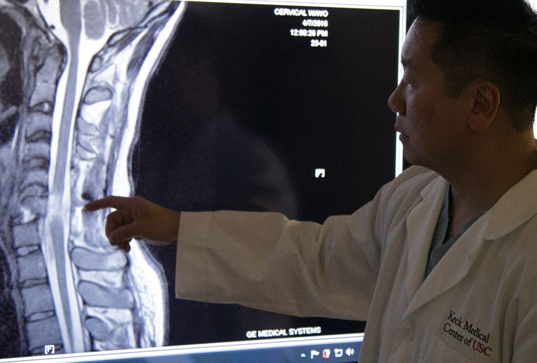
Dr. Liu points out cervical spine injury (credit: USC)

On March 6, Boesen suffered a traumatic injury to his cervical spine when his car fishtailed on a wet road, hit a tree and slammed into a telephone pole. Parents Rodney and Annette Boesen were warned there was a good chance their son would be permanently paralyzed from the neck down.

“As of 90 days post-treatment, Kris has gained significant improvement in his motor function, up to two spinal cord levels,” said Liu.  “In Kris’ case, two spinal cord levels means the difference between using your hands to brush your teeth, operate a computer, or do other things you wouldn’t otherwise be able to do, so having this level of functional independence cannot be overstated.”

The pioneering surgery is the latest example of how the emerging fields of neurorestoration and regenerative medicine may have the potential to improve the lives of thousands of patients who have suffered a severe spinal cord injury.

Keck is one of six sites** in the U.S. authorized to enroll subjects and administer the clinical trial dosage. To qualify for the clinical trial, enrollees must be between the age of 18 and 69, and their condition must be stable enough to receive an injection of AST-OPC1 between the fourteenth and thirtieth days following injury.

* The stem cell procedure is part of a Phase 1/2a clinical trial that is evaluating the safety and efficacy of escalating doses of AST-OPC1 cells developed by Asterias Biotherapeutics. AST-OPC1 cells are made from embryonic stem cells by carefully converting them into oligodendrocyte progenitor cells (OPCs), which are cells found in the brain and spinal cord that support the healthy functioning of nerve cells.  In previous laboratory studies, AST-OPC1 was shown to produce neurotrophic factors, stimulate vascularization and induce remyelination of denuded axons. All are critical factors in the survival, regrowth and conduction of nerve impulses through axons at the injury site in the “SCiStar” clinical trial according the researchers.

** The SCiStar clinical trial participating institutions include Indiana University in Indianapolis, Medical College of Wisconsin in Milwaukee, Rush University Medical Center in Chicago, Shepherd Center in Atlanta and, in California, Stanford University/Santa Clara Valley Medical Center, and Rancho Los Amigos/Keck Medicine of USC.

Designing new ultrasound imaging tools with Lego-like proteins

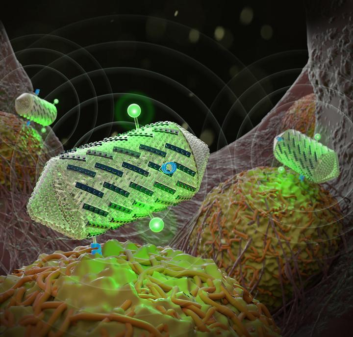
Protein-shelled structures called gas vesicles, illustrated here, can be engineered with Lego-like proteins to improve ultrasound methods. The gas vesicles can help detect specific cell types and create multicolor images. (credit: Barth van Rossum for Caltech)

The next step in ultrasound imaging will let doctors view specific cells and molecules deeper in the body, such as those associated with tumors or bacteria in our gut.

A new study from Caltech outlines how protein engineering techniques might help achieve this milestone. The researchers engineered protein-shelled nanostructures called gas vesicles (which reflect sound waves) to exhibit new properties useful for ultrasound technologies. In the future, these gas vesicles could be administered to a patient to visualize tissues of interest.

The modified gas vesicles were shown to give off more distinct signals (making them easier to image), target specific cell types, and help create color ultrasound images.

“It’s somewhat like engineering with molecular Legos,” says assistant professor of chemical engineering and Heritage Principal Investigator Mikhail Shapiro, who is the senior author of a new paper about the research published in this month’s issue of the journal ACS Nano and featured on the journal’s cover. “We can swap different protein ‘pieces’ on the surface of gas vesicles to alter their targeting properties and to visualize multiple molecules in different colors.”

“Gas vehicle” proteins reflect sound waves

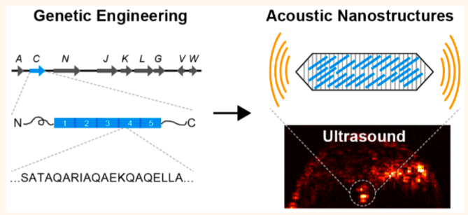
Genetic engineering of gas vesicles — genetically encoded protein nanostructures isolated from buoyant photosynthetic microbes — results in nanostructures with new mechanical, acoustic, surface, and functional properties to enable harmonic, multiplexed, and multimodal ultrasound imaging as well as cell-specific molecular targeting. (credit: Anupama Lakshmanan et al./ACS Nano)

In 2014, Shapiro first discovered the potential use of gas vesicles in ultrasound imaging. These gas-filled structures are naturally occurring in water-dwelling single-celled organisms, such as Anabaena flos-aquae, a species of cyanobacteria that forms filamentous clumps of multicell chains.

The gas vesicles help the organisms control how much they float and thus their exposure to sunlight at the water’s surface. Shapiro realized that the vesicles would readily reflect sound waves during ultrasound imaging, and ultimately demonstrated this using mice.

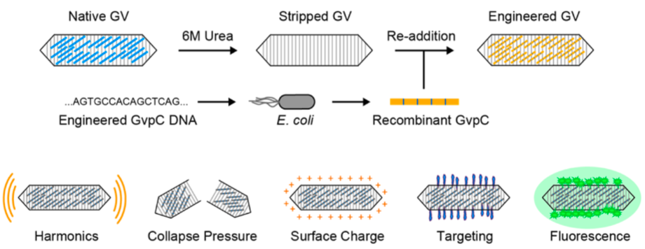
Genetic engineering a type of protein called GvpC (gas vesicle protein C) can be used to modify the properties of acoustic gas-vesicle nanostructures. (credit: Anupama Lakshmanan et al./ACS Nano)

In the latest research, Shapiro and his team set out to give the gas vesicles new properties by engineering gas vesicle protein C, or GvpC, a protein naturally found on the surface of vesicles that gives them mechanical strength and prevents them from collapsing. The protein can be engineered to have different sizes, with longer versions of the protein producing stronger and stiffer nanostructures.

In one experiment, the scientists removed the strengthening protein from gas vesicles and then administered the engineered vesicles to mice and performed ultrasound imaging. Compared to normal vesicles, the modified vesicles vibrated more in response to sound waves, and thus resonated with harmonic frequencies.

Harmonics are created when sound waves bounce around, for instance in a violin, and form new waves with doubled and tripled frequencies. Harmonics are not readily created in natural tissues, making the vesicles stand out in ultrasound images.

In another set of experiments, the researchers demonstrated how the gas vesicles could be made to target certain tissues in the body. They genetically engineered the vesicles to display various cellular targets, such as an amino acid sequence that recognizes proteins called integrins that are overproduced in tumor cells.

Multicolor ultrasound images

The team also showed how multicolor ultrasound images might be created. Conventional ultrasound images appear black and white. Shapiro’s group created an approach for imaging three different types of gas vesicles as separate “colors” based on their differential ability to resist collapse under pressure. The vesicles themselves do not appear in different colors, but they can be assigned colors based on their different properties.

To demonstrate this, the team made three different versions of the vesicles with varying strengths of the GvpC protein. They then increased the ultrasound pressures, causing the variant populations to successively collapse one by one.

As each population collapsed, the overall ultrasound signal decreased in proportion to the amount of that variant in the sample, and this signal change was then mapped to a specific color. In the future, if each variant population targeted a specific cell type, researchers would be able to visualize the cells in multiple colors.

“You might be able to see tumor cells versus the immune cells attacking the tumor, and thus monitor the progress of a medical treatment,” says Shapiro.

The ACS Nano paper, entitled “Molecular Engineering Of Acoustic Protein Nanostructures,” was funded by the National Institutes of Health, the Defense Advanced Research Projects Agency, the Heritage Research Institute for the Advancement of Medicine and Science at Caltech, and the Burroughs Wellcome Fund.


Abstract of Molecular Engineering of Acoustic Protein Nanostructures

Ultrasound is among the most widely used biomedical imaging modalities, but has limited ability to image specific molecular targets due to the lack of suitable nanoscale contrast agents. Gas vesicles—genetically encoded protein nanostructures isolated from buoyant photosynthetic microbes—have recently been identified as nanoscale reporters for ultrasound. Their unique physical properties give gas vesicles significant advantages over conventional microbubble contrast agents, including nanoscale dimensions and inherent physical stability. Furthermore, as a genetically encoded material, gas vesicles present the possibility that the nanoscale mechanical, acoustic, and targeting properties of an imaging agent can be engineered at the level of its constituent proteins. Here, we demonstrate that genetic engineering of gas vesicles results in nanostructures with new mechanical, acoustic, surface, and functional properties to enable harmonic, multiplexed, and multimodal ultrasound imaging as well as cell-specific molecular targeting. These results establish a biomolecular platform for the engineering of acoustic nanomaterials.

Seeing the invisible: visible-light metamaterial superlens made from nanobeads

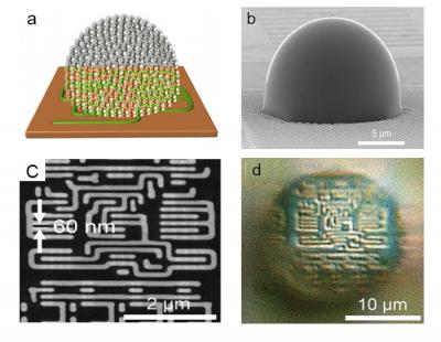
(a) Conceptual drawing of nanoparticle-based metamaterial solid immersion lens (mSIL) (b) Lab-made mSIL using titanium dioxide nanoparticles (c) SEM image of 60 nm size imaging sample (d) corresponding superlens imaging of the 60 nm sample by the developed mSIL. (credit: BangorUniversity/Fudan University)

A team of British and Chinese scientists has developed a new “metamaterial-based solid immersion lens” (mSIL) microscope lens design that can extend the magnification of an optical microscope to see objects smaller than the approximately 200 nanometers Abbe diffraction limit, the smallest size of bacteria.

Led by Zengbo Wang, PhD, at Bangor University UK and Prof Limin Wu at Fudan University, China, the team created minute droplet-like lens structures on the surface to be examined. These act as an additional lens to magnify the surface features previously invisible to a normal microscope lens, adding 5x magnification to existing microscopes.

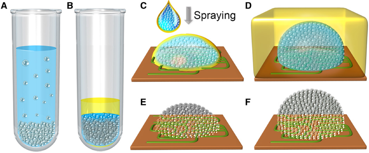
Schematic illustration of the assembly of the all-dielectric TiO2 mSIL. (A) Anatase TiO2 nanoparticles (15 nm) were centrifuged into a tightly packed precipitate. (B) The supernatant was replaced by an organic solvent mixture consisting of hexane and tetrachloroethylene to form a TiO2 nano–solid-fluid. (C) To prepare a hemispherical mSIL, the nano–solid-fluid was directly sprayed onto the sample surface. (D) To prepare a super-hemispherical mSIL, the nano–solid-fluid was sprayed onto the sample surface covered by a thin layer of organic solvent mixture. (E and F) After evaporation of the solvents, the nanoparticles underwent a phase transition to form a more densely packed structure. (credit: Wen Fan et al./Science Advances)

Made of millions of nanobeads, the spheres break up the light beam. Acting as individual minute beams, each bead refracts the light. “We’ve used high-index titanium dioxide (TiO2) nanoparticles as the building element of the lens,” Wang says. “These nanoparticles are able to bend light to a higher degree than water.”

“Each sphere bends the light to a high magnitude and splits the light beam, creating millions of individual beams of light. It is these tiny light beams which enable us to view previously unseen detail.”

Wang believes that the results will be easily replicable and that other labs will soon be adopting the technology and using it for themselves. Titanium dioxide is cheap and readily available, so rather than buying a new microscope, the lenses are applied to the material to be viewed, rather than to the microscope.

“The next challenge is to adapt the technology for use in biology and medicine. This would not require the current use of a combination of dyes and stains and laser light, which change the samples being viewed,” he says.

The lens is described in a paper in the open-access journal Science Advances today (August 12).


Abstract of Three-dimensional all-dielectric metamaterial solid immersion lens for subwavelength imaging at visible frequencies

Although all-dielectric metamaterials offer a low-loss alternative to current metal-based metamaterials to manipulate light at the nanoscale and may have important applications, very few have been reported to date owing to the current nanofabrication technologies. We develop a new “nano–solid-fluid assembly” method using 15-nm TiO2 nanoparticles as building blocks to fabricate the first three-dimensional (3D) all-dielectric metamaterial at visible frequencies. Because of its optical transparency, high refractive index, and deep-subwavelength structures, this 3D all-dielectric metamaterial-based solid immersion lens (mSIL) can produce a sharp image with a super-resolution of at least 45 nm under a white-light optical microscope, significantly exceeding the classical diffraction limit and previous near-field imaging techniques. Theoretical analysis reveals that electric field enhancement can be formed between contacting TiO2 nanoparticles, which causes effective confinement and propagation of visible light at the deep-subwavelength scale. This endows the mSIL with unusual abilities to illuminate object surfaces with large-area nanoscale near-field evanescent spots and to collect and convert the evanescent information into propagating waves. Our all-dielectric metamaterial design strategy demonstrates the potential to develop low-loss nanophotonic devices at visible frequencies.

Miniature portable device produces biopharmaceuticals on demand at point-of-care

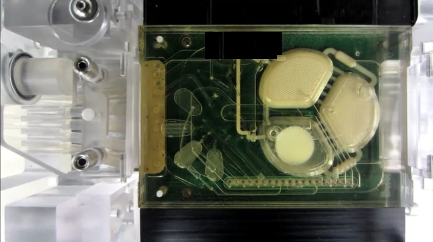
The new portable production system is designed to manufacture a range of biopharmaceuticals on-demand. The microbioreactor contains a polycarbonate-PDMS membrane-polycarbonate sandwiched chip with active microfluidic components (thin white lines) that are equipped for pneumatic routing of reagents, precise peristaltic injection, growth chamber mixing, and fluid extraction. (credit: Pablo Perez-Pinera et al./Nature Communications)

MIT researchers with DARPA funding have developed a portable device for manufacturing a range of biopharmaceuticals on demand, virtually anywhere.

For medics on the battlefield and doctors in remote or developing parts of the world, getting rapid access to the drugs needed to treat patients can be challenging. That’s because biopharmaceutical drugs, which are used in a wide range of therapies including vaccines and treatments for diabetes and cancer, are currently produced in large, centralized fermentation plants. Then they must be transported to the treatment site, which can be expensive, time-consuming, and difficult to execute in areas with poor supply chains.

In an open-access paper published Friday July 29 in the journal Nature Communications, the researchers demonstrate that the system can be used to produce a single dose of treatment from a compact device containing just a small droplet of cells in a liquid.

The system could ultimately be carried onto the battlefield and used to produce treatments at the point of care. It could also be used to manufacture a vaccine to prevent a disease outbreak in a remote village, according to senior author Tim Lu, an associate professor of biological engineering and electrical engineering and computer science, and head of the Synthetic Biology Group at MIT’s Research Laboratory of Electronics. “Imagine you were on Mars or in a remote desert, without access to a full formulary; you could program the yeast to produce drugs on demand locally,” Lu says.

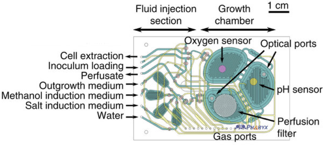
The microbioreactor contains microfluidic circuits (green), sensors for monitoring oxygen and acidity, and a filter to retain the cells while the therapeutic protein is extracted. (credit: Pablo Perez-Pinera et al./Nature Communications)

The prototype system is based on a programmable strain of yeast, Pichia pastoris, which can be induced to express (generate) one of two therapeutic proteins when exposed to a particular chemical trigger. The researchers chose P. pastoris because it can grow to very high densities on simple and inexpensive carbon sources, and is able to express large amounts of protein. “We altered the yeast so it could be more easily genetically modified, and could include more than one therapeutic in its repertoire,” Lu says.

In an experiment, when the researchers exposed the modified yeast to estrogen β-estradiol, the cells expressed recombinant human growth hormone (rHGH). But when they exposed the same cells to methanol, the yeast expressed the protein interferon.

How to create a DIY biopharmaceutical drug

(credit: Pablo Perez-Pinera et al./Nature Communications)

  1. Put the yeast cells into the millimeter-scale table-top microbioreactor.
  2. Feed a  liquid containing the desired chemical trigger (such as methanol) into the reactor to mix with the cells.
  3. Gently massage the liquid droplet to ensure its contents are fully mixed together.
  4. Pressurize the gas in the reactor. That causes oxygen to flow through a silicone rubber membrane and allows carbon dioxide to be extracted.
  5. The device continuously monitors conditions within microfluidic chip, including monitors cell density, oxygen levels, temperature, and pH, to ensure the optimum environment for cell growth.
  6. If you need a different protein, just flush the liquid through a filter, leaving the cells behind*. Then add fresh liquid containing a new chemical trigger to stimulate production of the next protein.

The researchers are now investigating how to use the system in combinatorial treatments, in which multiple therapeutics, such as antibodies, are used together. Combining multiple therapeutics in this way can be expensive if each requires its own production line, Lu says. “But if you could engineer a single strain, or maybe even a consortia of strains that grow together, to manufacture combinations of biologics or antibodies, that could be a very powerful way of producing these drugs at a reasonable cost,” he says.

The portable microbioreactor system operating in an ambulance (credit: Pablo Perez-Pinera et al./Nature Communications)

* Other research teams have previously attempted to build microbioreactors, but these could not retain the protein-producing cells while flushing out the liquid they are mixed with.


Abstract of Synthetic biology and microbioreactor platforms for programmable production of biologics at the point-of-care

Current biopharmaceutical manufacturing systems are not compatible with portable or distributed production of biologics, as they typically require the development of single biologic-producing cell lines followed by their cultivation at very large scales. Therefore, it remains challenging to treat patients in short time frames, especially in remote locations with limited infrastructure. To overcome these barriers, we developed a platform using genetically engineered Pichia pastoris strains designed to secrete multiple proteins on programmable cues in an integrated, benchtop, millilitre-scale microfluidic device. We use this platform for rapid and switchable production of two biologics from a single yeast strain as specified by the operator. Our results demonstrate selectable and near-single-dose production of these biologics in <24 h with limited infrastructure requirements. We envision that combining this system with analytical, purification and polishing technologies could lead to a small-scale, portable and fully integrated personal biomanufacturing platform that could advance disease treatment at point-of-care.

Americans worried about gene editing, brain chip implants, and synthetic blood

(iStock Photo)

Many in the general U.S. public are concerned about technologies to make people’s minds sharper and their bodies stronger and healthier than ever before, according to a new Pew Research Center survey of more than 4,700 U.S. adults.

The survey covers broad public reaction to scientific advances and examines public attitudes about the potential use of three specific emerging technologies for human enhancement.

The nationally representative survey centered on public views about gene editing that might give babies a lifetime with much reduced risk of serious disease, implantation of brain chips that potentially could give people a much improved ability to concentrate and process information, and transfusions of synthetic blood that might give people much greater speed, strength, and stamina.

A majority of Americans would be “very” or “somewhat” worried about gene editing (68%); brain chips (69%); and synthetic blood (63%), while no more than half say they would be enthusiastic about each of these developments.

Among the key data:

  • More say they would not want enhancements of their brains and their blood–66% and 63%, respectively–than say they would want them (32% and 35%). U.S. adults are closely split on the question of whether they would want gene editing to help prevent diseases for their babies (48% would, 50% would not).
  • Majorities say these enhancements could exacerbate the divide between haves and have-nots. For instance, 73% believe inequality will increase if brain chips become available because initially they will be obtainable only by the wealthy. At least seven-in-ten predict each of these technologies will become available before they have been fully tested or understood.
  • Substantial shares say they are not sure whether these interventions are morally acceptable. But among those who express an opinion, more people say brain and blood enhancements would be morally unacceptable than say they are acceptable.
  • More adults say the downsides of brain and blood enhancements would outweigh the benefits for society than vice versa. Americans are a bit more positive about the impact of gene editing to reduce disease; 36% think it will have more benefits than downsides, while 28% think it will have more downsides than benefits.
  • Opinion is closely divided when it comes to the fundamental question of whether these potential developments are “meddling with nature” and cross a line that should not be crossed, or whether they are “no different” from other ways that humans have tried to better themselves over time. For example, 49% of adults say transfusions with synthetic blood for much improved physical abilities would be “meddling with nature,” while a roughly equal share (48%) say this idea is no different than other ways human have tried to better themselves.

The survey data reveal several patterns surrounding Americans’ views about these ideas:

  • People’s views about these human enhancements are strongly linked with their religiosity.
  • People are less accepting of enhancements that produce extreme changes in human abilities. And, if an enhancement is permanent and cannot be undone, people are less inclined to support it.
  • Women tend to be more wary than men about these potential enhancements from cutting-edge technologies.

The survey also finds some similarities between what Americans think about these three potential, future enhancements and their attitudes toward the kinds of enhancements already widely available today. As a point of comparison, this study examined public thinking about a handful of current enhancements, including elective cosmetic surgery, laser eye surgery, skin or lip injections, cosmetic dental procedures to improve one’s smile, hair replacement surgery and contraceptive surgery.

  • 61% of Americans say people are too quick to undergo cosmetic procedures to change their appearance in ways that are not really important, while 36% “it’s understandable that more people undergo cosmetic procedures these days because it’s a competitive world and people who look more attractive tend to have an advantage.”
  • When it comes to views about elective cosmetic surgery, in particular, 34% say elective cosmetic surgery is “taking technology too far,” while 62% say it is an “appropriate use of technology.” Some 54% of U.S. adults say elective cosmetic surgery leads to about equal benefits and downsides for society, while 26% express the belief that there are more downsides than benefits, and just 16% say society receives more benefits than downsides from cosmetic surgery.

The survey data is drawn from a nationally representative survey of 4,726 U.S. adults conducted by Pew Research Center online and by mail from March 2-28, 2016.

Pew Research Center is a nonpartisan “fact tank” that informs the public about the issues, attitudes and trends shaping America and the world. It does not take policy positions. The center is a subsidiary of The Pew Charitable Trusts, its primary funder.

New nanomaterial mimics cell membranes

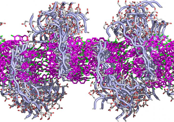
This simulated cross-section shows how the lipid-like peptoids interact to form a membrane. Each peptoid has two sections: a fatty-like region that interacts via benzene rings (shown in pink) with its neighbors to form a sheet, and a water-loving region that juts above or below the flat sheet. Each region can be designed to have specific functions. (credit: Chun-Long Chen/PNNL)

Materials scientists at the Department of Energy’s Pacific Northwest National Laboratory have created a new material that performs like a biological cell membrane — a material that has long been sought for applications like water purification and drug delivery.

The “peptoid” material can assemble itself into a sheet that’s thinner, but more stable, than a soap bubble, the researchers report this week in Nature Communications. The assembled sheet can withstand being submerged in a variety of liquids and can even repair itself after damage.

“We believe these materials have potential in water filters, sensors, drug delivery, and especially fuel cells or other energy applications,” said chemist Chun-Long Chen.

Biological cell membranes, which are made from thin sheets of fatty molecules called lipids, are at least ten times thinner than an iridescent soap bubble and yet allow cells to collectively form organisms as diverse at bacteria, trees and people.

Cell membranes are also very selective about what they let pass through, using tiny embedded proteins as gatekeepers. Membranes repair dings to their structure automatically and change thickness to pass signals from the outside environment to the cell’s interior, where most of the action is.

Scientists would like to take advantage of these gatekeeping and other membrane properties to make filters. A cell-membrane-like material would have advantages over other thin materials such as graphene. For example, mimicking a cell membrane’s efficient gatekeeping could result in water purifying membranes that don’t require a lot of pressure or energy to push the water through.

How to design imitation biological cell membranes

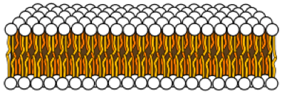
Lipid bilayer sheet (credit: Mariana Ruiz Villarreal/Wikipedia)

Synthetic molecules called peptoids have caught the interest of researchers because they are cheap, versatile and customizable. They are like natural proteins, including those that embed themselves in cell membranes, and can be designed to have very specific forms and functions. So Chen and colleagues decided to see if they could design peptoids to make them more lipid-like (that is, more like fats).

Lipid molecules are long and mostly straight: They have a fatty end that prefers to hang out with other fats, and a water-like end that prefers the comfort of water. Because of this chemistry, lipid molecules arrange themselves with the fatty ends pointed toward each other, sandwiched between the water-loving ends pointed out. Scientists call this a lipid bilayer, essentially a sheet that envelops the contents of a cell. Proteins or carbohydrate molecules embed themselves in the membranous sheet.

Inspired by this, Chen and colleagues designed peptoids in which each base peptoid was a long molecule with one end water-loving and the other end fat-loving. They chose chemical features that they hoped would encourage the individual molecules to pack together. They examined the resulting structures using a variety of analysis methods, including some at the Advanced Light Source and the Molecular Foundry, two DOE Office of Science User Facilities at Lawrence Berkeley National Laboratory.

Forming nanomembranes

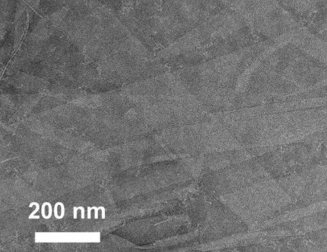
Peptoid nanomembrane (credit: Haibao Jin et al./Nature Communications)

The team found that after putting the lipid-like peptoids into a liquid solution, the molecules spontaneously crystallized and formed “nanomembranes” — straight-edged sheets as thin as cell membranes — floating in the beaker. These nanomembranes maintained their structure in water or alcohol, at different temperatures, in solutions with high or low pH, or high concentrations of salts, a feat that few cell membranes could accomplish.

To better understand the nanomembranes, the team simulated how single peptoid molecules interacted with each other using molecular dynamics software. The simulated peptoids formed a membrane reminiscent of a lipid bilayer: The fat-loving ends lined up in the middle, and their water-loving ends pointed outward either above or below.

They also confirmed the ability of the synthetic membranes to hold proteins that have specific functions, such as ones that let water, and only water, through, and to repair themselves.

The results showed the researchers that they are on the right path to making synthetic cell membrane-like materials. The next step, Chen said, is to build biomimetic membranes by incorporating natural membrane proteins or other synthetic water channels such as carbon nanotubes into these sheet matrices. The team is also looking into ways to make the peptoid membranes conductive for energy uses.

This work was supported by the Department of Energy Office of Science and PNNL.


Abstract of Highly stable and self-repairing membrane-mimetic 2D nanomaterials assembled from lipid-like peptoids

An ability to develop sequence-defined synthetic polymers that both mimic lipid amphiphilicity for self-assembly of highly stable membrane-mimetic 2D nanomaterials and exhibit protein-like functionality would revolutionize the development of biomimetic membranes. Here we report the assembly of lipid-like peptoids into highly stable, crystalline, free-standing and self-repairing membrane-mimetic 2D nanomaterials through a facile crystallization process. Both experimental and molecular dynamics simulation results show that peptoids assemble into membranes through an anisotropic formation process. We further demonstrated the use of peptoid membranes as a robust platform to incorporate and pattern functional objects through large side-chain diversity and/or co-crystallization approaches. Similar to lipid membranes, peptoid membranes exhibit changes in thickness upon exposure to external stimuli; they can coat surfaces in single layers and self-repair. We anticipate that this new class of membrane-mimetic 2D nanomaterials will provide a robust matrix for development of biomimetic membranes tailored to specific applications.

Why your immune system may control your social behavior

Normal brain activity (credit: University of Virginia Health System)

In a discovery that raises fundamental questions about human behavior, researchers at the University of Virginia School of Medicine have found that the immune system directly affects — and even controls — our social behavior, such as our desire to interact with others. That finding could have significant implications for neurological diseases such as autism-spectrum disorders and schizophrenia, the researchers suggest.

“The brain and the adaptive immune system were thought to be isolated from each other, and any immune activity in the brain was perceived as sign of a pathology. And now, not only are we showing that they are closely interacting, but some of our behavior traits might have evolved because of our immune response to pathogens,” explained Jonathan Kipnis, chair of UVA’s Department of Neuroscience.

“It’s crazy, but maybe we are just multicellular battlefields for two ancient forces: pathogens and the immune system. Part of our personality may actually be dictated by the immune system.”

Evolutionary forces linking brains and pathogens

KurzweilAI has cited supporting evidence for that idea. For example, permanent stress may affect immune cells in the brain, leading to mental disorders and protective immune microglia cells also have direct involvement in creating the cellular networks at the core of brain behavior.

Last year, Kipnis, the director of UVA’s Center for Brain Immunology and Glia, and his team discovered that meningeal membranes (covering the brain and spinal cord) in the brain directly link the brain with the lymphatic system. That overturned decades of textbook teaching that the brain lacks a direct connection to the immune system.

Now, the researchers suggest, the relationship between people and pathogens could have directly affected the development of our social behavior. Social behavior (which is necessary for the survival of the species) allows pathogens to spread, so our immune systems may have developed to protect us from the diseases that accompany those interactions.

Specifically, the UVA researchers have now shown that a specific immune molecule, interferon gamma, seems to be critical for social behavior, and that a variety of creatures, such as flies, zebrafish, mice and rats, activate interferon gamma responses (as protection) when they are social.

Normally, this molecule is produced by the immune system in response to bacteria, viruses or parasites. But blocking the molecule in mice using genetic modification also made regions of the brain hyperactive, causing the mice to become less social. Restoring the molecule restored the brain connectivity and behavior to normal.

A hyperactive brain, triggered by a blocked immune system, may lead to less-social behavior (credit: University of Virginia Health System)

In a Nature paper outlining their findings, the researchers note the immune molecule plays a “profound role in maintaining proper social function.”

“It’s extremely critical for an organism to be social for the survival of the species. It’s important for foraging, sexual reproduction, gathering, hunting,” said Anthony J. Filiano, Hartwell postdoctoral fellow in the Kipnis lab and lead author of the study. “The hypothesis is that when organisms come together, you have a higher propensity to spread infection — you need to be social, but [in doing so] you have a higher chance of spreading pathogens.” Which explains why “interferon gamma, in evolution, has been used as a more efficient way to both boost social behavior while boosting an anti-pathogen response.”

Immune-system failure leads to social deficits

The researchers explain that a malfunctioning immune system may be responsible for “social deficits in numerous neurological and psychiatric disorders.” But exactly what this might mean for autism and other specific conditions requires further investigation.

It is unlikely that any one molecule will be responsible for disease or the key to a cure. The researchers believe that the causes are likely to be much more complex. But the discovery that the immune system — and possibly pathogens, by extension — can control our interactions raises many exciting avenues for scientists to explore, both in terms of battling neurological disorders and understanding human behavior.

Kipnis and his team worked closely with UVA’s Department of Pharmacology and with Vladimir Litvak’s research group at the University of Massachusetts Medical School. Litvak’s team developed a computational approach to investigate the complex dialogue between immune signaling and brain function in health and disease.

“Using this approach we predicted a role for interferon gamma, an important cytokine secreted by T lymphocytes, in promoting social brain functions,” Litvak said. “Our findings contribute to a deeper understanding of social dysfunction in neurological disorders, such as autism and schizophrenia, and may open new avenues for therapeutic approaches.”

The work was supported by NIH grants and the Hartwell Foundation.


Medicine Virginia | Shocking New Role Found for the Immune System: Controlling Social Interactions


Abstract of Unexpected role of interferon-γ in regulating neuronal connectivity and social behaviour

Immune dysfunction is commonly associated with several neurological and mental disorders. Although the mechanisms by which peripheral immunity may influence neuronal function are largely unknown, recent findings implicate meningeal immunity influencing behaviour, such as spatial learning and memory1. Here we show that meningeal immunity is also critical for social behaviour; mice deficient in adaptive immunity exhibit social deficits and hyper-connectivity of fronto-cortical brain regions. Associations between rodent transcriptomes from brain and cellular transcriptomes in response to T-cell-derived cytokines suggest a strong interaction between social behaviour and interferon-γ (IFN-γ)-driven responses. Concordantly, we demonstrate that inhibitory neurons respond to IFN-γ and increase GABAergic (γ-aminobutyric-acid) currents in projection neurons, suggesting that IFN-γ is a molecular link between meningeal immunity and neural circuits recruited for social behaviour. Meta-analysis of the transcriptomes of a range of organisms reveals that rodents, fish, and flies elevate IFN-γ/JAK-STAT-dependent gene signatures in a social context, suggesting that the IFN-γ signalling pathway could mediate a co-evolutionary link between social/aggregation behaviour and an efficient anti-pathogen response. This study implicates adaptive immune dysfunction, in particular IFN-γ, in disorders characterized by social dysfunction and suggests a co-evolutionary link between social behaviour and an anti-pathogen immune response driven by IFN-γ signalling.