FDA places hold on clinical trial of cancer treatment previously reported on KurzweilAI — UPDATE: FDA releases hold

Juno Therapeutics, Inc. announced July 7 that it has received notice from the FDA that it has placed a clinical hold on an immune-cell cancer treatment known as the “ROCKET” trial, which was reported on KurzweilAI on Mar. 10, 2016.

The clinical hold was initiated after two patient deaths, which followed the recent addition of fludarabine to the pre-conditioning regimen. Juno has proposed to the FDA to continue the ROCKET trial using JCAR015 with cyclophosphamide pre-conditioning alone.

Further information on Forbes.

UPDATE JULY 13, 2016: Juno Therapeutics announced Tuesday July 12 that the FDA has removed the hold and “the ROCKET trial will continue enrollment using JCAR015 with cyclophosphamide pre-conditioning only.” In related news, the Law Offices of Howard G. Smith announced an investigation on behalf of investors of Juno Therapeutics, Inc. (NASDAQ: JUNO) concerning the Company and its officers’ possible violations of federal securities laws and noted that “Juno’s share price fell significantly in value, dropping almost 32%.”

This deadly soil bug can reach your brain in a day, end up in spinal cord

B. pseudomallei soil-dwelling bacterium endemic in tropical and subtropical regions worldwide, particularly in Thailand and northern Australia (credit: Wikipedia CC)

Imagine a  deadly bacteria that can be picked up by a simple sniff and can travel to your brain and spinal cord in just 24 hours. Or one that could just be quietly sitting there, waiting for an opportune moment. Or maybe just doing small incremental damage ever day over a lifetime … as you lose the function in your brain incrementally.

That’s the grisly finding (in mice), published in Immunity and Infection this week, of a new study by Australian Griffith University and Bond University scientists.

The pathogenic bacteria Burkholderia pseudomallei, which causes the potentially fatal disease melioidosis, kills 89,000 people around the world each year and is prevalent in northern Australia, where a person with melioidosis has a 20–50 per cent chance of dying once it infects the brain. The bacterium is found in the northern parts of the Northern Territory, including Darwin.

In Southeast Asia 50 per cent of the population may be positive for melioidosis, and in places like Cambodia the mortality rate is as high as 50 per cent.

But for the rest of us, the findings could also lead to discoveries of how the common staphylococcus and acne bacterium also end up in the spinal cord, as well as how chlamydia travels to the brain in Alzheimer’s patients. Or even explain common back problems, which could be where bacteria have infected your bone, causing pain that could be simply treated with antibiotics, according to the researchers.

Tracing the bacteria in mice brains and spinal cords

(Top) A schematic drawing of a mouse brain showing the location of various images. (Bottom) D: A B. pseudomallei rod (arrow) present in trigeminal nerve near the connection between the trigeminal nerve in the brain and the brainstem. (E) B. pseudomallei rod (arrow) with a fluorescent particle (arrow with tail) after the merge between the trigeminal nerve and brainstem. Scale bars in μm. (credit: James A. St John et al./Infection and Immunity)

The olfactory mucosa, located in the nose, is very close to the brain and it has long been known that viruses could reach the brain from the olfactory mucosa. But researchers have not understand exactly how the bacteria traveled to the brain and spinal cord, or just how quickly.

To find out, James St. John, PhD., Head of Griffith’s Clem Jones Centre for Neurobiology and Stem Cell Research, and associates infected mice with B. pseudomallei. They were able to trace the bacteria travels from the nerves in the nasal cavity before moving to the brain stem and then into the spinal cord. (He noted that this could also be a pathway for many other common bacteria.)

“Our latest results represent the first direct demonstration of transit of a bacterium from the olfactory mucosa to the central nervous system (CNS) via the trigeminal nerve; bacteria were found a considerable distance from the olfactory mucosa, in the brain stem, and even more remarkably in the spinal cord,” said professor Ifor Beacham from the Griffith Institute for Glycomics.

Researchers will now work on ways to stimulate supporting cells that could remove the bacteria. St. John said the work was important because the bacteria had the potential to be used as a bioweapon and knowing how to combat it was extremely important.

“Bacteria have been implicated as a major causative agent of some types of back pain. We now need to work out whether the bacteria that cause back pain also can enter the brainstem and spinal cord via the trigeminal nerve,” he added.


Abstract of Burkholderia pseudomallei rapidly infects the brainstem and spinal cord via the trigeminal nerve after intranasal inoculation

Infection with Burkholderia pseudomallei causes melioidosis, a disease with a high mortality rate (20% in Australia and 40% in south-east Asia). Neurological melioidosis is particularly prevalent in northern Australian patients and involves brainstem infection, which can progress to the spinal cord; however, the route by which the bacteria invade the central nervous system (CNS) is unknown. We have previously demonstrated that B. pseudomallei can infect the olfactory and trigeminal nerves within the nasal cavity following intranasal inoculation. As the trigeminal nerve projects into the brainstem, we investigated whether the bacteria could continue along this nerve to penetrate the CNS. After intranasal inoculation of mice, B. pseudomallei caused low-level localised infection within the nasal cavity epithelium, prior to invasion of the trigeminal nerve in small numbers. B. pseudomallei rapidly invaded the trigeminal nerve and crossed the astrocytic barrier to enter the brainstem within 24 hours and then rapidly progressed over 2000 μm into the spinal cord. To rule out that the bacteria used a haematogenous route, we used a capsule-deficient mutant of B. pseudomallei, which does not survive in the blood, and found that it also entered the CNS via the trigeminal nerve. This suggests that the primary route of entry is via the nerves that innervate the nasal cavity. We found that actin-mediated motility could facilitate initial infection of the olfactory epithelium. Thus, we have demonstrated that B. pseudomallei can rapidly infect the brain and spinal cord via the trigeminal nerve branches that innervate the nasal cavity.

Robot mimics vertebrate motion

Pleurobot (credit: EPFL)

École polytechnique fédérale de Lausanne (EPFL) scientists have invented a new robot called “Pleurobot” that mimics the way salamanders walk and swim with unprecedented detail.

Aside from being cool (and a likely future Disney attraction), the researchers believe designing the robot will provide a new tool for understanding the evolution of vertebrate locomotion. That could lead to better understanding of how the spinal cord controls the body’s locomotion, which may help develop future therapies and neuroprosthetic devices for paraplegic patients and amputees.

Pleurobot mimics Salamander. Neurobiologists say electrical stimulation of the spinal cord is what determines whether the salamander walks, crawls or swims: At lowest level of stimulation, the salamander walks; with higher stimulation, its pace increases, and beyond some threshold the salamander begins to swim. (credit: EPFL)

Simulating the 3D motion of the salamander’s locomotion requires exceptional precision. The Biorobotics Laboratory scientists started by shooting detailed x-ray videos of the salamander species Pleurodeles waltl from the top and the side, tracking up to 64 points along its skeleton while it performed different types of motion in water and on the ground.

Auke Ijspeert and his team at EPFL then 3D-printed bones and motorized joints, and even created a “nervous system” using electronic circuitry, allowing the Pleurobot to walk, crawl, and even swim underwater.*

Ijspeert thinks that the design methodology used for the Pleurobot can help develop other types of “biorobots,” which could become important tools in neuroscience and biomechanics.

The research, described in the Royal Society journal Interface, received funding from the Swiss National Center of Competence in Research (NCCR) in Robotics and from the Swiss National Science Foundation.


École polytechnique fédérale de Lausanne | A new robot mimics vertebrate motion

* In the design process, the researchers identified the minimum number of motorized segments required, as well as the optimal placement along the robot’s body, to replicate many of the salamander’s types of movement. That made it possible to construct Pleurobot with fewer bones and joints than the real-life creature — only 27 motors and 11 segments along its spine (the real animal has 40 vertebrae and multiple joints, some of which can even rotate freely and move side-to-side or up and down). 


Abstract of From cineradiography to biorobots: an approach for designing robots to emulate and study animal locomotion

Robots are increasingly used as scientific tools to investigate animal locomotion. However, designing a robot that properly emulates the kinematic and dynamic properties of an animal is difficult because of the complexity of musculoskeletal systems and the limitations of current robotics technology. Here, we propose a design process that combines high-speed cineradiography, optimization, dynamic scaling, three-dimensional printing, high-end servomotors and a tailored dry-suit to construct Pleurobot: a salamander-like robot that closely mimics its biological counterpart, Pleurodeles waltl. Our previous robots helped us test and confirm hypotheses on the interaction between the locomotor neuronal networks of the limbs and the spine to generate basic swimming and walking gaits. With Pleurobot, we demonstrate a design process that will enable studies of richer motor skills in salamanders. In particular, we are interested in how these richer motor skills can be obtained by extending our spinal cord models with the addition of more descending pathways and more detailed limb central pattern generator networks. Pleurobot is a dynamically scaled amphibious salamander robot with a large number of actuated degrees of freedom (DOFs: 27 in total). Because of our design process, the robot can capture most of the animal’s DOFs and range of motion, especially at the limbs. We demonstrate the robot’s abilities by imposing raw kinematic data, extracted from X-ray videos, to the robot’s joints for basic locomotor behaviours in water and on land. The robot closely matches the behaviour of the animal in terms of relative forward speeds and lateral displacements. Ground reaction forces during walking also resemble those of the animal. Based on our results, we anticipate that future studies on richer motor skills in salamanders will highly benefit from Pleurobot’s design.

The top 10 emerging technologies of 2016

(credit: WEF)

The World Economic Forum’s annual list of this year’s breakthrough technologies, published today, includes “socially aware” openAI, grid-scale energy storage, perovskite solar cells, and other technologies with the potential to “transform industries, improve lives, and safeguard the planet.” The WEF’s specific interest is to “close gaps in investment and regulation.”

“Horizon scanning for emerging technologies is crucial to staying abreast of developments that can radically transform our world, enabling timely expert analysis in preparation for these disruptors. The global community needs to come together and agree on common principles if our society is to reap the benefits and hedge the risks of these technologies,” said Bernard Meyerson, PhD, Chief Innovation Officer of IBM and Chair of the WEF’s Meta-Council on Emerging Technologies.

The list also provides an opportunity to debate human, societal, economic or environmental risks and concerns that the technologies may pose — prior to widespread adoption.

One of the criteria used by council members during their deliberations was the likelihood that 2016 represents a tipping point in the deployment of each technology. So the list includes some technologies that have been known for a number of years, but are only now reaching a level of maturity where their impact can be meaningfully felt.

The top 10 technologies that make this year’s list are:

  1. Nanosensors and the Internet of Nanothings  — With the Internet of Things expected to comprise 30 billion connected devices by 2020, one of the most exciting areas of focus today is now on nanosensors capable of circulating in the human body or being embedded in construction materials. They could use DNA and proteins to recognize specific chemical targets, store a few bits of information, and then report their status by changing color or emitting some other easily detectable signal.
  2. Next-Generation Batteries — One of the greatest obstacles holding renewable energy back is matching supply with demand, but recent advances in energy storage using sodium, aluminum, and zinc based batteries makes mini-grids feasible that can provide clean, reliable, around-the-clock energy sources to entire villages.
  3. The Blockchain — With venture investment related to the online currency Bitcoin exceeding $1 billion in 2015 alone, the economic and social impact of blockchain’s potential to fundamentally change the way markets and governments work is only now emerging.
  4. 2D Materials — Plummeting production costs mean that 2D materials like graphene are emerging in a wide range of applications, from air and water filters to new generations of wearables and batteries.
  5. Autonomous Vehicles — The potential of self-driving vehicles for saving lives, cutting pollution, boosting economies, and improving quality of life for the elderly and other segments of society has led to rapid deployment of key technology forerunners along the way to full autonomy.
  6. Organs-on-chips — Miniature models of human organs could revolutionize medical research and drug discovery by allowing researchers to see biological mechanism behaviors in ways never before possible.
  7. Perovskite Solar Cells — This new photovoltaic material offers three improvements over the classic silicon solar cell: it is easier to make, can be used virtually anywhere and, to date, keeps on generating power more efficiently.
  8. Open AI Ecosystem — Shared advances in natural language processing and social awareness algorithms, coupled with an unprecedented availability of data, will soon allow smart digital assistants to help with a vast range of tasks, from keeping track of one’s finances and health to advising on wardrobe choice.
  9. Optogenetics — Recent developments mean light can now be delivered deeper into brain tissue, something that could lead to better treatment for people with brain disorders.
  10. Systems Metabolic Engineering — Advances in synthetic biology, systems biology, and evolutionary engineering mean that the list of building block chemicals that can be manufactured better and more cheaply by using plants rather than fossil fuels is growing every year.

To compile this list, the World Economic Forum’s Meta-Council on Emerging Technologies, a panel of global experts, “drew on the collective expertise of the Forum’s communities to identify the most important recent technological trends. By doing so, the Meta-Council aims to raise awareness of their potential and contribute to closing gaps in investment, regulation and public understanding that so often thwart progress.”

You can read 10 expert views on these technologies here or download the series as a PDF.

Bionic leaf 2.0

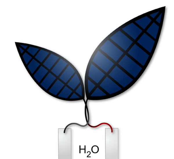
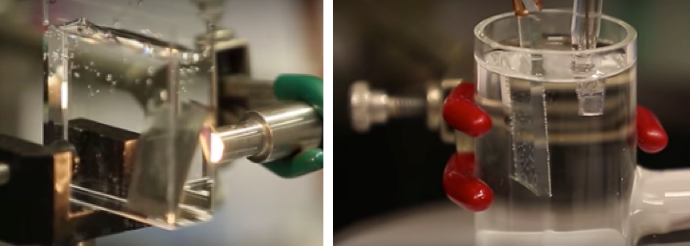
Bionic leaf 2.0: An artificial photosynthesis system (credit: Jessica Polka)

Harvard scientists have created a system a system that uses solar energy plus hydrogen-eating bacteria to produce liquid fuels with 10 percent efficiency, compared to the 1 percent seen in the fastest-growing plants.

The system, co-created by Daniel Nocera, the Patterson Rockwood Professor of Energy at Harvard University, and Pamela Silver, the Elliott T. and Onie H. Adams Professor of Biochemistry and Systems Biology at Harvard Medical School, uses solar energy to split water molecules into hydrogen and oxygen molecules.

A paper on the research was published June 3 in Science.

“This is a true artificial photosynthesis system,” Nocera said. “Before, people were using artificial photosynthesis for water-splitting, but this is a true A-to-Z system, and we’ve gone well over the efficiency of photosynthesis in nature.”

“What we’ve invented is an artificial leaf. You just drop it in water and sunlight hits it, and out one side comes hydrogen and out the other side comes oxygen.” — Daniel Nocera

“The beauty of biology is it’s the world’s greatest chemist: Biology can do chemistry we can’t do easily,” said Silver, who is also a founding core member of the Wyss Institute at Harvard University. “In principle, we have a platform that can make any downstream carbon-based molecule. So this has the potential to be incredibly versatile.”

Dubbed “bionic leaf 2.0,” the new system builds on previous work by Nocera, Silver and others, which faced a number of challenges. Mainly, the catalyst they used to produce hydrogen (a nickel-molybdenum-zinc alloy) also created reactive oxygen species — molecules that attacked and destroyed the bacteria’s DNA. To avoid that problem, researchers were forced to run the system at abnormally high voltages, resulting in reduced efficiency.

Ready for commercial applications, with a new model

“For this paper, we designed a new cobalt-phosphorus alloy catalyst, which we showed does not make reactive oxygen species,” Nocera said. “That allowed us to lower the voltage, and that led to a dramatic increase in efficiency.”

Nocera and colleagues were also able to expand the portfolio of the system to include isobutanol (a solvent) and isopentanol (used in geothermal power production to drive turbines), along with PHB, a bioplastic precursor.

“Instead of having a gas station, the Sun is hitting your house, you have the artificial leaf, you could be generating your own fuel.” — Daniel Nocera (credit: Rose Lincoln/Harvard Staff Photographer)

The new catalyst’s chemical design also allows it to “self-heal,” meaning it won’t leach material into solution — it’s biologically compatible.

Nocera said the system is already effective enough to consider possible commercial applications but within a different model for technology translation. “It’s an important discovery… [that] can do better than photosynthesis,” Nocera said. “But I also want to bring this technology to the developing world.”

Working in conjunction with the First 100 Watts Project at Harvard, which helped fund the research, Nocera hopes to continue developing the technology and its applications in nations such as India with the help of that country’s scientists.

In many ways, Nocera said, the new system marks fulfillment of the promise of his “artificial leaf,” which used solar power to split water and make hydrogen fuel (see ‘Artificial leaf’ harnesses sunlight for efficient, safe hydrogen fuel production).

“If you think about it, photosynthesis is amazing,” he said. “It takes sunlight, water and air—and then look at a tree. That’s exactly what we did, but we do it significantly better, because we turn all that energy into a fuel.”

The work, a direct result of the First 100 Watts Project established at Harvard University, was was supported by Office of Naval Research Multidisciplinary University, Research Initiative Award, Air Force Office of Scientific Research Grant, and the Wyss Institute for Biologically Inspired Engineering. The Harvard University Climate Change Solutions Fund is supporting ongoing research into the “bionic leaf” platform.


Harvard University | Bionic Leaf Turns Sunlight Into Liquid Fuel


Abstract of Water splitting–biosynthetic system with CO2 reduction efficiencies exceeding photosynthesis

Artificial photosynthetic systems can store solar energy and chemically reduce CO2. We developed a hybrid water splitting–biosynthetic system based on a biocompatible Earth-abundant inorganic catalyst system to split water into molecular hydrogen and oxygen (H2 and O2) at low driving voltages. When grown in contact with these catalysts, Ralstonia eutropha consumed the produced H2 to synthesize biomass and fuels or chemical products from low CO2 concentration in the presence of O2. This scalable system has a CO2 reduction energy efficiency of ~50% when producing bacterial biomass and liquid fusel alcohols, scrubbing 180 grams of CO2 per kilowatt-hour of electricity. Coupling this hybrid device to existing photovoltaic systems would yield a CO2 reduction energy efficiency of ~10%, exceeding that of natural photosynthetic systems.

New material kills E. coli bacteria in 30 seconds

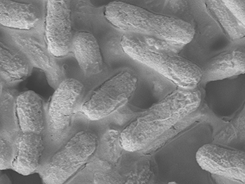
A microscopic image of the E. coli bacteria (credit: Institute of Bioengineering and Nanotechnology)

A new material that can kill E. coli bacteria within 30 seconds has been developed by researchers at the Institute of Bioengineering and Nanotechnology (IBN) of A*STAR in Singapore.

Triclosan, a common antibacterial ingredient found in many products such as toothpastes, soaps, and detergents to reduce or prevent bacterial infections, has been linked to making bacteria resistant to antibiotics, with adverse health effects. The European Union has restricted the use of triclosan in cosmetics, and the U.S. FDA is conducting an ongoing review of this ingredient.

Destroying the cell membrane to prevent resistance to antibiotics

To find a more suitable alternative, IBN Group Leader Yugen Zhang, PhD, and his team synthesized a chemical compound made up of molecules linked together in a chain (“imidazolium oligomers”), which they found can kill 99.7% of the E. coli bacteria within 30 seconds. The chain-like structure helps to penetrate the cell membrane and destroy the bacteria.

SEM image of E. coli before (left) and after treatment with IBN-C8 (right) (credit: S.N. Riduan et al./Small)

In contrast, current antibiotics only kill the bacteria but fail to destroy the cell membrane, allowing new antibiotic-resistant bacteria to grow.

“Our unique material can kill bacteria rapidly and inhibit the development of antibiotic-resistant bacteria. Computational chemistry studies supported our experimental findings that the chain-like compound works by attacking the cell membrane. This material is also safe for use because it carries a positive charge that targets the more negatively charged bacteria, without destroying red blood cells,” said Zhang.

The imidazolium oligomers come in the form of a white powder that is soluble in water. The researchers also found that once this was dissolved in alcohol, it formed gels spontaneously. So this material could be incorporated in alcohol-based sprays used for sterilization in hospitals or homes.

E. coli is a type of bacteria found in the intestines of humans and animals, and some strains can cause severe diarrhea, abdominal pain, and fever. Such infection is contagious and can spread through contaminated food or water, or from contact with people or animals.

Besides E. coli, IBN’s material was tested against other common strains of antibiotic-resistant bacteria and fungi such as Staphylococcus aureusPseudomonas aeruginosa, and Candida albicans (which can cause conditions ranging from skin infections to pneumonia and toxic shock syndrome) and was able to kill 99.9% of these microbes within two minutes.

The finding was published in the peer-reviewed journal Small.


Abstract of Ultrafast Killing and Self-Gelling Antimicrobial Imidazolium Oligomers

Infectious diseases and the increasing threat of worldwide pandemics have underscored the importance of antibiotics and hygiene. Intensive efforts have been devoted to developing new antibiotics to meet the rapidly growing demand. In particular, advancing the knowledge of the structure–property–activity relationship is critical to expedite the design and development of novel antimicrobial with the needed potential and efficacy. Herein, a series of new antimicrobial imidazolium oligomers are developed with the rational manipulation of terminal group’s hydrophobicity. These materials exhibit superior activity, excellent selectivity, ultrafast killing (>99.7% killing within 30 s), and desirable self-gelling properties. Molecular dynamic simulations reveal the delicate effect of structural changes on the translocation motion across the microbial cell membrane. The energy barrier of the translocation process analyzed by free energy calculations provides clear kinetic information to suggest that the spontaneous penetration requires a very short timescale of seconds to minutes for the new imidazolium oligomers.

Chronic stroke patients safely recover after injection of human stem cells


Injecting specially prepared human adult stem cells directly into the brains of chronic stroke patients proved safe and effective in restoring motor (muscle) function in a small clinical trial led by Stanford University School of Medicine investigators.

The 18 patients had suffered their first and only stroke between six months and three years before receiving the injections, which involved drilling a small hole through their skulls.

For most patients, at least a full year had passed since their stroke — well past the time when further recovery might be hoped for.  In each case, the stroke had taken place beneath the brain’s outermost layer, or cortex, and had severely affected motor function. “Some patients couldn’t walk,” Steinberg said. “Others couldn’t move their arm.”
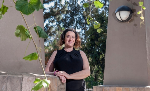
Sonia Olea Coontz had a stroke in 2011 that affected the movement of her right arm and leg. After modified stem cells were injected into her brain as part of a clinical trial, she says her limbs “woke up.” (credit: Mark Rightmire/Stanford University School of Medicine)

One of those patients, Sonia Olea Coontz, of Long Beach, California, now 36, had a stroke in May 2011. “My right arm wasn’t working at all,” said Coontz. “It felt like it was almost dead. My right leg worked, but not well.” She walked with a noticeable limp. “I used a wheelchair a lot. After my surgery, they woke up,” she said of her limbs.

‘Clinically meaningful’ results

The promising results set the stage for an expanded trial of the procedure now getting underway. They also call for new thinking regarding the permanence of brain damage, said Gary Steinberg, MD, PhD, professor and chair of neurosurgery.

“This was just a single trial, and a small one,” cautioned Steinberg, who led the 18-patient trial and conducted 12 of the procedures himself. (The rest were performed at the University of Pittsburgh.) “It was designed primarily to test the procedure’s safety. But patients improved by several standard measures, and their improvement was not only statistically significant, but clinically meaningful. Their ability to move around has recovered visibly. That’s unprecedented. At six months out from a stroke, you don’t expect to see any further recovery.”

The trial’s results are detailed in a paper published online June 2 in Stroke. Steinberg, who has more than 15 years’ worth of experience in work with stem cell therapies for neurological indications, is the paper’s lead and senior author.

The procedure involved injecting SB623 mesenchymal stem cells, derived from the bone marrow of two donors and then modified to beneficially alter the cells’ ability to restore neurologic function.*

Motor-function improvements

Substantial improvements were seen in patients’ scores on several widely accepted metrics of stroke recovery. Perhaps most notably, there was an overall 11.4-point improvement on the motor-function component of the Fugl-Meyer test, which specifically gauges patients’ movement deficits. “Patients who were in wheelchairs are walking now,” said Steinberg, who is the Bernard and Ronni Lacroute-William Randolph Hearst Professor in Neurosurgery and Neurosciences.

“We know these cells don’t survive for more than a month or so in the brain,” he added. “Yet we see that patients’ recovery is sustained for greater than one year and, in some cases now, more than two years.”

Importantly, the stroke patients’ postoperative improvement was independent of their age or their condition’s severity at the onset of the trial. “Older people tend not to respond to treatment as well, but here we see 70-year-olds recovering substantially,” Steinberg said. “This could revolutionize our concept of what happens after not only stroke, but traumatic brain injury and even neurodegenerative disorders. The notion was that once the brain is injured, it doesn’t recover — you’re stuck with it. But if we can figure out how to jump-start these damaged brain circuits, we can change the whole effect.

“We thought those brain circuits were dead. And we’ve learned that they’re not.”

New trial now recruiting 156 patients

A new randomized, double-blinded multicenter phase-2b trial aiming to enroll 156 chronic stroke patients is now actively recruiting patients. Steinberg is the principal investigator of that trial. For more information, you can e-mail stemcellstudy@stanford.edu. “There are close to 7 million chronic stroke patients in the United States,” Steinberg said. “If this treatment really works for that huge population, it has great potential.”

Some 800,000 people suffer a stroke each year in the United States alone. About 85 percent of all strokes are ischemic: They occur when a clot forms in a blood vessel supplying blood to part of the brain, with subsequent intensive damage to the affected area. The specific loss of function incurred depends on exactly where within the brain the stroke occurs, and on its magnitude.

Although approved therapies for ischemic stroke exist, to be effective they must be applied within a few hours of the event — a time frame that often is exceeded by the amount of time it takes for a stroke patient to arrive at a treatment center.

Consequently, only a small fraction of patients benefit from treatment during the stroke’s acute phase. The great majority of survivors end up with enduring disabilities. Some lost functionality often returns, but it’s typically limited. And the prevailing consensus among neurologists is that virtually all recovery that’s going to occur comes within the first six months after the stroke.

* Mesenchymal stem cells are the naturally occurring precursors of muscle, fat, bone and tendon tissues. In preclinical studies, though, they’ve not been found to cause problems by differentiating into unwanted tissues or forming tumors. Easily harvested from bone marrow, they appear to trigger no strong immune reaction in recipients even when they come from an unrelated donor. In fact, they may actively suppress the immune system. For this trial, unlike the great majority of transplantation procedures, the stem cell recipients received no immunosuppressant drugs.

During the procedure, patients’ heads were held in fixed positions while a hole was drilled through their skulls to allow for the injection of SB623 cells, accomplished with a syringe, into a number of spots at the periphery of the stroke-damaged area, which varied from patient to patient.

Afterward, patients were monitored via blood tests, clinical evaluations and brain imaging. Interestingly, the implanted stem cells themselves do not appear to survive very long in the brain. Preclinical studies have shown that these cells begin to disappear about one month after the procedure and are gone by two months. Yet, patients showed significant recovery by a number of measures within a month’s time, and they continued improving for several months afterward, sustaining these improvements at six and 12 months after surgery. Steinberg said it’s likely that factors secreted by the mesenchymal cells during their early postoperative presence near the stroke site stimulates lasting regeneration or reactivation of nearby nervous tissue.

No relevant blood abnormalities were observed. Some patients experienced transient nausea and vomiting, and 78 percent had temporary headaches related to the transplant procedure.


Abstract of Clinical Outcomes of Transplanted Modified Bone Marrow–Derived Mesenchymal Stem Cells in Stroke: A Phase 1/2a Study

Background and Purpose—Preclinical data suggest that cell-based therapies have the potential to improve stroke outcomes.

Methods—Eighteen patients with stable, chronic stroke were enrolled in a 2-year, open-label, single-arm study to evaluate the safety and clinical outcomes of surgical transplantation of modified bone marrow–derived mesenchymal stem cells (SB623).

Results—All patients in the safety population (N=18) experienced at least 1 treatment-emergent adverse event. Six patients experienced 6 serious treatment-emergent adverse events; 2 were probably or definitely related to surgical procedure; none were related to cell treatment. All serious treatment-emergent adverse events resolved without sequelae. There were no dose-limiting toxicities or deaths. Sixteen patients completed 12 months of follow-up at the time of this analysis. Significant improvement from baseline (mean) was reported for: (1) European Stroke Scale: mean increase 6.88 (95% confidence interval, 3.5–10.3;P<0.001), (2) National Institutes of Health Stroke Scale: mean decrease 2.00 (95% confidence interval, −2.7 to −1.3; P<0.001), (3) Fugl-Meyer total score: mean increase 19.20 (95% confidence interval, 11.4–27.0; P<0.001), and (4) Fugl-Meyer motor function total score: mean increase 11.40 (95% confidence interval, 4.6–18.2;P<0.001). No changes were observed in modified Rankin Scale. The area of magnetic resonance T2 fluid-attenuated inversion recovery signal change in the ipsilateral cortex 1 week after implantation significantly correlated with clinical improvement at 12 months (P<0.001 for European Stroke Scale).

Conclusions—In this interim report, SB623 cells were safe and associated with improvement in clinical outcome end points at 12 months.

Deep learning applied to drug discovery and repurposing

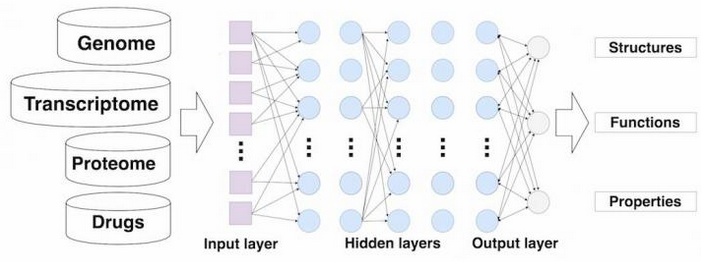
Deep neural networks for drug discovery (credit: Insilico Medicine, Inc.)

Scientists from Insilico Medicine, Inc. have trained deep neural networks (DNNs) to predict the potential therapeutic uses of 678 drugs, using gene-expression data obtained from high-throughput experiments on human cell lines from Broad Institute’s LINCS databases and NIH MeSH databases.

The supervised deep-learning drug-discovery engine used the properties of small molecules, transcriptional data, and literature to predict efficacy, toxicity, tissue-specificity, and heterogeneity of response.

“We used LINCS data from Broad Institute to determine the effects on cell lines before and after incubation with compounds, co-author and research scientist Polina Mamoshina explained to KurzweilIAI.

“We used gene expression data of total mRNA from cell lines extracted and measured before incubation with compound X and after incubation with compound X to identify the response on a molecular level. The goal is to understand how gene expression (the transcriptome) will change after drug uptake. It is a differential value, so we need a reference (molecular state before incubation) to compare.”

The research is described in a paper in the upcoming issue of the journal Molecular Pharmaceutics.

Helping pharmas accelerate R&D

Alex Zhavoronkov, PhD, Insilico Medicine CEO, who coordinated the study, said the initial goal of their research was to help pharmaceutical companies significantly accelerate their R&D and increase the number of approved drugs. “In the process we came up with more than 800 strong hypotheses in oncology, cardiovascular, metabolic, and CNS spaces and started basic validation,” he said.

The team measured the “differential signaling pathway activation score for a large number of pathways to reduce the dimensionality of the data while retaining biological relevance.” They then used those scores to train the deep neural networks.*

“This study is a proof of concept that DNNs can be used to annotate drugs using transcriptional response signatures, but we took this concept to the next level,” said Alex Aliper, president of research, Insilico Medicine, Inc., lead author of the study.

Via Pharma.AI, a newly formed subsidiary of Insilico Medicine, “we developed a pipeline for in silico drug discovery — which has the potential to substantially accelerate the preclinical stage for almost any therapeutic — and came up with a broad list of predictions, with multiple in silico validation steps that, if validated in vitro and in vivo, can almost double the number of drugs in clinical practice.”

Despite the commercial orientation of the companies, the authors agreed not to file for intellectual property on these methods and to publish the proof of concept.

Deep-learning age biomarkers

According to Mamoshina, earlier this month, Insilico Medicine scientists published the first deep-learned biomarker of human age — aiming to predict the health status of the patient — in a paper titled “Deep biomarkers of human aging: Application of deep neural networks to biomarker development” by Putin et al, in Aging; and an overview of recent advances in deep learning in a paper titled “Applications of Deep Learning in Biomedicine” by Mamoshina et al., also in Molecular Pharmaceutics.

Insilico Medicine is located in the Emerging Technology Centers at Johns Hopkins University in Baltimore, Maryland, in collaboration with Datalytic Solutions and Mind Research Network.

* In this study, scientists used the perturbation samples of 678 drugs across A549, MCF-7 and PC-3 cell lines from the Library of Integrated Network-Based Cellular Signatures (LINCS) project developed by the National Institutes of Health (NIH) and linked those to 12 therapeutic use categories derived from MeSH (Medical Subject Headings) developed and maintained by the National Library of Medicine (NLM) of the NIH.

To train the DNN, scientists utilized both gene level transcriptomic data and transcriptomic data processed using a pathway activation scoring algorithm, for a pooled dataset of samples perturbed with different concentrations of the drug for 6 and 24 hours. Cross-validation experiments showed that DNNs achieve 54.6% accuracy in correctly predicting one out of 12 therapeutic classes for each drug.

One peculiar finding of this experiment was that a large number of drugs misclassified by the DNNs had dual use, suggesting possible application of DNN confusion matrices in drug repurposing.


FutureTechnologies Media Group | Video presentation Insilico medicine


Abstract of Deep learning applications for predicting pharmacological properties of drugs and drug repurposing using transcriptomic data

Deep learning is rapidly advancing many areas of science and technology with multiple success stories in image, text, voice and video recognition, robotics and autonomous driving. In this paper we demonstrate how deep neural networks (DNN) trained on large transcriptional response data sets can classify various drugs to therapeutic categories solely based on their transcriptional profiles. We used the perturbation samples of 678 drugs across A549, MCF-7 and PC-3 cell lines from the LINCS project and linked those to 12 therapeutic use categories derived from MeSH. To train the DNN, we utilized both gene level transcriptomic data and transcriptomic data processed using a pathway activation scoring algorithm, for a pooled dataset of samples perturbed with different concentrations of the drug for 6 and 24 hours. When applied to normalized gene expression data for “landmark genes,” DNN showed cross-validation mean F1 scores of 0.397, 0.285 and 0.234 on 3-, 5- and 12-category classification problems, respectively. At the pathway level DNN performed best with cross-validation mean F1 scores of 0.701, 0.596 and 0.546 on the same tasks. In both gene and pathway level classification, DNN convincingly outperformed support vector machine (SVM) model on every multiclass classification problem. For the first time we demonstrate a deep learning neural net trained on transcriptomic data to recognize pharmacological properties of multiple drugs across different biological systems and conditions. We also propose using deep neural net confusion matrices for drug repositioning. This work is a proof of principle for applying deep learning to drug discovery and development.

Triggering the protein that programs cancer cells to kill themselves


WEHI | Apoptosis

Researchers at the Walter and Eliza Hall Institute in Australia have discovered a new way to trigger cell death that could lead to drugs to treat cancer and autoimmune disease.

Programmed cell death (a.k.a. apoptosis) is a natural process that removes unwanted cells from the body. Failure of apoptosis can allow cancer cells to grow unchecked or immune cells to inappropriately attack the body.

The protein known as Bak is central to apoptosis. In healthy cells, Bak sits in an inert state but when a cell receives a signal to die, Bak transforms into a killer protein that destroys the cell.

Triggering the cancer-apoptosis trigger

Institute researchers Sweta Iyer, PhD, Ruth Kluck, PhD, and colleagues unexpectedly discovered that an antibody they had produced to study Bak actually bound to the Bak protein and triggered its activation. They hope to use this discovery to develop drugs that promote cell death.

The researchers used information about Bak’s three-dimensional structure to find out precisely how the antibody activated Bak. “It is well known that Bak can be activated by a class of proteins called ‘BH3-only proteins’ that bind to a groove on Bak. We were surprised to find that despite our antibody binding to a completely different site on Bak, it could still trigger activation,” Kluck said.  “The advantage of our antibody is that it can’t be ‘mopped up’ and neutralized by pro-survival proteins in the cell, potentially reducing the chance of drug resistance occurring.”

Drugs that target this new activation site could be useful in combination with other therapies that promote cell death by mimicking the BH3-only proteins. The researchers are now working with collaborators to develop their antibody into a drug that can access Bak inside cells.

Their findings have just been published in the open-access journal Nature Communications. The research was supported by the National Health and Medical Research Council, the Australian Research Council, the Victorian State Government Operational Infrastructure Support Scheme, and the Victorian Life Science Computation Initiative.


Abstract of Identification of an activation site in Bak and mitochondrial Bax triggered by antibodies

During apoptosis, Bak and Bax are activated by BH3-only proteins binding to the α2–α5 hydrophobic groove; Bax is also activated via a rear pocket. Here we report that antibodies can directly activate Bak and mitochondrial Bax by binding to the α1–α2 loop. A monoclonal antibody (clone 7D10) binds close to α1 in non-activated Bak to induce conformational change, oligomerization, and cytochrome c release. Anti-FLAG antibodies also activate Bak containing a FLAG epitope close to α1. An antibody (clone 3C10) to the Bax α1–α2 loop activates mitochondrial Bax, but blocks translocation of cytosolic Bax. Tethers within Bak show that 7D10 binding directly extricates α1; a structural model of the 7D10 Fab bound to Bak reveals the formation of a cavity under α1. Our identification of the α1–α2 loop as an activation site in Bak paves the way to develop intrabodies or small molecules that directly and selectively regulate these proteins.