No longer ‘junk DNA’ — shedding light on the ‘dark matter’ of the genome

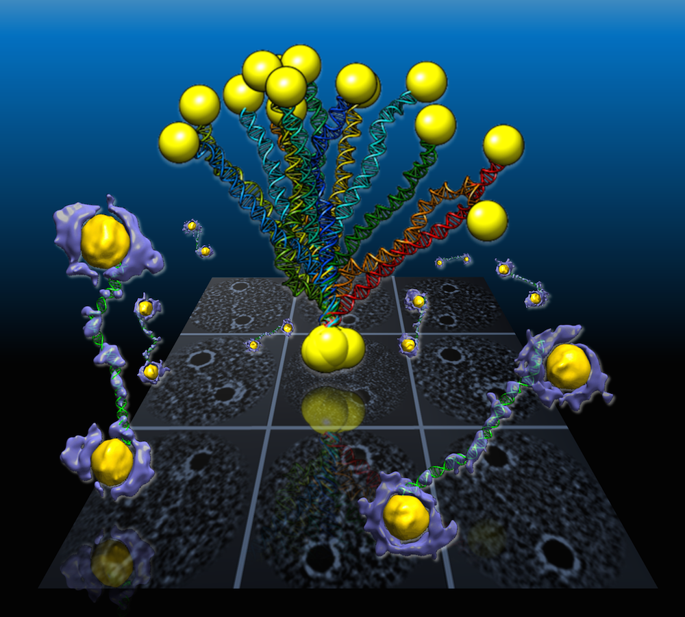
The LIGR-seq method for global-scale mapping of RNA-RNA interactions in vivo to reveal unexpected functions for uncharacterized RNAs that act via base-pairing interactions (credit: University of Toronto)

What used to be dismissed by many as “junk DNA” has now become vitally important, as accelerating genomic data points to the importance of non-coding RNAs (ncRNAs) — a genome’s messages that do not specifically code for proteins — in development and disease.

But our progress in understanding these molecules has been slow because of the lack of technologies that allow for systematic mapping of their functions.

Now, professor Benjamin Blencowe’s team at the University of Toronto’s Donnelly Centre has developed a method called “LIGR-seq” that enables scientists to explore in depth what ncRNAs do in human cells.

The study, described in Molecular Cell, was published on May 19, along with two other papers, in Molecular Cell and Cell, respectively, from Yue Wan’s group at the Genome Institute of Singapore and Howard Chang’s group at Stanford University in California, who developed similar methods to study RNAs in different organisms.

So what exactly do ncRNAs do?

mRNAs vs. ncRNAs (credit: Thomas Shafee/CC)

Of the 3 billion letters in the human genome, only two per cent make up the protein-coding genes. The genes are copied, or transcribed, into messenger RNA (mRNA) molecules, which provide templates for building proteins that do most of the work in the cell. Much of the remaining 98 per cent of the genome was initially considered by some as lacking in functional importance. However, large swaths of the non-coding genome — between half and three quarters of it — are also copied into RNA.

So then what might the resulting ncRNAs do? That depends on whom you ask. Some researchers believe that most ncRNAs have no function, that they are just a by-product of the genome’s powerful transcription machinery that makes mRNA. However, it is emerging that many ncRNAs do have important roles in gene regulation — some ncRNAs act as carriages for shuttling the mRNAs around the cell, or provide a scaffold for other proteins and RNAs to attach to and do their jobs.

But the majority of available data has trickled in piecemeal or through serendipitous discovery. And with emerging evidence that ncRNAs could drive disease progression, such as cancer metastasis, there was a great need for a technology that would allow a systematic functional analysis of ncRNAs.

Up until now, with existing methods, you had to know what you are looking for because they all require you to have some information about the RNA of interest. The power of our method is that you don’t need to preselect your candidates; you can see what’s occurring globally in cells, and use that information to look at interesting things we have not seen before and how they are affecting biology,” says Eesha Sharma, a PhD candidate in Blencowe’s group who, along with postdoctoral fellow Tim Sterne-Weiler, co-developed the method.

A new ncRNA identification tool

The human RNA-RNA interactome, showing interactions detected by LIGR-seq (credit: University of Toronto)

The new ‘‘LIGation of interacting RNA and high-throughput sequencing’’ (LIGR-seq) tool captures interactions between different RNA molecules. When two RNA molecules have matching sequences — strings of letters copied from the DNA blueprint — they will stick together like Velcro. With LIGR-seq, the paired RNA structures are removed from cells and analyzed by state-of-the-art sequencing methods to precisely identify the RNAs that are stuck together.

Most researchers in the life sciences agree that there’s an urgent need to understand what ncRNAs do. This technology will open the door to developing a new understanding of ncRNA function,” says Blencowe, who is also a professor in the Department of Molecular Genetics.

Not having to rely on pre-existing knowledge will boost the discovery of RNA pairs that have never been seen before. Scientists can also, for the first time, look at RNA interactions as they occur in living cells, in all their complexity, unlike in the juices of mashed up cells that they had to rely on before. This is a bit like moving on to explore marine biology from collecting shells on the beach to scuba-diving among the coral reefs, where the scope for discovery is so much bigger.

Actually, ncRNAs come in multiple flavors: there’s rRNA, tRNA, snRNA, snoRNA, piRNA, miRNA, and lncRNA, to name a few, where prefixes reflect the RNA’s place in the cell or some aspect of its function. But the truth is that no one really knows the extent to which these ncRNAs control what goes on in the cell, or how they do this.

Discoveries

Nonetheless, the new technology developed by Blencowe’s group has been able to pick up new interactions involving all classes of RNAs and has already revealed some unexpected findings.

The team discovered new roles for small nucleolar RNAs (snoRNAs), which normally guide chemical modifications of other ncRNAs. It turns out that some snoRNAs can also regulate stability of a set of protein-coding mRNAs. In this way, snoRNAs can also directly influence which proteins are made, as well as their abundance, adding a new level of control in cell biology.

And this is only the tip of the iceberg; the researchers plan to further develop and apply their technology to investigate the ncRNAs in different settings.

“We would like to understand how ncRNAs function during development. We are particularly interested in their role in the formation of neurons. But we will also use our method to discover and map changes in RNA-RNA interactions in the context of human diseases,” says Blencowe.


Abstract of Global Mapping of Human RNA-RNA Interactions

The majority of the human genome is transcribed into non-coding (nc)RNAs that lack known biological functions or else are only partially characterized. Numerous characterized ncRNAs function via base pairing with target RNA sequences to direct their biological activities, which include critical roles in RNA processing, modification, turnover, and translation. To define roles for ncRNAs, we have developed a method enabling the global-scale mapping of RNA-RNA duplexes crosslinked in vivo, “LIGation of interacting RNA followed by high-throughput sequencing” (LIGR-seq). Applying this method in human cells reveals a remarkable landscape of RNA-RNA interactions involving all major classes of ncRNA and mRNA. LIGR-seq data reveal unexpected interactions between small nucleolar (sno)RNAs and mRNAs, including those involving the orphan C/D box snoRNA, SNORD83B, that control steady-state levels of its target mRNAs. LIGR-seq thus represents a powerful approach for illuminating the functions of the myriad of uncharacterized RNAs that act via base-pairing interactions.

Gene helps prevent heart attack, stroke; may also block effects of aging

An atherosclerotic lesion. Such lesions can rupture and cause heart attacks and strokes. (credit: UVA School of Medicine)

University of Virginia School of Medicine have discovered that a gene called Oct4 — which scientific dogma insists is inactive in adults — actually plays a vital role in preventing ruptured atherosclerotic plaques inside blood vessels, the underlying cause of most heart attacks and strokes.

The researchers found that Oct4 controls the conversion of smooth muscle cells into protective fibrous “caps” inside plaques, making the plaques less likely to rupture. They also discovered that the gene promotes many changes in gene expression that are beneficial in stabilizing the plaques. In addition, the researchers believe it may be possible to develop drugs or other therapeutic agents that target the Oct4 pathway as a way to reduce the incidence of heart attacks or stroke.

Could impact many human diseases, regenerative medicine

The researchers are also currently testing Oct4′s possible role in repairing cellular damage and healing wounds, which would make it useful for regenerative medicine.

Oct4 is one of the “stem cell pluripotency factors” described by Shinya Yamanaka, PhD, of Kyoto University, for which he received the 2012 Nobel Prize. His lab and many others have shown that artificial over-expression of Oct4 within somatic cells grown in a lab dish is essential for reprogramming these cells into induced pluripotential stem cells, which can then develop into any cell type in the body or even an entire organism.

“Finding a way to reactivate this pathway may have profound implications for health and aging,” said researcher Gary K. Owens, director of UVA’s Robert M. Berne Cardiovascular Research Center. “This could impact many human diseases and the field of regenerative medicine. [It may also] end up being the ‘fountain-of-youth gene,’ a way to revitalize old and worn-out cells.”

The discovery is described in a paper published online in Nature Medicine. The work was funded by the National Institutes of Health, the Russian Science Foundation, the Russian Federal Agency of Scientific Organization, and the U.S. Department of Defense.


Abstract of Activation of the pluripotency factor OCT4 in smooth muscle cells is atheroprotective

Although somatic cell activation of the embryonic stem cell (ESC) pluripotency factor OCT4 has been reported, this previous work has been controversial and has not demonstrated a functional role for OCT4 in somatic cells. Here we demonstrate that smooth muscle cell (SMC)-specific conditional knockout of Oct4 in Apoe−/− mice resulted in increased lesion size and changes in lesion composition that are consistent with decreased plaque stability, including a thinner fibrous cap, increased necrotic core area, and increased intraplaque hemorrhage. Results of SMC-lineage-tracing studies showed that these effects were probably the result of marked reductions in SMC numbers within lesions and SMC investment within the fibrous cap, which may result from impaired SMC migration. The reactivation of Oct4 within SMCs was associated with hydroxymethylation of the Oct4promoter and was hypoxia inducible factor-1α (HIF-1α, encoded by HIF1A) and Krüppel-like factor-4 (KLF4)-dependent. These results provide the first direct evidence that OCT4 has a functional role in somatic cells, and they highlight the potential role of OCT4 in normal and diseased somatic cells.

Scientists digitally mimic evolution to create novel proteins

Overview of the SEWING method. (A) Continuous SEWING workflow. (B) Discontinuous SEWING workflow. Left: Parental Protein Data Banks are shown with extracted substructures; graph schematic — colored nodes indicate substructures contained in the final design model, and superimposed structures show structural similarity indicated by adjacent edges. Center: design model before sequence optimization and loop design. Right: final design models. (credit: T. M. Jacobs et al./Science)

Here’s an innovative idea: create new proteins by simply “sewing” together pieces of existing proteins. That’s exactly what researchers at the University of North Carolina School of Medicine have done to design new “cellular machines” needed to understand and battle diseases.

Published today in the journal Science, the new technique, called SEWING, was inspired by natural evolutionary mechanisms that also recombine portions of the 100,000 different known proteins in the body.

First, throw out the blueprint

Traditionally, researchers have used computational protein design to recreate in the laboratory what already exists in the natural world. But in recent years, their focus has shifted toward inventing novel proteins with new functionality. Those design projects all start with a specific structural “blueprint” in mind, and as a result are limited.

Senior study author Brian Kuhlman, PhD, professor of biochemistry and biophysics, and his colleagues believe that by removing the limitations of a pre-determined blueprint and taking cues from evolution, they can more easily create new functional proteins.

“We can now begin to think about engineering proteins to do things that nothing else is capable of doing,” he said. “The structure of a protein determines its function, so if we are going to learn how to design new functions, we have to learn how to design new structures. Our study is a critical step in that direction and provides tools for creating proteins that haven’t been seen before in nature.”

Mimicking nature

To mimic the mechanisms of natural protein evolution, they developed a computer design strategy called SEWING (Structure Extension With Native-substructure Graphs):

  1. The researchers digitally chopped up naturally occurring proteins into well-defined pieces.
  2. They performed a series of computational tests to figure out which pieces would fit well together. In nature, this step would involve looking for stretches of amino acid sequences that are similar between proteins. On the computer, it involved searching for regions of structural similarity to make things fit in the right place.
  3. They whittled down the list to the top 21 proteins, which they produced in the lab.
  4. To confirm what they created, they took pictures of these proteins using x-ray crystallography and nuclear magnetic resonance (NMR), and found that the proteins contained all the unique structural varieties they had designed on the computer.

Currently, the researchers are using SEWING to create proteins that can bind to several other proteins at a time. Many of the most important natural proteins are similar multitaskers, including the blood protein hemoglobin, which carries four copies of oxygen from the lungs to the body’s tissues.


Abstract of Design of structurally distinct proteins using strategies inspired by evolution

Natural recombination combines pieces of preexisting proteins to create new tertiary structures and functions. We describe a computational protocol, called SEWING, which is inspired by this process and builds new proteins from connected or disconnected pieces of existing structures. Helical proteins designed with SEWING contain structural features absent from other de novo designed proteins and, in some cases, remain folded at more than 100°C. High-resolution structures of the designed proteins CA01 and DA05R1 were solved by x-ray crystallography (2.2 angstrom resolution) and nuclear magnetic resonance, respectively, and there was excellent agreement with the design models. This method provides a new strategy to rapidly create large numbers of diverse and designable protein scaffolds.

Scientists turn skin cells into heart and brain cells using only drugs — no stem cells required

Neurons created from chemically induced neural stem cells. The cells were created from skin cells that were reprogrammed into neural stem cells using a cocktail of only nine chemicals. This is the first time cellular reprogramming has been accomplished without adding external genes to the cells. (credit: Mingliang Zhang, PhD, Gladstone Institutes)

Scientists at the Gladstone Institutes have used chemicals to transform skin cells into heart cells and brain cells, instead of adding external genes — making this accomplishment a breakthrough, according to the scientists.

The research lays the groundwork for one day being able to regenerate lost or damaged cells directly with pharmaceutical drugs — a more efficient and reliable method to reprogram cells and one that avoids medical concerns surrounding genetic engineering.

Instead, in two studies published in an open-access paper in Science and in Cell Stem Cell, the team of scientists at the Roddenberry Center for Stem Cell Biology and Medicine at Gladstone used chemical cocktails to gradually coax skin cells to change into organ-specific stem-cell-like cells and ultimately into heart or brain cells.

“This method brings us closer to being able to generate new cells at the site of injury in patients,” said Gladstone senior investigator Sheng Ding, PhD, the senior author on both studies. “Our hope is to one day treat diseases like heart failure or Parkinson’s disease with drugs that help the heart and brain regenerate damaged areas from their own existing tissue cells. This process is much closer to the natural regeneration that happens in animals like newts and salamanders, which has long fascinated us.”

Chemically Repaired Hearts

A human heart cell that was chemically reprogrammed from a human skin cell (credit: Nan Cao/Gladstone Institutes)

Transplanted adult heart cells do not survive or integrate properly into the heart and few stem cells can be coaxed into becoming heart cells.

Instead, in the Science study, the researchers used a cocktail of nine chemicals to change human skin cells into beating heart cells. By trial and error, they found the best combination of chemicals to begin the process by changing the cells into a state resembling multipotent stem cells (cells that can turn into many different types of cells in a particular organ). A second cocktail of chemicals and growth factors then helped transition the cells to become heart muscle cells.

With this method, more than 97% of the cells began beating, a characteristic of fully developed, healthy heart cells. The cells also responded appropriately to hormones, and molecularly, they resembled heart muscle cells, not skin cells. What’s more, when the cells were transplanted into a mouse heart early in the process, they developed into healthy-looking heart muscle cells within the organ.

“The ultimate goal in treating heart failure is a robust, reliable way for the heart to create new muscle cells,” said Srivastava, co-senior author on the Science paper. “Reprogramming a patient’s own cells could provide the safest and most efficient way to regenerate dying or diseased heart muscle.”

Rejuvenating the brain with neural stem cell-like cells

In the second study, authored by Gladstone postdoctoral scholar Mingliang Zhang, PhD, and published in Cell Stem Cell, the scientists created neural stem-cell-like cells from mouse skin cells using a similar approach.

The chemical cocktail again consisted of nine molecules, some of which overlapped with those used in the first study. Over ten days, the cocktail changed the identity of the cells, until all of the skin-cell genes were turned off and the genes of the neural stem-cell-like cells were gradually turned on.

When transplanted into mice, the neural stem-cell-like cells spontaneously developed into the three basic types of brain cells: neurons, oligodendrocytes, and astrocytes. The neural stem-cell-like cells were also able to self-replicate, making them ideal for treating neurodegenerative diseases or brain injury.

With their improved safety, these neural stem-cell-like cells could one day be used for cell replacement therapy in neurodegenerative diseases like Parkinson’s disease and Alzheimer’s disease, according to co-senior author Yadong Huang, MD, PhD, a senior investigator at Gladstone. “In the future, we could even imagine treating patients with a drug cocktail that acts on the brain or spinal cord, rejuvenating cells in the brain in real time.”


Gladstone Institutes | Chemically Reprogrammed Beating Heart Cell


Abstract of Conversion of human fibroblasts into functional cardiomyocytes by small molecules

Reprogramming somatic fibroblasts into alternative lineages would provide a promising source of cells for regenerative therapy. However, transdifferentiating human cells to specific homogeneous, functional cell types is challenging. Here we show that cardiomyocyte-like cells can be generated by treating human fibroblasts with a combination of nine compounds (9C). The chemically induced cardiomyocyte-like cells (ciCMs) uniformly contracted and resembled human cardiomyocytes in their transcriptome, epigenetic, and electrophysiological properties. 9C treatment of human fibroblasts resulted in a more open-chromatin conformation at key heart developmental genes, enabling their promoters/enhancers to bind effectors of major cardiogenic signals. When transplanted into infarcted mouse hearts, 9C-treated fibroblasts were efficiently converted to ciCMs. This pharmacological approach for lineage-specific reprogramming may have many important therapeutic implications after further optimization to generate mature cardiac cells.


Abstract of Pharmacological Reprogramming of Fibroblasts into Neural Stem Cells by Signaling-Directed Transcriptional Activation

Cellular reprogramming using chemically defined conditions, without genetic manipulation, is a promising approach for generating clinically relevant cell types for regenerative medicine and drug discovery. However, small-molecule approaches for inducing lineage-specific stem cells from somatic cells across lineage boundaries have been challenging. Here, we report highly efficient reprogramming of mouse fibroblasts into induced neural stem cell-like cells (ciNSLCs) using a cocktail of nine components (M9). The resulting ciNSLCs closely resemble primary neural stem cells molecularly and functionally. Transcriptome analysis revealed that M9 induces a gradual and specific conversion of fibroblasts toward a neural fate. During reprogramming specific transcription factors such as Elk1 and Gli2 that are downstream of M9-induced signaling pathways bind and activate endogenous master neural genes to specify neural identity. Our study provides an effective chemical approach for generating neural stem cells from mouse fibroblasts and reveals mechanistic insights into underlying reprogramming processes.

Super-stretchy, self-healing material could lead to artificial muscle

This extremely stretchable polymer film can repair itself when punctured, suggesting potential applications in artificial muscle. (credit: Bao Research Group)

Stanford researchers have developed a new material that can stretch to 100 times its original length by exposing it to an electric field, and even repair itself if punctured, making it potentially useful as an artificial muscle.

Artificial muscles currently have applications in some consumer technology and robotics, but small holes or defects in the materials currently used make them less resilient, and they can’t self-repair if punctured or scratched, according to Stanford chemical engineering professor Zhenan Bao.

Self-healing

The new material (Fe-Hpdca-PDMS polymer film) has remarkable self-healing characteristics. Damaged polymers typically require a solvent or heat treatment to restore their properties, but the new material can heal itself at room temperature (even if the damaged pieces are aged for days) and at temperatures as low as negative 4 degrees Fahrenheit (-20 C).

The team attributes the extreme stretching and self-healing ability to a chemical bonding process known as crosslinking. To create it, they designed special organic molecules to attach to the short polymer strands in their crosslink to create a series of structure called ligands. These ligands joined together to form longer polymer chains (spring-like coils with inherent stretchiness).

Then they added metal ions, which have a chemical affinity for the ligands. When this combined material is strained, the knots loosen and allow the ligands to separate. But when relaxed, the affinity between the metal ions and the ligands pulls the fishnet taut. The result is a strong, stretchable and self-repairing elastomer.

The team found that they could tune the polymer to be stretchier or heal faster by varying the amount or type of metal ion included. The version that exceeded the measuring machine’s limits, for example, was created by decreasing the ratio of iron atoms to the polymers and organic molecules in the material.

Artificial skin

They have to do more work to increase the degree to which the material expands and contracts and control it more precisely.

This research dovetails with Bao’s efforts to create artificial skin that might be used to restore some sensory capabilities to people with prosthetic limbs.

This work in the development of strong, flexible, electronically active polymers could also spawn a new generation of wearable electronics, or medical implants that would last a long time without being repaired or replaced, says Bao.

The research is described in the current issue of Nature Chemistry. It was supported by the Air Force Office of Scientific Research, Samsung Electronics, and the Major State Basic Research Development Program of China.


Stanford University School of Engineering | Super-stretchy polymer could lead to artificial muscle


Abstract of A highly stretchable autonomous self-healing elastomer

It is a challenge to synthesize materials that possess the properties of biological muscles—strong, elastic and capable of self-healing. Herein we report a network of poly(dimethylsiloxane) polymer chains crosslinked by coordination complexes that combines high stretchability, high dielectric strength, autonomous self-healing and mechanical actuation. The healing process can take place at a temperature as low as −20 °C and is not significantly affected by surface ageing and moisture. The crosslinking complexes used consist of 2,6-pyridinedicarboxamide ligands that coordinate to Fe(III) centres through three different interactions: a strong pyridyl–iron one, and two weaker carboxamido–iron ones through both the nitrogen and oxygen atoms of the carboxamide groups. As a result, the iron–ligand bonds can readily break and re-form while the iron centres still remain attached to the ligands through the stronger interaction with the pyridyl ring, which enables reversible unfolding and refolding of the chains. We hypothesize that this behaviour supports the high stretchability and self-healing capability of the material.

Microscope uses nanosecond-speed laser and deep learning to detect cancer cells more efficiently

This microscope uses specially designed optics that boost image clarity and slows them enough to be detected and digitized at a rate of 36 million images per second. It then uses deep learning to distinguish cancer cells from healthy white blood cells. (credit: Tunde Akinloye/CNSI)

Scientists at the California NanoSystems Institute at UCLA have developed a new technique for identifying cancer cells in blood samples faster and more accurately than the current standard methods.

In one common approach to testing for cancer, doctors add biochemicals to blood samples. Those biochemicals attach biological “labels” to the cancer cells, and those labels enable instruments to detect and identify them. However, the biochemicals can damage the cells and render the samples unusable for future analyses. There are other current techniques that don’t use labeling but can be inaccurate because they identify cancer cells based only on one physical characteristic.

Time-stretch quantitative phase imaging (TS-QPI) and analytics system

The new technique images cells without destroying them and can identify 16 physical characteristics — including size, granularity and biomass — instead of just one.

Time-stretch quantitative phase imaging (TS-QPI) and analytics system (credit: Claire Lifan Chen et al./Nature Scientific Reports)

The new technique combines two components that were invented at UCLA:

  • A “photonic time stretch” microscope, which is capable of quickly imaging cells in blood samples. Invented by Barham Jalali, professor and Northrop-Grumman Optoelectronics Chair in electrical engineering, it works by taking pictures of flowing blood cells using laser bursts (similar to how a camera uses a flash). Each flash only lasts nanoseconds (billionths of a second) to avoid damage to cells, but that normally means the images are both too weak to be detected and too fast to be digitized by normal instrumentation. The new microscope overcomes those challenges by using specially designed optics that amplify and boost the clarity of the images, and simultaneously slow them down enough to be detected and digitized at a rate of 36 million images per second.
  • A deep learning computer program, which identifies cancer cells with more than 95 percent accuracy. Deep learning is a form of artificial intelligence that uses complex algorithms to extract patterns and knowledge from rich multidimenstional datasets, with the goal of achieving accurate decision making.

The study was published in the open-access journal Nature Scientific Reports. The researchers write in the paper that the system could lead to data-driven diagnoses by cells’ physical characteristics, which could allow quicker and earlier diagnoses of cancer, for example, and better understanding of the tumor-specific gene expression in cells, which could facilitate new treatments for disease.

The research was supported by NantWorks, LLC.


Abstract of Deep Learning in Label-free Cell Classification

Label-free cell analysis is essential to personalized genomics, cancer diagnostics, and drug development as it avoids adverse effects of staining reagents on cellular viability and cell signaling. However, currently available label-free cell assays mostly rely only on a single feature and lack sufficient differentiation. Also, the sample size analyzed by these assays is limited due to their low throughput. Here, we integrate feature extraction and deep learning with high-throughput quantitative imaging enabled by photonic time stretch, achieving record high accuracy in label-free cell classification. Our system captures quantitative optical phase and intensity images and extracts multiple biophysical features of individual cells. These biophysical measurements form a hyperdimensional feature space in which supervised learning is performed for cell classification. We compare various learning algorithms including artificial neural network, support vector machine, logistic regression, and a novel deep learning pipeline, which adopts global optimization of receiver operating characteristics. As a validation of the enhanced sensitivity and specificity of our system, we show classification of white blood T-cells against colon cancer cells, as well as lipid accumulating algal strains for biofuel production. This system opens up a new path to data-driven phenotypic diagnosis and better understanding of the heterogeneous gene expressions in cells.

Chinese team genetically modifies human embryo, using CRISPR gene-editing technique

Photos showing injection of a human 3PN zygote (left) and development to 8 to 16-cell stage in vitro (right). Scale bar = 100 μm. (credit: Xiangjin Kang et al./Journal of Assisted Reproduction and Genetics)

Chinese researchers have genetically modified a human embryo using CRISPR/Cas9, the gene editing technique, using embryos that carried an extra set of chromosomes (so they were not viable) — hoping to learn more about the possibility of producing human babies that would be immune to HIV.

The Chinese team reports in the Journal of Assisted Reproduction and Genetics that they obtained 213 fertilized eggs from a fertility clinic, which had been deemed unsuitable for in vitro therapy.* They used the eggs to study a mutation that causes damage to an immune cell gene called CCR5 (this type of cell, when damaged naturally, has been found to lead to HIV resistance).

Aside from a previous study in China last year that involved editing human embryo genes (see Researchers in China have created genetically modified human embryos), most of the world has decided to hold off doing this controversial research. “We believe that any attempt to generate genetically modified humans through the modification of early embryos needs to be strictly prohibited until we can resolve both ethical and scientific issues,” the researchers say in their paper.

Failed experiment

The team reports that just 4 out of 26 of the embryos that were edited were modified successfully — some still contained genes that had not been modified, and others had resulted in unexpected gene mutations.

“This paper doesn’t look like it offers much more than anecdotal evidence that it works in human embryos, which we already knew,” George Daley, a stem-cell biologist at Children’s Hospital Boston, told Nature. “It’s certainly a long way from realizing the intended potential” — a human embryo with all its copies of CCR5 inactivated.

“It just emphasizes that there are still a lot of technical difficulties to doing precision editing in human embryo cells,” Xiao-Jiang Li, a neuroscientist at Emory University, also said in the Nature article . He thinks that researchers should work out these kinks in non-human primates, for example, before continuing to modify the genomes of human embryos using techniques such as CRISPR.

* The women who had donated the eggs all gave permission for the embryos to be used for genetic research, on condition that the embryos would not be allowed to mature into a human being (all of the embryos were destroyed after three days).


Abstract of Introducing precise genetic modifications into human 3PN embryos by CRISPR/Cas-mediated genome editing

Purpose

As a powerful technology for genome engineering, the CRISPR/Cas system has been successfully applied to modify the genomes of various species. The purpose of this study was to evaluate the technology and establish principles for the introduction of precise genetic modifications in early human embryos.

Methods

3PN zygotes were injected with Cas9 messenger RNA (mRNA) (100 ng/μl) and guide RNA (gRNA) (50 ng/μl). For oligo-injections, donor oligo-1 (99 bp) or oligo-2 (99 bp) (100 ng/μl) or dsDonor (1 kb) was mixed with Cas9 mRNA (100 ng/μl) and gRNA (50 ng/μl) and injected into the embryos.

Results

By co-injecting Cas9 mRNA, gRNAs, and donor DNA, we successfully introduced the naturally occurring CCR5Δ32 allele into early human 3PN embryos. In the embryos containing the engineeredCCR5Δ32 allele, however, the other alleles at the same locus could not be fully controlled because they either remained wild type or contained indel mutations.

Conclusions

This work has implications for the development of therapeutic treatments of genetic disorders, and it demonstrates that significant technical issues remain to be addressed. We advocate preventing any application of genome editing on the human germline until after a rigorous and thorough evaluation and discussion are undertaken by the global research and ethics communities.

Best textile manufacturing methods for creating human tissues with stem cells

All four textile manufacturing processes and corresponding scaffold (structure) types studied exhibited the presence of lipid vacuoles (small red spheres, right column, indicating stem cells undergoing random differentiation), compared to control (left). Electrospun scaffolds (row a) exhibited only a monolayer of lipid vacuoles in a single focal plane, while meltblown, spunbond, and carded scaffolds (rows b, c, d) exhibited vacuoles in multiple planes throughout the fabric thickness. Scale bars: 100 μm (credit: S. A. Tuin et al./Biomedical Materials)

Elizabeth Loboa, dean of the Missouri University College of Engineering, and her team have tested new tissue- engineering methods (based on textile manufacturing) to find ones that are most cost-effective and can be produced in larger quantities.

Tissue engineering is a process that uses novel biomaterials seeded with stem cells to grow and replace missing tissues. When certain types of materials are used, the “scaffolds” that are created to hold stem cells eventually degrade, leaving natural tissue in its place. The new tissues could help patients suffering from wounds caused by diabetes and circulation disorders, patients in need of cartilage or bone repair, and women who have had mastectomies by replacing their breast tissue. The challenge is creating enough of the material on a scale that clinicians need to treat patients.

Comparing textile manufacturing techniques

Electrospinning experiment: nanofibers are collected into an ethanol bath and removed at predefined time intervals (credit: J. M. Coburn et al./The Johns Hopkins University/PNAS)

In typical tissue engineering approaches that use fibers as scaffolds, non-woven materials are often bonded together using an electrostatic field. This process, called electrospinning (see Nanoscale scaffolds and stem cells show promise in cartilage repair and Improved artificial blood vessels), creates the scaffolds needed to attach to stem cells.

However, large-scale production with electrospinning is not cost-effective. “Electrospinning produces weak fibers, scaffolds that are not consistent, and pores that are too small,” Loboa said. “The goal of ‘scaling up’ is to produce hundreds of meters of material that look the same, have the same properties, and can be used in clinical settings. So we investigated the processes that create textiles, such as clothing and window furnishings like drapery, to scale up the manufacturing process.”

The group published two papers using three industry-standard, high-throughput manufacturing techniques — meltblowing, spunbonding, and carding — to determine if they would create the materials needed to mimic native tissue.

Meltblowing is a technique during which nonwoven materials are created using a molten polymer to create continuous fibers. Spunbond materials are made much the same way but the fibers are drawn into a web while in a solid state instead of a molten one. Carding involves the separation of fibers through the use of rollers, forming the web needed to hold stem cells in place.

Schematic of gilled fiber multifilament spinning and carded scaffold fabrication (credit: Stephen A. Tuin et al./Acta Biomaterialia)

Cost-effective methods

Loboa and her colleagues tested these techniques to create polylactic acid (PLA) scaffolds (a Food and Drug Administration-approved material used as collagen fillers), seeded with human stem cells. They then spent three weeks studying whether the stem cells remained healthy and if they began to differentiate into fat and bone pathways, which is the goal of using stem cells in a clinical setting when new bone and/or new fat tissue is needed at a defect site. Results showed that the three textile manufacturing methods proved as viable if not more so than electrospinning.

“These alternative methods are more cost-effective than electrospinning,” Loboa said. “A small sample of electrospun material could cost between $2 to $5. The cost for the three manufacturing methods is between $.30 to $3.00; these methods proved to be effective and efficient. Next steps include testing how the different scaffolds created in the three methods perform once implanted in animals.”

Researchers at North Carolina State University and the University of North Carolina at Chapel Hill were also involved in the two studies, which were published in Biomedical Materials (open access) and Acta Biomaterialia. The National Science Foundation, the National Institutes of Health, and the Nonwovens Institute provided funding for the studies.


Abstract of Creating tissues from textiles: scalable nonwoven manufacturing techniques for fabrication of tissue engineering scaffolds

Electrospun nonwovens have been used extensively for tissue engineering applications due to their inherent similarities with respect to fibre size and morphology to that of native extracellular matrix (ECM). However, fabrication of large scaffold constructs is time consuming, may require harsh organic solvents, and often results in mechanical properties inferior to the tissue being treated. In order to translate nonwoven based tissue engineering scaffold strategies to clinical use, a high throughput, repeatable, scalable, and economic manufacturing process is needed. We suggest that nonwoven industry standard high throughput manufacturing techniques (meltblowing, spunbond, and carding) can meet this need. In this study, meltblown, spunbond and carded poly(lactic acid) (PLA) nonwovens were evaluated as tissue engineering scaffolds using human adipose derived stem cells (hASC) and compared to electrospun nonwovens. Scaffolds were seeded with hASC and viability, proliferation, and differentiation were evaluated over the course of 3 weeks. We found that nonwovens manufactured via these industry standard, commercially relevant manufacturing techniques were capable of supporting hASC attachment, proliferation, and both adipogenic and osteogenic differentiation of hASC, making them promising candidates for commercialization and translation of nonwoven scaffold based tissue engineering strategies.


Abstract of Fabrication of novel high surface area mushroom gilled fibers and their effects on human adipose derived stem cells under pulsatile fluid flow for tissue engineering applications

The fabrication and characterization of novel high surface area hollow gilled fiber tissue engineering scaffolds via industrially relevant, scalable, repeatable, high speed, and economical nonwoven carding technology is described. Scaffolds were validated as tissue engineering scaffolds using human adipose derived stem cells (hASC) exposed to pulsatile fluid flow (PFF). The effects of fiber morphology on the proliferation and viability of hASC, as well as effects of varied magnitudes of shear stress applied via PFF on the expression of the early osteogenic gene marker runt related transcription factor 2 (RUNX2) were evaluated. Gilled fiber scaffolds led to a significant increase in proliferation of hASC after seven days in static culture, and exhibited fewer dead cells compared to pure PLA round fiber controls. Further, hASC-seeded scaffolds exposed to 3 and 6 dyn/cm2 resulted in significantly increased mRNA expression of RUNX2 after one hour of PFF in the absence of soluble osteogenic induction factors. This is the first study to describe a method for the fabrication of high surface area gilled fibers and scaffolds. The scalable manufacturing process and potential fabrication across multiple nonwoven and woven platforms makes them promising candidates for a variety of applications that require high surface area fibrous materials.

Statement of Significance

We report here for the first time the successful fabrication of novel high surface area gilled fiber scaffolds for tissue engineering applications. Gilled fibers led to a significant increase in proliferation of human adipose derived stem cells after one week in culture, and a greater number of viable cells compared to round fiber controls. Further, in the absence of osteogenic induction factors, gilled fibers led to significantly increased mRNA expression of an early marker for osteogenesis after exposure to pulsatile fluid flow. This is the first study to describe gilled fiber fabrication and their potential for tissue engineering applications. The repeatable, industrially scalable, and versatile fabrication process makes them promising candidates for a variety of scaffold-based tissue engineering applications.

Berkeley Lab captures first high-res 3D images of DNA segments

In a Berkeley Lab-led study, flexible double-helix DNA segments (purple, with green DNA models) connected to gold nanoparticles (yellow) are revealed from the 3D density maps reconstructed from individual samples using a Berkeley Lab-developed technique called individual-particle electron tomography (IPET). Projections of the structures are shown in the green background grid. (credit: Berkeley Lab)

An international research team working at the Lawrence Berkeley National Laboratory (Berkeley Lab) has captured the first high-resolution 3D images of double-helix DNA segments attached at either end to gold nanoparticles — which could act as building blocks for molecular computer memory and electronic devices (see World’s smallest electronic diode made from single DNA molecule), nanoscale drug-delivery systems, and as markers for biological research and for imaging disease-relevant proteins.

The researchers connected coiled DNA strands between polygon-shaped gold nanoparticles and then reconstructed 3D images, using a cutting-edge electron microscope technique coupled with a protein-staining process and sophisticated software that provided structural details at the scale of about 2 nanometers.

“We had no idea about what the double-strand DNA would look like between the gold nanoparticles,” said Gang “Gary” Ren, a Berkeley Lab scientist who led the research. “This is the first time for directly visualizing an individual double-strand DNA segment in 3D,” he said.

The results were published in an open-access paper in the March 30 edition of Nature Communications.

The method developed by this team, called individual-particle electron tomography (IPET), had earlier captured the 3-D structure of a single protein that plays a key role in human cholesterol metabolism. By grabbing 2D images of an object from different angles, the technique allows researchers to assemble a 3D image of that object.

The team has also used the technique to uncover the fluctuation of another well-known flexible protein, human immunoglobulin 1, which plays a role in the human immune system.


Berkeley Lab | 3-D Reconstructions of Double strand DNA and Gold Nanoparticle Structures

For this new study of DNA nanostructures, Ren used an electron-beam study technique called cryo-electron microscopy (cryo-EM) to examine frozen DNA-nanogold samples, and used IPET to reconstruct 3-D images from samples stained with heavy metal salts. The team also used molecular simulation tools to test the natural shape variations (“conformations”) in the samples, and compared these simulated shapes with observations.

First visualization of DNA strand dynamics without distorting x-ray crystallography

Ren explained that the naturally flexible dynamics of samples, like a man waving his arms, cannot be fully detailed by any method that uses an average of many observations.

A popular way to view the nanoscale structural details of delicate biological samples is to form them into crystals and zap them with X-rays, but that destroys their natural shape, especially fir the DNA-nanogold samples in this study, which the scientists say are incredibly challenging to crystallize. Other common research techniques may require a collection of thousands of near-identical objects, viewed with an electron microscope, to compile a single, averaged 3-D structure. But an averaged 3D image may not adequately show the natural shape fluctuations of a given object.

The samples in the latest experiment were formed from individual polygon gold nanostructures, measuring about 5 nanometers across, connected to single DNA-segment strands with 84 base pairs. Base pairs are basic chemical building blocks that give DNA its structure. Each individual DNA segment and gold nanoparticle naturally zipped together with a partner to form the double-stranded DNA segment with a gold particle at either end.


Berkeley Lab | These views compare the various shape fluctuations obtained from different samples of the same type of double-helix DNA segment (DNA renderings in green, 3D reconstructions in purple) connected to gold nanoparticles (yellow).

The samples were flash-frozen to preserve their structure for study with cryo-EM imaging. The distance between the two gold nanoparticles in individual samples varied from 20 to 30 nanometers, based on different shapes observed in the DNA segments.

Researchers used a cryo-electron microscope at Berkeley Lab’s Molecular Foundry for this study. They collected a series of tilted images of the stained objects, and reconstructed 14 electron-density maps that detailed the structure of individual samples using the IPET technique.

Sub-nanometer images next

Ren said that the next step will be to work to improve the resolution to the sub-nanometer scale.

“Even in this current state we begin to see 3-D structures at 1- to 2-nanometer resolution,” he said. “Through better instrumentation and improved computational algorithms, it would be promising to push the resolution to that visualizing a single DNA helix within an individual protein.”

In future studies, researchers could attempt to improve the imaging resolution for complex structures that incorporate more DNA segments as a sort of “DNA origami,” Ren said. Researchers hope to build and better characterize nanoscale molecular devices using DNA segments that can, for example, store and deliver drugs to targeted areas in the body.

“DNA is easy to program, synthesize and replicate, so it can be used as a special material to quickly self-assemble into nanostructures and to guide the operation of molecular-scale devices,” he said. “Our current study is just a proof of concept for imaging these kinds of molecular devices’ structures.”

The team included researchers at UC Berkeley, the Kavli Energy NanoSciences Institute at Berkeley Lab and UC Berkeley, and Xi’an Jiaotong University in China. This work was supported by the National Science Foundation, DOE Office of Basic Energy Sciences, National Institutes of Health, the National Natural Science Foundation of China, Xi’an Jiaotong University in China, and the Ministry of Science and Technology in China. View more about Gary Ren’s research group here.


Abstract of Three-dimensional structural dynamics and fluctuations of DNA-nanogold conjugates by individual-particle electron tomography

DNA base pairing has been used for many years to direct the arrangement of inorganic nanocrystals into small groupings and arrays with tailored optical and electrical properties. The control of DNA-mediated assembly depends crucially on a better understanding of three-dimensional structure of DNA-nanocrystal-hybridized building blocks. Existing techniques do not allow for structural determination of these flexible and heterogeneous samples. Here we report cryo-electron microscopy and negative-staining electron tomography approaches to image, and three-dimensionally reconstruct a single DNA-nanogold conjugate, an 84-bp double-stranded DNA with two 5-nm nanogold particles for potential substrates in plasmon-coupling experiments. By individual-particle electron tomography reconstruction, we obtain 14 density maps at ~2-nm resolution. Using these maps as constraints, we derive 14 conformations of dsDNA by molecular dynamics simulations. The conformational variation is consistent with that from liquid solution, suggesting that individual-particle electron tomography could be an expected approach to study DNA-assembling and flexible protein structure and dynamics.

A stem-cell repair system that can regenerate any kind of human tissue

UNSW researchers say the therapy has enormous potential for treating spinal disc injury and joint and muscle degeneration and could also speed up recovery following complex surgeries where bones and joints need to integrate with the body (credit: UNSW TV)

A stem cell therapy system capable of regenerating any human tissue damaged by injury, disease, or aging could be available within a few years, say University of New South Wales (UNSW Australia) researchers.

Their new repair system*, similar to the method used by salamanders to regenerate limbs, could be used to repair everything from spinal discs to bone fractures, and could transform current treatment approaches to regenerative medicine.

The UNSW-led research was published this week in the Proceedings of the National Academy of Sciences journal.

Reprogramming bone and fat cells

The system reprograms bone and fat cells into induced multipotent stem cells (iMS), which can regenerate multiple tissue types and has been successfully demonstrated in mice, according to study lead author, haematologist, and UNSW Associate Professor John Pimanda.

“This technique is a significant advance on many of the current unproven stem cell therapies, which have shown little or no objective evidence they contribute directly to new tissue formation,” Pimanda said. “We have taken bone and fat cells, switched off their memory and converted them into stem cells so they can repair different cell types once they are put back inside the body.”

“We are currently assessing whether adult human fat cells reprogrammed into iMS cells can safely repair damaged tissue in mice, with human trials expected to begin in late 2017.”

(credit: UNSW Media/Michael Whitehead)

Advantages over stem-cell types

There are different types of stem cells including embryonic stem (ES) cells, which during embryonic development generate every type of cell in the human body, and adult stem cells, which are tissue-specific, but don’t regenerate multiple tissue types. Embryonic stem cells cannot be used to treat damaged tissues because of their tumor forming capacity. The other problem when generating stem cells is the requirement to use viruses to transform cells into stem cells, which is clinically unacceptable, the researchers note.

Research shows that up to 20% of spinal implants either don’t heal or there is delayed healing. The rates are higher for smokers, older people and patients with diseases such diabetes or kidney disease.

Human trials are planned next year once the safety and effectiveness of the technique using human cells in mice has been demonstrated.

* The technique involves extracting adult human fat cells and treating them with the compound 5-Azacytidine (AZA), along with platelet-derived growth factor-AB (PDGF-AB) for about two days. The cells are then treated with the growth factor alone for a further two-three weeks.

AZA is known to induce cell plasticity, which is crucial for reprogramming cells. The AZA compound relaxes the hard-wiring of the cell, which is expanded by the growth factor, transforming the bone and fat cells into iMS cells. When the stem cells are inserted into the damaged tissue site, they multiply, promoting growth and healing.

The new technique is similar to salamander limb regeneration, which is also dependent on the plasticity of differentiated cells, which can repair multiple tissue types, depending on which body part needs replacing.

Along with confirming that human adult fat cells reprogrammed into iMS stem cells can safely repair damaged tissue in mice, the researchers said further work is required to establish whether iMS cells remain dormant at the sites of transplantation and retain their capacity to proliferate on demand.


UNSW Australia | Breakthrough for stem cell therapies


Abstract of PDGF-AB and 5-Azacytidine induce conversion of somatic cells into tissue-regenerative multipotent stem cells

Current approaches in tissue engineering are geared toward generating tissue-specific stem cells. Given the complexity and heterogeneity of tissues, this approach has its limitations. An alternate approach is to induce terminally differentiated cells to dedifferentiate into multipotent proliferative cells with the capacity to regenerate all components of a damaged tissue, a phenomenon used by salamanders to regenerate limbs. 5-Azacytidine (AZA) is a nucleoside analog that is used to treat preleukemic and leukemic blood disorders. AZA is also known to induce cell plasticity. We hypothesized that AZA-induced cell plasticity occurs via a transient multipotent cell state and that concomitant exposure to a receptive growth factor might result in the expansion of a plastic and proliferative population of cells. To this end, we treated lineage-committed cells with AZA and screened a number of different growth factors with known activity in mesenchyme-derived tissues. Here, we report that transient treatment with AZA in combination with platelet-derived growth factor–AB converts primary somatic cells into tissue-regenerative multipotent stem (iMS) cells. iMS cells possess a distinct transcriptome, are immunosuppressive, and demonstrate long-term self-renewal, serial clonogenicity, and multigerm layer differentiation potential. Importantly, unlike mesenchymal stem cells, iMS cells contribute directly to in vivo tissue regeneration in a context-dependent manner and, unlike embryonic or pluripotent stem cells, do not form teratomas. Taken together, this vector-free method of generating iMS cells from primary terminally differentiated cells has significant scope for application in tissue regeneration.