Synapse-like memristor-based electronic device detects brain spikes in real time

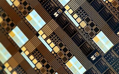
Memristor chip (credit: University of Southampton)

A bio-inspired electronic device called a memristor could allow for real-time processing of neuronal signals (spiking events), new research led by the University of Southampton has demonstrated.

The research could lead to using multi-electrode array implants for detecting spikes in the brain’s electrical signals from more than 1,000 recording channels to help treat neurological conditions, without requiring expensive, high-bandwidth, bulky systems for processing data. The research could lead to future autonomous, fully implantable neuroprosthetic devices.

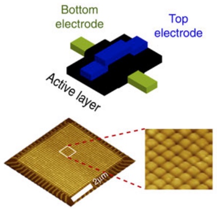
Schematic illustration of a solid-state titanium-oxide memristive device and atomic force microscopic (AFM) image a portion of a 32 × 32 crossbar array of memristors (credit: Isha Gupta/Nature Communications)

A memristors is an electronic component that limits or regulates the flow of electrical current in a circuit, can remember the amount of charge that was flowing through it, and retain that data, even when the power is turned off. The researchers used an array of memristors.

The research team designed a new nanoscale device they called a “memristive integrating sensor” (MIS) based on a memristors and associated electronic circuits for detecting spikes.*

Acting like synapses in the brain, the MIS was able to encode and compress (up to 200 times) neuronal spiking activity recorded by multi-electrode arrays. Besides addressing the bandwidth constraints, this approach was also very power-efficient; the power needed per recording channel was up to 100 times less when compared to current best practice.

The research was published in the open-access journal Nature Communications.

The Prodromakis Group at the University of Southampton collaborated among others with Leon Chua (a Diamond Jubilee Visiting Academic at the University of Southampton), who theoretically predicted the existence of memristors in 1971.

This interdisciplinary work was supported by an FP7 project (the European Union’s Research and Innovation funding) and brought together engineers from the Nanoelectronics and Nanotechnology Group at the University of Southampton with biologists from the University of Padova and the Max Planck Institute, Germany, using the state-of-art facilities of the Southampton Nanofabrication Centre.

* The paper explains that signals from an array of neural electrodes are fed into the MIS system as a series of voltage-time samples. “The MIS begins by pre-amplifying the incoming signal to voltage levels suitable for operating the memristor sitting at the core of the MIS and then proceeding to apply the pre-amplified signals to the memristor in real-time. The memristor’s resistive state is assessed periodically and when a significant change in comparison to the previous state is detected, the system registers a spiking event.”


Abstract of Real-time encoding and compression of neuronal spikes by metal-oxide memristors

Advanced brain-chip interfaces with numerous recording sites bear great potential for investigation of neuroprosthetic applications. The bottleneck towards achieving an efficient bio-electronic link is the real-time processing of neuronal signals, which imposes excessive requirements on bandwidth, energy and computation capacity. Here we present a unique concept where the intrinsic properties of memristive devices are exploited to compress information on neural spikes in real-time. We demonstrate that the inherent voltage thresholds of metal-oxide memristors can be used for discriminating recorded spiking events from background activity and without resorting to computationally heavy off-line processing. We prove that information on spike amplitude and frequency can be transduced and stored in single devices as non-volatile resistive state transitions. Finally, we show that a memristive device array allows for efficient data compression of signals recorded by a multi-electrode array, demonstrating the technology’s potential for building scalable, yet energy-efficient on-node processors for brain-chip interfaces.

A thought-controlled robotic exoskeleton for the hand

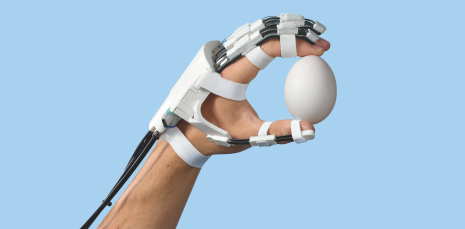
A robotic hand exoskeleton helps stroke patients integrate rehabilitation exercises into their everyday lives. (credit: Gerber Loesch Photography)

Scientists at ETH Rehabilitation Engineering Laboratory in Switzerland have invented a robotic system that they say could fundamentally change the daily lives of stroke patients.

According to the ETH scientists, one in six people will suffer a stroke in their lifetime; two thirds of those affected suffer from paralysis of the arm. Intensive clinical training, including robot-assisted therapy, can help patients regain a degree of limited control over their arms and hands.

But now Roger Gassert, Professor of Rehabilitation Engineering at ETH Zurich, has a better idea. “My vision is that instead of performing exercises in an abstract situation at the clinic, patients will be able to integrate them into their daily life at home, supported in some cases by a robot” — using an exoskeleton mounted on the hand.

A lightweight exoskeleton that extends the patient’s hand

The problem: existing exoskeletons are heavy, so patients can’t lift their hands, Gassert says, and patients have difficulty feeling objects and exerting the right amount of force. “That’s why we wanted to develop a model that leaves the palm of the hand more or less free, allowing patients to perform daily activities that support not only motor (movement) functions but somatosensory functions as well.”

The initial solution, developed with Professor Jumpei Arata from Kyushu University (Japan), was a mechanism for the finger featuring three overlapping leaf springs. A motor moves the middle spring, which transmits the force to the different segments of the finger through the other two springs. The fingers thus automatically adapt to the shape of the object the patient wants to grasp. But the motors brought the weight of the exoskeleton to 250 grams, which in clinical tests proved too heavy for patients.

The new solution: remove the motors from the hand and fix them to the patient’s back. The force is transmitted to the exoskeleton using a bicycle brake cable. The hand module now weighs slightly less than 120 grams and is strong enough to lift a liter bottle of mineral water.

A hand exoskeleton with motors that can be fixed to the patient’s back: A bicycle brake cable transmits enough force to lift a liter bottle of mineral water. (credit: Stefan Schneller)

Strengthening existing neural connections between brain and hand

Another problem was making sure commands from the brain can reach the extremities after a stroke. “Especially with seriously affected patients, the connection between the brain and the hand is often severely or completely disrupted,” Gassert explains.

The idea is to enable the brain to detect a patient’s intention to move his or her hand and directly pass this information on to the exoskeleton.

Gassert says a number of studies show that it is possible to strengthen existing neural connections between the brain and the hand with regular exercise if the brain can receive somatosensory feedback from the hand when it produces a command to move.

Gassert is using electroencephalography (EEG) and functional near-infrared spectroscopy (fNIRS) to study this. An interaction between the brain and the exoskeleton could lead to a device that is ideally suited for therapy — without requiring brain implants.

Even if the deficits are permanent, a robotic device could still offer vital long-term support.

Highest-resolution map of the entire human brain created

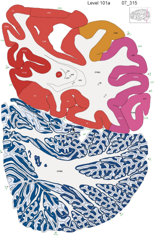
Allen Human Brain Reference Atlas image (credit: Allen Institute for Brain Science)

The Allen Institute for Brain Science has published the highest-resolution atlas of the human brain to date in a stand-alone issue of the Journal of Comparative Neurology. This digital human brain atlas allows researchers to investigate the structural basis of human brain function and is freely available as part of the suite of Allen Brain Atlas tools at brain-map.org.

“To understand the human brain, we need to have a detailed description of its underlying structure,” says Ed Lein, Ph.D., Investigator at the Allen Institute for Brain Science. “Human brain atlases have long lagged behind atlases of the brain of worms, flies or mice, both in terms of spatial resolution and in terms of completeness due to technical limitations related to the enormous size and complexity of the human brain. This large-scale focused effort aimed to create a large resource combining different data types at high resolution, and use these data to generate a comprehensive mapping of brain regions.”

Combining neuroimaging with cellular resolution histological analysis and expert structural mapping, “This is the most structurally complete atlas to date and we hope it will serve as a new reference standard for the human brain across different disciplines,” says Lein.

Cellular microscopic resolution

To create this modern atlas, the team at the Allen Institute partnered with Bruce Fischl, Ph.D. at Massachusetts General Hospital to perform magnetic resonance and diffusion tensor imaging on an intact brain before it was cut into slabs and serially sectioned to allow histological staining of individual sections. This imaging on the same brain created opportunities for linking fine molecular and cellular studies of the brain in health and disease with non-invasive neuroimaging studies.

The Allen Human Brain Reference Atlas digitized the histological data at true cellular microscopic resolution, creating a complete ontology of brain regions, and delineating all brain regions on a series of cross-sections through the brain. To image these sections, the Allen Institute team had to develop a new tile-based scanner that could image tissue sections the size of a complete human brain hemisphere at the resolution of 1 micrometer per pixel.

The Allen Human Brain Reference Atlas is an integrated, freely accessible online resource that allows users to navigate the brain and travel from the macroscopic scale of full brain sections to the level of individual neurons.

The Allen Human Brain Reference is “the only brain atlas to date to combine the rigor of a peer-reviewed scientific research paper with a presentation as a book format that includes the full set of annotated plates, open-access online availability of the resource with the ability to navigate and explore the details of any given area, and have programmatic access to the underlying data,” said Patrick Hof, M.D., Editor-in-Chief of the Journal of Comparative Neurology and a participant in the project.


Abstract of Comprehensive cellular-resolution atlas of the adult human brain

Detailed anatomical understanding of the human brain is essential for unraveling its functional architecture, yet current reference atlases have major limitations such as lack of whole-brain coverage, relatively low image resolution, and sparse structural annotation. We present the first digital human brain atlas to incorporate neuroimaging, high-resolution histology, and chemoarchitecture across a complete adult female brain, consisting of magnetic resonance imaging (MRI), diffusion-weighted imaging (DWI), and 1,356 large-format cellular resolution (1 µm/pixel) Nissl and immunohistochemistry anatomical plates. The atlas is comprehensively annotated for 862 structures, including 117 white matter tracts and several novel cyto- and chemoarchitecturally defined structures, and these annotations were transferred onto the matching MRI dataset. Neocortical delineations were done for sulci, gyri, and modified Brodmann areas to link macroscopic anatomical and microscopic cytoarchitectural parcellations. Correlated neuroimaging and histological structural delineation allowed fine feature identification in MRI data and subsequent structural identification in MRI data from other brains. This interactive online digital atlas is integrated with existing Allen Institute for Brain Science gene expression atlases and is publicly accessible as a resource for the neuroscience community. J. Comp. Neurol. 524:3127-3481, 2016. © 2016 The Authors The Journal of Comparative Neurology Published by Wiley Periodicals, Inc.

Ultrasound jump-starts brain of man in coma

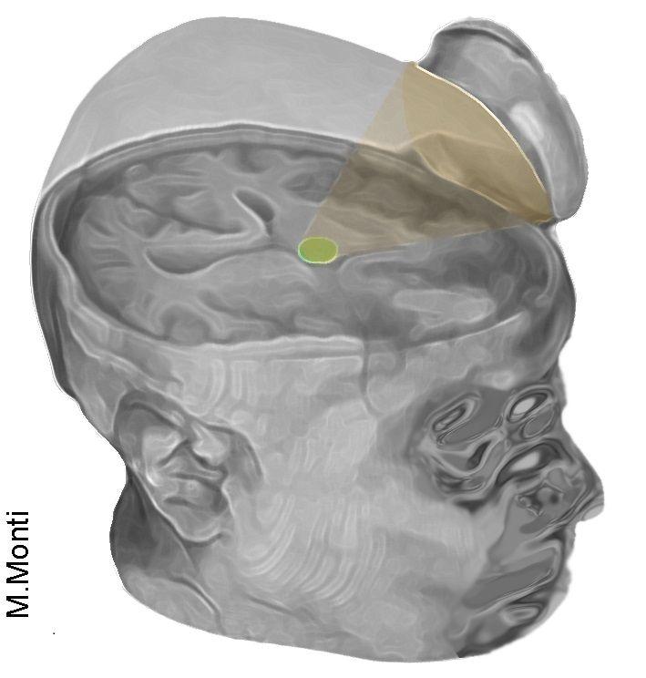
The non-invasive technique uses ultrasound to target the brain’s thalamus (credit: Martin Monti/UCLA)

UCLA neurosurgeons used ultrasound to “jump-start” the brain of a 25-year-old man from a coma, and he has made remarkable progress following the treatment.

The technique, called “low-intensity focused ultrasound pulsation” (LIFUP), works non-invasively and without affecting intervening tissues. It excites neurons in the thalamus, an egg-shaped structure that serves as the brain’s central hub for processing information.

“It’s almost as if we were jump-starting the neurons back into function,” said Martin Monti, the study’s lead author and a UCLA associate professor of psychology and neurosurgery. “Until now, the only way to achieve this was a risky surgical procedure known as deep brain stimulation, in which electrodes are implanted directly inside the thalamus,” he said. “Our approach directly targets the thalamus but is noninvasive.”

What about using it on vegetative or minimally conscious patients?

Monti cautioned that the procedure requires further study on additional patients before the scientists can determine whether it could be used consistently to help other people recovering from comas.

“It is possible that we were just very lucky and happened to have stimulated the patient just as he was spontaneously recovering,” Monti said.

If the technology helps other people recovering from coma, Monti said, it could eventually be used to build a portable device — perhaps incorporated into a helmet — as a low-cost way to help “wake up” patients, perhaps even those who are in a vegetative or minimally conscious state (MCS). Currently, there is almost no effective treatment for such patients, he said.


Israel Stinson (credit: Life Legal Defense Foundation)

On Thursday August 25, two year-old Israel Stinson, whose fight for life gained international attention, died Thursday after doctors at Children’s Hospital of Los Angeles removed him from a breathing ventilator against his parents’ wishes, after a Los Angeles Superior Court judge removed a restraining order, the Los Angeles Times reports.

It’s not clear, going forward, why doctors or the FDA could ethically refuse to provide “compassionate access” to a treatment such as LIFUP as a last resort before pulling the plug.

Speculation: brain hackers will start (have started?) experimenting with LIFUP as a brain stimulant.

“Foggy from all-night cramming for midterms? LIFUP it!”


Safer than DBS and tDCS

report on the treatment is published in the journal Brain Stimulation. This is the first time the approach has been used to treat severe brain injury.

The authors note that the new technique combines the advantages of highly invasive deep brain stimulation (DBS) and transcranial direct current stimulation (tDCS) while avoiding their respective disadvantages.

Low-intensity focused ultrasound pulsation (credit: Brainsonix)

The procedure was pioneered by Alexander Bystritsky, a UCLA professor of psychiatry and biobehavioral sciences in the Semel Institute for Neuroscience and Human Behavior and a co-author of the study.

Bystritsky is also a founder of Brainsonix, a Sherman Oaks, California-based company that provided the device (BXPulsar 1001) the researchers used in the study.

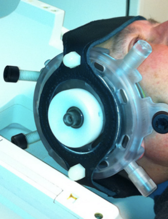
That device, about the size of a coffee cup saucer, creates a small sphere of acoustic energy that can be aimed at different regions of the brain to excite brain tissue.

For the new study, researchers placed it by the side of the man’s head and activated it 10 times for 30 seconds each, in a 10-minute period.

Monti said the device is safe because it emits only a small amount of energy — less than a conventional Doppler ultrasound.

“First-in-man” clinical trial

The patient was brought to the Ronald Reagan Medical Center (RRMC) at UCLA after suffering a road-traffic-related severe brain injury, with a field Glasgow Coma Scale (GCS) of 3 (“severe”). The patient had severe traumatic brain injury with prolonged loss of consciousness (more than 24 hours) post-injury.

Before the procedure began, the man showed only minimal signs of being conscious and of understanding speech. For example, he could perform small, limited movements when asked. By the day after the treatment, his responses had improved measurably.

Three days later, the patient had regained full consciousness and full language comprehension, and he could reliably communicate by nodding his head “yes” or shaking his head “no,” consistent with emergence from MCS (eMCS). He even made a fist-bump gesture to say goodbye to one of his doctors.

“The changes were remarkable,” Monti said.

The technique targets the thalamus because, in people whose mental function is deeply impaired after a coma, thalamus performance is typically diminished. Medications that are commonly prescribed to people who are coming out of a coma only indirectly target the thalamus.

Under the direction of Paul Vespa, a UCLA professor of neurology and neurosurgery at the David Geffen School of Medicine at UCLA, the researchers plan to test the procedure on several more people beginning this fall at the Ronald Reagan UCLA Medical Center. Those tests will be conducted in partnership with the UCLA Brain Injury Research Center and funded in part by the Dana Foundation and the Tiny Blue Dot Foundation.

UPDATE 8/29/2016: clarified depth of coma


Abstract of Non-Invasive Ultrasonic Thalamic Stimulation in Disorders of Consciousness after Severe Brain Injury: A First-in-Man Report

Modern intensive care medicine has greatly increased the rates of survival after severe brain injury (BI). Nonetheless, a number of patients fail to fully recover from coma, and awaken to a disorder of consciousness (DOC) such as the vegetative state (VS) or the minimally conscious state (MCS) [1]. In these conditions, which can be transient or last indefinitely, patients can lose virtually all autonomy and have almost no treatment options [1,2]. In addition, these conditions place great emotional and financial strain on families, lead to increased burn-out rates among care-takers, impose financial stress on medical structures and public finances due to the costs of prolonged intensive care, and raise difficult legal and ethical questions [3].

Implantable cell-size ‘neural pixel’ device senses and blocks epileptic seizures

A biochemical system for reducing epileptic activity (experimentally generated chemically) in mice hippocampus brain tissue. The miniature “neural pixel” device (bottom) sensed the epileptic attack and then delivered the natural calming neurotransmitter GABA via PEDOT:PSS electrodes, which also  recorded the subsequent electrophysiological activity to confirm effectiveness. (credit: Amanda Jonsson et al./PNAS)

Researchers at Linköping University in Sweden and in France have developed a “neural pixel” device that when implanted in a mouse hippocampus brain slice detects the initial signal of an epileptic attack and also locally administers the exact dose of the natural neurotransmitter GABA needed to stop the attack.

The researchers used a conducting polymer called poly(3,4-ethylenedioxythiophene) doped with poly(styrenesulfonate) (PEDOT:PSS) for electrodes. It has ten times better conductivity than gold, platinum, and iridium electrodes.  A tiny organic electronic ion pump* was used to pump the GABA neurotransmitter through a selective membrane, enabling high spatiotemporal delivery resolution (tiny and works fast) without requiring liquid flow (which is hard to control).

The idea is to have local, real-time measurement and precision delivery directly to specific neurons, which could pave the way in the future to closed-loop, fully automatic, miniature therapeutic devices. Combining electronic detection and release in the same electrode is a major advance, according to the researchers.

The development offers an alternative to drugs taken orally, which may be toxic outside the brain, may not cross the blood−brain barrier, or may have deleterious side effects when they penetrate the brain’s “healthy” regions, affecting physiological functions such as memory and learning. It also opens up a range of opportunities in basic neuroscience.

The research results have been published in the journal Proceedings of the National Academy of Sciences (PNAS).

* The implantable ion pump was developed at Linköping University’s Laboratory for Organic Electronics and announced in 2015. It was used initially to deliver exact dosages of painkiller GABA to the exact location where the pain signals reach the spinal cord for further transmission to the brain, and could be in clinical use in five to ten years, the researchers say.

The neural sensor for the initial signal of an epileptic attack was developed by the LiU researchers’ collaborators at the École Nationale Supérieure des Mines in Gardanne, France. The mouse experiments were performed at Aix-Marseille University. The entire device is manufactured from conductive, biocompatible plastic.

The Swedish part of the research was funded by Vinnova, the Swedish Research Council, and the Knut and Alice Wallenberg Foundation. The work took place at the OBOE center, under the leadership of Asst. Prof. Daniel Simon and Professor Magnus Berggren.


Abstract of Bioelectronic neural pixel: Chemical stimulation and electrical sensing at the same site

Local control of neuronal activity is central to many therapeutic strategies aiming to treat neurological disorders. Arguably, the best solution would make use of endogenous highly localized and specialized regulatory mechanisms of neuronal activity, and an ideal therapeutic technology should sense activity and deliver endogenous molecules at the same site for the most efficient feedback regulation. Here, we address this challenge with an organic electronic multifunctional device that is capable of chemical stimulation and electrical sensing at the same site, at the single-cell scale. Conducting polymer electrodes recorded epileptiform discharges induced in mouse hippocampal preparation. The inhibitory neurotransmitter, γ-aminobutyric acid (GABA), was then actively delivered through the recording electrodes via organic electronic ion pump technology. GABA delivery stopped epileptiform activity, recorded simultaneously and colocally. This multifunctional “neural pixel” creates a range of opportunities, including implantable therapeutic devices with automated feedback, where locally recorded signals regulate local release of specific therapeutic agents.

Neuroscientists identify cortical links to adrenal medulla (mind-body connection)

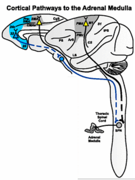
Cortical pathways to the adrenal medulla. Cortical areas on the lateral surface and the medial wall of the hemisphere are the source of neurons that influence the adrenal medulla. Gray shading: cortical motor areas; blue shading: medial prefrontal areas. (credit: Richard P. Dum et al./PNAS)

Neuroscientists at the University of Pittsburgh have identified the neural networks that connect the cerebral cortex to the adrenal medulla — the inner part of the adrenal gland, located above each kidney, which is responsible for the body’s rapid response in stressful situations.

These findings, reported in the online Early Edition of the journal Proceedings of the National Academy of Sciences PNAS), provide evidence for the neural basis of a mind-body connection. They also shed new light on how stress, depression, and other mental states can alter organ function, and show that there is a real anatomical basis for psychosomatic illness.

The research also identifies a concrete neural substrate that may help explain why meditation and certain exercises such as yoga and Pilates can be so helpful in modulating the body’s responses to physical, mental and emotional stress, according to senior author Peter L. Strick, Ph.D., Thomas Detre Chair of the Department of Neurobiology and scientific director of the University of Pittsburgh Brain Institute.

Why it matters which cortical areas influence the adrenal medulla

In their experiments with monkeys, the scientists traced the neural circuitry that links areas of the cerebral cortex to the adrenal medulla, using a unique tracing method that involves rabies virus. This approach is capable of revealing long chains of interconnected neurons.

Different hormones produced in different zones of the cortex and medulla of the gland (credit: Regents of University of Michigan Medical School)

Another surprising result of the research was that motor areas in the cerebral cortex, which are involved in the planning and performance of movement, provide a substantial input to the adrenal medulla. One of these areas is a portion of the primary motor cortex that is concerned with the control of axial body movement and posture. This input to the adrenal medulla may explain why core body exercises are so helpful in modulating responses to stress. Calming practices such as Pilates, yoga, tai chi and even dancing in a small space all require proper skeletal alignment, coordination, and flexibility to avoid harm to the body.

The PNAS study also revealed that the areas of the cortex that are active when we sense conflict, or are aware that we have made an error, are a source of influence over the adrenal medulla. “This observation,” said Dr. Strick, “raises the possibility that activity in these cortical areas when you re-imagine an error, or beat yourself up over a mistake, or think about a traumatic event, results in descending signals that influence the adrenal medulla in just the same way as the actual event.”

These anatomical findings have relevance for therapies that deal with post-traumatic stress.

Additional links with the adrenal medulla were discovered in cortical areas that are active during mindful mediation and areas that show changes in bipolar familial depression. “One way of summarizing our results is that we may have uncovered the stress and depression connectome,” says Strick.

Overall, these results indicate that circuits exist to link movement, cognition and affect to the function of the adrenal medulla and the control of stress. This circuitry may mediate the effects of internal states like chronic stress and depression on organ function and thus provide a concrete neural substrate for some psychosomatic illness.

This research was funded by National Institutes of Health and the Pennsylvania Department of Health.


Abstract of Motor, cognitive, and affective areas of the cerebral cortex influence the adrenal medulla

Modern medicine has generally viewed the concept of “psychosomatic” disease with suspicion. This view arose partly because no neural networks were known for the mind, conceptually associated with the cerebral cortex, to influence autonomic and endocrine systems that control internal organs. Here, we used transneuronal transport of rabies virus to identify the areas of the primate cerebral cortex that communicate through multisynaptic connections with a major sympathetic effector, the adrenal medulla. We demonstrate that two broad networks in the cerebral cortex have access to the adrenal medulla. The larger network includes all of the cortical motor areas in the frontal lobe and portions of somatosensory cortex. A major component of this network originates from the supplementary motor area and the cingulate motor areas on the medial wall of the hemisphere. These cortical areas are involved in all aspects of skeletomotor control from response selection to motor preparation and movement execution. The second, smaller network originates in regions of medial prefrontal cortex, including a major contribution from pregenual and subgenual regions of anterior cingulate cortex. These cortical areas are involved in higher-order aspects of cognition and affect. These results indicate that specific multisynaptic circuits exist to link movement, cognition, and affect to the function of the adrenal medulla. This circuitry may mediate the effects of internal states like chronic stress and depression on organ function and, thus, provide a concrete neural substrate for some psychosomatic illness.

Anti-inflammatory drug reverses memory loss in Alzheimer’s-disease-model mice

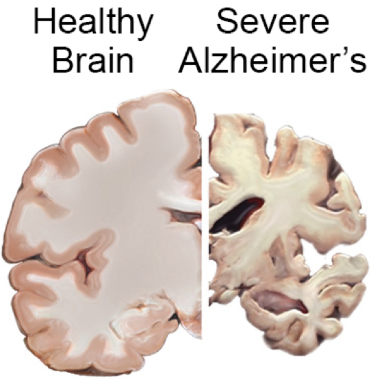
(credit: NIH National Institute on Aging)

Anti-inflammatory drug mefenamic acid completely reversed memory loss and brain inflammation in mice genetically engineered to develop symptoms of Alzheimer’s disease and amyloid beta-induced memory loss, a team led by David Brough, PhD, from the University of Manchester has discovered.

The non-steroidal anti-inflammatory drug (NSAID) drug targets an important inflammatory pathway called the NLRP3 inflammasome, which damages brain cells, according to Brough. This is the first time a drug has been shown to target this inflammatory pathway, highlighting its importance in the disease model, Brough said.

“Because this drug is already available and the toxicity and pharmacokinetics of the drug is known, the time for it to reach patients should, in theory, be shorter than if we were developing completely new drugs. We are now preparing applications to perform early phase II trials to determine a proof-of-concept that the molecules have an effect on neuroinflammation in humans.”

“There is experimental evidence now to strongly suggest that inflammation in the brain makes Alzheimer’s disease worse. Until now, no drug has been available to target this pathway, so we are very excited by this result.”

The research, funded by the Medical Research Council and the Alzheimer’s Society, paves the way for human trials that the team hopes to conduct in the future, but Brough cautions that more research is needed to identify its impact on humans and the long-term implications of its use.

The findings were published Thursday Aug. 11 in an open-access paper authored by Brough and colleagues in the journal Nature Communications.


Abstract of Fenamate NSAIDs inhibit the NLRP3 inflammasome and protect against Alzheimer’s disease in rodent models

Non-steroidal anti-inflammatory drugs (NSAIDs) inhibit cyclooxygenase-1 (COX-1) and COX-2 enzymes. The NLRP3 inflammasome is a multi-protein complex responsible for the processing of the proinflammatory cytokine interleukin-1β and is implicated in many inflammatory diseases. Here we show that several clinically approved and widely used NSAIDs of the fenamate class are effective and selective inhibitors of the NLRP3 inflammasome via inhibition of the volume-regulated anion channel in macrophages, independently of COX enzymes. Flufenamic acid and mefenamic acid are efficacious in NLRP3-dependent rodent models of inflammation in air pouch and peritoneum. We also show therapeutic effects of fenamates using a model of amyloid beta induced memory loss and a transgenic mouse model of Alzheimer’s disease. These data suggest that fenamate NSAIDs could be repurposed as NLRP3 inflammasome inhibitors and Alzheimer’s disease therapeutics.

Ultrasonic wireless ‘neural dust’ sensors monitor nerves, muscles in real time

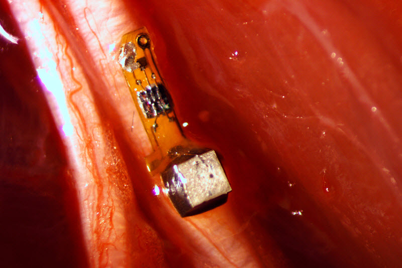
Prototype wireless battery-less “neural dust” mote (3 x 1 x 1 millimeters) with electrodes attached to a nerve fiber in a rat. The mote contains a piezoelectric crystal (silver cube) that converts ultrasonic signals to electrical current, powering a simple electronic circuit containing a transistor (black square) that responds to the voltage generated by a nerve firing and triggers the piezoelectric crystal to create ultrasonic backscatter, which indicates detection of a neural signal. (photo credit: Ryan Neely/UC Berkeley)

University of California, Berkeley engineers have designed and built millimeter-scale device wireless, batteryless “neural dust” sensors and implanted them in muscles and peripheral nerves of rats to make in vivo electrophysiological recordings.

The new technology opens the door to “electroceuticals” — bioelectronic methods to monitor and record wireless electromyogram (EMG) signals from muscle membranes and electroneurogram (ENG) signals from local neuron electrical activity, and to stimulate the immune system, reduce inflammation, and treat disorders such as epilepsy.

The technology could also improve neural control of prosthetics (allowing a paraplegic to control a computer or a robotic arm, for example) by stimulating nerves and muscles directly, instead of requiring implanted wires.

The neural-dust sensors use ultrasound technology to both power the sensors and read out measurements. Ultrasound is already well-developed for hospital use and can penetrate nearly anywhere in the body, unlike radio waves.

The researchers reported their findings August 3 in an open-access paper in the journal Neuron.

How a neural dust “mote” sensor monitors neural and muscle signals

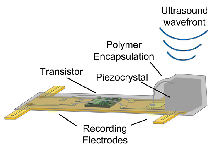
Diagram showing the components of the neural-dust mote (sensor). The entire device is covered in a biocompatible gel. (credit: Dongjin Seo et al./Neuron)

1. A team implants the neural dust mote. In the reported study, the mote was implanted in the rat sciatic nerve to do ENG recordings and in the gastrocnemius muscle to do EMG recordings. The tether-less connection also avoids potential infections and adverse biological responses due to micro-motion of the implant within the tissue.

2. An external ultrasonic generator sends a ultrasound signal to a piezoelectric crystal, which converts the sound energy into an electrical voltage, used to power a transistor circuit — no battery required.

3. When neurons or muscle fibers fire, they generate a tiny voltage (action potential) that the two electrodes pick up and send to the transistor.

4. The transistor amplifies the signal and drives the piezoelectric crystal to vibrate at an ultrasonic rate.

5. That vibration interferes with the transmitted ultrasonic signal, causing a modified “backscatter” signal that communicates information about the voltage across the sensor’s two electrodes.

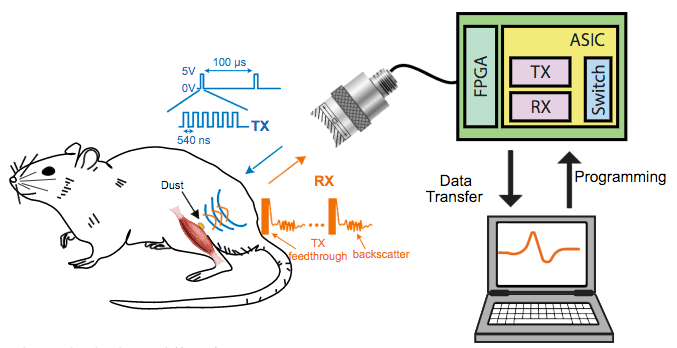
In vivo experimental setup for a tether-less neural dust electromyography (EMG) recording from the gastrocnemius muscle in rats. The neural dust mote was placed on the exposed muscle surface, and the wound was closed with surgical suture. The ASIC chip switches between ultrasound transmit and receive modules.Triggered by the FPGA chip, the external transducer sends ultrasound (TX, green) to the mote. The backscatter received signal (RX, red) carries information about the EMG voltage that is decoded and recorded/displayed on a computer. The dust mote was pinged every 100 microseconds with six 540-nanosecond ultrasound pulses. (credit: Dongjin Seo et al./Neuron)

6. The backscatter ultrasound signal is decoded to extract EMG or ENG data.

7. A computer displays and records the information.

Microscale motes: future research

The experiments so far have involved the peripheral nervous system and muscles, using an external ultrasonic patch over the implanted site to acquire  information from the motes for the desired diagnosis or therapy.

But according to the researchers, neural dust motes can be implanted anywhere in the body, including the central nervous system and brain to control prosthetics. This would be an alternative to today’s implantable electrodes (for Parkinson’s disease, for example), which require wires that pass through holes in the skull and degrade within one to two years.

The researchers are now building motes from biocompatible thin films, which would potentially last in the body without degradation for a decade or more. Up to hundreds of wireless sensors could be sealed in, avoiding infection and unwanted movement of the electrodes, and could last a timeline, according to the researchers.

The team is also now working to miniaturize the device further and they plan to use beam-steering technology to focus the ultrasonic signals on individual motes. The team is also building little backpacks for rats to hold the ultrasound transceiver that will record data from implanted motes. And the researchers are working to expand the motes’ ability to detect non-electrical signals, such as oxygen or hormone levels.

The researchers estimate that they could eventually shrink the sensors down to a cube 50 micrometers on a side. At that size, the motes could monitor a few specific nerve axons and continually record their electrical activity.

The researchers conceived of the idea of neural dust about five years ago, but initial attempts to power an implantable device and read out the data using radio waves were disappointing. Radio attenuates very quickly with distance in tissue, so communicating with devices deep in the body would be difficult without using potentially damaging high-intensity radiation. In 2013, the researchers published an open-access arXiv paper that described how a neural-dust system with ultrasonic signals might work.

The ongoing research is supported by the U.S. Defense Advanced Research Projects Agency as part of DARPA’s Electrical Prescriptions (ElectRx) program, which is focused in part on developing interface technologies that are suitable for chronic use for biosensing and neuromodulation of specific peripheral nerves.


UC Berkeley | “Neural dust” sensor


Abstract of Wireless Recording in the Peripheral Nervous System with Ultrasonic Neural Dust

The emerging field of bioelectronic medicine seeks methods for deciphering and modulating electrophysiological activity in the body to attain therapeutic effects at target organs. Current approaches to interfacing with peripheral nerves and muscles rely heavily on wires, creating problems for chronic use, while emerging wireless approaches lack the size scalability necessary to interrogate small-diameter nerves. Furthermore, conventional electrode-based technologies lack the capability to record from nerves with high spatial resolution or to record independently from many discrete sites within a nerve bundle. Here, we demonstrate neural dust, a wireless and scalable ultrasonic backscatter system for powering and communicating with implanted bioelectronics. We show that ultrasound is effective at delivering power to mm-scale devices in tissue; likewise, passive, battery-less communication using backscatter enables high-fidelity transmission of electromyogram (EMG) and electroneurogram (ENG) signals from anesthetized rats. These results highlight the potential for an ultrasound-based neural interface system for advancing future bioelectronics-based therapies.

No, exercise does not wipe out previous memories

2016 Rio Games Opening Ceremony promo (credit: NBC)

A week ago on KurzweilAI, we learned that prolonged sitting may increase risk of death, but that an hour of moderate exercise a day is enough to counter health risks. Now new research suggests that such exercise results in larger brain size and lowered dementia risk, while other new research suggests that the new neurons created in that exercise preserve old memories, contrary to previous research.

Exercise results in larger brain size and lowered dementia risk

UCLA researchers have found an association between low physical activity and a higher risk for dementia in older individuals, based on data from the landmark Framingham Heart Study.

The researchers found that physical activity particularly affected the size of the hippocampus, involved in short-term memory. They also found the protective effect of regular physical activity against dementia was strongest in people age 75 and older.

This suggests that regular physical activity for older adults could lead to higher brain volumes and a reduced risk for developing dementia.

The Framingham study was begun in 1948 primarily as a way to trace factors and characteristics leading to cardiovascular disease, but also examining dementia and other physiological conditions. For this study, the UCLA researchers followed an older, community-based cohort from the Framingham study for more than a decade to examine the association between physical activity and the risk for incident dementia and subclinical brain MRI markers of dementia.

The study appears in the Journals of Gerontology Series A: Biological Sciences and Medical Sciences. It was supported by an NIH/National Heart, Lung, and Blood Institute contract and training grant, the National Institute on Aging, the National Institute of Neurological Disorders and Stroke, and the American Heart Association.

New neurons created through exercise don’t cause you to forget old memories

Meanwhile, Texas A&M College of Medicine scientists have found in a study recently published in the Journal of Neuroscience that exercise causes more new neurons to be formed in a critical brain region — and contrary to an earlier study, these new neurons do not cause the individual to forget old memories.

Exercise is well known for its cognitive benefits, thought to occur because it causes neurogenesis, or the creation of new neurons, in the hippocampus, which is a key brain region for learning, memory and mood regulation. So it was a surprise in 2014 when a research study, published in the journal Science, found that exercise caused mice to forget what they’d already learned.

“It stunned the field of hippocampal neurogenesis,” said Ashok K. Shetty, PhD, a professor in the Texas A&M College of Medicine Department of Molecular and Cellular Medicine, associate director of the Institute for Regenerative Medicine, and research career scientist at the Central Texas Veterans Health Care System.

The animal models in the exercise group — in the previous study — showed far more neurogenesis than the control group, but these additional neurons seemed to erase memories that were formed before they started the exercise regimen. To test this, the researchers removed the extra neurons, and the mice suddenly were able to remember again.

Replicating the research with rats reversed the outcome

Shetty and his team decided to replicate this earlier research, using rats instead of mice. Rats are thought to be more like humans physiologically, with more-similar neuronal workings. They found that these animal models showed no such degradation in memories.

The researchers trained their animal models to complete a task over the course of four days, followed by several days of memory consolidation by performing the task over and over again. Then, half the trained animal models were put into cages with running wheels for several weeks, while the control group remained sedentary.

The rats who ran further over the course of that time had much greater neurogenesis in their hippocampus, and all rats who had access to a wheel (and therefore ran at least some), had greater neurogenesis than the sedentary group.

Importantly, despite differing levels of increased neurogenesis, both moderate runners and brisk runners (those who ran further than average) in Shetty’s study showed the same ability as the sedentary runners to recall the task they learned before they began to exercise.

This means even a large amount of running (akin to people who perform significant amount of exercise on a daily basis) doesn’t interfere with the recall of memory.

Why fidgeting can protect leg arteries and improve learning

(credit: Dave Clark/National Education Association)

But not everybody has that extra hour — or is motivated to exercise. For all of us couch/desk-bound folks, new research suggests a simple sitting exercise that may tide you over until you can go for a walk or run: fidgeting.

Ignore that teacher advice to “sit in your seat and don’t fidget!” — which the National Education Association says in this article actually improves learning — and suggests footrests using old tires to make giant rubber bands to facilitate fidgeting.

University of Missouri researchers have found that fidgeting while sitting can also protect the arteries in legs and potentially help prevent the arterial disease that may be brought on by binge TV watching or working at a computer.

In the study, the researchers compared the leg vascular function of 11 healthy young men and women before and after three hours of sitting. While sitting, the participants were asked to fidget one leg intermittently, tapping one foot for one minute and then resting it for four minutes, while the other leg remained still throughout. On average, the participants moved their feet 250 times per minute.

The researchers then measured the blood flow of the popliteal — an artery in the lower leg — and found that the fidgeting leg had a significant increase in blood flow, as expected, while the stationary leg experienced a reduction in blood flow.

Research has shown that increased blood flow and its associated shear stress — the friction of the flowing blood on the artery wall — is an important stimulus for vascular health.

While only one leg was exposed to fidgeting during the experiment, in a real-world scenario the researchers recommend tapping both legs to maximize the beneficial effects.

But not a substitute for walking and exercise

“Many of us sit for hours at a time, whether it’s binge watching our favorite TV show or working at a computer,” said Jaume Padilla, PhD, an assistant professor of nutrition and exercise physiology at MU and lead author of the study.

“We wanted to know whether a small amount of leg fidgeting could prevent a decline in leg vascular function caused by prolonged sitting. While we expected fidgeting to increase blood flow to the lower limbs, we were quite surprised to find this would be sufficient to prevent a decline in arterial function.”

But fidgeting is not a substitute for walking and exercise, which produce more overall cardiovascular benefits, the researchers caution.

“You should attempt to break up sitting time as much as possible by standing or walking,” Padilla said. “But if you’re stuck in a situation in which walking just isn’t an option, fidgeting can be a good alternative. Any movement is better than no movement.”

The study was recently published in the American Journal of Physiology Heart and Circulatory Physiology and was supported by the NIH and the Japan Society for the Promotion of Science.


Abstract of Physical Activity, Brain Volume, and Dementia Risk: The Framingham Study

Background: Several longitudinal studies found an inverse relationship between levels of physical activity and cognitive decline, dementia, and/or Alzheimer’s disease (AD), but results have been inconsistent. We followed an older, community-based cohort for over a decade to examine the association of physical activity with the risk of incident dementia and subclinical brain MRI markers of dementia.

Methods: The physical activity index (PAI) was assessed in the Framingham Study Original and Offspring cohorts, aged 60 years or older. We examined the association between PAI and risk of incident all-cause dementia and AD in participants of both cohorts who were cognitively intact and had available PAI (n = 3,714; 54% women; mean age = 70±7 years). We additionally examined the association between PAI and brain MRI in the Offspring cohort (n = 1,987).

Results: Over a decade of follow-up, 236 participants developed dementia (188 AD). Participants in the lowest quintile of PAI had an increased risk of incident dementia compared with those in higher quintiles (hazard ratio [HR] = 1.50, 95% confidence interval [CI] = 1.04–1.97, p = .028) in a multivariable-adjusted model. Secondary analysis revealed that this relation was limited to participants who were apolipoprotein (APO)E ε4 allele noncarriers (HR = 1.58, 95% CI = 1.08–2.32; p = .018) and strongest in participants aged 75 years or older. PAI was also linearly related to total brain and hippocampal volumes (β ± SE = 0.24±0.06; p < .01 and 0.004±0.001; p = .003, respectively).

Conclusion: Low physical activity is associated with a higher risk for dementia in older individuals, suggesting that a reduced risk of dementia and higher brain volumes may be additional health benefits of maintaining physical activity into old age.


Abstract of Voluntary Running Exercise-Mediated Enhanced Neurogenesis Does Not Obliterate Retrograde Spatial Memory

Running exercise (RE) improves cognition, formation of anterograde memories, and mood, alongside enhancing hippocampal neurogenesis. A previous investigation in a mouse model showed that RE-induced increased neurogenesis erases retrograde memory (Akers et al., 2014). However, it is unknown whether RE-induced forgetting is common to all species. We ascertained whether voluntary RE-induced enhanced neurogenesis interferes with the recall of spatial memory in rats. Young rats assigned to either sedentary (SED) or running exercise (RE) groups were first subjected to eight learning sessions in a water maze. A probe test (PT) conducted 24 h after the final training session confirmed that animals in either group had a similar ability for the recall of short-term memory. Following this, rats in the RE group were housed in larger cages fitted with running wheels, whereas rats in the SED group remained in standard cages. Animals in the RE group ran an average of 78 km in 4 weeks. A second PT performed 4 weeks after the first PT revealed comparable ability for memory recall between animals in the RE and SED groups, which was evidenced through multiple measures of memory retrieval function. The RE group displayed a 1.5- to 2.1-fold higher hippocampal neurogenesis than SED rats. Additionally, both moderate and brisk RE did not interfere with the recall of memory, although increasing amounts of RE proportionally enhanced neurogenesis. In conclusion, RE does not impair memory recall ability in a rat model despite substantially increasing neurogenesis.

SIGNIFICANCE STATEMENT Running exercise (RE) improves new memory formation along with an increased neurogenesis in the hippocampus. In view of a recent study showing that RE-mediated increased hippocampal neurogenesis promotes forgetfulness in a mouse model, we ascertained whether a similar adverse phenomenon exists in a rat model. Memory recall ability examined 4 weeks after learning confirmed that animals that had run a mean of 78 km and displayed a 1.5- to 2.1-fold increase in hippocampal neurogenesis demonstrated similar proficiency for memory recall as animals that had remained sedentary. Furthermore, both moderate and brisk RE did not interfere with memory recall, although increasing amounts of RE proportionally enhanced neurogenesis, implying that RE has no adverse effects on memory recall.


Abstract of Prolonged sitting-induced leg endothelial dysfunction is prevented by fidgeting

Prolonged sitting impairs endothelial function in the leg vasculature, and this impairment is thought to be largely mediated by a sustained reduction in blood flow-induced shear stress. Indeed, preventing the marked reduction of shear stress during sitting with local heating abolishes the impairment in popliteal artery endothelial function. Herein, we tested the hypothesis that sitting-induced reductions in shear stress and ensuing endothelial dysfunction would be prevented by periodic leg movement, or “fidgeting.” In 11 young, healthy subjects, bilateral measurements of popliteal artery flow-mediated dilation (FMD) were performed before and after a 3-h sitting period during which one leg was subjected to intermittent fidgeting (1 min on/4 min off) while the contralateral leg remained still throughout and served as an internal control. Fidgeting produced a pronounced increase in popliteal artery blood flow and shear rate (prefidgeting, 33.7 ± 2.6 s−1 to immediately postfidgeting, 222.7 ± 28.3 s−1; mean ± SE; P < 0.001) that tapered off during the following 60 s. Fidgeting did not alter popliteal artery blood flow and shear rate of the contralateral leg, which was subjected to a reduction in blood flow and shear rate throughout the sitting period (presit, 71.7 ± 8.0 s−1 to 3-h sit, 20.2 ± 2.9 s−1; P < 0.001). Popliteal artery FMD was impaired after 3 h of sitting in the control leg (presit, 4.5 ± 0.3% to postsit: 1.6 ± 1.1%; P = 0.039) but improved in the fidgeting leg (presit, 3.7 ± 0.6% to postsit, 6.6 ± 1.2%; P = 0.014). Collectively, the present study provides evidence that prolonged sitting-induced leg endothelial dysfunction is preventable with small amounts of leg movement while sitting, likely through the intermittent increases in vascular shear stress.