Mental, physical exercises found to produce different brain benefits

(credit: iStock)

Cognitive brain training improves executive function while aerobic activity improves memory, according to a new study by the Center for BrainHealth at The University of Texas at Dallas.

The study, published in an open-access paper in Frontiers in Human Neuroscience, compared cerebral blood flow and cerebrovascular reactivity data, obtained via MRI, for two groups of healthy sedentary adults ages 56–75 years. The members of both groups participated in training three hours per week over 12 weeks.

Cognitive training group

This group participated in cognitive training called Strategic Memory Advanced Reasoning Training (SMART), developed at the Center for BrainHealth. It focuses on three executive functions: strategic attention (prioritizing brain resources); integrative reasoning (synthesizing information at a deeper level); and innovation (encouraging fluid thinking, diverse perspective-taking, and problem solving).

The group demonstrated positive changes in executive brain function and a 7.9 percent increase in global brain flow.

“We can lose 1–2 percent in global brain blood flow every decade, starting in our 20s. To see almost an 8 percent increase in brain blood flow may be seen as regaining decades of brain health, since blood flow is linked to neural health,” said Sandra Bond Chapman, PhD, study lead author, founder and chief director of the Center for BrainHealth, and Dee Wyly Distinguished University Professor.

“We believe the reasoning training triggered neural plasticity by engaging the brain networks involved in staying focused on a goal, such as writing a brief business proposal, while continuously adapting to new information, such as feedback from a collaborator,” Chapman said.

Aerobic exercise group

The aerobic exercise group completed three, 60-minute sessions per week that included five minutes of warmup and cool down with 50 minutes of either walking on a treadmill or cycling on a stationary bike while maintaining 50–75 percent of maximum heart rate. It was designed to meet health guidelines for adults.

The group showed increases in immediate and delayed memory performance, with higher cerebral blood flow in the bilateral hippocampi, an area underlying memory function and particularly vulnerable to aging and dementia. But the group did not show significant global blood flow gains.

This work was supported by a grant from the National Institutes of Health and by grants from the Lyda Hill Foundation, T. Boone Pickens Foundation, and the Dee Wyly Distinguished University Endowment.


Abstract of Distinct Brain and Behavioral Benefits from Cognitive vs. Physical Training: A Randomized Trial in Aging Adults

Insidious declines in normal aging are well-established. Emerging evidence suggests that non-pharmacological interventions, specifically cognitive and physical training, may counter diminishing age-related cognitive and brain functions. This randomized trial compared effects of two training protocols: cognitive training (CT) vs. physical training (PT) on cognition and brain function in adults 56–75 years. Sedentary participants (N = 36) were randomized to either CT or PT group for 3 h/week over 12 weeks. They were assessed at baseline-, mid-, and post-training using neurocognitive, MRI, and physiological measures. The CT group improved on executive function whereas PT group’s memory was enhanced. Uniquely deploying cerebral blood flow (CBF) and cerebral vascular reactivity (CVR) MRI, the CT cohort showed increased CBF within the prefrontal and middle/posterior cingulate cortex (PCC) without change to CVR compared to PT group. Improvements in complex abstraction were positively associated with increased resting CBF in dorsal anterior cingulate cortex (dACC). Exercisers with higher CBF in hippocampi bilaterally showed better immediate memory. The preliminary evidence indicates that increased cognitive and physical activity improves brain health in distinct ways. Reasoning training enhanced frontal networks shown to be integral to top-down cognitive control and brain resilience. Evidence of increased resting CBF without changes to CVR implicates increased neural health rather than improved vascular response. Exercise did not improve cerebrovascular response, although CBF increased in hippocampi of those with memory gains. Distinct benefits incentivize testing effectiveness of combined protocols to strengthen brain health.

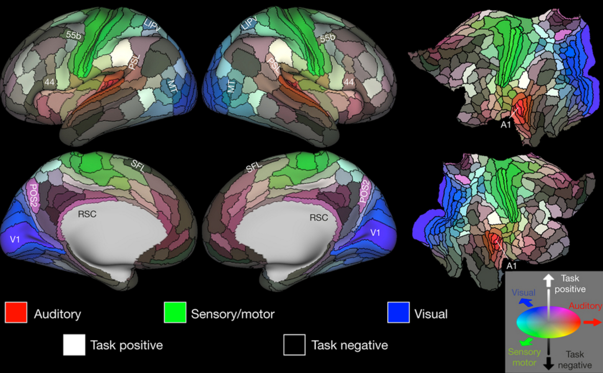
New brain map provides unprecedented detail in 180 areas of the cerebral cortex

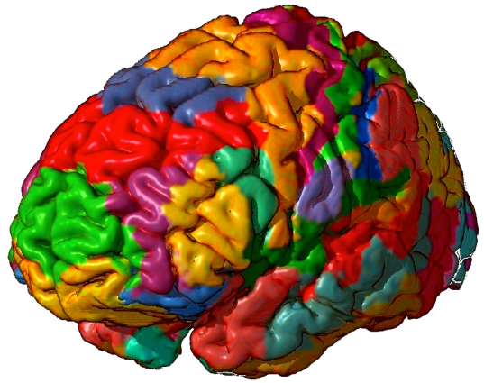
A detailed new map by researchers at Washington University School of Medicine in St. Louis lays out the landscape of the cerebral cortex. The 180 areas delineated and identified in both left and right hemispheres are displayed on inflated and flattened cortical surfaces. Black outlines indicate areal borders. Colors indicate the extent to which the areas are associated in the resting state with auditory (red), somatosensory (green), visual (blue), task positive (towards white), or task negative (towards black) groups of areas. The legend on the bottom right illustrates the 3D color space used in the figure. Data at http://balsa.wustl.edu/WN56. (credit: Matthew F. Glasser et al./Nature)

A detailed new map by researchers at Washington University School of Medicine in St. Louis and associates* lays out the landscape of 180 areas of the cerebral cortex in painstaking detail; 97 of these areas have never been previously described.

The new map is intended to help researchers studying brain disorders such as autism, schizophrenia, dementia and epilepsy. They will be able to use it to understand differences in the brains of patients with these diseases, compared with adults who are healthy. It also will accelerate progress in deciphering the workings of the healthy brain and elucidating what makes us unique as a species, the researchers say. The new map will also be vital for neurosurgeons.

The work was published today, July 20, in Nature.


Nature Video | The ultimate brain map

The researchers drew upon data and methods generated by the Human Connectome Project, a five-year, multimillion dollar study led by David Van Essen, PhD, the senior author on this paper. The Human Connectome Project used a powerful, custom-built MRI machine to map the brains of 1,200 young adults. This new study complements the Human Connectome Project by carefully delineating the brain regions so that their connections can be more accurately mapped.

The new map divides both the left and right cerebral hemispheres into 180 areas based on physical differences (such as the thickness of the cortex), functional distinctions (such as which areas respond to language stimuli), and differences in the connections of the areas.

“The brain is not like a computer that can support any operating system and run any software,” said Van Essen, the Alumni Endowed Professor of Neuroscience at Washington University Medical School. “Instead, the software — how the brain works — is intimately correlated with the brain’s structure, its hardware, so to speak. If you want to find out what the brain can do, you have to understand how it is organized and wired.”

The researchers mapped the cortex, a layer of neural tissue that encases the rest of the brain like a crumpled sheet of paper. The cortex is important for sensation, attention, memory, perception, thought, language and consciousness.

Beyond Brodmann’s areas

Brodmann areas (credit: Mark Dow/Wikipedia)

The new study is intended to replace previous maps, such as Korbinian Brodmann’s map of the human cortex, created in the first decade of the 20th century, which identified 50 regions. “My early work on language connectivity involved taking that 100-year-old map and trying to guess where Brodmann’s areas were in relation to the pathways underneath them,” said Matthew Glasser, PhD, of Washington University Medical School. “It quickly became obvious to me that we needed a better way to map the areas in the living brains that we were studying.”

Until now, most brain maps have been based on a single type of measurement. To make the new map, the researchers pooled data from 210 healthy young adults of both sexes, combining measures of the thickness of the cortex and the amount of insulation around neuronal cables, using MRI scans of the resting brain and of the brain performing simple tasks, such as listening to a story. The information also included measurements of brain function, connectivity between regions, topographic organization of cells in brain tissue, and levels of myelin, which speeds up neural signaling.

In the new map, some of the 180 areas identified are clearly involved in particular tasks, such as 55b, which lights up with activity when a person hears a story. Others contain a map of a person’s field of vision, or are involved in controlling movement. But most areas probably will never be identified with a single function, because they don’t do just one thing, but instead coordinate information from many different signals.

Many other maps of the cortex have been drawn, showing anywhere from 50 to 200 different areas. But the researchers say they improved on previous maps by precisely aligning the brains to a common coordinate system before analysis, using an algorithm developed by colleagues at Oxford University, and incorporating the highest-quality MRI data available. The researchers also verified that their method could be applied to individuals by producing maps of the brains of a different set of 210 healthy young adults.

Guiding neurosurgeons

The results are a precise map with unusually crisp borders and an algorithm capable of locating the areas in individual brains, even though each individual is unique in terms of the pattern of cortical folds and in the size and shape of areas on the cortical map.

“In the past, it was not always clear whether the results from two separate neuroimaging studies referred to the same area or not,” Glasser said. By using the new map and alignment algorithm, results of separate studies could be more accurately compared.

Better individual maps of the brain could help neurosurgeons avoid damaging the most important areas, such as those involved in language or motor function, and could guide treatment for neurological or psychiatric illnesses. Different types of dementia, for example, are characterized by degeneration in different areas of the brain. Clinicians could use the individual maps to personalize treatment, based on the areas affected, or to monitor response to treatment.

Like cartographers of old, brain cartographers primarily are providing a tool for others to use in exploration and discovery.

“We were able to persuade Nature to put online almost 200 extra pages of detailed information on each of the 180 regions as well as all of the algorithms we used to align the brains and create the map,” Van Essen said. “We think it will serve the scientific community best if they can dive down and get these maps onto their computer screens and explore as they see fit.”

* University of Oxford; Imperial College, London; University of Minnesota; Radboud University; Radboud University Medical Centre.

Middle-age-plus memory decline may just be a matter of changing focus

When middle-aged and older adults were shown a series of faces, red regions of the brain were more active; these include an area in the medial prefrontal cortex that is associated with self-referential thinking. In young adults, by contrast, blue regions — which include areas important for memory and attention –+ were more active during this task. (credit: N. Rajah, McGill University)

Are you middle-aged or older and having problems remembering details, like where you left the keys or parked your car?

Cheer up, it may simply be the result of a change in what information your brain focuses on during memory formation and retrieval, rather than a decline in brain function, according to a study by McGill University researchers.

In the study, published in the journal, NeuroImage, 112 healthy adults ranging in age from 19 to 76 years were shown a series of faces. Participants were then asked to recall where a particular face appeared on the screen (left or right) and when it appeared (least or most recently). The researchers used functional MRI to analyze which parts of brain were activated during recall of these details.

Different parts of the brain involved

Senior author Natasha Rajah, Director of the Brain Imaging Centre, and colleagues found that young adults activated their visual cortex while successfully performing this task.

But for middle-aged and older adults, their medial prefrontal cortex was activated instead. That’s a part of the brain known to be involved with information having to do with one’s own life and introspection. This may reflect changes in what adults deem “important information” as they age, she said.

Rajah says middle-aged and older adults can improve their recall abilities by learning to focus on external rather than internal information, using mindfulness meditation, for example.*

Rajah is currently analyzing data from a similar study to discern if there are any gender differences in middle-aged brain function as it relates to memory. “At mid-life women are going through a lot of hormonal change. So we’re wondering how much of these results is driven by post-menopausal women.”

The research was supported by the Canadian Institutes of Health Research and by a grant from the Alzheimer’s Society of Canada.

* Other options to improve memory include:

  • Peppermint tea or rosemary essential oil, scientists at Northumbria University found in studies with subjects over age 65, presented at the British Psychological Society’s Annual Conference in Nottingham in April. Rosemary aroma significantly enhanced prospective memory (for things you plan to do).
  • Eight nutrients to protect the aging brain: cocoa flavanols, omega-3 fatty acids, phosphatidylserine and phosphatidic Acid, walnuts, citicoline, choline (found especially in eggs) and magnesium (avocado, soy beans, bananas and dark chocolate), and blueberries, according to a study published in the journal Food Technology. Details here.

Abstract of Changes in the modulation of brain activity during context encoding vs. context retrieval across the adult lifespan

Age-related deficits in context memory may arise from neural changes underlying both encoding and retrieval of context information. Although age-related functional changes in the brain regions supporting context memory begin at midlife, little is known about the functional changes with age that support context memory encoding and retrieval across the adult lifespan. We investigated how age-related functional changes support context memory across the adult lifespan by assessing linear changes with age during successful context encoding and retrieval. Using functional magnetic resonance imaging (fMRI), we compared young, middle-aged and older adults during both encoding and retrieval of spatial and temporal details of faces. Multivariate behavioral partial least squares (B-PLS) analysis of fMRI data identified a pattern of whole brain activity that correlated with a linear age term, and a pattern of whole brain activity that was associated with an age-by-memory phase (encoding vs. retrieval) interaction. Further investigation of this latter effect identified three main findings: 1) reduced phase-related modulation in bilateral fusiform gyrus, left superior/anterior frontal gyrus and right inferior frontal gyrus that started at midlife and continued to older age, 2) reduced phase-related modulation in bilateral inferior parietal lobule that occurred only in older age, and 3) changes in phase-related modulation in older but not younger adults in left middle frontal gyrus and bilateral parahippocampal gyrus that was indicative of age-related over-recruitment. We conclude that age-related reductions in context memory arise in midlife and are related to changes in perceptual recollection and changes in fronto-parietal retrieval monitoring.

Why your immune system may control your social behavior

Normal brain activity (credit: University of Virginia Health System)

In a discovery that raises fundamental questions about human behavior, researchers at the University of Virginia School of Medicine have found that the immune system directly affects — and even controls — our social behavior, such as our desire to interact with others. That finding could have significant implications for neurological diseases such as autism-spectrum disorders and schizophrenia, the researchers suggest.

“The brain and the adaptive immune system were thought to be isolated from each other, and any immune activity in the brain was perceived as sign of a pathology. And now, not only are we showing that they are closely interacting, but some of our behavior traits might have evolved because of our immune response to pathogens,” explained Jonathan Kipnis, chair of UVA’s Department of Neuroscience.

“It’s crazy, but maybe we are just multicellular battlefields for two ancient forces: pathogens and the immune system. Part of our personality may actually be dictated by the immune system.”

Evolutionary forces linking brains and pathogens

KurzweilAI has cited supporting evidence for that idea. For example, permanent stress may affect immune cells in the brain, leading to mental disorders and protective immune microglia cells also have direct involvement in creating the cellular networks at the core of brain behavior.

Last year, Kipnis, the director of UVA’s Center for Brain Immunology and Glia, and his team discovered that meningeal membranes (covering the brain and spinal cord) in the brain directly link the brain with the lymphatic system. That overturned decades of textbook teaching that the brain lacks a direct connection to the immune system.

Now, the researchers suggest, the relationship between people and pathogens could have directly affected the development of our social behavior. Social behavior (which is necessary for the survival of the species) allows pathogens to spread, so our immune systems may have developed to protect us from the diseases that accompany those interactions.

Specifically, the UVA researchers have now shown that a specific immune molecule, interferon gamma, seems to be critical for social behavior, and that a variety of creatures, such as flies, zebrafish, mice and rats, activate interferon gamma responses (as protection) when they are social.

Normally, this molecule is produced by the immune system in response to bacteria, viruses or parasites. But blocking the molecule in mice using genetic modification also made regions of the brain hyperactive, causing the mice to become less social. Restoring the molecule restored the brain connectivity and behavior to normal.

A hyperactive brain, triggered by a blocked immune system, may lead to less-social behavior (credit: University of Virginia Health System)

In a Nature paper outlining their findings, the researchers note the immune molecule plays a “profound role in maintaining proper social function.”

“It’s extremely critical for an organism to be social for the survival of the species. It’s important for foraging, sexual reproduction, gathering, hunting,” said Anthony J. Filiano, Hartwell postdoctoral fellow in the Kipnis lab and lead author of the study. “The hypothesis is that when organisms come together, you have a higher propensity to spread infection — you need to be social, but [in doing so] you have a higher chance of spreading pathogens.” Which explains why “interferon gamma, in evolution, has been used as a more efficient way to both boost social behavior while boosting an anti-pathogen response.”

Immune-system failure leads to social deficits

The researchers explain that a malfunctioning immune system may be responsible for “social deficits in numerous neurological and psychiatric disorders.” But exactly what this might mean for autism and other specific conditions requires further investigation.

It is unlikely that any one molecule will be responsible for disease or the key to a cure. The researchers believe that the causes are likely to be much more complex. But the discovery that the immune system — and possibly pathogens, by extension — can control our interactions raises many exciting avenues for scientists to explore, both in terms of battling neurological disorders and understanding human behavior.

Kipnis and his team worked closely with UVA’s Department of Pharmacology and with Vladimir Litvak’s research group at the University of Massachusetts Medical School. Litvak’s team developed a computational approach to investigate the complex dialogue between immune signaling and brain function in health and disease.

“Using this approach we predicted a role for interferon gamma, an important cytokine secreted by T lymphocytes, in promoting social brain functions,” Litvak said. “Our findings contribute to a deeper understanding of social dysfunction in neurological disorders, such as autism and schizophrenia, and may open new avenues for therapeutic approaches.”

The work was supported by NIH grants and the Hartwell Foundation.


Medicine Virginia | Shocking New Role Found for the Immune System: Controlling Social Interactions


Abstract of Unexpected role of interferon-γ in regulating neuronal connectivity and social behaviour

Immune dysfunction is commonly associated with several neurological and mental disorders. Although the mechanisms by which peripheral immunity may influence neuronal function are largely unknown, recent findings implicate meningeal immunity influencing behaviour, such as spatial learning and memory1. Here we show that meningeal immunity is also critical for social behaviour; mice deficient in adaptive immunity exhibit social deficits and hyper-connectivity of fronto-cortical brain regions. Associations between rodent transcriptomes from brain and cellular transcriptomes in response to T-cell-derived cytokines suggest a strong interaction between social behaviour and interferon-γ (IFN-γ)-driven responses. Concordantly, we demonstrate that inhibitory neurons respond to IFN-γ and increase GABAergic (γ-aminobutyric-acid) currents in projection neurons, suggesting that IFN-γ is a molecular link between meningeal immunity and neural circuits recruited for social behaviour. Meta-analysis of the transcriptomes of a range of organisms reveals that rodents, fish, and flies elevate IFN-γ/JAK-STAT-dependent gene signatures in a social context, suggesting that the IFN-γ signalling pathway could mediate a co-evolutionary link between social/aggregation behaviour and an efficient anti-pathogen response. This study implicates adaptive immune dysfunction, in particular IFN-γ, in disorders characterized by social dysfunction and suggests a co-evolutionary link between social behaviour and an anti-pathogen immune response driven by IFN-γ signalling.

A biocompatible, transparent therapeutic window to the brain

An illustration showing how the “window to the brain” transparent skull implant created by UC Riverside researchers would work (credit: UC Riverside)

Researchers at the University of California, Riverside have developed a transparent “window to the brain” — a skull implant that is biocompatible, infection-resistant, and does not need to be repetitively replaced.

Part of the ongoing “Window to the Brain” project, a multi-institution, cross-disciplinary effort, the idea is to use transparent skull implants to provide laser diagnosis and treatment of a wide variety of brain pathologies, including brain cancers, traumatic brain injury, stroke, and neurodegenerative diseases, without requiring repeated craniotomies (a surgical operation in which a bone flap is temporarily removed from the skull to access the brain). Such operations are vulnerable to bacterial infections.

A biocompatible transparent material

The researchers have developed a transparent version of the material yttria-stabilized zirconia (YSZ), a ceramic material used in hip implants and dental crowns.

The researchers implanted the material in a hamster, where it integrated into the host tissue without causing an immune response or other adverse effects, as they describe in a paper in the journal Nanomedicine: Nanotechnology, Biology and Medicine. The internal toughness of YSZ, which is more impact-resistant and biocompatible than the titanium, thermoplastic polymers, and glass-based materials developed by other researchers, makes it “the only transparent skull implant that could conceivably be used in humans,” according to the researchers.

Treating bacterial infections

Schematic diagram showing treatment of biofilm formation with near-infrared laser light via a transparent YSZ material, monitored by a thermal IR camera (credit: Yasaman Damestani et al./Lasers in Surgery and Medicine)

The scientists also developed a way to use the same laser light used in brain treatments to treat incidental bacterial infections. In a lab study, described in a paper in the journal Lasers in Surgery and Medicine, the researchers treated E. Coli infections by aiming laser light through the transparent implant, without having to remove the implant and without causing an immune response or other adverse effects to surrounding tissue.

“This was an important finding because it showed that the combination of our transparent implant and laser-based therapies enables us to treat not only brain disorders, but also to tackle bacterial infections that are common after cranial implants. These infections are especially challenging to treat because many antibiotics do not penetrate the blood brain barrier,” said Devin Binder, M.D., a neurosurgeon and neuroscientist in UCR’s School of Medicine and a collaborator on the project.

The Window to the Brain project is a multi-institution, interdisciplinary partnership led by Guillermo Aguilar, professor of mechanical engineering in UCR’s Bourns College of Engineering, and Santiago Camacho-López, from the Centro de Investigación Científica y de Educación Superior de Ensenada (CICESE) in Mexico.

Last October, the team received $3.6 million from the National Science Foundation’s Partnerships in International Research and Education (PIRE) program, which pairs U.S. universities with others around the world. An additional $1 million was from Consejo Nacional de Ciencia y Tecnología (CONACYT), Mexico’s entity in charge of promoting scientific and technological activities. The remainder of the money came from in-kind contributions from the Mexican universities.

The team’s long-term goal is to see the technology become the standard of care for patients with brain disorders.


Abstract of Inflammatory response to implantation of transparent nanocrystalline yttria-stabilized zirconia using a dorsal window chamber model

The long-range goal of the windows to the brain (WttB) is to improve patient care by providing a technique for delivery and/or collection of light into/from the brain, on demand, over large areas, and on a chronically-recurring basis without the need for repeated craniotomies. To evaluate the potential of nanocrystalline yttria-stabilized-zirconia (nc-YSZ) cranial implant for optical therapy and imaging, in vivo biocompatibility was studied using the dorsal window chamber model in comparison with control (no implant) and commercially available cranial implant materials (PEEK and PEKK). The host tissue response to implant was characterized by using transillumination and fluorescent microscopy to measure leukocyte adhesion, blood vessel diameter, blood flow rate, and vascular permeability over two weeks. The results indicated the lack of inflammatory reaction of the host tissue to nc-YSZ at the microscopic level, suggesting that nc-YSZ is a good alternative material for cranial implants.


Abstract of Evaluation of laser bacterial anti-fouling of transparent nanocrystalline yttria-stabilized-zirconia cranial implant

Background and Objective: The development and feasibility of a novel nanocrystalline yttria-stabilized-zirconia (nc-YSZ) cranial implant has been recently established. The purpose of what we now call “window to the brain (WttB)” implant (or platform), is to improve patient care by providing a technique for delivery and/or collection of light into/from the brain, on demand, over large areas, and on a chronically recurring basis without the need for repeated craniotomies. WttB holds the transformative potential for enhancing light-based diagnosis and treatment of a wide variety of brain pathologies including cerebral edema, traumatic brain injury, stroke, glioma, and neurodegenerative diseases. However, bacterial adhesion to the cranial implant is the leading factor for biofilm formation (fouling), infection, and treatment failure. Escherichia coli (E. coli), in particular, is the most common isolate in gram-negative bacillary meningitis after cranial surgery or trauma. The transparency of our WttB implant may provide a unique opportunity for non-invasive treatment of bacterial infection under the implant using medical lasers.

Study Design/Materials and Methods: A drop of a diluted overnight culture of BL21-293 E. coli expressing luciferase was seeded between the nc-YSZ implant and the agar plate. This was followed by immediate irradiation with selected laser. After each laser treatment the nc-YSZ was removed, and cultures were incubated for 24 hours at 37 °C. The study examined continuous wave (CW) and pulsed wave (PW) modes of near-infrared (NIR) 810 nm laser wavelength with a power output ranging from 1 to 3 W. During irradiation, the temperature distribution of nc-YSZ surface was monitored using an infrared thermal camera. Relative luminescence unit (RLU) was used to evaluate the viability of bacteria after the NIR laser treatment.

Results: Analysis of RLU suggests that the viability of E. coli biofilm formation was reduced with NIR laser treatment when compared to the control group (P < 0.01) and loss of viability depends on both laser fluence and operation mode (CW or PW). The results demonstrate that while CW laser reduces the biofilm formation more than PW laser with the same power, the higher surface temperature of the implant generated by CW laser limits its medical efficacy. In contrast, with the right parameters, PW laser produces a more moderate photothermal effect which can be equally effective at controlling bacterial growth.

Conclusions: Our results show that E. coli biofilm formation across the thickness of the nc-YSZ implant can be disrupted using NIR laser treatment. The results of this in vitro study suggest that using nc-YSZ as a cranial implant in vivo may also allow for locally selective, non-invasive, chronic treatment of bacterial layers (fouling) that might form under cranial implants, without causing adverse thermal damage to the underlying host tissue when appropriate laser parameters are used. Lasers Surg. Med. © 2016 Wiley Periodicals, Inc.

Allen Brain Observatory launched

The Allen Institute for Brain Science today announced the release of the Allen Brain Observatory.

This is standardized survey of cellular-level activity in the mouse visual system. The goal is to empower scientists to “investigate how circuits in the behaving mouse brain coordinate to drive activity and perception, and lays a crucial foundation for understanding perception, cognition and ultimately consciousness.”

The Allen Institute is known for its comprehensive brain atlases, with deep, high-quality data sets revealing where genes are expressed and how cells and connections are arranged in the mouse and human brain, according to Allan Jones, Ph.D., CEO of the Allen Institute.

With this new project, the researchers took a clever approach, combining a large variety of visual stimuli, including film clips and moving images, with the associated responses of neurons in four areas of the mouse visual cortex at multiple depths, sampling more than 18,000 neurons in total. The goal is to determine the “tuning,” or preference, of each individual neuron to visual features like motion and shape orientation, as well as complex images like natural scenes and movies that reveal integrative dynamics of visual processing.


Allen Institute | Allen Brain Observatory: Visualizing the brain in action

The data are presented as part of the suite of Allen Brain Atlas tools in the uniform and shareable Neurodata Without Borders file format, which allows scientists anywhere to easily mine and model the data. (The mouse is an important model system often used to understand the far less accessible and far larger human brain.) The data are presented in a novel visualization format through the Allen Brain Atlas data portal, and are accompanied by analysis tools and access to all raw data, which allows scientists to deeply explore the rules that govern how networks of cells in the visual cortex communicate.

“The Allen Brain Observatory is a stunning window into the visual brain of the mouse,” says Christof Koch, Ph.D., President and Chief Scientific Officer of the Allen Institute for Brain Science. “No one has ever taken this kind of standardized approach to surveying the active brain at cellular resolution in order to measure how the brain processes information in real time. This is a milestone in our quest to decode how the brain’s computations give rise to perception, behavior, and consciousness.

“Just like in astronomy, modelers and theoreticians worldwide can now study this wealth of data using their own analysis tools. If we want to understand higher-order brain functions, we need to understand not just the individual components of the brain but how they all work together.”

Understanding visual processing is a key gateway to understanding how other parts of the brain process information, and future releases of the Allen Brain Observatory will also explore the neural circuits that underlie more complex behaviors like decision-making, according to the researchers.


Allen Institute | Inside the Allen Brain Observatory


How to detect early signs of Alzheimer’s with a simple eye exam before symptoms appear

Monochromatic images of normal mouse retina at four wavelengths from blue (upper left) to red (lower right). Boxes in lower right show locations typical of amyloid (a substance found in the brain associated with Alzheimer’s). (credit: Swati S. More et al./IOVS)

University of Minnesota (UMN) scientists and associates have developed new technology that can detect signs of Alzheimer’s before the onset of symptoms — early enough to give drugs a chance to work — in mice and humans by simply examining the back of their eyes.

Looking at Alzheimer’s effects through the eye is a key advantage of the new technology. “The retina of the eye is not just ‘connected’ to the brain — it is part of the central nervous system,” said Swati More, PhD, of the Center for Drug Design at UMN, co-author of a paper recently published in Investigative Ophthalmology & Visual Science (IOVS).

The brain and retina undergo similar changes due to Alzheimer’s disease, he said, but “unlike the brain, the retina is easily accessible to us. We saw changes in the retinas of Alzheimer’s mice before the typical age at which neurological signs are observed.”

Human clinical trials are set to start in July to test the technology in humans who are 40–75 years old (for more information on participating in the clinical trial, you can visit the trial website).

Optical spectra recorded from human and mouse retina samples. Upper two lines show the intensity of light for different wavelengths of light from red (right side) to blue (left side) for a normal human retina (heavy line) and for an Alzheimer’s (AD) patients retina. Comparable mouse plots, shown below (WT = normal mouse; APP1/PS1 = Alzheimer’s model mouse), show a similar pattern. (credit: Swati S. More et al./IOVS)

Early detection of Alzheimer’s is critical for two reasons. “First, effective treatments need to be administered well before patients show actual neurological signs,” said co-author Robert Vince, PhD, of the Center for Drug Design at the University of Minnesota (UMN). “Second, since there are no available early detection techniques, drugs currently cannot be tested to determine if they are effective against early Alzheimer’s disease. An early diagnostic tool like ours could help the development of drugs as well.”


Abstract of Early Detection of Amyloidopathy in Alzheimer’s Mice by Hyperspectral Endoscopy

Purpose: To describe a spectral imaging system for small animal studies based on noninvasive endoscopy of the retina, and to present time-resolved spectral changes from live Alzheimer’s mice prior to cognitive decline, corroborating our previous in vitro findings.

Methods: Topical endoscope fundus imaging was modified to use a machine vision camera and tunable wavelength system for acquiring monochromatic images across the visible to near-infrared spectral range. Alzheimer’s APP/PS1 mice and age-matched, wild-type mice were imaged monthly from months 3 through 8 to assess changes in the fundus reflection spectrum. Optical changes were fit to Rayleigh light scatter models as measures of amyloid aggregation.

Results: Good quality spectral images of the central retina were obtained. Short-wavelength reflectance from Alzheimer’s mice retinae showed significant reduction over time compared to wild-type mice. Optical changes were consistent with an increase in Rayleigh light scattering in neural retina due to soluble Aβ1–42aggregates. The changes in light scatter showed a monotonic increase in soluble amyloid aggregates over a 6-month period, with significant build up occurring at 7 months.

Conclusions: Hyperspectral imaging technique can be brought inexpensively to the study of retinal changes caused by Alzheimer’s disease progression in live small animals. A similar previous finding of reduction in the light reflection over a range of wavelengths in isolated Alzheimer’s mice retinae, was reproducible in the living Alzheimer’s mice. The technique presented here has a potential for development as an early Alzheimer’s retinal diagnostic test in humans, which will support the treatment outcome.

This deadly soil bug can reach your brain in a day, end up in spinal cord

B. pseudomallei soil-dwelling bacterium endemic in tropical and subtropical regions worldwide, particularly in Thailand and northern Australia (credit: Wikipedia CC)

Imagine a  deadly bacteria that can be picked up by a simple sniff and can travel to your brain and spinal cord in just 24 hours. Or one that could just be quietly sitting there, waiting for an opportune moment. Or maybe just doing small incremental damage ever day over a lifetime … as you lose the function in your brain incrementally.

That’s the grisly finding (in mice), published in Immunity and Infection this week, of a new study by Australian Griffith University and Bond University scientists.

The pathogenic bacteria Burkholderia pseudomallei, which causes the potentially fatal disease melioidosis, kills 89,000 people around the world each year and is prevalent in northern Australia, where a person with melioidosis has a 20–50 per cent chance of dying once it infects the brain. The bacterium is found in the northern parts of the Northern Territory, including Darwin.

In Southeast Asia 50 per cent of the population may be positive for melioidosis, and in places like Cambodia the mortality rate is as high as 50 per cent.

But for the rest of us, the findings could also lead to discoveries of how the common staphylococcus and acne bacterium also end up in the spinal cord, as well as how chlamydia travels to the brain in Alzheimer’s patients. Or even explain common back problems, which could be where bacteria have infected your bone, causing pain that could be simply treated with antibiotics, according to the researchers.

Tracing the bacteria in mice brains and spinal cords

(Top) A schematic drawing of a mouse brain showing the location of various images. (Bottom) D: A B. pseudomallei rod (arrow) present in trigeminal nerve near the connection between the trigeminal nerve in the brain and the brainstem. (E) B. pseudomallei rod (arrow) with a fluorescent particle (arrow with tail) after the merge between the trigeminal nerve and brainstem. Scale bars in μm. (credit: James A. St John et al./Infection and Immunity)

The olfactory mucosa, located in the nose, is very close to the brain and it has long been known that viruses could reach the brain from the olfactory mucosa. But researchers have not understand exactly how the bacteria traveled to the brain and spinal cord, or just how quickly.

To find out, James St. John, PhD., Head of Griffith’s Clem Jones Centre for Neurobiology and Stem Cell Research, and associates infected mice with B. pseudomallei. They were able to trace the bacteria travels from the nerves in the nasal cavity before moving to the brain stem and then into the spinal cord. (He noted that this could also be a pathway for many other common bacteria.)

“Our latest results represent the first direct demonstration of transit of a bacterium from the olfactory mucosa to the central nervous system (CNS) via the trigeminal nerve; bacteria were found a considerable distance from the olfactory mucosa, in the brain stem, and even more remarkably in the spinal cord,” said professor Ifor Beacham from the Griffith Institute for Glycomics.

Researchers will now work on ways to stimulate supporting cells that could remove the bacteria. St. John said the work was important because the bacteria had the potential to be used as a bioweapon and knowing how to combat it was extremely important.

“Bacteria have been implicated as a major causative agent of some types of back pain. We now need to work out whether the bacteria that cause back pain also can enter the brainstem and spinal cord via the trigeminal nerve,” he added.


Abstract of Burkholderia pseudomallei rapidly infects the brainstem and spinal cord via the trigeminal nerve after intranasal inoculation

Infection with Burkholderia pseudomallei causes melioidosis, a disease with a high mortality rate (20% in Australia and 40% in south-east Asia). Neurological melioidosis is particularly prevalent in northern Australian patients and involves brainstem infection, which can progress to the spinal cord; however, the route by which the bacteria invade the central nervous system (CNS) is unknown. We have previously demonstrated that B. pseudomallei can infect the olfactory and trigeminal nerves within the nasal cavity following intranasal inoculation. As the trigeminal nerve projects into the brainstem, we investigated whether the bacteria could continue along this nerve to penetrate the CNS. After intranasal inoculation of mice, B. pseudomallei caused low-level localised infection within the nasal cavity epithelium, prior to invasion of the trigeminal nerve in small numbers. B. pseudomallei rapidly invaded the trigeminal nerve and crossed the astrocytic barrier to enter the brainstem within 24 hours and then rapidly progressed over 2000 μm into the spinal cord. To rule out that the bacteria used a haematogenous route, we used a capsule-deficient mutant of B. pseudomallei, which does not survive in the blood, and found that it also entered the CNS via the trigeminal nerve. This suggests that the primary route of entry is via the nerves that innervate the nasal cavity. We found that actin-mediated motility could facilitate initial infection of the olfactory epithelium. Thus, we have demonstrated that B. pseudomallei can rapidly infect the brain and spinal cord via the trigeminal nerve branches that innervate the nasal cavity.

Neurons grown from stem cells in a dish reveal clues about autism

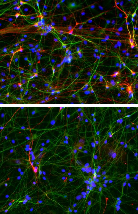
Salk researchers have turned the skin cells of people with autism spectrum disorder into neurons. These cells show specific defects (indicated by red and green dots in the neuron) compared with neurons derived from healthy people, including diminished ability to form excitatory connections with other neurons  (credit: Salk Institute)

Why do the brains of up to 30 percent of people with autism spectrum disorder grow faster than usual, early in life? A new study co-led by Salk Institute scientists has used a new stem cell reprogramming technique to find out.

Published July 6, 2016 in the journal Molecular Psychiatry, the Salk team found that stem cell-derived neurons made fewer connections in a dish compared to cells from healthy individuals. The scientists were also able to restore communication between the cells by adding IGF-1, a drug currently being evaluated in clinical trials of autism.*

Neurons derived from people with autism spectrum disorder, shown in the bottom panel, form fewer inhibitory connections, shown in the red stain, compared to those derived from healthy individuals (top panel). The total number of neurons that researchers were able to generate was about the same between the two groups. (credit: Salk Institute)

Autism affects approximately 1 out of every 68 children in the United States. It is characterized by problems communicating, difficulties interacting with others, and in repetitive behaviors, although the symptoms range dramatically in type and severity. There is no known cause of autism, according to the scientists.

Led by senior investigator Rusty Gage, a professor in Salk’s Laboratory of Genetics and holder of the Vi and John Adler Chair for Research on Age-Related Neurodegenerative Diseases, the researchers created stem cells from a subset of people with autism whose brains had grown as much as 23 percent faster than usual, but had subsequently normalized.

Improvement by adding IGF-1

The neuron precursor cells derived from the patients multiplied faster than those of typically developing individuals. The finding supports a theory some experts have put forth that brain enlargement is caused by disruptions to the cell’s normal cycle of division, the researchers say. In addition, the stem cell-derived neurons of individuals with autism behaved abnormally, bursting with activity less often compared with those cells of healthy people.

Those neurons’ activity seemed to improve by adding IGF-1, which is known to enhance the connections between neurons. The group plans to use the patient cells to investigate the molecular mechanisms behind IGF-1’s effects, in particular probing for changes in gene expression with treatment.

Other authors on the study include researchers from the University of California, San Francisco; the University of California, Los Angeles; the University of California San Diego; Case Western Reserve University, University of Rochester School of Medicine and Dentistry, and Icahn School of Medicine at Mount Sinai.

The research was supported by the California Institute for Regenerative Medicine, the National Institutes of Health, The International Rett Syndrome Foundation, a NARSAD Independent Investigator Award, a NIMH Autism Center of Excellence Program Project grant, The Leona M. and Harry B. Helmsley Charitable Trust, The JPB Foundation, the Robert and Mary Jane Engman Foundation, the CDMRP Autism Research Program, the University of California, San Diego Clinical and Translational Research Institute, and Autism Speaks.

* In 2010, Gage, Carol Marchetto of Salk’s Laboratory of Genetics, Alysson Muotri of the University of California, San Diego, and their collaborators showed they could recreate features of Rett syndrome—a rare disorder that shares features of autism but is caused by mutations in a single gene—in a petri dish.

They did so by taking skin cells from patients, adding a mix of chemicals that instructed those cells to form stem cells, and in turn, coaxing their new stem cells into neurons. The ability to form what’s called induced pluripotent stem cells (iPSCs) from human cells was pioneered by researchers in 2007, but some scientists were initially skeptical that the new technology could lend insight into complex heritable disorders such as autism.

“In that study, induced pluripotent stem cells gave us a window into the birth of a neuron that we would not otherwise have,” says Marchetto, a senior staff scientist and the study’s first author. “Seeing features of Rett syndrome in a dish gave us the confidence to next study classical autism.”


Abstract of Altered proliferation and networks in neural cells derived from idiopathic autistic individuals

Autism spectrum disorders (ASD) are common, complex and heterogeneous neurodevelopmental disorders. Cellular and molecular mechanisms responsible for ASD pathogenesis have been proposed based on genetic studies, brain pathology and imaging, but a major impediment to testing ASD hypotheses is the lack of human cell models. Here, we reprogrammed fibroblasts to generate induced pluripotent stem cells, neural progenitor cells (NPCs) and neurons from ASD individuals with early brain overgrowth and non-ASD controls with normal brain size. ASD-derived NPCs display increased cell proliferation because of dysregulation of a β-catenin/BRN2 transcriptional cascade. ASD-derived neurons display abnormal neurogenesis and reduced synaptogenesis leading to functional defects in neuronal networks. Interestingly, defects in neuronal networks could be rescued by insulin growth factor 1 (IGF-1), a drug that is currently in clinical trials for ASD. This work demonstrates that selection of ASD subjects based on endophenotypes unraveled biologically relevant pathway disruption and revealed a potential cellular mechanism for the therapeutic effect of IGF-1.

Surprising discovery of highly dynamic changes in olfactory region of the adult mouse brain

In light brown, in the center of the image, a new adult-born neuron. The neurons in blue are synaptic partner neurons, which connect to the new neurons. The neurons in dark brown are pre-existing neurons. (credit: Institut Pasteur/PM Lledo)

Scientists from the Institut Pasteur and the CNRS have made the surprising discovery that new neurons formed in the olfactory bulb of adult mice are constantly reorganizing the billions of synaptic contacts they establish among themselves (also described as constant structural plasticity).

The researchers found this puzzling because constant structural plasticity is normally confined to specific critical periods after birth, and “plasticity in neural circuits must strike a balance between flexibility and stability so new information can be acquired while previously learned information can be retained,” they note in a paper published in the journal Neuron.

The olfactory bulb in rodents and the hippocampus in humans are two of the areas of the brain capable of constantly regenerating their neurons in adulthood, but it had not been known that olfactory neurons were able to reorganize synaptic contacts.

Watching the brain change through a porthole

To observe the ongoing formation of neuronal circuits, the scientists marked the new granule cell (GC) inter-neurons with a green fluorescent protein (GFP), to view the dynamic changes, using two-photon microscopy imaging via a cranial window.

These experiments were carried out over a period of several months, following the entire life cycle of the new neurons. In the first three weeks of their life, these new neurons extended their cellular projections, known as dendrites, to form several ramifications, which subsequently became very stable.

Sample images of the same adult-born granule cell (GC) inter-neuron dendritic segment at a 2-day interval for 20–22, 40–42, and 60–62 days after virus injection (for monitoring) showing stable (closed arrowheads with numbers indexing stable spines), new (open arrowheads), and lost (asterisks) spines (credit: Kurt A. Sailor et al./Neuron)

They next observed neuronal spines — the structure where synapses form — and demonstrated that 20% of the synapses between new and pre-existing neurons were changed on a daily basis — a phenomenon that was also observed in their synaptic partners, the principal olfactory bulb neurons.

Adjusting efficiently and reliably to ongoing sensory changes

Using computer-based models, the authors showed that these dynamics enabled the synaptic network to adjust efficiently and reliably to ongoing sensory changes in the environment, enabling optimal processing of sensory information by the olfactory bulb.

“Our findings suggest that the plasticity of this constantly regenerating region of the brain occurs with continuous physical formation and elimination of synaptic connections. This structural plasticity reveals a unique dynamic mechanism that is vital for the regeneration and integration of new neurons within the adult brain circuit,” concluded the scientists.

More generally, they said, this study suggests a universal plasticity mechanism in brain regions that is closely associated with memory and learning.

This research was supported by the Institut Pasteur and the CNRS and was funded by AG2R-La Mondiale, the French National Research Agency, the “Revive” LabEx, and the “Biopsy” LabEx.


Abstract of Persistent Structural Plasticity Optimizes Sensory Information Processing in the Olfactory Bulb

In the mammalian brain, the anatomical structure of neural circuits changes little during adulthood. As a result, adult learning and memory are thought to result from specific changes in synaptic strength. A possible exception is the olfactory bulb (OB), where activity guides interneuron turnover throughout adulthood. These adult-born granule cell (GC) interneurons form new GABAergic synapses that have little synaptic strength plasticity. In the face of persistent neuronal and synaptic turnover, how does the OB balance flexibility, as is required for adapting to changing sensory environments, with perceptual stability? Here we show that high dendritic spine turnover is a universal feature of GCs, regardless of their developmental origin and age. We find matching dynamics among postsynaptic sites on the principal neurons receiving the new synaptic inputs. We further demonstrate in silico that this coordinated structural plasticity is consistent with stable, yet flexible, decorrelated sensory representations. Together, our study reveals that persistent, coordinated synaptic structural plasticity between interneurons and principal neurons is a major mode of functional plasticity in the OB.