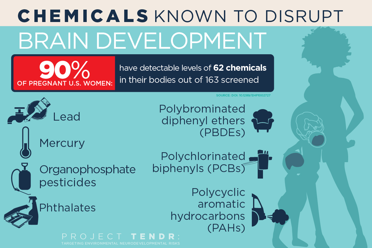
A host of common chemicals endanger child brain development, NIH journal reports

(credit: Graphic by Julie McMahon)

In a new open-access report in the NIH journal Environmental Health Perspectives, 47 scientists, health practitioners, and children’s health advocates have made a consensus statement in “Project TENDR: Targeting Environmental Neuro-Developmental Risks“ — endorsed by nine medical organizations — and issued a call to action for renewed attention to the growing evidence that many common and widely available chemicals endanger neurodevelopment in fetuses and children of all ages.

The list includes chemicals used extensively in consumer products and that have become widespread in the environment. Of most concern are lead and mercury; organophosphate pesticides used in agriculture and home gardens; phthalates, which are used in pharmaceuticals, plastics and personal care products; flame retardants known as polybrominated diphenyl ethers; and air pollutants produced by the combustion of wood and fossil fuels, said University of Illinois Comparative Biosciences professor Susan Schantz, one of dozens of individual signatories to the consensus statement.

The list provides “prime examples of toxic chemicals that can contribute to learning, behavioral, or intellectual impairment, as well as specific neurodevelopmental disorders such as ADHD or autism spectrum disorder,” according to the report.

Polychlorinated biphenyls

Polychlorinated biphenyls, once used as coolants and lubricants in transformers and other electrical equipment, also are of concern. PCBs were banned in the U.S. in 1977, but can persist in the environment for decades, she said.

“These chemicals are pervasive, not only in air and water, but in everyday consumer products that we use on our bodies and in our homes,” Schantz said. “Reducing exposures to toxic chemicals can be done, and is urgently needed to protect today’s and tomorrow’s children.”

“The human brain develops over a very long period of time, starting in gestation and continuing during childhood and even into early adulthood,” Schantz said. “But the biggest amount of growth occurs during prenatal development. The neurons are forming and migrating and maturing and differentiating. And if you disrupt this process, you’re likely to have permanent effects.”

Hormonal disrupters

Some of the chemicals of concern, such as phthalates and PBDEs, are known to interfere with normal hormone activity. For example, most pregnant women in the U.S. will test positive for exposure to phthalates and PBDEs, both of which disrupt thyroid hormone function.

“Thyroid hormone is involved in almost every aspect of brain development, from formation of the neurons to cell division, to the proper migration of cells and myelination of the axons after the cells are differentiated,” said Schantz. “It regulates many of the genes involved in nervous system development.”

Schantz and her colleagues at Illinois are studying infants and their mothers to determine whether prenatal exposure to phthalates and other endocrine disruptors leads to changes in the brain or behavior. This research, along with parallel studies in older children and animals, is a primary focus of the Children’s Environmental Health Research Center at Illinois, which Schantz directs.

Phthalates also interfere with steroid hormone activity. Studies link exposure to certain phthalates with attention deficits, lower IQ and conduct disorders in children. “Phthalates are everywhere; they’re in all kinds of different products. We’re exposed to them every day,” Schantz said.

The report criticizes current regulatory lapses that allow chemicals to be introduced into people’s lives with little or no review of their effects on fetal and child health. “For most chemicals, we have no idea what they’re doing to children’s neurodevelopment,” Schantz said. “They just haven’t been studied.

“And if it looks like something is a risk, we feel policymakers should be willing to make a decision that this or that chemical could be a bad actor and we need to stop its production or limit its use,” she said. “We shouldn’t have to wait 10 or 15 years — allowing countless children to be exposed to it in the meantime — until we’re positive it’s a bad actor.”

Project TENDR has a website with information about each of the chemicals of concern. The National Institute of Environmental Health Sciences at the National Institutes of Health and the U.S. Environmental Protection Agency fund the Children’s Environmental Health Research Center at the University of Illinois.

Project TENDR is an alliance of 48 of the nation’s top scientists, health professionals and health advocates. It was launched by Maureen Swanson of the Learning Disabilities Association of America and Irva Hertz-Picciotto of UC Davis, who brought together participants across many disciplines and sectors, including epidemiology, toxicology, exposure science, pediatrics, obstetrics and gynecology, nursing, public health, and federal and state chemical policy. Medical and scientific societies that have signed on in support include American Congress of Obstetricians and Gynecologists, American Nurses Association, Endocrine Society, National Association of Pediatric Nurse Practitioners, National Medical Association, National Hispanic Medical Association, Alliance of Nurses for Healthy Environments, Physicians for Social Responsibility and the National Council of Asian Pacific Island Physicians. TENDR’s long-term mission is to lower the incidence of neurodevelopmental disorders by reducing exposure levels to chemicals and pollutants that can contribute to these conditions, especially during fetal development and early childhood.


Steve Drake, Beckman Institute for Advanced Science and Technology | U. of I. biosciences professor Susan Schantz directs the Children’s Environmental Health Research Center at the University of Illinois, which is studying whether, and how, exposure to phthalates disrupts child brain development. Phthalates are used in some cosmetics, food packaging and products with fragrances.


Food Safety | From DDT to Glyphosate: Rachel Carson, We Need You Again


Abstract of Project TENDR: Targeting Environmental Neuro-Developmental Risks. The TENDR Consensus Statement

SUMMARY: Children in America today are at an unacceptably high risk of developing neurodevelopmental disorders that affect the brain and nervous system including autism, attention deficit hyperactivity disorder, intellectual disabilities, and other learning and behavioral disabilities. These are complex disorders with multiple causes—genetic, social, and environmental. The contribution of toxic chemicals to these disorders can be prevented. APPROACH: Leading scientific and medical experts, along with children’s health advocates, came together in 2015 under the auspices of Project TENDR: Targeting Environmental Neuro-Developmental Risks to issue a call to action to reduce widespread exposures to chemicals that interfere with fetal and children’s brain development. Based on the available scientific evidence, the TENDR authors have identified prime examples of toxic chemicals and pollutants that increase children’s risks for neurodevelopmental disorders. These include chemicals that are used extensively in consumer products and that have become widespread in the environment. Some are chemicals to which children and pregnant women are regularly exposed, and they are detected in the bodies of virtually all Americans in national surveys conducted by the U.S. Centers for Disease Control and Prevention. The vast majority of chemicals in industrial and consumer products undergo almost no testing for developmental neurotoxicity or other health effects. CONCLUSION: Based on these findings, we assert that the current system in the United States for evaluating scientific evidence and making health-based decisions about environmental chemicals is fundamentally broken. To help reduce the unacceptably high prevalence of neurodevelopmental disorders in our children, we must eliminate or significantly reduce exposures to chemicals that contribute to these conditions. We must adopt a new framework for assessing chemicals that have the potential to disrupt brain development and prevent the use of those that may pose a risk. This consensus statement lays the foundation for developing recommendations to monitor, assess, and reduce exposures to neurotoxic chemicals. These measures are urgently needed if we are to protect healthy brain development so that current and future generations can reach their fullest potential.

Prenatal exposure to acetaminophen may increase autism spectrum symptoms

Tylenol PM (left) and Tylenol (right) (credit: Ragesoss/CC)

A new study has found that paracetamol (aka acetaminophen; trade names include Tylenol and Panadol), which is used extensively during pregnancy, has a strong association with autism spectrum symptoms in boys and for both genders in relation to attention-related and hyperactivity symptoms.

The findings* were published this week in the International Journal of Epidemiology. This is the first study of its kind to report an independent association between the use of this drug in pregnancy and autism spectrum symptoms in children.

It is also the first study to report different effects on boys and girls. Comparing persistently to nonexposed children, the study has found an increase of 30 per cent in the risk of detriment to some attention functions, and an increase of two clinical symptoms of autism spectrum symptoms in boys. Boys also showed more autism spectrum symptoms when persistently exposed to paracetamol.

“Paracetamol could be harmful to neurodevelopment for several reasons,” said Co-author Dr. Jordi Júlvez, also a researcher at CREAL. “First of all, it relieves pain by acting on cannabinoid receptors in the brain.

Why boys are more likely to have autism

“Since these receptors normally help determine how neurons mature and connect with one another, paracetamol could alter these important processes. It can also affect the development of the immune system, or be directly toxic to some fetuses that may not have the same capacity as an adult to metabolize this drug, or by creating oxidative stress.”

There could also be an explanation for why boys are more likely to have autism spectrum symptoms: “The male brain may be more vulnerable to harmful influences during early life”, said Claudia Avella-Garcia. “Our differing gender results suggest that androgenic endocrine disruption, to which male brains could be more sensitive, may explain the association.”

The study concluded that the widespread exposure of infants to paracetamol in utero could increase the number of children with ADHD or autism spectrum symptoms. However, they stressed further studies should be conducted with more precise dosage measurements, and that the risks versus benefits of paracetamol use during pregnancy and early life should be assessed before treatment recommendations are made.

* Researchers in Spain recruited 2644 mother-child pairs in a birth cohort study during pregnancy. 88 per cent were evaluated when the child was one year old, and 79.9 per cent were evaluated when they were five years old. Mothers were asked about their use of paracetamol during pregnancy and the frequency of use was classified as never, sporadic, or persistent. Exact doses could not be noted due to mothers being unable to recall them exactly. 43 per cent of children evaluated at age one and 41 per cent assessed at age five were exposed to any paracetamol at some point during the first 32 weeks of pregnancy. When assessed at age five, exposed children were at higher risk of hyperactivity or impulsivity symptoms. Persistently exposed children in particular showed poorer performance on a computerised test measuring inattention, impulsivity and visual speed processing.


Abstract of Acetaminophen use in pregnancy and neurodevelopment: attention function and autism spectrum symptoms

Background: Acetaminophen is extensively used during pregnancy. But there is a lack of population-representative cohort studies evaluating its effects on a range of neuropsychological and behavioural endpoints. We aimed to assess whether prenatal exposure to acetaminophen is adversely associated with neurodevelopmental outcomes at 1 and 5 years of age.

Methods: This Spanish birth cohort study included 2644 mother-child pairs recruited during pregnancy. The proportion of liveborn participants evaluated at 1 and 5 years was 88.8% and 79.9%, respectively. Use of acetaminophen was evaluated prospectively in two structured interviews. Ever/never use and frequency of use (never, sporadic, persistent) were measured. Main neurodevelopment outcomes were assessed using Childhood Autism Spectrum Test (CAST), Conner’s Kiddie Continuous Performance Test (K-CPT) and ADHD-DSM-IV form list. Regression models were adjusted for social determinants and co-morbidities.

Results: Over 40% of mothers reported using acetaminophen. Ever-exposed offspring had higher risks of presenting more hyperactivity/impulsivity symptoms [incidence rate ratio (IRR) = 1.41, 95% confidence interval (CI) 1.01–1.98), K-CPT commission errors (IRR = 1.10, 1.03–1.17), and lower detectability scores (coefficient β = −0.75, −0.13–−0.02). CAST scores were increased in ever-exposed males (β = 0.63, 0.09–1.18). Increased effect sizes of risks by frequency of use were observed for hyperactivity/impulsivity symptoms (IRR = 2.01, 0.95–4.24) in all children, K-CPT commission errors (IRR = 1.32, 1.05–1.66) and detectability (β = −0.18, −0.36–0.00) in females, and CAST scores in males (β = 1.91, 0.44–3.38).

Conclusions: Prenatal acetaminophen exposure was associated with a greater number of autism spectrum symptoms in males and showed adverse effects on attention-related outcomes for both genders. These associations seem to be dependent on the frequency of exposure.

This remarkable fish has no cortex but intelligently switches between electrical and vision senses

The elephantnose fish (Gnathonemus petersii) explores objects in its surroundings by using its eyes or its electrical sense (credit: Timo Moritz)

With its tiny brain (and no cortex), the elephantnose fish (Gnathonemus petersii)* achieves performance comparable to that of humans or other mammals in certain tasks, according to zoologists at the University of Bonn and a colleague from Oxford.

To perceive objects in the water, the fish uses electrolocation (similar to the echolocation of bats) to perceive objects in the water, aided by an electrical organ in its tail, which emits electrical impulses, and numerous electrical sensor organs in its skin. It also uses its visual sense.

Curiously, in an experiment** in which the animals became familiar with an object in an aquarium with the visual sense, they were also able to recognize it again using the electrical sense, although they had never perceived it electrically before.

Experimental setup. Aquarium tanks were divided into two compartments by a partition with two closable gates, behind which were positioned two objects (indicated in red) 1 cm from the gate. Distance grids, which were placed directly behind the gates and which could be passed by pushing them aside, made sure that the fish kept the correct minimal distance to the objects. The larger compartment (experimental area) was again divided into two compartments, one gate leading to each compartment. (credit: S. Schumacher et al./PNAS)

In the experiment, when the two senses delivered different information in the close range of up to two centimeters, the fish trusted only the electrical information and were then “blind” to the visual stimuli. In contrast, for more distant objects, the animals relied above all on their eyes. And they perceived the environment best by using their visual and electrical senses in combination.

“This ability has only been found in mammals, suggesting such a high-level function might be associated with complex mammalian brain structures. Furthermore, the modality-specific inputs are weighted dynamically according to their reliability at different ranges,” the researchers note in a paper published online on PNAS.

“A transfer between the different senses was previously known only for certain highly developed mammals, such as monkeys, dolphins, rats, and humans”, says Professor Gerhard von der Emde at the Institute of Zoology at the University of Bonn. “In a dark, unfamiliar apartment, people feel their way forward to avoid stumbling. When the light goes on, the obstacles felt are recognized by the eye without any problem.”

The secret is in the cerebellum

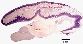
Parasagittal cresyl violet stained section through the brain of a Gnathonemus petersii showing the topography of the valvulla cerebelli covering most of the rest of the brain.
“Brain” on this figure includes, from left to right, parts of the telencephalon, diencephalon and mesencephalon. (credit: E. Gilissen, based on the material of Prof. J.M. Allman, Caltech)

So exactly how does Gnathonemus petersii achieve this surprising level of intelligence? A clue is provided by Emmanuel Gilissen on the website of The Royal Museum for Central Africa. He explains that the African mormyrids or elephant-nose fishes were noted for having unusually large brains already more than a century ago. “For a mean body mass of 26 g, the mean brain weight of Gnathonemus petersii reaches 0.53 g, almost three times its expected mean value of 0.19 g, as calculated from the relationship between brain size and body size in teleost fish (Kaufman, 2003).

“This character is probably, at least in part, related to their ability to sense prey and to communicate by generating and perceiving electric fields (Nieuwenhuys and Nicholson, 1969).  In contrast with mammals, it is the cerebellum, and not the telencephalon that is greatly enlarged in these fishes. … In elephant-nose fishes, the valvula cerebelli covers most of the rest of the brain.  In contrast, in another highly derived brain such as the human brain, it is the telencephalon, and more specifically the neocortex, a telencephalic structure unique to mammals, that entirely covers the rest of the brain. …

“In the electric fish Gnathonemus petersii, the brain is responsible for approximately 60% of body O2 consumption, a figure three times higher than that for any other vertebrate studied so far, including human.”

So are these electrically genius fish up there with the macaw?

Gnathonemus petersii is widespread in the flowing waters of West Africa and hunts insect larva at dawn and dusk.

** The elephantnose fish was in an aquarium connected to two different chambers; the animal could choose. Behind openings to the chambers there were differently shaped objects: a sphere or a cuboid. The fish learned to steer toward one of these objects by being rewarded with insect larvae. Subsequently, it searched for this object again, to obtain the reward again. When does the fish use a particular sense? To answer this question, the researchers repeated the experiments in absolute darkness. Now the fish could rely only on its electrical sense. As shown by images taken with an infrared camera, it was able to recognize the object only at short distances. With the light on the fish was most successful, because it was able to use its eyes and the electrical sense for the different distances. To find out when the fish used its eyes alone, the researchers made the objects invisible to the electrical sense. Now, the sphere and cuboid to be discriminated had the same electrical characteristics as the water.


Abstract of Cross-modal object recognition and dynamic weighting of sensory inputs in a fish

Most animals use multiple sensory modalities to obtain information about objects in their environment. There is a clear adaptive advantage to being able to recognize objects cross-modally and spontaneously (without prior training with the sense being tested) as this increases the flexibility of a multisensory system, allowing an animal to perceive its world more accurately and react to environmental changes more rapidly. So far, spontaneous cross-modal object recognition has only been shown in a few mammalian species, raising the question as to whether such a high-level function may be associated with complex mammalian brain structures, and therefore absent in animals lacking a cerebral cortex. Here we use an object-discrimination paradigm based on operant conditioning to show, for the first time to our knowledge, that a nonmammalian vertebrate, the weakly electric fish Gnathonemus petersii, is capable of performing spontaneous cross-modal object recognition and that the sensory inputs are weighted dynamically during this task. We found that fish trained to discriminate between two objects with either vision or the active electric sense, were subsequently able to accomplish the task using only the untrained sense. Furthermore we show that cross-modal object recognition is influenced by a dynamic weighting of the sensory inputs. The fish weight object-related sensory inputs according to their reliability, to minimize uncertainty and to enable an optimal integration of the senses. Our results show that spontaneous cross-modal object recognition and dynamic weighting of sensory inputs are present in a nonmammalian vertebrate.

Artificial synapse said to rival biological synapses in energy consumption and function

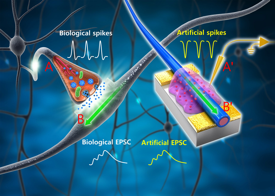
Schematic of biological neuronal network and an organic nanowire (ONW) synaptic transistor (ST) that emulates a biological synapse. The yellow conductive lines and probe (A′) mimic an axon (A) that delivers presynaptic spikes from a pre-neuron to the presynaptic membrane. The mobile ions in the ion gel move in the electrical field, analogous to the biological neuron transmitters in the synaptic cleft; the field later induces an excitatory postsynaptic current (EPSC, light blue line) in the biological dendrite (B). An ONW (B′) combined with a drain electrode (yellow surface) mimics a biological dendrite (B). EPSC (light green line) is generated in the ONW in response to presynaptic spikes and is delivered to a post-neuron through connections to the drain electrode. (credit: Wentao Xu et al./Science Advances)

An artificial synapse that emulates a biological synapse while requiring less energy has been developed by Pohang University Of Science & Technology (POSTECH) researchers* in Korea.

Energy consumption in Joules per synaptic event of currently available synaptic devices.*** NG, nanogap; PCM, phase change memory; RRAM, resistive switching random access memory. (credit: Wentao Xu et al./Science Advances)

A human synapse consumes an extremely small amount of energy (~10 fJ or femtojoules** per synaptic event).

The researchers have fabricated an organic nanofiber (ONF), or organic nanowire (ONW), electronic device that emulates the important working principles and energy consumption of biological synapses while requiring only ~1 fJ per synaptic event. The ONW also emulates the morphology (form) of a synapse.

Array of 144 ONW STs (organic nanowire synaptic transistors) fabricated on a 4-inch silicon wafer. Inset: Scanning electron microscopy (SEM) image of a typical ONW with a diameter of 200 nm. (credit: Wentao Xu et al./Science Advances)

The morphology of ONFs is similar to that of nerve fibers, which form crisscrossing grids to enable the high memory density of a human brain. The researchers say the highly-aligned ONFs can be massively produced with precise control over alignment and dimension; and this morphology may make possible the future construction of the high-density memory of a neuromorphic (brain-form-emulating) system.****

The researchers say they have emulated important working principles of a biological synapse, such as paired-pulse facilitation (PPF), short-term plasticity (STP), long-term plasticity (LTP), spike-timing dependent plasticity (STDP), and spike-rate dependent plasticity (SRDP).

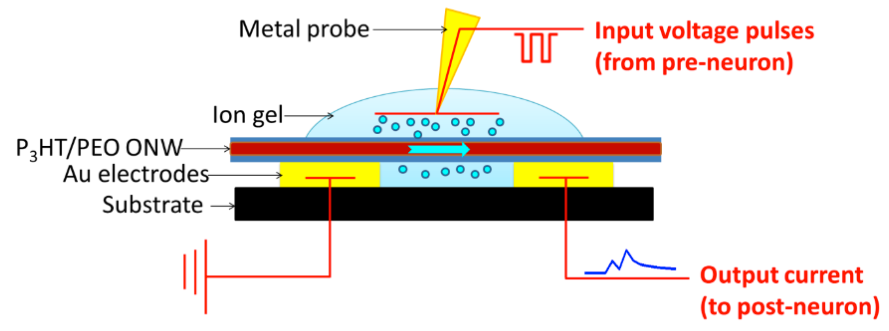
The ONW STs are three-terminal devices. Input voltage pulses that emulate presynaptic spikes from a pre-neuron are applied to the metal probe, which functions as a gate electrode. The input pulses cause ions to migrate in the ion gel, causing a change in the source-drain (yellow rectangles) current flowing through the semiconducting ONW (red). The output current signal — which emulates post-synaptic current flowing to a post-neuron in a biological synapse — is measured (during characterization) by recording the drain current. (credit: Wentao Xu et al./Science Advances)

The artificial synapse devices provide a new research direction in neuromorphic electronics and open a new era of organic electronics with high memory density and low energy consumption, the researchers claim. Potential applications include neuromorphic computing systems, AI systems for self-driving cars, analysis of big data, cognitive systems, robot control, medical diagnosis, stock-trading analysis, remote sensing, and other smart human-interactive systems and machines in the future, they suggest.

This research was supported by the Pioneer Research Center Program and Center for Advanced Soft-Electronics as a Global Frontier Project, funded by the Korean Ministry of Science, ICT, and Future Planning.

The research was published an open-access paper in Science Advances, a new sister journal of Science.

* Prof. Tae-Woo Lee, Wentao Xu, and Sung-Yong Min, PhD, with the Dept. of Materials Science and Engineering at POSTECH

** A fJ (femtojoule) is 10-15 Joule (one watt-second). 

*** ~1014 synapses.

**** Previous attempts to realize synaptic functions in single electronic devices include resistive random access memory (RRAM), phase change memory (PCM), conductive bridges, and synaptic transistors.


Abstract of Organic core-sheath nanowire artificial synapses with femtojoule energy consumption

Emulation of biological synapses is an important step toward construction of large-scale brain-inspired electronics. Despite remarkable progress in emulating synaptic functions, current synaptic devices still consume energy that is orders of magnitude greater than do biological synapses (~10 fJ per synaptic event). Reduction of energy consumption of artificial synapses remains a difficult challenge. We report organic nanowire (ONW) synaptic transistors (STs) that emulate the important working principles of a biological synapse. The ONWs emulate the morphology of nerve fibers. With a core-sheath–structured ONW active channel and a well-confined 300-nm channel length obtained using ONW lithography, ~1.23 fJ per synaptic event for individual ONW was attained, which rivals that of biological synapses. The ONW STs provide a significant step toward realizing low-energy–consuming artificial intelligent electronics and open new approaches to assembling soft neuromorphic systems with nanometer feature size.

Are you ready for mood-altering drugs precisely inserted into your brain?

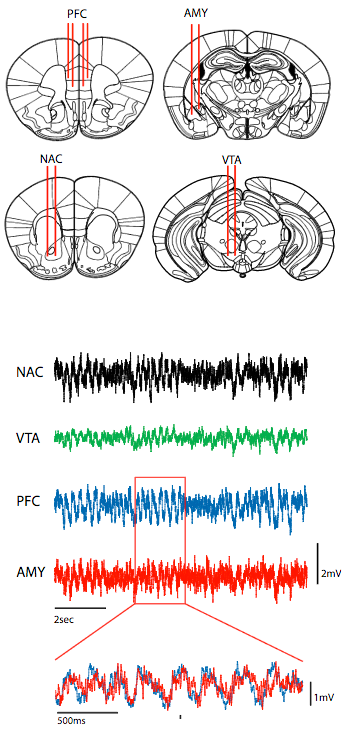
To directly probe the relationship between prefrontal cortex circuitry (PFC), limbic network oscillatory dysfunction, and the emergence of depression-related behavior, the researchers implanted mice with microwire recording electrodes in PFC and three relevant limbic brain regions implicated in major depressive disorder: nucleus accumbens (NAC), amygdala (AMY), and ventral tegmental area (VTA). Shown below: representative local field potential traces of neural signals. In the overlaid traces below, note that PFC oscillations (blue) tended to precede AMY oscillations (red). (credit: Rainbo Hultman et al./Neuron)

Imagine if doctors could precisely insert a tiny amount of a custom drug into a specific circuit in your brain and improve your depression (or other mood problems) — instead of treating the entire brain.

That’s exactly what Duke University researchers have explored in mice. Stress-susceptible animals that appeared depressed or anxious were restored to relatively normal behavior this way, according to a study appearing in the forthcoming July 20 issue of Neuron.

The plan was to define specific glitches in the neural circuits and then use a drug to fix them. The ambitious goal: go from a protein, to a signaling activity, to a cell, to a circuit, to activity that happens across the whole brain, to actual behavior.

1. Identify the key neurons in the prefrontal cortex

The researchers first determined how the prefrontal cortex is used as a pacemaker for the limbic system, said lead researcher Kafui Dzirasa, an assistant professor of psychiatry and behavioral sciences, and neurobiology.

The team started by precisely placing arrays of 32 electrodes in four brain areas of the mice (see illustration above). Then they recorded brain activity as these mice were subjected to a stressful situation called chronic social defeat.* This allowed the researchers to observe the activity between the prefrontal cortex and three areas of the limbic system that are implicated in major depression.

To interpret the complicated data coming from the electrodes, the team used machine learning algorithms — identifying which parts of the data seemed to be the timing control signal between the prefrontal cortex and the limbic system— and then zeroed in on the individual neurons involved in that cortical signal and its corresponding circuit.

2. Inject a drug to restore function

They then applied engineered molecules called DREADD (Designer Receptors Exclusively Activated by Designer Drug), developed by University of North Carolina at Chapel Hill pharmacologist Bryan Roth, in very tiny amounts (0.5 microliter). A drug that attaches only to that DREADD is then administered to give the researchers control over the circuit.

They found that direct stimulation of PFC-amygdala neural circuitry with DREADDs normalized PFC-dependent limbic synchrony in stress-susceptible animals and restored normal behavior.

The researchers suggest that their findings also demonstrate an interdisciplinary approach that can be used to identify the large-scale network changes that underlie complex emotional pathologies and the specific network nodes that can be used to develop targeted interventions.

“These subcortical circuits are the key regulators of our emotional life,” said Helen Mayberg, a professor of psychiatry, neurology and radiology at Emory University who was not involved in this research. “What’s great about this paper is that they use different approaches to see a circuit that’s relevant to a lot of disorders,” said Mayberg, who has been pioneering deep-brain stimulation of very specific sites in the human prefrontal cortex to treat mood disorders.

But she cautions that to assess anything like “mood” in a mouse, one can only infer from its behaviors. “It’s hard to do, even in a human,” she said.

This work was supported by funding from National Institutes of Mental Health and a research incubator award from the Duke Institute for Brain Sciences.

* The mice were repeatedly exposed to larger aggressive animals for 10–15 consecutive days. At the end of this protocol, animals exhibit multiple depressive endophenotypes including hedonic dysfunction, circadian dysregulation, anxiety, and psychomotor retardation.


Abstract of Dysregulation of Prefrontal Cortex-Mediated Slow-Evolving Limbic Dynamics Drives Stress-Induced Emotional Pathology

Circuits distributed across cortico-limbic brain regions compose the networks that mediate emotional behavior. The prefrontal cortex (PFC) regulates ultraslow (<1 Hz) dynamics across these networks, and PFC dysfunction is implicated in stress-related illnesses including major depressive disorder (MDD). To uncover the mechanism whereby stress-induced changes in PFC circuitry alter emotional networks to yield pathology, we used a multi-disciplinary approach including in vivo recordings in mice and chronic social defeat stress. Our network model, inferred using machine learning, linked stress-induced behavioral pathology to the capacity of PFC to synchronize amygdala and VTA activity. Direct stimulation of PFC-amygdala circuitry with DREADDs normalized PFC-dependent limbic synchrony in stress-susceptible animals and restored normal behavior. In addition to providing insights into MDD mechanisms, our findings demonstrate an interdisciplinary approach that can be used to identify the large-scale network changes that underlie complex emotional pathologies and the specific network nodes that can be used to develop targeted interventions.

How exercise improves memory

Experimental design (partial view). Study participants learned 90 picture-location associations over a period of approximately 40 minutes before being randomly assigned to one of three groups: one group performed exercise immediately, the second performed exercise four hours later, and the third did not perform any exercise. Forty-eight hours later, participants returned for a test to show how much they remembered while their brains were imaged via magnetic resonance imaging (MRI).

Physical exercise after learning improves memory and memory traces if the exercise is done four hours later, and not immediately after learning, according to findings recently reported (open-access) in the Cell Press journal Current Biology.

It’s not yet clear exactly how or why delayed exercise has this effect on memory. However, earlier studies of laboratory animals suggest that naturally occurring chemical compounds in the body known as catecholamines, including dopamine and norepinephrine, can improve memory consolidation, say the researchers at the Donders Institute at the Radboud University Medical Center in the Netherlands. One way to boost catecholamines is through physical exercise.

The researchers tested the effects of a single session of physical exercise after learning on memory consolidation and long-term memory. Seventy-two study participants learned 90 picture-location associations over a period of approximately 40 minutes before being randomly assigned to one of three groups: one group performed exercise immediately, the second performed exercise four hours later, and the third did not perform any exercise. The exercise consisted of 35 minutes of interval training on an exercise bike at an intensity of up to 80 percent of participants’ maximum heart rates. Forty-eight hours later, participants returned for a test to show how much they remembered while their brains were imaged via magnetic resonance imaging (MRI). The researchers found that those who exercised four hours after their learning session retained the information better two days later than those who exercised either immediately or not at all.

The researchers plan to follow up with another study of the timing and molecular underpinnings of exercise and its influence on learning and memory in more detail.

The researchers were supported by a grant from the European Research Council.

Brain chemicals 

Exercise induces synthesis of a chemical called DBHB in the liver. In the hippocampus, DBHB induces Bdnf gene expression, which in turn has positive effects on memory, cognition and synaptic transmission. (credit: Sama F. Sleiman et al./eLife)

In a related study published in eLife June 2 with mice, researchers note that exercise is known to be accompanied by an increase in brain-derived neurotrophic factor (BDNF) in the hippocampus, which is associated with cognitive improvement and the alleviation of depression and anxiety.

But how? It is known that a substance known as β-hydroxybutyrate (DBHB), produced in the liver from fatty acids, serves as an alternative energy source when glucose (blood sugar) levels are reduced. In their research (with mice on a running wheel for 30 days vs. no exercise), they found that the resulting increase of DBHB blocked the action of histone enzymes, which normally inhibit the production of BDNF.

Confirming that, injecting DBHB directly into the brains of mice also led to increase in BDNF.


Abstract of Physical Exercise Performed Four Hours after Learning Improves Memory Retention and Increases Hippocampal Pattern Similarity during Retrieval

Persistent long-term memory depends on successful stabilization and integration of new memories after initial encoding [ 1, 2 ]. This consolidation process is thought to require neuromodulatory factors such as dopamine, noradrenaline, and brain-derived neurotrophic factor [ 3–7 ]. Without the release of such factors around the time of encoding, memories will decay rapidly [ 3, 5, 6, 8 ]. Recent studies have shown that physical exercise acutely stimulates the release of several consolidation-promoting factors in humans [ 9–14 ], raising the question of whether physical exercise can be used to improve memory retention [ 15–17 ]. Here, we used a single session of physical exercise after learning to exogenously boost memory consolidation and thus long-term memory. Three groups of randomly assigned participants first encoded a set of picture-location associations. Afterward, one group performed exercise immediately, one 4 hr later, and the third did not perform any exercise. Participants otherwise underwent exactly the same procedures to control for potential experimental confounds. Forty-eight hours later, participants returned for a cued-recall test in a magnetic resonance scanner. With this design, we could investigate the impact of acute exercise on memory consolidation and retrieval-related neural processing. We found that performing exercise 4 hr, but not immediately, after encoding improved the retention of picture-location associations compared to the no-exercise control group. Moreover, performing exercise after a delay was associated with increased hippocampal pattern similarity for correct responses during delayed retrieval. Our results suggest that appropriately timed physical exercise can improve long-term memory and highlight the potential of exercise as an intervention in educational and clinical settings.

Abstract of Exercise promotes the expression of brain derived neurotrophic factor (BDNF) through the action of the ketone body β-hydroxybutyrate

Exercise induces beneficial responses in the brain, which is accompanied by an increase in BDNF, a trophic factor associated with cognitive improvement and the alleviation of depression and anxiety. However, the exact mechanisms whereby physical exercise produces an induction in brain Bdnf gene expression are not well understood. While pharmacological doses of HDAC inhibitors exert positive effects on Bdnf gene transcription, the inhibitors represent small molecules that do not occur in vivo. Here, we report that an endogenous molecule released after exercise is capable of inducing key promoters of the Mus musculus Bdnf gene. The metabolite β-hydroxybutyrate, which increases after prolonged exercise, induces the activities of Bdnf promoters, particularly promoter I, which is activity-dependent. We have discovered that the action of β-hydroxybutyrate is specifically upon HDAC2 and HDAC3, which act upon selective Bdnf promoters. Moreover, the effects upon hippocampal Bdnfexpression were observed after direct ventricular application of β-hydroxybutyrate. Electrophysiological measurements indicate that β-hydroxybutyrate causes an increase in neurotransmitter release, which is dependent upon the TrkB receptor. These results reveal an endogenous mechanism to explain how physical exercise leads to the induction of BDNF.


The top 10 emerging technologies of 2016

(credit: WEF)

The World Economic Forum’s annual list of this year’s breakthrough technologies, published today, includes “socially aware” openAI, grid-scale energy storage, perovskite solar cells, and other technologies with the potential to “transform industries, improve lives, and safeguard the planet.” The WEF’s specific interest is to “close gaps in investment and regulation.”

“Horizon scanning for emerging technologies is crucial to staying abreast of developments that can radically transform our world, enabling timely expert analysis in preparation for these disruptors. The global community needs to come together and agree on common principles if our society is to reap the benefits and hedge the risks of these technologies,” said Bernard Meyerson, PhD, Chief Innovation Officer of IBM and Chair of the WEF’s Meta-Council on Emerging Technologies.

The list also provides an opportunity to debate human, societal, economic or environmental risks and concerns that the technologies may pose — prior to widespread adoption.

One of the criteria used by council members during their deliberations was the likelihood that 2016 represents a tipping point in the deployment of each technology. So the list includes some technologies that have been known for a number of years, but are only now reaching a level of maturity where their impact can be meaningfully felt.

The top 10 technologies that make this year’s list are:

  1. Nanosensors and the Internet of Nanothings  — With the Internet of Things expected to comprise 30 billion connected devices by 2020, one of the most exciting areas of focus today is now on nanosensors capable of circulating in the human body or being embedded in construction materials. They could use DNA and proteins to recognize specific chemical targets, store a few bits of information, and then report their status by changing color or emitting some other easily detectable signal.
  2. Next-Generation Batteries — One of the greatest obstacles holding renewable energy back is matching supply with demand, but recent advances in energy storage using sodium, aluminum, and zinc based batteries makes mini-grids feasible that can provide clean, reliable, around-the-clock energy sources to entire villages.
  3. The Blockchain — With venture investment related to the online currency Bitcoin exceeding $1 billion in 2015 alone, the economic and social impact of blockchain’s potential to fundamentally change the way markets and governments work is only now emerging.
  4. 2D Materials — Plummeting production costs mean that 2D materials like graphene are emerging in a wide range of applications, from air and water filters to new generations of wearables and batteries.
  5. Autonomous Vehicles — The potential of self-driving vehicles for saving lives, cutting pollution, boosting economies, and improving quality of life for the elderly and other segments of society has led to rapid deployment of key technology forerunners along the way to full autonomy.
  6. Organs-on-chips — Miniature models of human organs could revolutionize medical research and drug discovery by allowing researchers to see biological mechanism behaviors in ways never before possible.
  7. Perovskite Solar Cells — This new photovoltaic material offers three improvements over the classic silicon solar cell: it is easier to make, can be used virtually anywhere and, to date, keeps on generating power more efficiently.
  8. Open AI Ecosystem — Shared advances in natural language processing and social awareness algorithms, coupled with an unprecedented availability of data, will soon allow smart digital assistants to help with a vast range of tasks, from keeping track of one’s finances and health to advising on wardrobe choice.
  9. Optogenetics — Recent developments mean light can now be delivered deeper into brain tissue, something that could lead to better treatment for people with brain disorders.
  10. Systems Metabolic Engineering — Advances in synthetic biology, systems biology, and evolutionary engineering mean that the list of building block chemicals that can be manufactured better and more cheaply by using plants rather than fossil fuels is growing every year.

To compile this list, the World Economic Forum’s Meta-Council on Emerging Technologies, a panel of global experts, “drew on the collective expertise of the Forum’s communities to identify the most important recent technological trends. By doing so, the Meta-Council aims to raise awareness of their potential and contribute to closing gaps in investment, regulation and public understanding that so often thwart progress.”

You can read 10 expert views on these technologies here or download the series as a PDF.

Brain markers of numeric, verbal, and spatial reasoning abilities found

A new study found that higher concentrations of NAA (N-acetyl aspartate) in two areas of the brain were associated with better performance on verbal and spatial tests. NAA is a byproduct of glucose metabolism and an indicator of brain health. (credit: Julie McMahon and Erick Paul)

A new study helps explain how brain structure and chemistry relate to “fluid intelligence” — the ability to adapt to new situations and solve problems one has never encountered before.

The study, reported in an open-access paper in the journal NeuroImage, observed two facets of fluid intelligence*:

  • Verbal or spatial reasoning was linked to higher concentrations of a compound called NAA (N-acetyl aspartate) in the medial parietal and posterior cingulate cortices of the brain. NAA is a byproduct of glucose metabolism and a marker of energy production in the brain. It was measured with magnetic resonance spectroscopy.
  • Number-related problem-solving was linked to brain volume in all subjects, measured using magnetic resonance imaging (MRI).

The analysis involved 211 research subjects, making it the largest study to date linking brain chemistry and intelligence in living humans. A follow-up analysis revealed that this pattern of findings was observed for males and females when analyzed separately.

A similar separation of reasoning abilities has been demonstrated in previous studies, but more studies will be needed to confirm and extend the findings, the researchers said.

The study tested participants’ performance on a number of intelligence tests, including questions, similar to this one, testing subjects’ spatial reasoning. (credit: Graphic by Ryan Larsen and Julie McMahon)

The work was conducted in the Decision Neuroscience Laboratory at the Beckman Institute for Advanced Science and Technology, University of Illinois at Urbana-Champaign.

“Our findings contribute to a growing body of evidence to suggest that intelligence reflects multiple levels of organization in the brain — spanning neuroanatomy, for example, brain size; and neurophysiology, such as brain metabolism — and that specific properties of the brain provide a powerful lens to investigate and understand the nature of specific intellectual abilities,” said Aron Barbey, an affiliate of the Beckman Institute and of the Carl R. Woese Institute for Genomic Biology.

The Intelligence Advanced Research Projects Activity at the Office of the Director of National Intelligence supported this research.

* Three canonical tests of Gf (BOMAT, Number Series, and Letter Sets) and three working memory tasks (Reading, Rotation, and Symmetry span tasks).


Abstract of Dissociable brain biomarkers of fluid intelligence

Cognitive neuroscience has long sought to understand the biological foundations of human intelligence. Decades of research have revealed that general intelligence is correlated with two brain-based biomarkers: the concentration of the brain biochemical N-acetyl aspartate (NAA) measured by proton magnetic resonance spectroscopy (MRS) and total brain volume measured using structural MR imaging (MRI). However, the relative contribution of these biomarkers in predicting performance on core facets of human intelligence remains to be well characterized. In the present study, we sought to elucidate the role of NAA and brain volume in predicting fluid intelligence (Gf). Three canonical tests of Gf (BOMAT, Number Series, and Letter Sets) and three working memory tasks (Reading, Rotation, and Symmetry span tasks) were administered to a large sample of healthy adults (n = 211). We conducted exploratory factor analysis to investigate the factor structure underlying Gf independent from working memory and observed two Gf components (verbal/spatial and quantitative reasoning) and one working memory component. Our findings revealed a dissociation between two brain biomarkers of Gf (controlling for age and sex): NAA concentration correlated with verbal/spatial reasoning, whereas brain volume correlated with quantitative reasoning and working memory. A follow-up analysis revealed that this pattern of findings is observed for males and females when analyzed separately. Our results provide novel evidence that distinct brain biomarkers are associated with specific facets of human intelligence, demonstrating that NAA and brain volume are independent predictors of verbal/spatial and quantitative facets of Gf.

Are you smarter than a macaw?

The macaw has a brain the size of an unshelled walnut, compared to the macaque monkey’s lemon-sized brain. But the macaw has more neurons in its forebrain — the portion of the brain associated with intelligent behavior — than the macaque. (credit: Vanderbilt University)

The first study to systematically measure the number of neurons in the brains of more than two dozen species of birds has found that the birds that were studied consistently have more neurons packed into their small brains than those in mammalian or even primate brains of the same mass.

The study results were published online in an open-access paper in the Proceedings of the National Academy of Sciences early edition on the week of June 13.

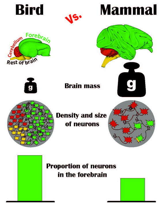
Graphic summary of the results of the avian brain study (credit: Pavel Nemec, Charles University of Prague)

“For a long time having a ‘bird brain’ was considered to be a bad thing. Now it turns out that it should be a compliment,” said Vanderbilt University neuroscientist Suzana Herculano-Houzel, senior author of the paper with Pavel Němec at the Charles University in Prague.

The study answers a puzzle that comparative neuroanatomists have been wrestling with for more than a decade: How can birds with their small brains perform complicated cognitive behaviors?

The conundrum was created by a series of studies beginning in the previous decade that directly compared the cognitive abilities of parrots and crows with those of primates. The studies found that the birds could manufacture and use tools, use insight to solve problems, make inferences about cause-effect relationships, recognize themselves in a mirror, and plan for future needs, among other cognitive skills previously considered the exclusive domain of primates.

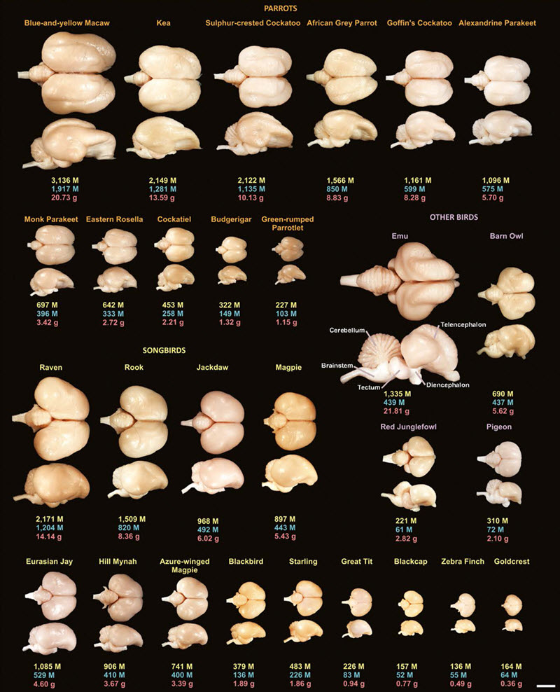
The collection of avian brains that the scientists analyzed. For each species, the total number of neurons (in millions) in each brain is shown in yellow, the number of neurons (in millions) in the forebrain (pallium) is shown in blue and the brain mass (in grams) is shown in red. The scale bar in the lower right is 10 mm. (credit: Suzana Herculano-Houzel, Vanderbilt University)

So scientists assumed avian brains must  be wired differently from primate brains. Two years ago, even this hypothesis was knocked down by a detailed study of pigeon brains, which concluded that they are, in fact, organized along quite similar lines to those of primates.

More neurons in the forebrain than previously thought

Top ten in number of whole-brain neurons and pallium (forebrain) neurons for the avian and mammalian species examined (credit: Seweryn Olkowicz et al./PNAS)

The new study provides a plausible explanation: Birds can perform these complex behaviors because birds’ forebrains contain a lot more neurons than any one had previously thought — as many as in mid-sized primates.

“We found that birds, especially songbirds and parrots, have surprisingly large numbers of neurons in their pallium: the part of the brain that corresponds to the cerebral cortex, which supports higher cognition functions such as planning for the future or finding patterns. That explains why they exhibit levels of cognition at least as complex as primates,” said Herculano-Houzel.

That’s because the neurons in avian brains are much smaller and more densely packed than those in mammalian brains, the study found. Parrot and songbird brains, for example, contain about twice as many neurons as primate brains of the same mass and two to four times as many neurons as equivalent rodent brains.

Also, the proportion of neurons in the forebrain is significantly higher, the study found.

More than one way to build better brains

“In designing brains, nature has two parameters it can play with: the size and number of neurons and the distribution of neurons across different brain centers,” said Herculano-Houzel, “and in birds we find that nature has used both of them.”

Although she acknowledges that the relationship between intelligence and neuron count has not yet been firmly established, Herculano-Houzel and her colleagues argue that having the same or greater forebrain neuron counts than primates with much larger brains can potentially provide the birds with much higher “cognitive power” per pound than mammals.

In other words, there’s more than one way to build better brains. Previously, neuroanatomists thought that as brains grew larger, neurons had to grow bigger as well because they had to connect over longer distances. “But bird brains show that there are other ways to add neurons: Keep most neurons small and locally connected and only allow a small percentage to grow large enough to make the longer connections. This keeps the average size of the neurons down,” she explained.

But that raises troubling questions:

  • Does the surprisingly large number of neurons in bird brains comes at a correspondingly large energetic cost?
  • Are the small neurons in bird brains a response to selection for small body size due to flight, or possibly the ancestral way of adding neurons to the brain — from which mammals, not birds, may have diverged.

Herculano-Houzel hopes that the results of the study and the questions it raises will stimulate other neuroscientists to begin exploring the mysteries of the avian brain, especially how their behavior compares to that of mammals of similar numbers of neurons or brain size.

Researchers at Charles University in Prague and the University of Vienna were also involved in the study.


Vanderbilt University | Bird Brain: Smarter Than You Think


Vanderbilt University | Study gives new meaning to the term “bird brain”


Abstract of Birds have primate-like numbers of neurons in the forebrain

Some birds achieve primate-like levels of cognition, even though their brains tend to be much smaller in absolute size. This poses a fundamental problem in comparative and computational neuroscience, because small brains are expected to have a lower information-processing capacity. Using the isotropic fractionator to determine numbers of neurons in specific brain regions, here we show that the brains of parrots and songbirds contain on average twice as many neurons as primate brains of the same mass, indicating that avian brains have higher neuron packing densities than mammalian brains. Additionally, corvids and parrots have much higher proportions of brain neurons located in the pallial telencephalon compared with primates or other mammals and birds. Thus, large-brained parrots and corvids have forebrain neuron counts equal to or greater than primates with much larger brains. We suggest that the large numbers of neurons concentrated in high densities in the telencephalon substantially contribute to the neural basis of avian intelligence.

Mobilizing mitochondria to regenerate damaged neurons

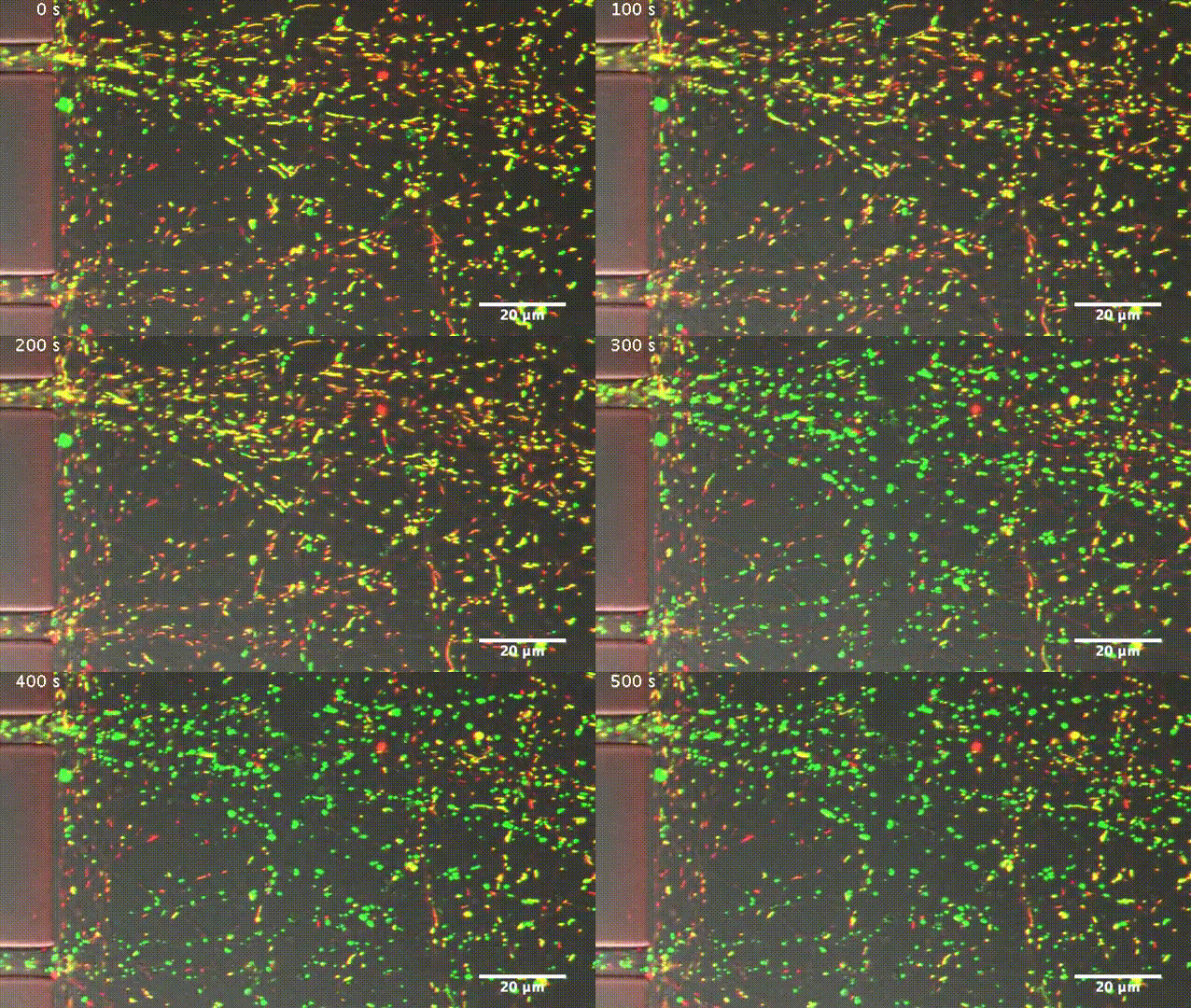
After axonal injury, nearby mitochondria become incapable of producing ATP, as indicated by their change in color from yellow (healthy) to green (damaged) (credit: Zhou et al., 2016)

Boosting the transport of mitochondria (cell energy suppliers) along neuronal axons enhances the ability of mouse nerve cells to repair themselves and regrow after injury or disease, researchers at the National Institute of Neurological Disorders and Stroke report in The Journal of Cell Biology.

Neurons need large amounts of energy to extend their axons long distances through the body. This energy — in the form of adenosine triphosphate (ATP) — is provided by mitochondria.

During development, mitochondria are transported up and down growing axons to generate ATP wherever it is needed. In adults, however, mitochondria become less mobile as mature neurons produce a protein called syntaphilin that anchors the mitochondria in place.

Zu-Hang Sheng and colleagues at the National Institute of Neurological Disorders and Stroke wondered whether this decrease in mitochondrial transport might explain why adult neurons are typically unable to regrow after injury.

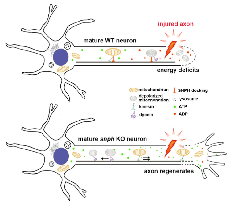
Repairing damaged neurons (top) by enhancing mitochondrial transport (bottom) (credit: Bing Zhou et al./JCB)

They initially found that when mature mouse axons are severed, nearby mitochondria are damaged and become unable to provide sufficient ATP to support injured nerve regeneration. However, when the researchers experimentally removed syntaphilin from the nerve cells (by using a genetically modified mouse), mitochondrial transport was enhanced, allowing the damaged mitochondria to be replaced by healthy mitochondria capable of producing ATP.

The Syntaphilin-deficient mature neurons therefore regained the ability to regrow after injury, just like young neurons.

“Our in vivo and in vitro studies suggest that activating an intrinsic growth program requires the coordinated modulation of mitochondrial transport and recovery of energy deficits. Such combined approaches may represent a valid therapeutic strategy to facilitate regeneration in the central and peripheral nervous systems after injury or disease,” Sheng says.


Abstract of Facilitation of axon regeneration by enhancing mitochondrial transport and rescuing energy deficits

Although neuronal regeneration is a highly energy-demanding process, axonal mitochondrial transport progressively declines with maturation. Mature neurons typically fail to regenerate after injury, thus raising a fundamental question as to whether mitochondrial transport is necessary to meet enhanced metabolic requirements during regeneration. Here, we reveal that reduced mitochondrial motility and energy deficits in injured axons are intrinsic mechanisms controlling regrowth in mature neurons. Axotomy induces acute mitochondrial depolarization and ATP depletion in injured axons. Thus, mature neuron-associated increases in mitochondria-anchoring protein syntaphilin (SNPH) and decreases in mitochondrial transport cause local energy deficits. Strikingly, enhancing mitochondrial transport via genetic manipulation facilitates regenerative capacity by replenishing healthy mitochondria in injured axons, thereby rescuing energy deficits. An in vivo sciatic nerve crush study further shows that enhanced mitochondrial transport in snph knockout mice accelerates axon regeneration. Understanding deficits in mitochondrial trafficking and energy supply in injured axons of mature neurons benefits development of new strategies to stimulate axon regeneration.