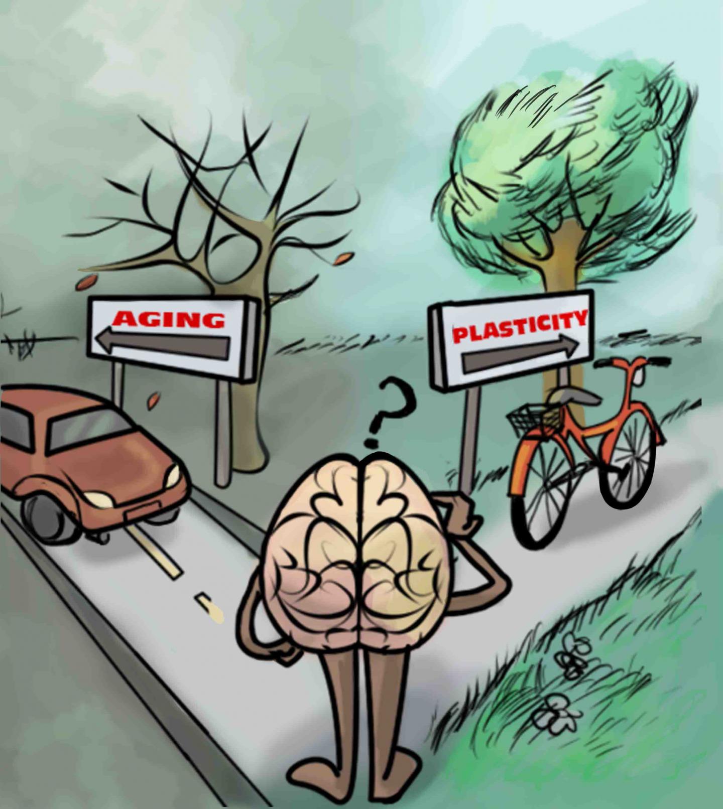
Can physical activity make you learn better?

This is an artistic representation of the take home messages in Lunghi and Sale: “A cycling lane for brain rewiring,” which is that physical activity (such as cycling) is associated with increased brain plasticity. (credit: Dafne Lunghi Art)

Exercise may enhance plasticity of the adult brain — the ability of our neurons to change with experience — which is essential for learning, memory, and brain repair, Italian researchers report in an open-access paper in the Cell Press journal Current Biology.

Their research, which focused on the the visual cortex, may offer hope for people with traumatic brain injury or eye conditions such as amblyopia, the researchers suggest. “We provide the first demonstration that moderate levels of physical activity enhance neuroplasticity in the visual cortex of adult humans,” says Claudia Lunghi of the University of Pisa in Italy.

Brain plasticity is generally thought to decline with age, especially in the sensory region of the brain (such as vision). But previous studies by research colleague Alessandro Sale of the National Research Council’s Neuroscience Institute  showed that animals performing physical activity — for example rats running on a wheel — showed elevated levels of plasticity in the visual cortex and had improved recovery from amblyopia compared  to more sedentary animals.

Binocular rivalry test

Binocular rivalry before and after “monocular deprivation” (reduced vision due to a patch) for inactive and active groups (credit: Claudia Lunghi and Alessandro Sale/Current Biology)

To find out whether the same might hold true for people, the researchers used a simple test of binocular rivalry. When people have one eye patched for a short period of time, the closed eye becomes stronger as the visual brain attempts to compensate for the lack of visual input. This recovered strength (after the eye patch is removed) is a measure of the brain’s visual plasticity.

In the new study, Lunghi and Sale put 20 adults through this test twice. In one test, participants with the dominant eye patched with a translucent material watched a movie while relaxing in a chair. In the other test, participants with one eye patched also watched a movie, but while exercising on a stationary bike for ten-minute intervals during the movie.

Exercise enhances brain plasticity (at least for vision)

Result: brain plasticity in the patched eye was enhanced by the exercise. After physical activity, the patched eye was strengthened more quickly (indicating increased levels of brain plasticity) than with the couch potatoes.

While further study is needed, the researchers think this stronger vision may have resulted from a decrease in an inhibitory neurotransmitter called GABA caused by exercise, allowing the brain to become more responsive.

The findings suggest that exercise may play an important role in brain health and recovery. This could be especially good news for people with amblyopia (called “lazy eye” because the brain “turns off” the visual processing of the weak eye to prevent double vision) — generally considered to be untreatable in adults.

Lunghi and Sale say they now plan to investigate the effects of moderate levels of physical exercise on visual function in amblyopic adult patients and to look deeper into the underlying neural mechanisms.

Time for a walk or bike ride?

UPDATE Dec. 10, 2o15: title wording changed from “smarter” to “learn better.”


Abstract of A cycling lane for brain rewiring

Brain plasticity, defined as the capability of cerebral neurons to change in response to experience, is fundamental for behavioral adaptability, learning, memory, functional development, and neural repair. The visual cortex is a widely used model for studying neuroplasticity and the underlying mechanisms. Plasticity is maximal in early development, within the so-called critical period, while its levels abruptly decline in adulthood. Recent studies, however, have revealed a significant residual plastic potential of the adult visual cortex by showing that, in adult humans, short-term monocular deprivation alters ocular dominance by homeostatically boosting responses to the deprived eye. In animal models, a reopening of critical period plasticity in the adult primary visual cortex has been obtained by a variety of environmental manipulations, such as dark exposure, or environmental enrichment, together with its critical component of enhanced physical exercise. Among these non-invasive procedures, physical exercise emerges as particularly interesting for its potential of application to clinics, though there has been a lack of experimental evidence available that physical exercise actually promotes visual plasticity in humans. Here we report that short-term homeostatic plasticity of the adult human visual cortex induced by transient monocular deprivation is potently boosted by moderate levels of voluntary physical activity. These findings could have a bearing in orienting future research in the field of physical activity application to clinical research.

As the worm turns: research tracks how an embryo’s brain is assembled

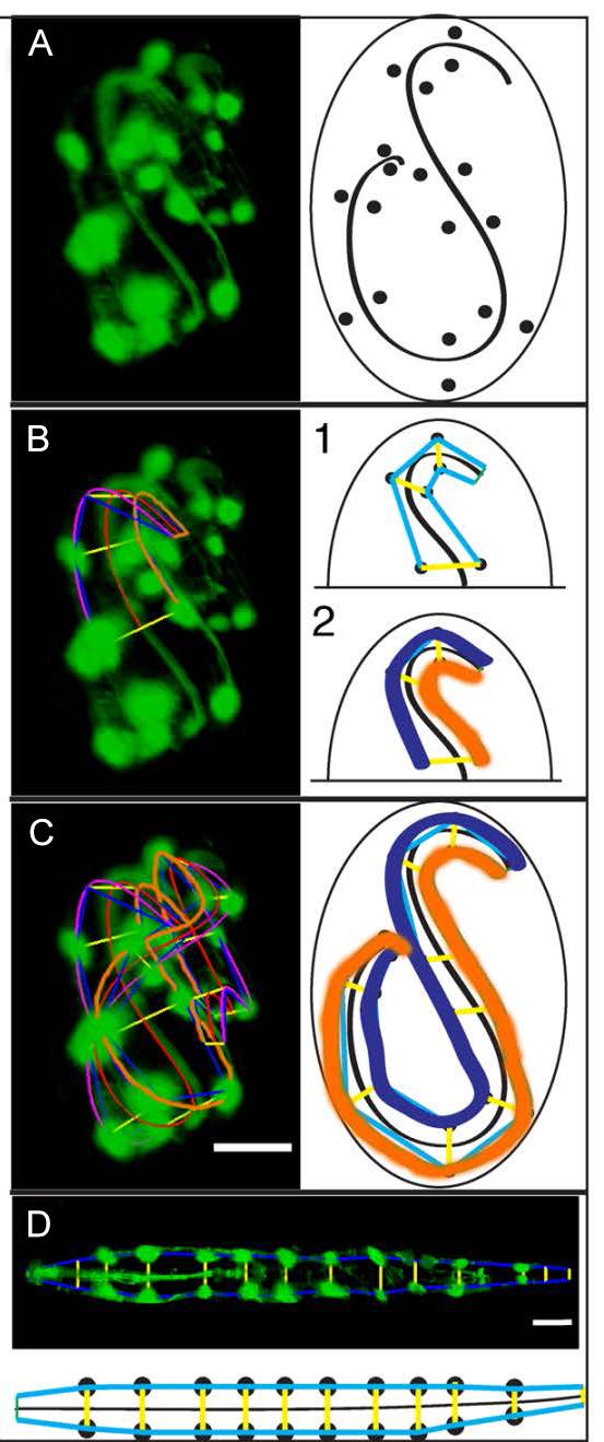
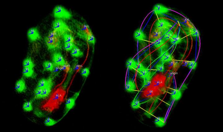
The image on the left shows skin cells (green dots) and neurons (red cell) marking the shape of the embryo. The image on the right shows the skin cells connected by the software to make a computerized model of how the embryo folds and twists. (credit: Hari Shroff, National Institute of Biomedical Imaging and Bioengineering)

New open-source software that can help track the embryonic development and movement of neuronal cells throughout the body of a worm is now available to scientists. The software is described in a paper published in the open access journal, eLife on December 3rd by a research team*.

One significant challenge is determining the formation of complex neuronal structures made up of billions of cells in the human brain. As with many biological challenges, researchers are first examining this question in simpler organisms, such as worms.

Although scientists have identified a number of important proteins that determine how neurons navigate during brain formation, it’s largely unknown how all of these proteins interact in a living organism.

Model animals, despite their differences from humans, have already revealed much about human physiology because they are much simpler and easier to understand. In this case, researchers chose Caenorhabditis elegans (C. elegans), because it has only 302 neurons, 222 of which form while the worm is still an embryo.

While some of these neurons go to the worm nerve ring (brain) they also spread along the ventral nerve cord, which is broadly analogous to the spinal cord in humans.  The worm even has its own versions of many of the same proteins used to direct brain formation in more complex organisms such as flies, mice, or humans.

Tracking neurons: a complex task
“Understanding why and how neurons form and the path they take to reach their final destination could one day give us valuable information about how proteins and other molecular factors interact during neuronal development,” said Hari Shroff, Ph.D., head of the NIBIB research team.
We don’t yet understand neurodevelopment even in the context of the humble worm, but we’re using it as a simple model of how these factors work together to drive the development of the worm brain and neuronal structure. We’re hoping that by doing so, some of the lessons will translate all the way up to humans.”

These four images show the step by step process the computer program goes through to the “untwist” a worm image. First, (Image D) the computer identifies the cells to track. Then (Image E) the computer begins to create a lattice that traces the shape of the worm. Once the computer has traced the lattice, it can create a 3D model of the worm embryo (Image F). Finally, it can untwist the model (Image G). (credit: Hari Shroff, National Institute of Biomedical Imaging and Bioengineering)

However, following neurons as they travel through the worm during its embryonic development is not as simple as it might seem. The first challenge was to create new microscopes that could record the embryogenesis of these worms without damaging them through too much light exposure while still getting the resolution needed to clearly see individual cells.

Shroff and his team at NIBIB, in collaboration with Daniel Colon-Ramos at Yale University and Zhirong Bao at Sloan-Kettering, tackled this problem by developing new microscopes that improved the speed and resolution at which they could image worm embryonic development.  (Read more)

The second problem was that during development, the worm begins to “twitch,” moving around inside the egg. The folding and twisting makes it hard to track cells and parse out movement. For example, if a neuron moves in the span of a couple of minutes, is it because the embryo twisted or because the neuron actually changed position within the embryo?

Understanding the mechanisms that move neurons to their final destination is an important factor in understanding how brains form–and is difficult to determine without knowing where and how a neuron is moving. Finally, it can be challenging to determine where a neuron is in 3D space while looking at a two-dimensional image–especially of a worm that’s folded up.

The worm embryo is normally transparent, but the researchers made several cells in the embryo glow with fluorescent proteins to act as markers.

When a microscopic image of these cells is fed into the program, the computer identifies each cell and uses the information to create a model of the worm, which it then computationally “untwists” to generate a straightened image. The program also enables a user to check the accuracy of the computer model and edit it when any mistakes are discovered.

“In addition, users can also mark cells or structures within the worm embryo they want the program to track, allowing the users to follow the position of a cell as it moves and grows in the developing embryo. This feature could help scientists understand how certain cells develop into neurons, as opposed to other types of cells, and what factors influence the development of the brain and neuronal structure.

A worm atlas

Shroff and his colleagues say that such technology will be pivotal in their project to create a 4D neurodevelopmental “worm atlas,” (see also http://www.wormguides.org) that attempts to catalog the formation of the worm nervous system.

This catalog will be the first comprehensive view of how an entire nervous system develops, and the researchers believe that it will be helpful in understanding the fundamental mechanisms by which all nervous systems, including ours, assemble. They also expect that some of the concepts developed, such as the approach taken to combine neuronal data from multiple embryos, can be applied to additional model organisms besides the worm.

* Researchers at the National Institute of Biomedical Imaging and Bioengineering (NIBIB) and the Center for Information Technology (CIT); along with Memorial Sloan-Kettering Institute, New York City; Yale University, New Haven, Connecticut; Zhejiang University, China; and the University of Connecticut Health Center, Farmington. NIBIB is part of the National Institutes of Health.


NIBIB | C. Elegans Embryo Development


Abstract of Untwisting the Caenorhabditis elegans embryo

The nematode Caenorhabditis elegans possesses a simple embryonic nervous system comprising 222 neurons, a number small enough that the growth of each cell could be followed to provide a systems-level view of development. However, studies of single cell development have largely been conducted in fixed or pre-twitching live embryos, because of technical difficulties associated with embryo movement in late embryogenesis. We present open source untwisting and annotation software which allows the investigation of neurodevelopmental events in post-twitching embryos, and apply them to track the 3D positions of seam cells, neurons, and neurites in multiple elongating embryos. The detailed positional information we obtained enabled us to develop a composite model showing movement of these cells and neurites in an “average” worm embryo. The untwisting and cell tracking capability we demonstrate provides a foundation on which to catalog C. elegans neurodevelopment, allowing interrogation of developmental events in previously inaccessible periods of embryogenesis.

How robots can learn from babies

A collaboration between UW developmental psychologists and computer scientists aims to enable robots to learn in the same way that children naturally do. The team used research on how babies follow an adult’s gaze to “teach” a robot to perform the same task. (credit: University of Washington)

Babies learn about the world by exploring how their bodies move in space, grabbing toys, pushing things off tables and by watching and imitating what adults are doing. So instead of laboriously writing code (or moving a robot’s arm or body to show it how to perform an action), why not just let them learn like babies?

That’s exactly what University of Washington (UW) developmental psychologists and computer scientists have now demonstrated in experiments that suggest that robots can “learn” much like kids — by amassing data through exploration, watching a human do something, and determining how to perform that task on its own.

That new method would allow someone who doesn’t know anything about computer programming to be able to teach a robot by demonstration — showing the robot how to clean your dishes, fold your clothes, or do household chores.

“But to achieve that goal, you need the robot to be able to understand those actions and perform them on their own,” said Rajesh Rao, a UW professor of computer science and engineering and senior author of an open-access paper in the journal PLoS ONE.

In the paper, the UW team developed a new probabilistic model aimed at solving a fundamental challenge in robotics: building robots that can learn new skills by watching people and imitating them. The roboticists collaborated with UW psychology professor and I-LABS co-director Andrew Meltzoff, whose seminal research has shown that children as young as 18 months can infer the goal of an adult’s actions and develop alternate ways of reaching that goal themselves.

In one example, infants saw an adult try to pull apart a barbell-shaped toy, but the adult failed to achieve that goal because the toy was stuck together and his hands slipped off the ends. The infants watched carefully and then decided to use alternate methods — they wrapped their tiny fingers all the way around the ends and yanked especially hard — duplicating what the adult intended to do.

Machine-learning algorithms based on play

This robot used the new UW model to imitate a human moving toy food objects around a tabletop. By learning which actions worked best with its own geometry, the robot could use different means to achieve the same goal — a key to enabling robots to learn through imitation. (credit: University of Washington)

Children acquire intention-reading skills, in part, through self-exploration that helps them learn the laws of physics and how their own actions influence objects, eventually allowing them to amass enough knowledge to learn from others and to interpret their intentions. Meltzoff thinks that one of the reasons babies learn so quickly is that they are so playful.

“Babies engage in what looks like mindless play, but this enables future learning. It’s a baby’s secret sauce for innovation,” Meltzoff said. “If they’re trying to figure out how to work a new toy, they’re actually using knowledge they gained by playing with other toys. During play they’re learning a mental model of how their actions cause changes in the world. And once you have that model you can begin to solve novel problems and start to predict someone else’s intentions.”

Rao’s team used that infant research to develop machine learning algorithms that allow a robot to explore how its own actions result in different outcomes. Then the robot uses that learned probabilistic model to infer what a human wants it to do and complete the task, and even to “ask” for help if it’s not certain it can.

How to follow a human’s gaze

The team tested its robotic model in two different scenarios: a computer simulation experiment in which a robot learns to follow a human’s gaze, and another experiment in which an actual robot learns to imitate human actions involving moving toy food objects to different areas on a tabletop.

In the gaze experiment, the robot learns a model of its own head movements and assumes that the human’s head is governed by the same rules. The robot tracks the beginning and ending points of a human’s head movements as the human looks across the room and uses that information to figure out where the person is looking. The robot then uses its learned model of head movements to fixate on the same location as the human.

The team also recreated one of Meltzoff’s tests that showed infants who had experience with visual barriers and blindfolds weren’t interested in looking where a blindfolded adult was looking, because they understood the person couldn’t actually see. Once the team enabled the robot to “learn” what the consequences of being blindfolded were, it no longer followed the human’s head movement to look at the same spot.

Smart movements: beyond mimicking

In the second experiment, the team allowed a robot to experiment with pushing or picking up different objects and moving them around a tabletop. The robot used that model to imitate a human who moved objects around or cleared everything off the tabletop. Rather than rigidly mimicking the human action each time, the robot sometimes used different means to achieve the same ends.

“If the human pushes an object to a new location, it may be easier and more reliable for a robot with a gripper to pick it up to move it there rather than push it,” said lead author Michael Jae-Yoon Chung, a UW doctoral student in computer science and engineering. “But that requires knowing what the goal is, which is a hard problem in robotics and which our paper tries to address.”

Though the initial experiments involved learning how to infer goals and imitate simple behaviors, the team plans to explore how such a model can help robots learn more complicated tasks.

“Babies learn through their own play and by watching others,” says Meltzoff, “and they are the best learners on the planet — why not design robots that learn as effortlessly as a child?”

That raises a question: can babies also learn from robots they’ve taught — in a closed loop? And where might that eventually take education — and civilization?


Abstract of A Bayesian Developmental Approach to Robotic Goal-Based Imitation Learning

A fundamental challenge in robotics today is building robots that can learn new skills by observing humans and imitating human actions. We propose a new Bayesian approach to robotic learning by imitation inspired by the developmental hypothesis that children use self-experience to bootstrap the process of intention recognition and goal-based imitation. Our approach allows an autonomous agent to: (i) learn probabilistic models of actions through self-discovery and experience, (ii) utilize these learned models for inferring the goals of human actions, and (iii) perform goal-based imitation for robotic learning and human-robot collaboration. Such an approach allows a robot to leverage its increasing repertoire of learned behaviors to interpret increasingly complex human actions and use the inferred goals for imitation, even when the robot has very different actuators from humans. We demonstrate our approach using two different scenarios: (i) a simulated robot that learns human-like gaze following behavior, and (ii) a robot that learns to imitate human actions in a tabletop organization task. In both cases, the agent learns a probabilistic model of its own actions, and uses this model for goal inference and goal-based imitation. We also show that the robotic agent can use its probabilistic model to seek human assistance when it recognizes that its inferred actions are too uncertain, risky, or impossible to perform, thereby opening the door to human-robot collaboration.

Possible biochemical mechanism underlying long-term memories identified

It’s a nagging question: why do some of our memories fade away, while others last forever? Now scientists at the Stowers Institute for Medical Research have identified a possible biochemical mechanism: a specific synaptic protein called Orb2 can either block or maintain neural synapses (connections between neurons), which create and maintain long-term memories.

So for a memory to persist, the synaptic connections must be kept strong. But how? The researchers previously identified a synaptic protein called CPEB that is responsible for maintaining the strength of such connections in the sea slug (a model organism used in memory research). Recently, they identified a similar protein, called Orb2, in the fruit fly.

Now, using a fruit fly model system, they found that the synaptic connections are kept strong by the transformation of Orb2 from one molecular state to another. And that transformation causes Orb2 molecules to solidify and strengthen the memory connections in the brain.

The authors conclude their paper, published in the current issue of the journal Cell, with several questions. How and what triggers this transformation, how long does it persist? Is the continued presence of a prion-like state necessary for the persistence of memory, and is it correlated with or predictive of long-lasting memory? And most interestingly: can a transient memory about to be forgotten be stabilized by artificial recruitment of the prion-like state (perhaps by a neurotropic compound)?

And what about that ironic link with prions, associated with neurodegenerative disorders? Are prions some twisted form of memory that could one day even have value? We’ll be keeping an eye on where this fascinating research leads.

Technical details: the memory switch

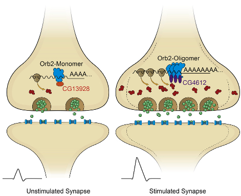
In their latest study, the researchers determined that Orb2 exists in two distinct physical states: monomeric (a single molecule that can bind to other molecules) and oligomeric (a molecular complex).

Like CPEB, oligomeric Orb2 is prion-like — that is, it’s a self-copying cluster. (But unlike prions, oligomeric Orb2 and CPEB are not toxic.) Monomeric Orb2 represses, and oligomeric Orb2 activates a crucial step in the complex cellular process that leads to protein synthesis.

During this crucial step, messenger RNA (mRNA), which is an RNA copy of a gene’s recipe for a protein, is translated by the cell’s ribosome into the sequence of amino acids that will make up a newly synthesized protein. The monomeric form of Orb2 binds to the target mRNA, keeping it in a repressed state.

The Stowers scientists also determined that prion-like Orb2 not only activates translation into amino acids but imparts its translational state to nearby monomer forms of Orb2. As a result, monomeric Orb2 is transformed into prion-like Orb2, so its role in translation switches from repression to activation.

Self-sustaining activation maintains synaptic activity

Stowers Associate Investigator Kausik Si, Ph.D. thinks this switch is the possible mechanism by which fleeting experiences create an enduring memory. “Because of the self-sustaining nature of the prion-like state, this creates a local and self-sustaining translation activation of Orb2-target mRNA, which maintains the changed state of synaptic activity over time,” says Si.

The discovery that the two distinct states of Orb2 have opposing roles in the translation process provides “for the first time a biochemical mechanism of synapse-specific persistent translation and long-lasting memory,” he states.

“To our knowledge, this is the first example of a prion-based protein switch that turns a repressor into an activator,” Si adds. “The recruitment of distinct protein complexes at the non-prion and prion-like forms to create altered activity states indicates the prion-like behavior is in essence a protein conformation-based switch.

“Through this switch, a protein can lose or gain a function that can be maintained over time in the absence of the original stimuli. Although such a possibility has been anticipated prior to this study, there was no direct evidence.”

The research builds upon previous studies by Si and Eric Kandel, M.D., of Columbia University and other scientists. These studies revealed that both short-term and long-term memories are created in synapses.


Abstract of Amyloidogenic Oligomerization Transforms Drosophila Orb2 from a Translation Repressor to an Activator

Memories are thought to be formed in response to transient experiences, in part through changes in local protein synthesis at synapses. In Drosophila, the amyloidogenic (prion-like) state of the RNA binding protein Orb2 has been implicated in long-term memory, but how conformational conversion of Orb2 promotes memory formation is unclear. Combining in vitro and in vivo studies, we find that the monomeric form of Orb2 represses translation and removes mRNA poly(A) tails, while the oligomeric form enhances translation and elongates the poly(A) tails and imparts its translational state to the monomer. The CG13928 protein, which binds only to monomeric Orb2, promotes deadenylation, whereas the putative poly(A) binding protein CG4612 promotes oligomeric Orb2-dependent translation. Our data support a model in which monomeric Orb2 keeps target mRNA in a translationally dormant state and experience-dependent conversion to the amyloidogenic state activates translation, resulting in persistent alteration of synaptic activity and stabilization of memory.

First direct evidence for synaptic plasticity in fruit fly brain

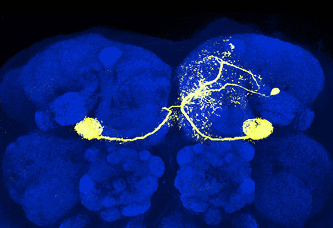
A singe dopamine neuron (yellow) in the mushroom body of the fruit fly Drosophila. Glenn Turner and colleagues trained flies to avoid certain odors by pairing them with stimulations of dopamine neurons signaling punishment. They found that this form of associative learning is driven by changes in synaptic strength between mushroom body neurons that process odors and downstream neurons that generate behavioral responses. (credit: Turner Lab, CSHL)

Scientists at Cold Spring Harbor Laboratory  (CSHL) have resolved a decades-long debate about how the brain is modified when an animal learns.

Using newly developed tools for manipulating specific populations of neurons, the researchers have for the first time observed direct evidence of synaptic plasticity — changes in the strength of synapse connections between neurons — in the fruit fly brain while flies are learning.

Due to the relative simplicity of fruit fly neural anatomy — there are just two synapses separating odor-detecting antenna from an olfactory-memory brain center called the mushroom body — the flies have provided a powerful model organism for studying learning.

Historically, researchers have monitored neurons in the mushroom body, as well as others to which they send signals, using a technique called calcium imaging. This approach enabled previous researchers to observe changes in neural activity that accompany learning. However this technique doesn’t reveal precisely how the electrical activity of the neurons is modified, since calcium is not the only ion involved in neuronal signaling.

Additionally, it was unclear how the changes that had been seen were related to the behavior of the animal.

CSHL Associate Professor Glenn Turner and colleagues at CSHL and the Howard Hughes Medical Institute’s Janelia Research Campus were able to make electrophysiological recordings to directly examine changes in synaptic strength at this site before and after learning for the first time.

Technical details: the experiment

The researchers exposed fruit flies to a specific test odor and a very short time later subjected them to an artificial aversive cue. To do so they fired tiny beams of laser light at dopamine-releasing neurons in the mushroom body that were genetically engineered to become active in response to the light. Just like our own neurons, dopamine-releasing neurons in the fly are involved in reward and punishment. “Presenting the smell of cherries, for example, which is normally an attractive odor to flies, while at the same time stimulating a particular dopamine neuron, trains the fly to avoid cherry odor,” Turner explains.

In addition to the dopamine neurons, the team identified neurons that represented the test odor and neurons that represented the flies’ behavioral response to that odor. These neurons are connected to each other, while the dopamine neurons, which represent the punishment signal, modulate that connection. The team then made recordings of the neurons representing the behavior. This enabled them to discover any changes to the synaptic inputs those neurons received from the odor-representing neurons before and after learning.

Strikingly, the team found a dramatic reduction in the synaptic inputs upon subsequent presentations of the test odor, but not control odors. This drop reflected the decrease in the attractiveness of the odor that resulted from the learning. “The average drop in synaptic strength was around 80 percent—that’s huge,” says Turner.

In future studies, Turner plans to exploit powerful tools available for studying fruit fly genetics to better understand the genetic components of learning. “We now have a way of investigating synaptic changes with genetic tools to identify molecules involved in learning and really understand the phenomenon at a level that bridges molecular and physiological mechanisms,” he says.

“That mechanistic level of understanding is going to be really important,” he adds. “It’s often at the level of molecules that you see really strong connections between Drosophila and other species, including humans.”

The results appeared online last week in the journal Neuron.


Abstract of Heterosynaptic Plasticity Underlies Aversive Olfactory Learning in Drosophila

Although associative learning has been localized to specific brain areas in many animals, identifying the underlying synaptic processes in vivo has been difficult. Here, we provide the first demonstration of long-term synaptic plasticity at the output site of the Drosophilamushroom body. Pairing an odor with activation of specific dopamine neurons induces both learning and odor-specific synaptic depression. The plasticity induction strictly depends on the temporal order of the two stimuli, replicating the logical requirement for associative learning. Furthermore, we reveal that dopamine action is confined to and distinct across different anatomical compartments of the mushroom body lobes. Finally, we find that overlap between sparse representations of different odors defines both stimulus specificity of the plasticity and generalizability of associative memories across odors. Thus, the plasticity we find here not only manifests important features of associative learning but also provides general insights into how a sparse sensory code is read out.

Do fish have emotions and consciousness?

Zebrafish (credit: Azul/CC)

Researchers in Spain and the U.K. have made the first observations infish of an increase in body temperature of 2–4 ºC when zebrafish were subjected to a stressful situation (they were confined in a net inside the tank at an uncomforable 27ºC for 15 minutes).*

This phenomenon is called “emotional fever” because it’s related to the emotions that animals feel in the face of an external stimulus, which been linked, controversially, with their consciousness. Until now, emotional fever had been observed in mammals, birds and certain reptiles, but never in fish, which is why fish have been regarded as animals without emotions or consciousness.

Does consciousness require a cerebral cortex?

Scientists differ on the degree to which fish can have consciousness. Some researchers argue that they cannot have consciousness as their brain is simple, lacking a cerebral cortex, and they have little capacity for learning and memory, a very simple behavioral repertoire, and no ability to experience suffering.

Others contest this view, pointing out that, despite the small size of the fish brain, detailed morphological and behavioral analyses have highlighted similarities between some fish brain structures and those seen in other vertebrates, such as the hippocampus (linked to learning and spatial memory) and the amygdala (linked to emotions) of mammals.

The research was published in an open-access paper recently in Proceedings of the Royal Society of London, Biological Sciences. It began three years ago at the Universitat Autònoma de Barcelona. Scientists from Stirling and Bristol universities helped with statistical analysis of the data.

* The researchers divided 72 zebrafish into two groups of 36 and placed them in a large tank with different interconnected compartments with temperatures ranging from 18ºC to 35ºC. The fish in one of these groups — the control group — were left undisturbed in the area where the temperature was at the level they prefer: 28ºC. The other group was subjected to a stressful situation: they were confined in a net inside the tank at 27ºC for 15 minutes. After this period the group was released.

While the control fish mainly stayed in the compartments at around 28ºC, the fish subjected to stress tended to move towards the compartments with a higher temperature, increasing their body temperature by two to four degrees. The researchers point to this as proof that these fish were displaying emotional fever.


Abstract of Fish can show emotional fever: stress-induced hyperthermia in zebrafish

Whether fishes are sentient beings remains an unresolved and controversial question. Among characteristics thought to reflect a low level of sentience in fishes is an inability to show stress-induced hyperthermia (SIH), a transient rise in body temperature shown in response to a variety of stressors. This is a real fever response, so is often referred to as ‘emotional fever’. It has been suggested that the capacity for emotional fever evolved only in amniotes (mammals, birds and reptiles), in association with the evolution of consciousness in these groups. According to this view, lack of emotional fever in fishes reflects a lack of consciousness. We report here on a study in which six zebrafish groups with access to a temperature gradient were either left as undisturbed controls or subjected to a short period of confinement. The results were striking: compared to controls, stressed zebrafish spent significantly more time at higher temperatures, achieving an estimated rise in body temperature of about 2–4°C. Thus, zebrafish clearly have the capacity to show emotional fever. While the link between emotion and consciousness is still debated, this finding removes a key argument for lack of consciousness in fishes.

Biologists induce flatworms to grow heads and brains of other species

Tufts biologists induced one species of flatworm —- G. dorotocephala, top left — to grow heads and brains characteristic of other species of flatworm, top row, without altering genomic sequence. Examples of the outcomes can be seen in the bottom row of the image. (credit: Center for Regenerative and Developmental Biology, School of Arts and Sciences, Tufts University.)

Tufts University biologists have electrically modified flatworms to grow heads and brains characteristic of another species of flatworm — without altering their genomic sequence. This suggests bioelectrical networks as a new kind of epigenetics (information existing outside of a genomic sequence) to determine large-scale anatomy.

Besides the overall shape of the head, the changes included the shape of the brain and the distribution of the worm’s adult stem cells.

The discovery could help improve understanding of birth defects and regeneration by revealing a new pathway for controlling complex pattern formation similar to how neural networks exploit bioelectric synapses to store and re-write information in the brain.

The findings are detailed in the open-access cover story of the November 2015 edition of the International Journal of Molecular Sciences, appearing online Nov. 24.

“These findings raise significant questions about how genes and bioelectric networks interact to build complex body structures,” said the paper’s senior author Michael Levin, Ph.D., who holds the Vannevar Bush Chair in biology and directs the Center for Regenerative and Developmental Biology in the School of Arts and Sciences at Tufts. Knowing how shape is determined and how to influence it is important because biologists could use that knowledge, for example, to fix birth defects or cause new biological structures to grow after an injury, he explained.

How they did it

The researchers worked with Girardia dorotocephala — free-living planarian flatworms, which have remarkable regenerative capacity. They induced the development of different species-specific head shapes by interrupting gap junctions, which are protein channels that enable cells to communicate with each other by passing electrical signals back and forth.

A conceptual model of shape change driven by physiological network dynamics. Planaria regeneration (B) parallels classical neural network behavior (A); both can be described in terms of free energy landscapes with multiple attractor states. (credit: Maya Emmons-Bell et al./Int. J. Mol. Sci.)

The ease with which a particular shape could be coaxed from a G. dorotocephala worm was proportional to the proximity of the target worm on the evolutionary timeline. The closer the two species were related, the easier it was to effect the change. This observation strengthens the connection to evolutionary history, suggesting that modulation of physiological circuits may be one more tool exploited by evolution to alter animal body plans.

However, this shape change was only temporary. Weeks after the planaria completed regeneration to the other species’ head shapes, the worms once again began remodeling and re-acquired their original head morphology. Additional research is needed to determine how this occurs. The authors also presented a computational model that explains how changes in cell-to-cell communication can give rise to the diverse shape types.

The interdisciplinary research involved U.S.- and Canada-based biologists and European mathematicians.


Abstract of Gap Junctional Blockade Stochastically Induces Different Species-Specific Head Anatomies in Genetically Wild-Type Girardia dorotocephala Flatworms

The shape of an animal body plan is constructed from protein components encoded by the genome. However, bioelectric networks composed of many cell types have their own intrinsic dynamics, and can drive distinct morphological outcomes during embryogenesis and regeneration. Planarian flatworms are a popular system for exploring body plan patterning due to their regenerative capacity, but despite considerable molecular information regarding stem cell differentiation and basic axial patterning, very little is known about how distinct head shapes are produced. Here, we show that after decapitation in G. dorotocephala, a transient perturbation of physiological connectivity among cells (using the gap junction blocker octanol) can result in regenerated heads with quite different shapes, stochastically matching other known species of planaria (S. mediterraneaD. japonica, and P. felina). We use morphometric analysis to quantify the ability of physiological network perturbations to induce different species-specific head shapes from the same genome. Moreover, we present a computational agent-based model of cell and physical dynamics during regeneration that quantitatively reproduces the observed shape changes. Morphological alterations induced in a genomically wild-type G. dorotocephala during regeneration include not only the shape of the head but also the morphology of the brain, the characteristic distribution of adult stem cells (neoblasts), and the bioelectric gradients of resting potential within the anterior tissues. Interestingly, the shape change is not permanent; after regeneration is complete, intact animals remodel back to G. dorotocephala-appropriate head shape within several weeks in a secondary phase of remodeling following initial complete regeneration. We present a conceptual model to guide future work to delineate the molecular mechanisms by which bioelectric networks stochastically select among a small set of discrete head morphologies. Taken together, these data and analyses shed light on important physiological modifiers of morphological information in dictating species-specific shape, and reveal them to be a novel instructive input into head patterning in regenerating planaria.

Master genetic switch for brain development discovered

Cells in which NeuroD1 is turned on are reprogrammed to become neurons. Cell nuclei are shown in blue and neurons, with their characteristic long processes, are shown in red. (credit: A. Pataskar, J. Jung & V. Tiwari)

Scientists at the Institute of Molecular Biology (IMB) in Mainz, Germany have unraveled a complex regulatory mechanism that explains how a single gene, NeuroD1, can drive the formation of brain cells. The research, published in The EMBO Journal, is an important step towards a better understanding of how the brain develops and may lead to breakthroughs in regenerative medicine.

Neurodegenerative disorders, such as Parkinson’s disease, are often characterized by an irreversible loss of brain cells. Unlike many other cell types in the body, these neurons are generally not able to regenerate by themselves, so if the brain is damaged, it stays damaged. One hope of developing treatments for this kind of damage is to understand how the brain develops in the first place, and then try to imitate the process. However, the brain is also one of the most complex organs in the body, and very little is understood about the molecular pathways that guide its development.

An epigenetic memory

Scientists in Dr. Vijay Tiwari’s group at the Institute of Molecular Biology at Johannes Gutenberg University Mainz have been investigating a central gene in brain development, NeuroD1. This gene is expressed in the developing brain and marks the onset of neurogenesis (neuron growth).

In their research article, Tiwari and his colleagues have shown that during brain development, it also acts as a master regulator of a large number of genes that cause these cells to develop into neurons. They used a combination of neurobiology, epigenetics, and computational biology approaches to show that these genes are normally turned off in development, but NeuroD1 activity changes their epigenetic state in order to turn them on.

Diagram showing how NeuroD1 influences the development of neurons. During brain development, expression of NeuroD1 marks the onset of neurogenesis. NeuroD1 accomplishes this via epigenetic reprogramming: neuronal genes are switched on, and the cells develop into neurons. TF: transcription factor; V: ventricle; P: pial surface. (credit: A. Pataskar, J. Jung & V. Tiwari)

Strikingly, the researchers show that these genes remain switched on even after NeuroD1 is later switched off. They further show that this is because NeuroD1 activity leaves permanent epigenetic marks on these genes that keep them turned on. In other words, it creates an epigenetic memory of neuronal differentiation in the cell.

“This is a significant step towards understanding the relationship between DNA sequence, epigenetic changes, and cell fate,” says Tiwari. “It not only sheds new light on the formation of the brain during embryonic development but also opens up novel avenues for regenerative therapy.”


Abstract of NeuroD1 reprograms chromatin and transcription factor landscapes to induce the neuronal program

Cell fate specification relies on the action of critical transcription factors that become available at distinct stages of embryonic development. One such factor is NeuroD1, which is essential for eliciting the neuronal development program and possesses the ability to reprogram other cell types into neurons. Given this capacity, it is important to understand its targets and the mechanism underlying neuronal specification. Here, we show that NeuroD1 directly binds regulatory elements of neuronal genes that are developmentally silenced by epigenetic mechanisms. This targeting is sufficient to initiate events that confer transcriptional competence, including reprogramming of transcription factor landscape, conversion of heterochromatin to euchromatin, and increased chromatin accessibility, indicating potential pioneer factor ability of NeuroD1. The transcriptional induction of neuronal fate genes is maintained via epigenetic memory despite a transient NeuroD1 induction during neurogenesis. NeuroD1 also induces genes involved in the epithelial‐to‐mesenchymal transition, thereby promoting neuronal migration. Our study not only reveals the NeuroD1‐dependent gene regulatory program driving neurogenesis but also increases our understanding of how cell fate specification during development involves a concerted action of transcription factors and epigenetic mechanisms.

An ultrafast 3-D imaging system to investigate traumatic brain injury

Still frame filmed at 200,000 frames/sec of a violently collapsing vapor bubble inside a brain-mimicking collagen gel (bubble size is approximately 100 microns). Inside the gel are thousands of brain cells (neurons). (credit: J. Estrada (Franck Lab)/Brown U)

Researchers at Brown University are using an ultrafast 3-D imaging system to investigate the effects of microcavitation bubbles on traumatic brain injury (TBI), experienced by some soldiers and football players.

In the fleeting moments after a liquid is subjected to a sudden change in pressure, microscopic bubbles rapidly form and collapse in a process known as cavitation.

In mechanical systems such as propellers, the resulting shock waves and jets can cause gradual wear, and in biological systems, they can shred and distort cells. In the human brain, this is believed to be a mechanistic cause of TBI, but the phenomenon has yet to be directly observed in brain tissue because the bubbles appear and disappear within microseconds.

To better understand the connection between microcavitation and traumatic brain injury, researchers at Brown University have developed a novel 3-D imaging system that allows them to film one million frames per second, with a single camera on a single microscope, and ultimately to explore mechanisms of damage to neurons in the laboratory.

Current 3-D image correlation methods typically involve making stereo projections of objects (projecting a sphere onto a plane) by capturing images with two or more cameras, but using multiple cameras carries a loss of spatial resolution. Instead, the researchers placed a diffraction grating in front of the imaging camera on the microscope. They then used a nanosecond-pulsed infrared laser to produce single cavitation bubbles within a model neural network made of collagen and biomimetic hydrogels embedded with neurons, thus simulating the action of a neural network subjected to a negative pressure surge.

It  generates two perspectives of the object — as if you had two cameras — without sacrificing spatial resolution. Currently, they can resolve motion fields down to the 10–100 nanometer scale, but they are aiming for single-nanometer scale using super-resolution microscopy techniques in 3-D.

The researchers are presenting their recent findings at the American Physical Society (APS) Division of Fluid Dynamics (DFD) 68th meeting, Nov. 22–24 in Boston, Mass.


Abstract of Microcavitation as a Neuronal Damage Mechanism in Blast Traumatic Brain Injury

Traumatic brain injury (TBI), usually the result of impact or blast to the head, affects about 1.5 million Americans annually. Diffuse axonal injury, the hallmark feature of blunt TBI, has been investigated in direct mechanical loading conditions. However, recent evidence suggests inertial cavitation as a possible bTBI mechanism, particularly in the case of armed forces exposed to concussive blasts. Cavitation damage to free surfaces has been well-studied in the fi eld of fl uid dynamics, but bubble interactions within confi ned 3D environments have not been largely investigated. Cavitation occurs via a low-pressure region caused by pressure waves and is strongly dependent on local geometric and mechanical properties. The structural damage features as the result of cavitation – in particular at the cellular level – are incompletely understood, in part due to the rapid bubble formation and strain rates of up to ~105 –106 s– 1 . This project aims to characterize material damage in 2D and 3D environments and cell cultures by utilizing digital image correlation at a speed of up to ten 6 frames per second.

First real-time imaging of neural activity invented

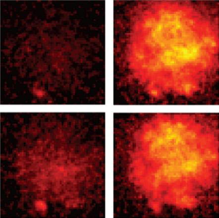
A series of images from a Duke engineering experiment show voltage spreading through a fruitfly neuron over a matter of just 4 milliseconds, a hundred times faster than the blink of an eye. The technology can see impulses as fleeting as 0.2 millisecond — 2000 times faster than a blink. (credit: Yiyang Gong, Duke University)

Researchers at Stanford University and Duke University have developed a new technique for watching the brain’s neurons in action with a temporal (time) resolution of about 0.2 milliseconds — a speed that is just fast enough to capture the action potentials in mammalian brains in real time for the first time.

The researchers combined genetically encoded voltage indicators, which can sense individual action potentials from many cell types in live animals, with a protein that can quickly sense neural voltage potentials with another protein that can amplify its signal output — the brightest fluorescing protein available.

The paper appeared online in Science on November 19, 2015.


Abstract of High-speed recording of neural spikes in awake mice and flies with a fluorescent voltage sensor

Genetically encoded voltage indicators (GEVIs) are a promising technology for fluorescence readout of millisecond-scale neuronal dynamics. Prior GEVIs had insufficient signaling speed and dynamic range to resolve action potentials in live animals. We coupled fast voltage-sensing domains from a rhodopsin protein to bright fluorophores via resonance energy transfer. The resulting GEVIs are sufficiently bright and fast to report neuronal action potentials and membrane voltage dynamics in awake mice and flies, resolving fast spike trains with 0.2-millisecond timing precision at spike detection error rates orders of magnitude better than prior GEVIs. In vivo imaging revealed sensory-evoked responses, including somatic spiking, dendritic dynamics, and intracellular voltage propagation. These results empower in vivo optical studies of neuronal electrophysiology and coding and motivate further advancements in high-speed microscopy.