Researchers discover signaling molecule that helps neurons find their way in the developing brain

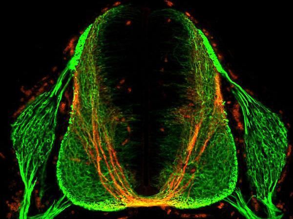
This image shows a section of the spinal cord of a mouse embryo. Neurons appear green. Commissural axons (which connect the two sides of the brain) appear as long, u-shaped threads, and the bottom, yellow segment of the structure represents the midline (between brain hemispheres). (credit: Laboratory of Brain Development and Repair/ The Rockefeller University)

Rockefeller University researchers have discovered a molecule secreted by cells in the spinal cord that helps guide axons (neuron extensions) during a critical stage of central nervous system development in the embryo. The finding helps solve the mystery: how do the billions of neurons in the embryo nimbly reposition themselves within the brain and spinal cord, and connect branches to form neural circuits?

Working in mice, the researchers identified an axon guidance factor, NELL2, and explained how it makes commissural axons (which connect the two sides of the brain).

The findings could help scientists understand what goes wrong in a rare disease called horizontal gaze palsy with progressive scoliosis. People affected by the condition often suffer from abnormal spine curvature, and are unable to move their eyes horizontally from side to side. The study was published Thursday Nov. 19 in the journal Science.


Abstract of Operational redundancy in axon guidance through the multifunctional receptor Robo3 and its ligand NELL2

Axon pathfinding is orchestrated by numerous guidance cues, including Slits and their Robo receptors, but it remains unclear how information from multiple cues is integrated or filtered. Robo3, a Robo family member, allows commissural axons to reach and cross the spinal cord midline by antagonizing Robo1/2–mediated repulsion from midline-expressed Slits and potentiating deleted in colorectal cancer (DCC)–mediated midline attraction to Netrin-1, but without binding either Slits or Netrins. We identified a secreted Robo3 ligand, neural epidermal growth factor-like-like 2 (NELL2), which repels mouse commissural axons through Robo3 and helps steer them to the midline. These findings identify NELL2 as an axon guidance cue and establish Robo3 as a multifunctional regulator of pathfinding that simultaneously mediates NELL2 repulsion, inhibits Slit repulsion, and facilitates Netrin attraction to achieve a common guidance purpose.

Exercise may protect against neurodegenerative diseases

(credit: iStock)

Exercise may protect aging brains against the neurodegenerative diseases resulting from energy-depleting stress caused by neurotoxins and other factors, according to researchers at the National Institute on Aging Intramural Research Program and Johns Hopkins University School of Medicine.

They found that running-wheel exercise increased the amount of SIRT3 in neurons of normal mice and protected them against degeneration.

However, mice models genetically modified to not produce SIRT3 became highly sensitive to stress when exposed to neurotoxins that cause neurodegeneration and epileptic seizures, and running failed to protect the neurons. In that case, neurons could be protected against stress by a gene-therapy technology to increase levels of SIRT3 in neurons, they found.

These findings suggest that bolstering mitochondrial function and stress resistance by increasing SIRT3 levels — either by exercise or gene therapy — may protect against age-related cognitive decline and brain diseases, the researchers say.

The research team report their findings online today (Nov. 19) in the journal Cell Metabolism. This work was supported by the Intramural Research Program of the National Institute on Aging and the Glenn Foundation for Biomedical Research.


Abstract of Mitochondrial SIRT3 mediates adaptive responses of neurons to exercise, and metabolic and excitatory challenges

The impact of mitochondrial protein acetylation status on neuronal function and vulnerability to neurological disorders is unknown. Here we show that the mitochondrial protein deacetylase SIRT3 mediates adaptive responses of neurons to bioenergetic, oxidative, and excitatory stress. Cortical neurons lacking SIRT3 exhibit heightened sensitivity to glutamate-induced calcium overload and excitotoxicity and oxidative and mitochondrial stress; AAV-mediated Sirt3 gene delivery restores neuronal stress resistance. In models relevant to Huntington’s disease and epilepsy, Sirt3-/- mice exhibit increased vulnerability of striatal and hippocampal neurons, respectively. SIRT3 deficiency results in hyperacetylation of several mitochondrial proteins, including superoxide dismutase 2 and cyclophilin D. Running wheel exercise increases the expression of Sirt3 in hippocampal neurons, which is mediated by excitatory glutamatergic neurotransmission and is essential for mitochondrial protein acetylation homeostasis and the neuroprotective effects of running. Our findings suggest that SIRT3 plays pivotal roles in adaptive responses of neurons to physiological challenges and resistance to degeneration.

Modulating brain’s stress circuity might prevent Alzheimer’s disease

Effect of drug treatment on AD mice in control group (left) or drug (right) on Ab plaque load. (credit: Cheng Zhang et al./Alzheimer’s & Dementia: The Journal of the Alzheimer’s Association)

In a novel animal study design that mimicked human clinical trials, researchers at University of California, San Diego School of Medicine report that long-term treatment using a small-molecule drug that reduces activity of  the brain’s stress circuitry significantly reduces Alzheimer’s disease (AD) neuropathology and prevents onset of cognitive impairment in a mouse model of the neurodegenerative condition.

The findings are described in the current online issue of the journal Alzheimer’s & Dementia: The Journal of the Alzheimer’s Association.

Previous research has shown a link between the brain’s stress signaling pathways and AD. Specifically, the release of a stress-coping hormone called corticotropin-releasing factor (CRF), which is widely found in the brain and acts as a neurotransmitter/neuromodulator, is dysregulated in AD and is associated with impaired cognition and with detrimental changes in tau protein and increased production of amyloid-beta protein fragments that clump together and trigger the neurodegeneration characteristic of AD.

“Our work and that of our colleagues on stress and CRF have been mechanistically implicated in Alzheimer’s disease, but agents that impact CRF signaling have not been carefully tested for therapeutic efficacy or long-term safety in animal models,” said the study’s principal investigator and corresponding author Robert Rissman, PhD, assistant professor in the Department of Neurosciences and Biomarker Core Director for the Alzheimer’s Disease Cooperative Study (ADCS).

The researchers determined that modulating the mouse brain’s stress circuitry mitigated generation and accumulation of amyloid plaques widely attributed with causing neuronal damage and death. As a consequence, behavioral indicators of AD were prevented and cellular damage was reduced.  The mice began treatment at 30-days-old — before any pathological or cognitive signs of AD were present — and continued until six months of age.

One particular challenge, Rissman noted, is limiting exposure of the drug to the brain so that it does not impact the body’s ability to respond to stress. “This can be accomplished because one advantage of these types of small molecule drugs is that they readily cross the blood-brain barrier and actually prefer to act in the brain,” Rissman said.

“Rissman’s prior work demonstrated that CRF and its receptors are integrally involved in changes in another AD hallmark, tau phosphorylation,” said William Mobley, MD, PhD, chair of the Department of Neurosciences and interim co-director of the Alzheimer’s Disease Cooperative Study at UC San Diego. “This new study extends those original mechanistic findings to the amyloid pathway and preservation of cellular and synaptic connections.  Work like this is an excellent example of UC San Diego’s bench-to-bedside legacy, whereby we can quickly move our basic science findings into the clinic for testing,” said Mobley.

Rissman said R121919 was well-tolerated by AD mice (no significant adverse effects) and deemed safe, suggesting CRF-antagonism is a viable, disease-modifying therapy for AD. Drugs like R121919 were originally designed to treat generalized anxiety disorder, irritable bowel syndrome and other diseases, but failed to be effective in treating those disorders.

Rissman noted that repurposing R121919 for human use was likely not possible at this point. He and colleagues are collaborating with the Sanford Burnham Prebys Medical Discovery Institute to design new assays to discover the next generation of CRF receptor-1 antagonists for testing in early phase human safety trials.

“More work remains to be done, but this is the kind of basic research that is fundamental to ultimately finding a way to cure — or even prevent —Alzheimer’s disease,” said David Brenner, MD, vice chancellor, UC San Diego Health Sciences and dean of UC San Diego School of Medicine. “These findings by Dr. Rissman and his colleagues at UC San Diego and at collaborating institutions on the Mesa suggest we are on the cusp of creating truly effective therapies.”


Abstract of Corticotropin-releasing factor receptor-1 antagonism mitigates beta amyloid pathology and cognitive and synaptic deficits in a mouse model of Alzheimer’s disease

Introduction: Stress and corticotropin-releasing factor (CRF) have been implicated as mechanistically involved in Alzheimer’s disease (AD), but agents that impact CRF signaling have not been carefully tested for therapeutic efficacy or long-term safety in animal models.

Methods: To test whether antagonism of the type-1 corticotropin-releasing factor receptor (CRFR1) could be used as a disease-modifying treatment for AD, we used a preclinical prevention paradigm and treated 30-day-old AD transgenic mice with the small-molecule, CRFR1-selective antagonist, R121919, for 5 months, and examined AD pathologic and behavioral end points.

Results: R121919 significantly prevented the onset of cognitive impairment in female mice and reduced cellular and synaptic deficits and beta amyloid and C-terminal fragment-β levels in both genders. We observed no tolerability or toxicity issues in mice treated with R121919.

Discussion: CRFR1 antagonism presents a viable disease-modifying therapy for AD, recommending its advancement to early-phase human safety trials.

Reprogramming neurons and rewiring the brain

Neurons of the cerebral cortex targeted for reprogramming are displayed in green. (credit: Caroline Rouaux/Arlotta Lab)

In previous research, Harvard Stem Cell Institute researchers disproved neurobiology dogma by “reprogramming” neurons — turning one form of neuron into another — in the brains of living animals. Now they’ve taken it a step further, showing that networks of communication among reprogrammed neurons and their neighbors can also be changed, or “rewired.”

The finding, by Paola Arlotta, a professor of stem cell and regenerative biology, in close collaboration with Takao Hensch, a professor of molecular and cellular biology, has implications for both a basic understanding of how neurons choose their synaptic partners when circuits wire during early development, and for developing strategies to change defective communication in psychiatric and neurodevelopmental diseases, such as schizophrenia and autism, the researchers suggest.

In the new research, the research team reprogrammed one type of excitatory neurons into another type of excitatory neurons, and then observed connections made by a type of inhibitory neurons, noticing that the inhibitory neurons then recognized the reprogrammed excitatory neurons as “new” cells and modified their circuitry as a result.*

“What we’ve shown here is that not only neurons can be dramatically changed from one type into another from within the brain,” said Arlotta, but also that “neighboring neurons recognize the reprogrammed cells as different and adapt by changing how they communicate with them. Transformed neurons were recognized as ‘new’ cells, with different properties, by neighboring inhibitory interneurons, which in turn created new circuitry appropriate for their ‘new’ neuronal target,” Arlotta said.

That demonstrates that “synaptic connections among neurons are not made randomly,” she added. “The brain is much more sophisticated, and different neurons have ways to control the behavior of neighboring circuits in their own unique way to ultimately change how much inhibition, for example, they receive from their synaptic partners.”

Rewiring the brain to fix a damaged circuit

One of the major focuses today in regenerative neurobiology is to use stem cells to produce and replace neurons killed off by disease, and then implant them into patients’ brains. “Instead of making neurons in a dish from stem cells, an alternative would be to reprogram the identity of other neurons,” Arlotta said.

“These are early but exciting days,” Arlotta said. “The work thus far has been done in the brains of young mice, which are far more plastic than adult brains,” she pointed out. The next frontier, she said, is to attempt to reprogram neurons and circuits in older brains. “If we could do it in an adult brain, it would be immensely powerful.”

Arlotta is also an associate member of the Stanley Center for Psychiatric Disease at the Broad Institute of Harvard and MIT.

The work is published in the latest edition of the journal Neuron.

* The researchers first reprogrammed “glutamatergic excitatory projection neurons (PNs)” (neurons of the cerebral cortex that normally would connect the two sides of the brain) into another type of PN neurons that instead connect far away, for example in the spinal cord. They then monitored the behavior of a third class of cells, “local GABAergic inhibitory interneurons (INs)” and showed that the INs change their synaptic connections to connect to the reprogrammed PN neurons.  


Abstract of Instructing Perisomatic Inhibition by Direct Lineage Reprogramming of Neocortical Projection Neurons

During development of the cerebral cortex, local GABAergic interneurons recognize and pair with excitatory projection neurons to ensure the fine excitatory-inhibitory balance essential for proper circuit function. Whether the class-specific identity of projection neurons has a role in the establishment of afferent inhibitory synapses is debated. Here, we report that direct in vivo lineage reprogramming of layer 2/3 (L2/3) callosal projection neurons (CPNs) into induced corticofugal projection neurons (iCFuPNs) increases inhibitory input onto the converted neurons to levels similar to that of endogenous CFuPNs normally found in layer 5 (L5). iCFuPNs recruit increased numbers of inhibitory perisomatic synapses from parvalbumin (PV)-positive interneurons, with single-cell precision and despite their ectopic location in L2/3. The data show that individual reprogrammed excitatory projection neurons extrinsically modulate afferent input by local PV+ interneurons, suggesting that projection neuron class-specific identity can actively control the wiring of the cortical microcircuit.

‘Golden window’ wavelength range for optimal deep-brain near-infrared imaging determined

Rayleigh scattering causes the reddening of the sun at sunset — an example of how longer wavelengths (yellow and red compared to blue in blue sky) penetrate matter (dust at sunset) better. (credit: Wikipedia/CC)

Researchers at The City College of New York (CCNY) have determined the optimal wavelengths for bioimaging of the brain at longer near-infrared wavelengths, which permit deeper imaging.

Near-infrared (NIR) radiation has been used for one- and two-photon fluorescence imaging at near-infrared wavelengths of 650–950 nm (nanometers) for deep brain imaging, but it is limited in penetration depth. (The CCNY researchers dubbed this Window I, also known as the therapeutic window.)

Longer infrared wavelengths penetrate deeper but are limited by Rayleigh and Mie scattering, which blur images, and absorption, which reduces the number of available photons (brightness). These limitations are based on the lack of suitable CMOS semiconductor imaging detectors or femtosecond laser sources.

The new CCNY study, led by biomedical engineer Lingyan Shi, studied three new optical windows in the near-infrared (NIR) region, in addition to Window I, for high-resolution deep brain imaging.

Their study built on a prior CCNY study* in 2014 using detectors based on indium gallium arsenide (GaAs) or indium antimonide-(InSb) and a femtosecond excitation source of IMRA fiber laser** to image rat brain tissue in window II (1,100– 1,350 nm), window III (1,600–1,870 nm), and window IV (1600 nm to 1870 nm, centered at 2,200 nm).
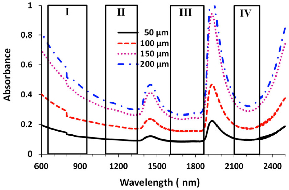
Absorbance for four windows at four brain-tissue thicknesses (credit: Lingyan Shi et al./J. Biophotonics)

The new CCNY research investigated the optimal wavelength band and optical properties of brain tissue with NIR, including the total attenuation coefficient (μt), absorption coefficient (μa), reduced scattering coefficient (μ0 s), and the scattering anisotropy coefficient (g) in these optical windows. The purpose of the study was to determine an optimal optical window in NIR in the 650 nm to 2500 nm range to reduce scattering, achieve optimal absorption, and reduce noise for deep-brain tissue imaging.

Golden Window: optimal wavelength range

Peak transmittance T (%) measured with each optical window for brain tissues with four thicknesses. Window III had the highest transmittance percentage for each of the thicknesses, followed by windows II and IV. (credit: Lingyan Shi et al./J. Biophotonics)

The researchers found that the “Golden Window” (1600 nm to 1870 nm) is an optimal wavelength range for light penetration in brain tissue, followed by Windows II and IV.

“This is a first for brain imaging and proved theoretically and experimentally that deep imaging of the brain is possible using light at longer wavelengths. It demonstrates these windows’ potential for deeper brain tissue imaging due to the reduction of scattering that causes blurring,” said Shi, a research associate in City College’s Institute for Ultrafast Spectroscopy and Lasers, and the biology department.

Published by the Journal of Biophotonics, her study sheds light on the development of the next generation of microscopy imaging technique, in which the “Golden Window” may be utilized for high-resolution deeper brain imaging. The next step in the research is in vivo imaging in mice using Golden Window wavelength light.

Shi’s team included Distinguished Professor of Physics Robert R. Alfano and Adrian Rodriguez-Contreras, an assistant professor of biology. Shi earned a Ph.D. in biomedical engineering from CCNY’s Grove School of Engineering in 2014.

* L. A. Sordillo, Y. Pu, S. Pratavieira, Y. Budansky, and R. R. Alfano, J. Biomed. Opt. 19, 056004 (2014) [link].

** Excitation wavelength 1680 nm, power > 200 mW, pulse width 100 fs, and 50 MHz repetition rate.


Abstract of Transmission in near-infrared optical windows for deep brain imaging

Near-infrared (NIR) radiation has been employed using one- and two-photon excitation of fluorescence imaging at wavelengths 650–950 nm (optical window I) for deep brain imaging; however, longer wavelengths in NIR have been overlooked due to a lack of suitable NIR-low band gap semiconductor imaging detectors and/or femtosecond laser sources. This research introduces three new optical windows in NIR and demonstrates their potential for deep brain tissue imaging. The transmittances are measured in rat brain tissue in the second (II, 1,100–1,350 nm), third (III, 1,600–1,870 nm), and fourth (IV, centered at 2,200 nm) NIR optical tissue windows. The relationship between transmission and tissue thickness is measured and compared with the theory. Due to a reduction in scattering and minimal absorption, window III is shown to be the best for deep brain imaging, and windows II and IV show similar but better potential for deep imaging than window I.
Blood-brain barrier opened non-invasively for the first time in humans, using focused ultrasound

Opening up the blood-brain barrier to deliver drugs (credit: Focused Ultrasound Foundation)

The blood-brain barrier has been non-invasively opened in a human patient for the first time. A team at Sunnybrook Health Sciences Centre in Toronto used focused ultrasound to temporarily open the blood-brain barrier (BBB), allowing for effective delivery of chemotherapy into a patient’s malignant brain tumor.

The team infused the chemotherapy agent doxorubicin, along with tiny gas-filled bubbles, into the bloodstream of a patient with a brain tumor. They then applied focused ultrasound to areas in the tumor and surrounding brain, causing the bubbles to vibrate, loosening the tight junctions of the cells comprising the BBB, and allowing high concentrations of the chemotherapy to enter targeted tissues.

This patient treatment is part of a pilot study of up to 10 patients to establish the feasibility, safety, and preliminary efficacy of focused ultrasound to temporarily open the blood-brain barrier to deliver chemotherapy to brain tumors. The Focused Ultrasound Foundation is currently funding this trial through their Cornelia Flagg Keller Memorial Fund for Brain Research. Based on these two pre-clinical studies, a pilot clinical trial using focused ultrasound to treat Alzheimer’s is being organized.

Dr. Kullervo Hynynen, senior scientist at the Sunnybrook Research Institute, has been performing similar pre-clinical studies for about a decade. In 2012, his team was able to bypass the BBB of a rat model non-invasively (see Bypassing the blood-brain barrier with MRI and ultrasound).

Previous methods where invasive, requiring an operation, such as an implanted mucosal graft in the nose (see A drug-delivery technique to bypass the blood-brain barrier and Researchers bypass the blood-brain barrier, widening treatment options for neurodegenerative and central nervous system disease) or inserting needle electrodes into the diseased tissue and applying multiple bursts of pulsed electric energy (see Blood-brain-barrier disruption with high-frequency pulsed electric fields).

Fighting disease

The researchers suggest that focused ultrasound could also be used to deliver other types of drugs, DNA-loaded nanoparticles, viral vectors, and antibodies to the brain to treat a range of neurological conditions, including various types of brain tumors, Parkinson’s, Alzheimer’s and some psychiatric diseases.

For example, the temporary opening of the blood-brain barrier appears to facilitate the brain’s clearance of a key pathologic protein related to Alzheimer’s and improves cognitive function, the researchers found. And a recent study at the Queensland Brain Institute in Australia demonstrated that opening the blood-brain barrier with focused ultrasound reduced brain plaques and improved memory in a mouse model of Alzheimer’s disease.


Focused Ultrasound Foundation


Abstract of Scanning ultrasound removes amyloid-β and restores memory in an Alzheimer’s disease mouse model

Transgenic mice with increased amyloid-β (Aβ) production show several aspects of Alzheimer’s disease, including Aβ deposition and memory impairment. By repeatedly treating these Aβ-forming mice with scanning ultrasound, Leinenga and Götz now demonstrate that Aβ is removed and memory is restored as revealed by improvement in three memory tasks. These improvements were achieved without the use of any therapeutic agent, and the scanning ultrasound treatment did not induce any apparent damage to the mouse brain. The authors then showed that scanning ultrasound activated resident microglial cells that took up Aβ into their lysosomes. These findings suggest that repeated scanning ultrasound may be a noninvasive method with potential for treating Alzheimer’s disease.

Fastest brain-computer-interface speller developed

System diagram of the advanced SSVEP-based BCI speller. It consists of four main procedures: visual stimulation, EEG recording, real-time data processing, and feedback presentation. The 5 × 8 stimulation matrix includes the 26 letters of the English alphabet, 10 numbers, and 4 symbols (space, comma, period, and backspace). The image of the screen stimulation matrix shown here is only for illustration. (credit: Xiaogang Chen et al./PNAS)

Brain–computer interface (BCI) spellers allow a paralyzed patient to spell out words by looking at letters on a screen. Paralyzed patients can communicate by gazing at different letters to spell out a word.

Currently, the most advanced systems for doing this use “steady state visually evoked potential” (SSVEP). This method tags different characters on a screen by flashing each character at a different frequency (from 3.5 Hz to 75 Hz in one system). When a patient looks at a specific flashing character, the brain generates evoked electrical activity at the same (or multiples of) the specific frequency of the visual stimulus. This video demonstrates how that works:


Nikolay Chumerin |SSVEP-based mindspeller

However, the low communication rate (low number of characters per minute) for existing SSVEP systems is a remaining obstacle to improving BCI-based communication. That’s because the tagged visual evoked potentials are difficult to detect due to interference from spontaneous EEG signals.

A new world record for BCI spellers claimed

Researchers at Tsinghua University in China and State Key Laboratory Integrated Optoelectronics, Institute of Semiconductors (IOS), Chinese Academy of Sciences have now developed a significantly improved SSVEP system. It can achieve rates of about 60 characters (∼12 words) per minute (5.32 bits per second) — a claimed new world record for BCI spellers, using either non-invasive or invasive methods.

To achieve that, the 40 characters in the stimulation matrix (used on the display) are tagged with a more sophisticated flickering frequency and phase coding scheme similar to that used in telecommunications systems, along with user-specific decoding. Real-time data analysis recognizes the target character through pre-processing, feature extraction, and classification.

The researchers suggest that the spelling speed achieved with this system (~1 character per second) seems close to the speed limit of human gaze control.

The research was published in Proceedings of the National Academy of Sciences. It was supported by the Chinese National Basic Research Program, the National High-Tech R&D Program, the National Natural Science Foundation, and the Recruitment Program for Young Professionals.


Abstract of High-speed spelling with a noninvasive brain–computer interface

The past 20 years have witnessed unprecedented progress in brain–computer interfaces (BCIs). However, low communication rates remain key obstacles to BCI-based communication in humans. This study presents an electroencephalogram-based BCI speller that can achieve information transfer rates (ITRs) up to 5.32 bits per second, the highest ITRs reported in BCI spellers using either noninvasive or invasive methods. Based on extremely high consistency of frequency and phase observed between visual flickering signals and the elicited single-trial steady-state visual evoked potentials, this study developed a synchronous modulation and demodulation paradigm to implement the speller. Specifically, this study proposed a new joint frequency-phase modulation method to tag 40 characters with 0.5-s-long flickering signals and developed a user-specific target identification algorithm using individual calibration data. The speller achieved high ITRs in online spelling tasks. This study demonstrates that BCIs can provide a truly naturalistic high-speed communication channel using noninvasively recorded brain activities.

Long-term aerobic exercise prevents age-related brain deterioration

Schematic illustration of age-related changes in the neurovascular unit that are prevented by exercise. In the aged cortex of sedentary mice, neurovascular dysfunction is evident by decreased numbers of pericytes (surrounding capillaries, pink), decline in basement membrane (BM) coverage (blue), increased transcytosis (a process that transports macromolecules across cells, allowing pathogens to invade) on endothelial cells (green), reduced expression of AQP4 in astrocytes, down-regulation of Apoe (an essential protein, light purple), decrease in synaptic proteins such as synaptophysin (SYN, green), and increased proinflammatory IBA1+ microglia/monocytes (indicating age-related neuroinflammation, yellow). These age-related changes were successfully prevented (horizontal T line, “Exercise”)  by 6 months of voluntary running during aging. (credit: Ileana Soto et al./PLOS Biology

A study of the brains of mice shows that structural deterioration associated with old age can be prevented by long-term aerobic exercise starting in mid-life, according to the authors of an open-access paper in the journal PLOS Biology yesterday (October 29).

Old age is the major risk factor for Alzheimer’s disease, like many other diseases, as the authors at The Jackson Laboratory in Bar Harbor, Maine, note. Age-related cognitive deficits are due partly to changes in neuronal function, but also correlate with deficiencies in the blood supply to the brain and with low-level inflammation.

“Collectively, our data suggests that normal aging causes significant dysfunction to the cortical neurovascular unit, including basement membrane reduction and pericyte (cells that wrap around blood capillaries) loss. These changes correlate strongly with an increase in microglia/monocytes in the aged cortex,” said Ileana Soto, lead author on the study.*

Benefits of aerobic exercise

However, the researchers found that if they let the mice run freely, the structural changes that make the blood-brain barrier leaky and result in inflammation of brain tissues in old mice can be mitigated. That suggests that there are also beneficial effects of exercise on dementia in humans.**

Further work will be required to establish the mechanism(s): what is the role of the complement-producing microglia/macrophages, how does Apoe decline contribute to age-related neurovascular decline, does the leaky blood-brain barrier allow the passage of damaging factors from the circulation into the brain?

This work was funded in part by The Jackson Laboratory Nathan Shock Center, the Fraternal Order of the Eagle, the Jane B Cook Foundation and NIH.

* The authors investigated the changes in the brains of normal young and aged laboratory mice by comparing by their gene expression profiles using a technique called RNA sequencing, and by comparing their structures at high-resolution by using fluorescence microscopy and electron microscopy. The gene expression analysis indicated age-related changes in the expression of genes relevant to vascular function (including focal adhesion, vascular smooth muscle and ECM-receptor interactions), and inflammation (especially related to the complement system, which clears foreign particles) in the brain cortex.

These changes were accompanied by a decline in the function of astrocytes (key support cells in the brain) and loss of pericytes (the contractile cells that surround small capillaries and venules and maintain the blood-brain barrier). There were also effects on the basement membrane, which forms an integral part of the blood-brain barrier, as well as an increase in the density and functional activation of the immune cells known as microglia/monocytes, which scavenge the brain for infectious agents and damaged cells.

** To investigate the impact of long-term physical exercise on the brain changes seen in the aging mice, the researchers provided the animals with a running wheel from 12 months old (equivalent to middle aged in humans) and assessed their brains at 18 months (equivalent to ~60yrs old in humans, when the risk of Alzheimer’s disease is greatly increased). Young and old mice alike ran about two miles per night, and this physical activity improved the ability and motivation of the old mice to engage in the typical spontaneous behaviors that seem to be affected by aging.

This exercise significantly reduced age-related pericyte loss in the brain cortex and improved other indicators of dysfunction of the vascular system and blood-brain barrier. Exercise also decreased the numbers of microglia/monocytes expressing a crucial initiating component of the complement pathway that others have shown previously to play are role in age-related cognitive decline. Interestingly, these beneficial effects of exercise were not seen in mice deficient in a gene called Apoe, variants of which are a major genetic risk factor for Alzheimer’s disease. The authors also report that Apoe expression in the brain cortex declines in aged mice and this decline can also be prevented by exercise.


Abstract of APOE Stabilization by Exercise Prevents Aging Neurovascular Dysfunction and Complement Induction

Aging is the major risk factor for neurodegenerative diseases such as Alzheimer’s disease, but little is known about the processes that lead to age-related decline of brain structures and function. Here we use RNA-seq in combination with high resolution histological analyses to show that aging leads to a significant deterioration of neurovascular structures including basement membrane reduction, pericyte loss, and astrocyte dysfunction. Neurovascular decline was sufficient to cause vascular leakage and correlated strongly with an increase in neuroinflammation including up-regulation of complement component C1QA in microglia/monocytes. Importantly, long-term aerobic exercise from midlife to old age prevented this age-related neurovascular decline, reduced C1QA+ microglia/monocytes, and increased synaptic plasticity and overall behavioral capabilities of aged mice. Concomitant with age-related neurovascular decline and complement activation, astrocytic Apoe dramatically decreased in aged mice, a decrease that was prevented by exercise. Given the role of APOE in maintaining the neurovascular unit and as an anti-inflammatory molecule, this suggests a possible link between astrocytic Apoe, age-related neurovascular dysfunction and microglia/monocyte activation. To test this, Apoe-deficient mice were exercised from midlife to old age and in contrast to wild-type (Apoe-sufficient) mice, exercise had little to no effect on age-related neurovascular decline or microglia/monocyte activation in the absence of APOE. Collectively, our data shows that neurovascular structures decline with age, a process that we propose to be intimately linked to complement activation in microglia/monocytes. Exercise prevents these changes, but not in the absence of APOE, opening up new avenues for understanding the complex interactions between neurovascular and neuroinflammatory responses in aging and neurodegenerative diseases such as Alzheimer’s disease.

Sleep disruptions similar to jet lag linked to memory and learning problems

(credit: iStock)

Chemical changes in brain cells caused by disturbances in the body’s day-night cycle may lead to the learning and memory loss associated with Alzheimer’s disease, according to a University of California, Irvine (UCI) study.

People with Alzheimer’s often have problems with sleeping or may experience changes in their slumber schedule. Scientists do not completely understand why these disturbances occur.

“The issue is whether poor sleep accelerates the development of Alzheimer’s disease or vice versa,” said UCI biomedical engineering professor Gregory Brewer, affiliated with UCI’s Institute for Memory Impairments and Neurological Disorders. “It’s a chicken-or-egg dilemma, but our research points to disruption of sleep as the accelerator of memory loss.”

Inducing jet lag in mice causes low glutathione levels

To examine the link between learning and memory and circadian disturbances, his team altered normal light-dark patterns, with an eight-hour shortening of the dark period every three days for two groups of mice: young mouse models of Alzheimer’s disease (mice genetically modified to have AD symptoms) and normal mice.

The resulting jet lag greatly reduced activity in both sets of mice. The researchers found that in water maze tests, the AD mouse models had significant learning impairments that were absent in the AD mouse models not exposed to light-dark variations or in normal mice with jet lag. However, memory three days after training was impaired in both types of mice.

In follow-up tissue studies, they saw that jet lag caused a decrease in glutathione levels in the brain cells of all the mice. But these levels were much lower in the AD mouse models and corresponded to poor performance in the water maze tests. Glutathione is a major antioxidant that helps prevent damage to essential cellular components.

Glutathione deficiencies produce redox changes in brain cells. Redox reactions involve the transfer of electrons, which leads to alterations in the oxidation state of atoms and may affect brain metabolism and inflammation.

Brewer pointed to the accelerated oxidative stress as a vital component in Alzheimer’s-related learning and memory loss and noted that potential drug treatments could target these changes in redox reactions.

“This study suggests that clinicians and caregivers should add good sleep habits to regular exercise and a healthy diet to maximize good memory,” he said.

Study results appear online in the Journal of Alzheimer’s Disease.

AD has emerged as a global public health issue, currently estimated to affect 4.4% of persons 65 years old and 22% of those aged 90 and older, with an estimated 5.4 million Americans affected, according to the paper.


Abstract of Circadian Disruption Reveals a Correlation of an Oxidative GSH/GSSG Redox Shift with Learning and Impaired Memory in an Alzheimer’s Disease Mouse Model

It is unclear whether pre-symptomatic Alzheimer’s disease (AD) causes circadian disruption or whether circadian disruption accelerates AD pathogenesis. In order to examine the sensitivity of learning and memory to circadian disruption, we altered normal lighting phases by an 8 h shortening of the dark period every 3 days (jet lag) in the APPSwDI NOS2–/– model of AD (AD-Tg) at a young age (4-5 months), when memory is not yet affected compared to non-transgenic (non-Tg) mice. Analysis of activity in 12-12 h lighting or constant darkness showed only minor differences between AD-Tg and non-Tg mice. Jet lag greatly reduced activity in both genotypes during the normal dark time. Learning on the Morris water maze was significantly impaired only in the AD-Tg mice exposed to jet lag. However, memory 3 days after training was impaired in both genotypes. Jet lag caused a decrease of glutathione (GSH) levels that tended to be more pronounced in AD-Tg than in non-Tg brains and an associated increase in NADH levels in both genotypes. Lower brain GSH levels after jet lag correlated with poor performance on the maze. These data indicate that the combination of the environmental stress of circadian disruption together with latent stress of the mutant amyloid and NOS2 knockout contributes to cognitive deficits that correlate with lower GSH levels.

What happens in the brain when we learn

Isolated cells in the visual cortex of a mouse (credit: Alfredo/Kirkwood (JHU))

A Johns Hopkins University-led research team has proven a working theory that explains what happens in the brain when we learn, as described in the current issue of the journal Neuron.

More than a century ago, Pavlov figured out that dogs fed after hearing a bell eventually began to salivate when they heard the bell ring. The team looked into the question of how Pavlov’s dogs (in “classical conditioning”) managed to associate an action with a delayed reward to create knowledge. For decades, scientists had a working theory of how it happened, but the team is now the first to prove it.

“If you’re trying to train a dog to sit, the initial neural stimuli, the command, is gone almost instantly — it lasts as long as the word sit,” said neuroscientist Alfredo Kirkwood, a professor with the university’s Zanvyl Krieger Mind/Brain Institute. “Before the reward comes, the dog’s brain has already turned to other things. The mystery was, ‘How does the brain link an action that’s over in a fraction of a second with a reward that doesn’t come until much later?’ ”

Eligibility traces

The working theory — which Kirkwood’s team has now validated experimentally — is that invisible “synaptic eligibility traces” effectively tag the synapses activated by the stimuli so that the learning can be cemented with the arrival of a reward. The reward is a neuromodulator* (neurochemical) that floods the dog’s brain with “good feelings.” Though the brain has long since processed the “sit” command, eligibility traces in the synapse respond to the neuromodulators, prompting a lasting synaptic change, a.k.a. “learning.”

The team was able to prove the eligibility-traces theory by isolating cells in the visual cortex of a mouse. When they stimulated the axon of one cell with an electrical impulse, they sparked a response in another cell. By doing this repeatedly, they mimicked the synaptic response between two cells as they process a stimulus and create an eligibility trace.

When the researchers later flooded the cells with neuromodulators, simulating the arrival of a delayed reward, the response between the cells strengthened (“long-term potentiation”) or weakened (“long-term depression”), showing that the cells had “learned” and were able to do so because of the eligibility trace.

“This is the basis of how we learn things through reward,” Kirkwood said, “a fundamental aspect of learning.”

In addition to a greater understanding of the mechanics of learning, these findings could enhance teaching methods and lead to treatments for cognitive problems, the researchers suggest.

Scientists at the University of Texas at Houston and the University of California, Davis were also involved in the research, which was supported by grants from JHU’s Science of Learning Institute and National Institutes of Health.

* The neuromodulators tested were norepinephrine, serotonin, dopamine, and acetylcholine, all of which have been implicated in cortical plasticity (ability to grow and form new connections to other neurons).


Abstract of Distinct Eligibility Traces for LTP and LTD in Cortical Synapses

In reward-based learning, synaptic modifications depend on a brief stimulus and a temporally delayed reward, which poses the question of how synaptic activity patterns associate with a delayed reward. A theoretical solution to this so-called distal reward problem has been the notion of activity-generated “synaptic eligibility traces,” silent and transient synaptic tags that can be converted into long-term changes in synaptic strength by reward-linked neuromodulators. Here we report the first experimental demonstration of eligibility traces in cortical synapses. We demonstrate the Hebbian induction of distinct traces for LTP and LTD and their subsequent timing-dependent transformation into lasting changes by specific monoaminergic receptors anchored to postsynaptic proteins. Notably, the temporal properties of these transient traces allow stable learning in a recurrent neural network that accurately predicts the timing of the reward, further validating the induction and transformation of eligibility traces for LTP and LTD as a plausible synaptic substrate for reward-based learning.