A drug-delivery technique to bypass the blood-brain barrier

Drugs used to treat a variety of central nervous system diseases may be administered through the nose and diffused through an implanted mucosal graft (left, in red) to gain access to the brain. Under normal circumstances, there are multiple layers within the nose that block the access of pharmaceutical agents from getting to the brain, including bone and the dura/arachnoid membrane, which represents part of the blood-brain barrier (top right). After endoscopic skull base surgery (bottom right), all of these layers are removed and replaced with a nasal mucosal graft, which is 1,000 times more porous than the native blood-brain barrier. So these grafts may be used to deliver very large drugs, including proteins, which would otherwise be blocked by the blood-brain barrier. (credit: Garyfallia Pagonis and Benjamin S. Bleier, M.D.)

Researchers at Massachusetts Eye and Ear/Harvard Medical School and Boston University have developed a new technique to deliver drugs across the blood-brain barrier and have successfully tested it in a Parkinson’s mouse model (a line of mice that has been genetically modified to express the symptoms and pathological features of Parkinson’s to various extents).

Their findings, published in the journal Neurosurgery, lend hope to patients with neurological conditions that are difficult to treat due to a barrier mechanism that prevents approximately 98 percent of drugs from reaching the brain and central nervous system.

“Although we are currently looking at neurodegenerative disease, there is potential for the technology to be expanded to psychiatric diseases, chronic pain, seizure disorders, and many other conditions affecting the brain and nervous system down the road,” said senior author Benjamin S. Bleier, M.D., of the department of otolaryngology at Mass. Eye and Ear/Harvard Medical School.

The nasal mucosal grafting solution

Researchers delivered glial derived neurotrophic factor (GDNF), a therapeutic protein in testing for treating Parkinson’s disease, to the brains of mice. They showed that their delivery method was equivalent to direct injection of GDNF, which has been shown to delay and even reverse disease progression of Parkinson’s disease in pre-clinical models.

Once they have finished the treatment, they use adjacent nasal lining to rebuild the hole in a permanent and safe way. Nasal mucosal grafting is a technique regularly used in the ENT (ear, nose, and throat) field to reconstruct the barrier around the brain after surgery to the skull base. ENT surgeons commonly use endoscopic approaches to remove brain tumors through the nose by making a window through the blood-brain barrier to access the brain.

The safety and efficacy of these methods have been well established through long-term clinical outcomes studies in the field, with the nasal lining protecting the brain from infection just as the blood brain barrier has done.

By functionally replacing a section of the blood-brain barrier with nasal mucosa, which is more than 1,000 times more permeable than the native barrier, surgeons could create a “screen door” to allow for drug delivery to the brain and central nervous system.

The technique has the potential to benefit a large population of patients with neurodegenerative disorders, where there is still a specific unmet need for blood-brain-penetrating therapeutic delivery strategies.

The study was funded by The Michael J. Fox Foundation for Parkinson’s Research (MJFF).


Abstract of Heterotopic Mucosal Grafting Enables the Delivery of Therapeutic Neuropeptides Across the Blood Brain Barrier

BACKGROUND: The blood-brain barrier represents a fundamental limitation in treating neurological disease because it prevents all neuropeptides from reaching the central nervous system (CNS). Currently, there is no efficient method to permanently bypass the blood-brain barrier.

OBJECTIVE: To test the feasibility of using nasal mucosal graft reconstruction of arachnoid defects to deliver glial-derived neurotrophic factor (GDNF) for the treatment of Parkinson disease in a mouse model.

METHODS: The Institutional Animal Care and Use Committee approved this study in an established murine 6-hydroxydopamine Parkinson disease model. A parietal craniotomy and arachnoid defect was repaired with a heterotopic donor mucosal graft. The therapeutic efficacy of GDNF (2 [mu]g/mL) delivered through the mucosal graft was compared with direct intrastriatal GDNF injection (2 [mu]g/mL) and saline control through the use of 2 behavioral assays (rotarod and apomorphine rotation). An immunohistological analysis was further used to compare the relative preservation of substantia nigra cell bodies between treatment groups.

RESULTS: Transmucosal GDNF was equivalent to direct intrastriatal injection at preserving motor function at week 7 in both the rotarod and apomorphine rotation behavioral assays. Similarly, both transmucosal and intrastriatal GDNF demonstrated an equivalent ratio of preserved substantia nigra cell bodies (0.79 +/- 0.14 and 0.78 +/- 0.09, respectively, P = NS) compared with the contralateral control side, and both were significantly greater than saline control (0.53 +/- 0.21; P = .01 and P = .03, respectively).

CONCLUSION: Transmucosal delivery of GDNF is equivalent to direct intrastriatal injection at ameliorating the behavioral and immunohistological features of Parkinson disease in a murine model. Mucosal grafting of arachnoid defects is a technique commonly used for endoscopic skull base reconstruction and may represent a novel method to permanently bypass the blood-brain barrier.

Creating an artificial sense of touch by electrical stimulation of the brain

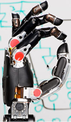
(credit: DARPA)

Neuroscientists in a project headed by the University of Chicago have determined some of the specific characteristics of electrical stimuli that should be applied to the brain to produce different sensations in an artificial upper limb intended to restore natural motor control and sensation in amputees.

The research is part of Revolutionizing Prosthetics, a multi-year Defense Advanced Research Projects Agency (DARPA).

Experimental setup for investigating the ability of monkeys to detect and discriminate trains of electrical pulses delivered to their somatosensory cortex through chronically implanted electrode arrays (credit: Sungshin Kima et al./PNAS)

For this study, the researchers used monkeys, whose sensory systems closely resemble those of humans. They implanted electrodes into the primary somatosensory cortex, the area of the brain that processes touch information from the hand. The animals were trained to perform two perceptual tasks: one in which they detected the presence of an electrical stimulus, and a second task in which they indicated which of two successive stimuli was more intense.

The sense of touch is made up of a complex and nuanced set of sensations, from contact and pressure to texture, vibration and movement. The goal of the research is to document the range, composition and specific increments of signals that create sensations that feel different from each other.

Chronically implanted electrode arrays in a monkey brain. (Left) one 96-electrode array (UEA) was implanted in area 1 (green) of the somatosensory cortex and two 16-electrode arrays (FMA) were implanted in area 3b (yellow). (Center) Colors correspond to the 96 and 16 electrodes. (Right) Colors indicate which electrodes mapped to corresponding hand areas. (credit: Sungshin Kima et al./PNAS)

To achieve that, the researchers manipulated various features of the electrical pulse train, such as its amplitude, frequency, and duration, and noted how the interaction of each of these factors affected the animals’ ability to detect the signal.

Of specific interest were the “just-noticeable differences” (JND),” — the incremental changes needed to produce a sensation that felt different. For instance, at a certain frequency, the signal may be detectable first at a strength of 20 microamps of electricity. If the signal has to be increased to 50 microamps to notice a difference, the JND in that case is 30 microamps.*

“When you grasp an object, for example, you can hold it with different grades of pressure. To recreate a realistic sense of touch, you need to know how many grades of pressure you can convey through electrical stimulation,” said Sliman Bensmaia, PhD, Associate Professor in the Department of Organismal Biology and Anatomy at the University of Chicago and senior author of the study, which was published today (Oct. 26) in the Proceedings of the National Academy of Sciences. “Ideally, you can have the same dynamic range for artificial touch as you do for natural touch.”

“This study gets us to the point where we can actually create real algorithms that work. It gives us the parameters as to what we can achieve with artificial touch, and brings us one step closer to having human-ready algorithms.”

Researchers from the University of Pittsburgh and Johns Hopkins University were also involved in the DARPA-supported study.

* The study also has important scientific implications beyond neuroprosthetics. In natural perception, a principle known as Weber’s Law states that the just-noticeable difference between two stimuli is proportional to the size of the stimulus. For example, with a 100-watt light bulb, you might be able to detect a difference in brightness by increasing its power to 110 watts. The JND in that case is 10 watts. According to Weber’s Law, if you double the power of the light bulb to 200 watts, the JND would also be doubled to 20 watts.

However, Bensmaia’s research shows that with electrical stimulation of the brain, Weber’s Law does not apply — the JND remains nearly constant, no matter the size of the stimulus. This means that the brain responds to electrical stimulation in a much more repeatable, consistent way than through natural stimulation.

“It shows that there is something fundamentally different about the way the brain responds to electrical stimulation than it does to natural stimulation,” Bensmaia said.


Abstract of Behavioral assessment of sensitivity to intracortical microstimulation of primate somatosensory cortex

Intracortical microstimulation (ICMS) is a powerful tool to investigate the functional role of neural circuits and may provide a means to restore sensation for patients for whom peripheral stimulation is not an option. In a series of psychophysical experiments with nonhuman primates, we investigate how stimulation parameters affect behavioral sensitivity to ICMS. Specifically, we deliver ICMS to primary somatosensory cortex through chronically implanted electrode arrays across a wide range of stimulation regimes. First, we investigate how the detectability of ICMS depends on stimulation parameters, including pulse width, frequency, amplitude, and pulse train duration. Then, we characterize the degree to which ICMS pulse trains that differ in amplitude lead to discriminable percepts across the range of perceptible and safe amplitudes. We also investigate how discriminability of pulse amplitude is modulated by other stimulation parameters—namely, frequency and duration. Perceptual judgments obtained across these various conditions will inform the design of stimulation regimes for neuroscience and neuroengineering applications.

Improving learning and memory in aged mice with cholesterol-binding membrane protein

SynCav1 gene delivery enhances granule cell neuron dendritic arborization (neuron branching in adult mice. Scale bar: 20 micrometers. (credit: Chitra D. Mandyam et al./Biological Psychiatry)

Using gene therapy to increase a crucial cholesterol-binding membrane protein called caveolin-1 (Cav-1) in neurons in the hippocampus* of the brain improved learning and memory in aged mice, according to findings from a new study led by scientists at The Scripps Research Institute (TSRI), the Veterans Affairs San Diego Healthcare System (VA) and University of California (UC) San Diego School of Medicine.

The result for treated mice was improved neuron growth and better retrieval of contextual memories — they froze in place, an indication of fear, when placed in a location where they’d once received small electric shocks.

The researchers believe that this type of gene therapy may be a path toward treating age-related memory loss, including loss resulting from alcohol and drug use. The researchers are now testing this gene therapy in mouse models of Alzheimer’s disease and expanding it to possibly treat injuries such as spinal cord injury and traumatic brain injury. ”

The study, published recently online ahead of print in the journal Biological Psychiatry, expands scientists’ understanding of neuroplasticity, the ability of neural pathways to grow in response to new stimuli.

* The hippocampus is a structure in the brain thought to participate in the formation of contextual memories — for example, if one remembers a past picnic when later visiting a park.


Abstract of Neuron-targeted caveolin-1 improves molecular signaling, plasticity and behavior dependent on the hippocampus in adult and aged mice

Background: Studies in vitro demonstrate that neuronal membrane/lipid rafts (MLRs) establish cell polarity by clustering pro-growth receptors and tethering cytoskeletal machinery necessary for neuronal sprouting. However, the effect of MLR and MLR-associated proteins on neuronal aging is unknown.

Methods: Here we assessed the impact of neuron-targeted overexpression of a MLR scaffold protein, caveolin-1 (via a synapsin promoter; SynCav1), in the hippocampus in vivo in adult (6-months-old) and aged (20-month-old) mice on biochemical, morphologic and behavioral changes.

Results: SynCav1 resulted in increased expression of Cav-1, MLRs, and MLR-localization of Cav-1 and tropomyosin-related kinase B (TrkB) receptor independent of age and time post gene transfer. Cav-1 overexpression in adult mice enhanced dendritic arborization within the apical dendrites of hippocampal CA1 and granule cell neurons, effects that were also observed in aged mice, albeit to a lesser extent, indicating preserved impact of Cav-1 on structural plasticity of hippocampal neurons with age. Cav-1 overexpression enhanced contextual fear memory in adult and aged mice demonstrating improved hippocampal function.

Conclusions: Neuron-targeted overexpression of Cav-1 in the adult and aged hippocampus enhances functional MLRs with corresponding roles in cell signaling and protein trafficking. The resultant structural alterations in hippocampal neurons in vivo are associated with improvements in hippocampal dependent learning and memory. Our findings suggest Cav-1 as a novel therapeutic strategy in disorders involving impaired hippocampal function.

Is your thinking chaotic? There’s a model for that.

A representation of a stable sequential working memory; different information items or memory patterns are shown in different colors. (credit: Image adopted from Rabinovich, M.I. et al. (2014))

Try to remember a phone number. You’re now using “sequential memory,” in which your mind processes a sequence of numbers, events, or ideas. It underlies how people think, perceive, and interact as social beings. To understand how sequential memory works, researchers have built mathematical models that mimic this process.

Cognitive modes

Taking this a step further, Mikhail Rabinovich, a physicist and neurocognitive scientist at the University of California, San Diego, and a group of researchers have now mathematically modeled how the mind switches among different ways of thinking about a sequence of objects, events, or ideas that are based on the activity of “cognitive modes.”

The new model, described in an open-access paper in the journal Chaos, may help scientists understand a variety of human psychiatric conditions that may involve sequential memory, including obsessive-compulsive disorder, bipolar, and attention deficit disorder, schizophrenia and autism.

Cognitive modes are the basic states of neural activity. Thinking, perceiving, and any other neural activity involve various parts of the brain that work together in concert, taking on well-defined patterns.

A pathological case (in particular, schizophrenia). The sequence is unstable — the initial sequence enters a chaotic valley after the purple unit. This happens when cognitive inhibition is weak. (credit: adopted from Rabinovich, M.I. et al. (2014))

Binding process

When the mind has sequential thoughts, the cognitive modes underlying neural activity switch among different modalities. This switching is called a binding process, because the mind “binds” each cognitive mode to a certain modality.

Limitless (credit: CBS)

Consider the TV show Limitless. In the show, FBI consultant Brian Finch, aided by the fictional cognitive enhancer NZT, is able to fluidly switch between complex sets of information (modalities), such as phone numbers, using different cognitive modes — rapidly processing a series of phone numbers of suspects on a screen, or analyzing a complex diagram showing potential criminal connections, then explaining it to colleagues, all without losing a beat.

In the new analysis, the mathematicians proved a theorem to show that in their model, this binding process is robust and able to withstand perturbations from the random disturbances in the brain. Your mind is full of other irregular neural signals — from things like other neural processes or external, sensory stimuli and distractions — but if they’re not too big, they don’t affect the thinking process.

This model could be used to better understand a variety of psychiatric disorders, such as obsessive-compulsive disorder, bipolar disorder, and attention deficit disorder, Rabinovich said. The way the mind binds to different modalities, and how such binding depends on time, may be related to conditions such as autism and schizophrenia. For example, some experiments suggest that for people with these conditions, the capacity of sequential binding memory is smaller.

Rabinovich worked with Valentin Afraimovich and Xue Gong, mathematicians at the Autonomous University of San Luis Potosi in Mexico and Ohio University, respectively.


Abstract of Sequential memory: Binding dynamics

Temporal order memories are critical for everyday animal and human functioning. Experiments and our own experience show that the binding or association of various features of an event together and the maintaining of multimodality events in sequential order are the key components of any sequential memories—episodic, semantic, working, etc. We study a robustness of binding sequential dynamics based on our previously introduced model in the form of generalized Lotka-Volterra equations. In the phase space of the model, there exists a multi-dimensional binding heteroclinic network consisting of saddle equilibrium points and heteroclinic trajectories joining them. We prove here the robustness of the binding sequential dynamics, i.e., the feasibility phenomenon for coupled heteroclinic networks: for each collection of successive heteroclinic trajectories inside the unified networks, there is an open set of initial points such that the trajectory going through each of them follows the prescribed collection staying in a small neighborhood of it. We show also that the symbolic complexity function of the system restricted to this neighborhood is a polynomial of degree L − 1, where L is the number of modalities.

Noninvasive imaging method can look twice as deep inside the living brain

In vivo 1.3-μm VCSEL SS-OCT imaging of a 12-week-old adult mouse with cranial window preparation. (a) Representative OCT image visualizing morphological details of the cerebral cortex and subsequent brain compartments. (b) OCT brain anatomy showing good correlation with photomicrograph of a Nissl-stained histology section of the same strain mouse brain. (credit: Allen Institute for Brain Science/Journal of Biomedical Optics)

University of Washington (UW) researchers have developed a noninvasive light-based imaging technology that can literally see inside the living brain at more than two times the depth, providing a new tool to study how diseases like dementia, Alzheimer’s, and brain tumors change brain tissue over time.

The work was reported Oct. 8 by Woo June Choi and Ruikang Wang of the UW Department of Bioengineering in the Journal of Biomedical Optics, published by SPIE, the international society for optics and photonics.

Noninvasive deep imaging

According to the authors, this new optical coherence tomography (OCT) approach to brain study may allow for examining acute and chronic morphological or functional vascular changes in the deep brain.

OCT is normally used to obtain sub-surface images of biological tissue at about the same resolution as a low-power microscope and can instantly deliver cross-section images of layers of tissue without invasive surgery or ionizing radiation. OCT images are based on light directly reflected from a sub-surface.

Widely used in clinical ophthalmology, OCT has recently been adapted for brain imaging in small animal models. Its application in neuroscience has been limited, however, because conventional OCT technology hasn’t been able to image more than 1 millimeter below the surface of biological tissue.

Portion of schematic of the 1.3-μm vertical cavity surface emitting laser (VCSEL) swept-source optical coherence tomography (SS-OCT) system. FC: optical fiber coupler; BD: dual balanced detector; DAQ: data acquisition. (credit: Woo June Choi and Ruikang K. Wang/Journal of Biomedical Optics)

In the paper, Choi and Wang describe how a new technique called “swept-source OCT” (SS-OCT) powered by a vertical-cavity surface-emitting laser (VCSEL) increases signal sensitivity, extending the imaging depth range to more than 2 millimeters. That may make it possible to do things that have been barely attempted in the OCT community, such as noninvasive imaging of the mouse hippocampus or full-length imaging of a human eye from cornea to retina.

It could also allow researchers to monitor deeper morphological changes caused by diseases such as Alzheimer’s disease and dementia, and even to study the effects of aging on the brain.


Abstract of Swept-source optical coherence tomography powered by a 1.3-μm vertical cavity surface emitting laser enables 2.3-mm-deep brain imaging in mice in vivo

We report noninvasive, in vivo optical imaging deep within a mouse brain by swept-source optical coherence tomography (SS-OCT), enabled by a 1.3-μm vertical cavity surface emitting laser (VCSEL). VCSEL SS-OCT offers a constant signal sensitivity of 105 dB throughout an entire depth of 4.25 mm in air, ensuring an extended usable imaging depth range of more than 2 mm in turbid biological tissue. Using this approach, we show deep brain imaging in mice with an open-skull cranial window preparation, revealing intact mouse brain anatomy from the superficial cerebral cortex to the deep hippocampus. VCSEL SS-OCT would be applicable to small animal studies for the investigation of deep tissue compartments in living brains where diseases such as dementia and tumor can take their toll.

Chemical transformation of human astroglial cells into neurons for brain repair

Astroglial cells before (top) and after (bottom) treatment with small-molecule cocktails (credit: Gong Chen lab, Penn State University)

Researchers have succeeded in transforming human support brain cells, called astroglial cells, into functioning neurons for brain repair.

The new technology opens the door to future development of drugs that patients could take as pills to regenerate neurons and to restore brain functions lost after traumatic injuries, stroke, or diseases such as Alzheimer’s.

Previous research, such as conventional stem-cell therapy, has required brain surgery, so it is much more invasive and prone to immune-system rejection and other problems.

The new research, led by Gong Chen, Professor of Biology and the Verne M. Willaman Chair in Life Sciences at Penn State University, was published online today (Oct. 15) in the journal Cell Stem Cell.

“We have discovered a cocktail of small molecules that can reprogram human brain astroglial cells into neuron-like cells after eight to ten days of chemical treatment,” Chen said. The reprogrammed nerves survived for more than five months in cell culture, where they formed functional synaptic networks.

The scientists also injected the reprogrammed human neurons into the brains of living mice, where they integrated into the neural circuits and survived there for at least one month.

“The small molecules are not only easy to synthesize and package into drug pills, but also much more convenient for use by patients than other methods now being developed,” Chen said.

Converting astroglial cells into neurons

Astroglial cells surround neurons and provide them with support, protection, oxygen, and nutrients. But when brain tissues are damaged by strokes or trauma, the astroglial cells react by multiplying — sometimes so much that they clog up the nervous system by forming a scar. These astroglial scars — a difficult research challenge for many decades — can cause health problems by preventing nerve regeneration and by blocking nerve-to-nerve communications between different regions of the brain.

Chen’s group previously invented a method to convert astroglial cells into neurons using viral particles. But Chen also wanted to investigate whether small chemical compounds, which could be packaged into swallowable pills, could also do the job.

Five students on Chen’s research team, led by graduate student Lei Zhang, tested hundreds of different conditions and eventually identified a cocktail of small molecules that can convert human astroglial cells into functional neurons in a cell-culture dish in the laboratory. The students found that adding small molecules in a certain sequence transformed the cultured human astroglial cells from a flat, polygon shape into a neuron-like shape with long “arms” called axons and dendrites.

“These chemically generated neurons are comparable to normal brain neurons in terms of firing electric activity and release of neurotransmitters,” Chen said. “Importantly, the human astroglial-converted neurons survived longer than five months in cell culture and longer than one month in the living mouse brain after transplantation.”

Chen acknowledges that further development, laboratory testing, and a series of clinical trials are still required, but he hopes that this new technology may have broad applications in the future treatment of stroke, Alzheimer’s disease, Parkinson’s disease, and other neurological disorders.

“Our dream is that, one day, patients with brain disorders can take drug pills at home to regenerate neurons inside their brains without any brain surgery and without any cell transplantation,” he said.

Scientists from Emory University School of Medicine were also involved in the research.


Abstract of Small Molecules Efficiently Reprogram Human Astroglial Cells into Functional Neurons

We have recently demonstrated that reactive glial cells can be directly reprogrammed into functional neurons by a single neural transcription factor, NeuroD1. Here we report that a combination of small molecules can also reprogram human astrocytes in culture into fully functional neurons. We demonstrate that sequential exposure of human astrocytes to a cocktail of nine small molecules that inhibit glial but activate neuronal signaling pathways can successfully reprogram astrocytes into neurons in 8-10 days. This chemical reprogramming is mediated through epigenetic regulation and involves transcriptional activation of NEUROD1 and NEUROGENIN2. The human astrocyte-converted neurons can survive for >5 months in culture and form functional synaptic networks with synchronous burst activities. The chemically reprogrammed human neurons can also survive for >1 month in the mouse brain in vivo and integrate into local circuits. Our study opens a new avenue using chemical compounds to reprogram reactive glial cells into functional neurons.

Surgeons reroute nerves to restore hand, arm movement to quadriplegic patients

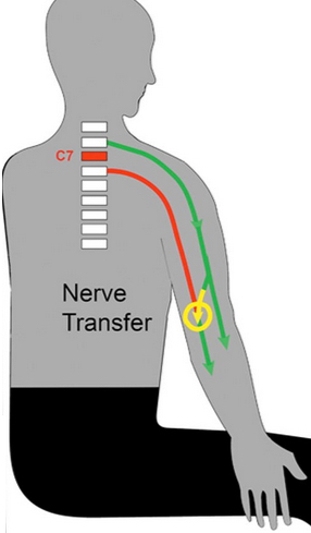
A nerve transfer bypasses the zone of a spinal cord injury (C7). Functional nerves (green) that are under volitional control are rerouted (yellow) to nerves (red) that come off below the spinal cord injury. (credit: Washington University in St. Louis)

A pioneering surgical technique has restored some hand and arm movement to nine patients immobilized by spinal cord injuries in the neck, reports a new study at Washington University School of Medicine in St. Louis.

Bypassing the spinal cord, the surgeons rerouted healthy nerves sitting above the injury site, usually in the shoulders or elbows, to paralyzed nerves in the hand or arm. Once a connection was established, patients underwent extensive physical therapy to train the brain to recognize the new nerve signals, a process that takes about 6–18 months.

The technique targets patients with injuries at the C6 or C7 vertebra, the lowest bones in the neck. It typically does not help patients who have lost all arm function due to higher injuries in vertebrae C1 through C5.

“Physically, nerve-transfer surgery provides incremental improvements in hand and arm function. However, psychologically, these small steps are huge for a patient’s quality of life,” said the study’s lead author, Ida K. Fox, MD, assistant professor of plastic and reconstructive surgery.

One of the most humbling effects of spine damage is the inability to manage bladder or bowel functions. “People with spinal cord injuries cannot control those functions because their brains can’t talk to the nerves in the lower body,” said Fox, who performs surgeries at Barnes-Jewish Hospital.

The study is published in an open-access paper in the October issue of the American Society of Plastic Surgeons’ journal, Plastic and Reconstructive Surgery.

Ultimately, medical professionals hope to discover a way to restore full movement to the estimated 250,000 people in the U.S. living with spinal cord injuries. More than half of such injuries involve the neck. However, until a cure is found, progress in regaining basic independence in routine tasks is important.


Abstract of Nerve transfers to restore upper extremity function in cervical spinal cord injury: Update and preliminary outcomes

Background: Cervical spinal cord injury can result in profound loss of upper extremity function. Recent interest in the use of nerve transfers to restore volitional control is an exciting development in the care of these complex patients. In this article, the authors review preliminary results of nerve transfers in spinal cord injury.

Methods: Review of the literature and the authors’ cases series of 13 operations in nine spinal cord injury nerve transfer recipients was performed. Representative cases were reviewed to explore critical concepts and preliminary outcomes.

Results: The nerve transfers used expendable donors (e.g., teres minor, deltoid, supinator, and brachialis) innervated above the level of the spinal cord injury to restore volitional control of missing function such as elbow extension, wrist extension, and/or hand function (posterior interosseous nerve or anterior interosseous nerve/finger flexors reinnervated). Results from the literature and the authors’ patients (after a mean postsurgical follow-up of 12 months) indicate gains in function as assessed by both manual muscle testing and patients’ self-reported outcomes measures.

Conclusions: Nerve transfers can provide an alternative and consistent means of reestablishing volitional control of upper extremity function in people with cervical level spinal cord injury. Early outcomes provide evidence of substantial improvements in self-reported function despite relatively subtle objective gains in isolated muscle strength. Further work to investigate the optimal timing and combination of nerve transfer operations, the combination of these with traditional treatments (tendon transfer and functional electrical stimulation), and measurement of outcomes is imperative for determining the precise role of these operations.

Hybrid bio-robotic system models physics of human leg locomotion

Schematic of bio-robotic modeling system (credit: Benjamin D. Robertson and Gregory S. Sawicki/PNAS)

North Carolina State University (NC State) researchers have developed a bio-inspired system that models how human leg locomotion works, by using a computer-controlled nerve stimulator (acting as the spinal cord) to activate a biological muscle-tendon.

The findings could help design robotic devices that begin to merge human and machine to assist human locomotion, serving as prosthetic systems for people with mobility impairments or exoskeletons for increasing the abilities of able-bodied individuals.

The model is based on the natural spring-like physics (mass, stiffness, and leverage) of the ankle’s primary muscle-tendon unit (using a bullfrog’s muscle). The system used a feedback-controlled servomotor, simulating the inertial/gravitational environment of terrestrial gait.

Tuning for natural resonance

The research showed that the natural resonance* of the system is a likely mechanism behind springy leg behavior during locomotion, according to Gregory Sawicki, associate professor at NC State and University of North Carolina at Chapel Hill Joint Department of Biomedical Engineering. He is also co-author of a paper on the work published in Proceedings of the National Academy of Sciences.

In this case, the electrical system — the body’s nervous system — drives the mechanical system (the leg’s muscle-tendon unit) at a frequency that provides maximum power output.

The researchers found that by matching the stimulation frequency to the natural resonance frequency of the passive biomechanical system, muscle-tendon interactions (resulting in spring-like behavior) occur naturally and do not require closed-loop neural control — simplifying system design.

“In locomotion, resonance comes from tuning the interaction between the nervous system and the leg so they work together,” said Sawicki. “It turns out that if I know the mass, leverage, and stiffness of a muscle-tendon unit, I can tell you exactly how often I should stimulate it to get resonance in the form of spring-like, elastic behavior.”

“In the end, we found that the same simple underlying principles that govern resonance in simple mechanical systems also apply to these extraordinarily complicated physiological systems,” said Temple University post-doctoral researcher Ben Robertson, corresponding author of the paper.

“This outcome points to mechanical resonance as an underlying principle governing muscle-tendon interactions and provides a physiology-based framework for understanding how mechanically simple elastic limb behavior may emerge from a complex biological system comprised of many simultaneously tuned muscle-tendons within the lower limb,” the researchers conclude in the paper.

* NC State biomedical engineer Greg Sawicki likened resonance tuning to interacting with a slinky toy. “When you get it oscillating well, you hardly have to move your hand — it’s the timing of the interaction forces that matters.


Abstract of Unconstrained muscle-tendon workloops indicate resonance tuning as a mechanism for elastic limb behavior during terrestrial locomotion

In terrestrial locomotion, there is a missing link between observed spring-like limb mechanics and the physiological systems driving their emergence. Previous modeling and experimental studies of bouncing gait (e.g., walking, running, hopping) identified muscle-tendon interactions that cycle large amounts of energy in series tendon as a source of elastic limb behavior. The neural, biomechanical, and environmental origins of these tuned mechanics, however, have remained elusive. To examine the dynamic interplay between these factors, we developed an experimental platform comprised of a feedback-controlled servo-motor coupled to a biological muscle-tendon. Our novel motor controller mimicked in vivo inertial/gravitational loading experienced by muscles during terrestrial locomotion, and rhythmic patterns of muscle activation were applied via stimulation of intact nerve. This approach was based on classical workloop studies, but avoided predetermined patterns of muscle strain and activation—constraints not imposed during real-world locomotion. Our unconstrained approach to position control allowed observation of emergent muscle-tendon mechanics resulting from dynamic interaction of neural control, active muscle, and system material/inertial properties. This study demonstrated that, despite the complex nonlinear nature of musculotendon systems, cyclic muscle contractions at the passive natural frequency of the underlying biomechanical system yielded maximal forces and fractions of mechanical work recovered from previously stored elastic energy in series-compliant tissues. By matching movement frequency to the natural frequency of the passive biomechanical system (i.e., resonance tuning), muscle-tendon interactions resulting in spring-like behavior emerged naturally, without closed-loop neural control. This conceptual framework may explain the basis for elastic limb behavior during terrestrial locomotion.

Imaging study shows you (and your fluid intelligence) can be identified by your brain activity

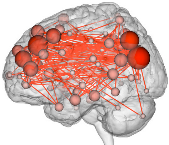
A connectome maps connections between different brain networks. (credit: Emily Finn)

Your brain activity appears to be as unique as your fingerprints, a new Yale-led “connectome fingerprinting” study published Monday (Oct. 12) in the journal Nature Neuroscience has found.

By analyzing* “connectivity profiles” (coordinated activity between pairs of brain regions) of fMRI (functional magnetic resonance imaging) images from 126 subjects, the Yale researchers were able to identify specific individuals from the fMRI data alone by their identifying “fingerprint.” The researchers could also assess the subjects’ “fluid intelligence.”

“In most past studies, data have been used to draw contrasts between, say, patients and healthy controls,” said Emily Finn, a Ph.D. student in neuroscience and co-first author of the paper. “We have learned a lot from these sorts of studies, but they tend to obscure individual differences, which may be important.”

Two frontoparietal networks networks — the medial frontal (purple) and the frontoparietal (teal) — out of the 268 brain regions were found best for identifying people and predicting fluid intelligence (credit: Emily S Finn/Xilin Shen, CC BY-ND)

The researchers looked specifically at areas that showed synchronized activity. The characteristic connectivity patterns were distributed throughout the brain, but notably, two frontoparietal networks emerged as most distinctive.

“These networks are comprised of higher-order association cortices rather than primary sensory regions; these cortical regions are also the most evolutionarily recent and show the highest inter-subject variance,” the researchers note in their paper. “These networks tend to act as flexible hubs, switching connectivity patterns according to task demands. Additionally, broadly distributed across-network connectivity has been reported in these same regions, suggesting a role in large-scale coordination of brain activity.”

Notably, the researchers were able to match the scan of a given individual’s brain activity during one imaging session to that person’s brain scan at another time — even when a person was engaged in a different task in each session, although in that case, the predictive accuracy dropped from 98–99% accuracy to 80–90%.

Predicting and treating neuropsychiatric illnesses (or criminal behavior?)

Finn said she hopes that this ability might one day help clinicians predict or even treat neuropsychiatric diseases based on individual brain connectivity profiles. The paper notes that “aberrant functional connectivity in the frontoparietal networks has been linked to a variety of neuropsychiatric illnesses.”

The study raises troubling questions. “Richard Haier, an intelligence researcher at the University of California, Irvine, [suggests that ] schools could scan children to see what sort of educational environment they’d thrive in, or determine who’s more prone to addiction, or screen prison inmates to figure out whether they’re violent or not,” Wired reports.

“Minority Report” Hawk-eye display (credit: Fox)

Or perhaps identify future criminals — or even predict future crimes, as in “Hawk-eye” technology (portrayed in Minority Report episode 3).

Identifying fluid intelligence

The researchers also discovered that the same two frontoparietal networks were most predictive of the level of fluid intelligence (the capacity for on-the-spot reasoning to discern patterns and solve problems, independent of acquired knowledge) shown on intelligence tests. That’s consistent with previous reports that structural and functional properties of these networks relate to intelligence.

Data for the study came from the Human Connectome Project led by the WU-Minn Consortium, which is funded by the 16 National Institutes of Health (NIH) Institutes and Centers that support the NIH Blueprint for Neuroscience Research, and by the McDonnell Center for Systems Neuroscience at Washington University. Primary funding for the Yale researchers was provided by the NIH.

* Finn and co-first author Xilin Shen, under the direction of R. Todd Constable, professor of diagnostic radiology and neurosurgery at Yale, compiled fMRI data from 126 subjects who underwent six scan sessions over two days. Subjects performed different cognitive tasks during four of the sessions. In the other two, they simply rested. Researchers looked at activity in 268 brain regions: specifically, coordinated activity between pairs of regions. Highly coordinated activity implies two regions are functionally connected. Using the strength of these connections across the whole brain, the researchers were able to identify individuals from fMRI data alone, whether the subject was at rest or engaged in a task. They were also able to predict how subjects would perform on tasks.


Abstract of Functional connectome fingerprinting: identifying individuals using patterns of brain connectivity

Functional magnetic resonance imaging (fMRI) studies typically collapse data from many subjects, but brain functional organization varies between individuals. Here we establish that this individual variability is both robust and reliable, using data from the Human Connectome Project to demonstrate that functional connectivity profiles act as a ‘fingerprint’ that can accurately identify subjects from a large group. Identification was successful across scan sessions and even between task and rest conditions, indicating that an individual’s connectivity profile is intrinsic, and can be used to distinguish that individual regardless of how the brain is engaged during imaging. Characteristic connectivity patterns were distributed throughout the brain, but the frontoparietal network emerged as most distinctive. Furthermore, we show that connectivity profiles predict levels of fluid intelligence: the same networks that were most discriminating of individuals were also most predictive of cognitive behavior. Results indicate the potential to draw inferences about single subjects on the basis of functional connectivity fMRI.

New ‘optoelectrode’ probe is potential neuroscience-technology breakthrough

Device for multichannel intracortical neural recording and optical stimulation. (a) A single optoelectrode structure. The zinc oxide (ZnO) shank is electrically insulated except for the active tip area, and shanks are isolated from each other by polymer adhesive. (b) Electron microscope image of the microscopically smooth tip with the recording area, covered by a final indium-tin oxide (ITO) conducting overlayer. (c) A 4 × 4 micro-optoelectrode array device flip-chip bonded on thin, flexible and semitransparent polyimide electrical cable. (credit: Joonhee Lee et al./Nature Methods)

Brown University School of Engineering and Seoul National University researchers have combined optoelectronics and intracortical neural recording for the first time — enabling neuroscientists to optically stimulate neuron activity while simultaneously recording the effects of the stimulation on associated neural microcircuits.

Described in the journal Nature Methods, the new compact, integrated device uses a semiconductor called zinc oxide, which is optically transparent yet able to conduct an electrical current. That makes it possible to both stimulate and detect with the same material.

The chip is just a few millimeters square with sixteen micrometer-sized pin-like “optoelectrodes,” each capable of both delivering light pulses at micrometer scale and sensing electrical current. The array of optoelectrodes also enables the device to couple to neural microcircuits composed of many neurons rather than single neurons and with millisecond precision.

“We think this is a window-opener,” said Joonhee Lee, a senior research associate in Professor Arto Nurmikko’s lab and one of the lead authors of the new paper. “The ability to rapidly perturb neural circuits according to specific spatial patterns and at the same time reconstruct how the circuits involved are perturbed, is in our view a substantial advance.”

First introduced around 2005, optogenetics involves genetically engineering neurons to express light-sensitive proteins on their membranes. With those proteins expressed, pulses of light can be used to either promote or suppress activity in those particular cells. The method gives researchers, in principle, unprecedented ability to control specific brain cells at specific times.

But until now, simultaneous optogenetic stimulation and recording of brain activity rapidly across multiple points within a brain microcircuit of interest has proven difficult. It requires a device that can both generate a spatial pattern of light pulses and detect the dynamical patterns of electrical reverberations generated by excited cellular activity.

Previous attempts to do this involved devices that cobbled together separate components for light emission and electrical sensing. Such probes were physically bulky, which is not ideal for insertion into a brain. And because the emitters and the sensors were necessarily at hundreds of micrometers apart, a sizable distance, the link between stimulation and recorded signal was not reliable.

The researchers’ next steps are developing a wireless version and using the technology as a chronic implant in non-human primates at potentially hundreds of points and, depending on future progress in worldwide research on optogenetics, perhaps even one day in humans.


Abstract of Transparent intracortical microprobe array for simultaneous spatiotemporal optical stimulation and multichannel electrical recording

Optogenetics, the selective excitation or inhibition of neural circuits by light, has become a transformative approach for dissecting functional brain microcircuits, particularly in in vivorodent models, owing to the expanding libraries of opsins and promoters. Yet there is a lack of versatile devices that can deliver spatiotemporally patterned light while performing simultaneous sensing to map the dynamics of perturbed neural populations at the network level. We have created optoelectronic actuator and sensor microarrays that can be used as monolithic intracortical implants, fabricated from an optically transparent, electrically highly conducting semiconductor ZnO crystal. The devices can perform simultaneous light delivery and electrical readout in precise spatial registry across the microprobe array. We applied the device technology in transgenic mice to study light-perturbed cortical microcircuit dynamics and their effects on behavior. The functionality of this device can be further expanded to optical imaging and patterned electrical microstimulation.