How to grow old brain cells for studying age-related diseases

Salk scientists developed a new technique to grow aged brain cells from patients’ skin. Fibroblasts (cells in connective tissue) from elderly human donors are directly converted into induced neurons, as shown here. (credit: Salk Institute)

Scientists have developed a first-ever technique for using skin samples from older patients to create brain cells — without first rolling back the youthfulness clock in the cells. The new technique, which yields cells resembling those found in older people’s brains, will be a boon to scientists studying age-related diseases like Alzheimer’s and Parkinson’s.

“This lets us keep age-related signatures in the cells so that we can more easily study the effects of aging on the brain,” says Rusty Gage, a professor in the Salk Institute’s Laboratory of Genetics and senior author of the paper, published yesterday (October 8, 2015) in Cell Stem Cell.

“By using this powerful approach, we can begin to answer many questions about the physiology and molecular machinery of human nerve cells — not just around healthy aging but pathological aging as well,” says Martin Hetzer, a Salk professor also involved in the work.

Over the past few years, researchers have increasingly turned to human stem cells (instead of animals) to study various diseases in humans. For example, scientists can take patients’ skin cells and turn them into induced pluripotent stem cells, which have the ability to become any cell in the body. From there, researchers can prompt the stem cells to turn into brain cells for further study. But this process — even when taking skin cells from an older human — doesn’t guarantee stem cells with “older” properties.

Epigenetic signatures in older cells —patterns of chemical marks on DNA that dictate what genes are expressed when — were reset to match younger signatures in the process. This made studying the aging of the human brain difficult, since researchers couldn’t create “old” brain cells with the approach.

Induced neurons

The researchers decided to try another approach, turning to an even newer technique that lets them directly convert skin cells to neurons, creating what’s called an induced neuron. “A few years ago, researchers showed that it’s possible to do this, completely bypassing the stem cell precursor state,” says Jerome Mertens, a postdoctoral research fellow and first author of the new paper.

The scientists collected skin cells from 19 people, aged from birth to 89, and prompted them to turn into brain cells using both the induced pluripotent stem cell technique and the direct conversion approach. Then, they compared the patterns of gene expression in the resulting neurons with cells taken from autopsied brains.

When the induced pluripotent stem cell method was used, as expected, the patterns in the neurons were indistinguishable between young and old derived samples. But brain cells that had been created using the direct conversion technique had different patterns of gene expression depending on whether they were created from young donors or older adults.

“The neurons we derived showed differences depending on donor age,” says Mertens. “And they actually show changes in gene expression that have been previously implicated in brain aging.” For instance, levels of a nuclear pore protein called RanBP17 — whose decline is linked to nuclear transport defects that play a role in neurodegenerative diseases — were lower in the neurons derived from older patients.

Now that the direct conversion of skin cells to neurons has been shown to retain these signatures of age, Gage expects the technique to become a valuable tool for studying aging. And, while the current work only tested its effectiveness in creating brain cells, he suspects a similar method will let researchers create aged heart and liver cells as well.

Scientists at Friederich-Alexander University Erlangen-Nuremberg and Tsinghau University were also involved in the study, which was supported by grants from the G. Harold & Leila Y. Mathers Charitable Foundation, the JPB Foundation, the Leona M. and Harry B. Helmsley Charitable Trust, Annette Merle-Smith, CIRM, the German Federal Ministry of Education and Research, and the Glenn Foundation for Medical Research.


Abstract of Human induced neurons retain aging transcriptome signatures that identify compromised nucleocytoplasmic compartmentalization during aging

Aging is a major risk factor for many human diseases, and in vitro generation of human neurons is an attractive approach for modeling aging-related brain disorders. However, modeling aging in differentiated human neurons has proved challenging. We generated neurons from human donors across a broad range of ages, either by iPSC-based reprogramming and differentiation or by direct conversion into induced neurons (iNs). While iPSCs and derived neurons did not retain aging-associated gene signatures, iNs displayed age-specific transcriptional profiles and revealed age-associated decreases in the nuclear transport receptor RanBP17. We detected an age-dependent loss of nucleocytoplasmic compartmentalization (NCC) in donor fibroblasts and corresponding iNs, and found that reduced RanBP17 impaired NCC in young cells while iPSC rejuvenation restored NCC in aged cells. These results show that iNs retain important aging-related signatures, thus allowing modeling of the aging process in vitro, and identify impaired NCC as an important factor in human aging.

A soft, bio-friendly ’3-D’ brain-implant electrode

A schematic of a “3-D” flexible electrode array. Note the Z-shaped part of the electrode array located between the cranial bone and the brain surface, and the tips (10 micrometers) of the protrusions at the bottom, which serve as recording sites. (credit: Johan Agorelius et al./Front. Neurosci.)

Researchers at Lund University have developed implantable multichannel electrodes that can capture signals from single neurons in the brain over a long period of time — without causing brain tissue damage, making it possible to better understand brain function in both healthy and diseased individuals.

Current flexible electrodes can’t maintain their shape when implanted, which is why they have to be attached to a solid chip. That limits their flexibility and irritates brain tissue, eventually killing surrounding nerve cells and making signals unreliable, says professor Jens Schouenborg.

He explains that recording neuronal signals from the brain requires an electrode that is bio-friendly (doesn’t cause any significant damage to brain tissue) and that is flexible in relation to the brain tissue (the brain floats in fluid inside the skull and moves around whenever a person breathes or turns their head).

“The electrode and the implantation technology that we have now developed have these properties,” he says. Described in an open-access paper in the journal Frontiers in Neuroscience, the new “3-D electrodes” are unique in that they are extremely soft (they even deflect against a water surface) and flexible in all three dimensions, enabling stable recordings from neurons over a long period of time.


Lund University | Breakthrough for electrode implants in the brain

How to implant soft electrodes

But the challenge was how to implant these electrodes in the brain. Visualize pushing spaghetti into a slab of meat. The solution: encapsulating the electrodes in a hard but dissolvable gelatin material, one that is also very gentle on the brain.

“This technology retains the electrodes in their original form inside the brain and can monitor what happens inside virtually undisturbed and normally functioning brain tissue,” said Johan Agorelius, a doctoral student in the project.

This allows for better understanding of what happens inside the brain and for developing more effective treatments for diseases such as Parkinson’s disease and chronic pain conditions, says Schouenborg.


Abstract of An array of highly flexible electrodes with a tailored configuration locked by gelatin during implantation—initial evaluation in cortex cerebri of awake rats

Background: A major challenge in the field of neural interfaces is to overcome the problem of poor stability of neuronal recordings, which impedes long-term studies of individual neurons in the brain. Conceivably, unstable recordings reflect relative movements between electrode and tissue. To address this challenge, we have developed a new ultra-flexible electrode array and evaluated its performance in awake non-restrained animals.

Methods:An array of eight separated gold leads (4 × 10 μm), individually flexible in 3D, were cut from a gold sheet using laser milling and insulated with Parylene C. To provide structural support during implantation into rat cortex, the electrode array was embedded in a hard gelatin based material, which dissolves after implantation. Recordings were made during 3 weeks. At termination, the animals were perfused with fixative and frozen to prevent dislocation of the implanted electrodes. A thick slice of brain tissue, with the electrode array still in situ, was made transparent using methyl salicylate to evaluate the conformation of the implanted electrode array.

Results: Median noise levels and signal/noise remained relatively stable during the 3 week observation period; 4.3–5.9 μV and 2.8–4.2, respectively. The spike amplitudes were often quite stable within recording sessions and for 15% of recordings where single-units were identified, the highest-SNR unit had an amplitude higher than 150 μV. In addition, high correlations (>0.96) between unit waveforms recorded at different time points were obtained for 58% of the electrode sites. The structure of the electrode array was well preserved 3 weeks after implantation.

Conclusions: A new implantable multichannel neural interface, comprising electrodes individually flexible in 3D that retain its architecture and functionality after implantation has been developed. Since the new neural interface design is adaptable, it offers a versatile tool to explore the function of various brain structures.

Sleep may strengthen long-term memories in the immune system

A model of memory formation in the central nervous system (upper section) and the immune system (lower section) (credit: Westermann et al./Trends in Neurosciences 2015)

Deep (slow-wave*) sleep, which helps retain memories in the brain, may also strengthen immunological memories of encountered pathogens, German and Dutch neuroscientists propose in an Opinion article published September 29 in Trends in Neurosciences.

The immune system “remembers” an encounter with a bacteria or virus by collecting fragments from the microbe to create memory T cells, which last for months or years and help the body recognize a previous infection and quickly respond. These memory T cells appear to abstract “gist information” about the pathogens, allowing memory T cells to detect new pathogens that are similar, but not identical, to previously encountered bacteria or viruses.

Studies in humans have shown that long-term increases in memory T cells are associated with deep slow-wave sleep on the nights after vaccination. Taken together, the findings support the view that slow-wave sleep contributes to the formation of long-term memories of abstract, generalized information, which leads to adaptive behavioral and immunological responses.

How lack of sleep puts your body at risk

The obvious implication is that sleep deprivation could put your body at risk. “If we didn’t sleep, then the immune system might focus on the wrong parts of the pathogen,” says senior author Jan Born of the University of Tuebingen.

“For example, many viruses can easily mutate some parts of their proteins to escape from immune responses. If too few antigen-recognizing cells [the cells that present the fragments to T cells] are available, then they might all be needed to fight off the pathogen. In addition to this, there is evidence that the hormones released during sleep benefit the crosstalk between antigen-presenting and antigen-recognizing cells, and some of these important hormones could be lacking without sleep.”

Born says that future research should examine what information is selected during sleep for storage in long-term memory, and how this selection is achieved. This research could have important clinical implications.

“In order to design effective vaccines against HIV, malaria, and tuberculosis, which are based on immunological memory, the correct memory model must be available,” Born says. “It is our hope that by comparing the concepts of neuronal and immunological memory, a model of immunological memory can be developed which integrates the available experimental data and serves as a helpful basis for vaccine development.”

* Slow wave sleep (SWS) is the constructive phase of sleep for recuperation of the mind-body system in which it rebuilds itself after each day. Substances that have been ingested into the body while an organism is awake are synthesized into complex proteins of living tissue. Growth hormones are also secreted to facilitate the healing of muscles as well as repairing damage to any tissues. Lastly, glial cells within the brain are restored with sugars to provide energy for the brain. Longer periods of SWS occur in the first part of the night, primarily in the first two sleep cycles (roughly three hours). — Wikipedia


Abstract of System Consolidation during Sleep — A Common Principle Underlying Psychological and Immunological Memory Formation

Sleep benefits the consolidation of psychological memory, and there are hints that sleep likewise supports immunological memory formation. Comparing psychological and immunological domains, we make the case for active system consolidation that is similarly established in both domains and partly conveyed by the same sleep-associated processes. In the psychological domain, neuronal reactivation of declarative memory during slow-wave sleep (SWS) promotes the redistribution of representations initially stored in hippocampal circuitry to extra-hippocampal circuitry for long-term storage. In the immunological domain, SWS seems to favor the redistribution of antigenic memories initially held by antigen-presenting cells, to persisting T cells serving as a long-term store. Because storage capacities are limited in both systems, system consolidation presumably reduces information by abstracting ‘gist’ for long-term storage.

DARPA selects research teams for its ElectRx neuron-sensing/stimulation program

DARPA announced Monday (Oct. 5, 2015) that it has selected seven teams of researchers to begin work on a radical new approach to healing called Electrical Prescriptions (ElectRx). It would involve a system that stimulates peripheral nerves to modulate functions in the brain, spinal cord, and internal organs, according to program manager Doug Weber.

DARPA envisions a closed-loop system aimed at monitoring and treating conditions such as chronic pain, inflammatory disease, post-traumatic stress, and other illnesses that may not be responsive to traditional treatments, using optical, acoustic, electromagnetic, or engineered biology strategies to achieve precise targeting, possibly at single-axon resolution.

Pacemakers for other organs

The oldest and simplest example of this concept is the cardiac pacemaker, which uses brief pulses of electricity to stimulate the heart to beat at a healthy rate. DARPA aims to extend this concept to other organs, like the spleen, and treat inflammatory diseases such as rheumatoid arthritis.

Fighting inflammation may also provide new treatments for depression, which growing evidence suggests might be caused in part by excess levels of inflammatory biomolecules. Peripheral nerve stimulation may also be used to regulate production of neurochemicals that regulate learning and memory in the brain, offering new treatments for post-traumatic stress and other mental health disorders.

In phase 1, the ElectRx program will focus on fundamental studies to map the neural circuits governing the physiology of diseases of interest to DARPA, and also on preliminary development of novel, minimally invasive neural and bio-interface technologies with unprecedented levels of precision, targeting, and scale.

The teams

The seven teams include a mix of first-time and prior DARPA performers.

For example, an MIT team led by Polina Anikeeva will aim to advance its research in stimulating brain tissue using external magnetic fields and injected magnetic nanoparticles to treat neurological diseases such as Parkinson’s disease, replacing surgically implanted electrodes, as KurzweilAI reported in March. When exposed to a low-frequency (100 kHz — 1 MHz) external alternating magnetic field — which can penetrate deep inside biological tissues — these nanoparticles rapidly heat up and trigger heat-sensitive capsaicin (the “hot” in peppers) receptors to stimulate neurons.


MIT | Wireless brain stimulation

The other teams are:

  • Circuit Therapeutics (Menlo Park, Calif.), a start-up co-founded by Stanford University scientists Karl Deisseroth and Scott Delp, plans to further develop its experimental optogenetic methods for treating neuropathic pain, building toward testing in animal models first.
  • A team at Columbia University (New York), led by Elisa Konofagou, will pursue fundamental science to support the use of non-invasive, targeted ultrasound for neuromodulation. The team aims to elucidate the underlying mechanisms that may make ultrasound an option for chronic intervention, including activation and inhibition of nerves.
  • A team at the Florey Institute of Neuroscience and Mental Health (Parkville, Australia), led by John Furness, will seek to map the nerve pathways that underlie intestinal inflammation, with a focus on determining the correlations between animal models and human neural circuitry. They will also explore the use of neurostimulation technologies based on the cochlear implant — developed by Cochlear, Inc. to treat hearing loss but adapted to modulate activity of the vagus nerve in response to biofeedback signals — as a possible treatment for inflammatory bowel disease.
  • A team at the Johns Hopkins University (Baltimore), led by Jiande Chen, aims to explore the root mechanisms of inflammatory bowel disease and the impact of sacral nerve stimulation on its progression. The team will apply a first-of-its-kind approach to visualize intestinal responses to neuromodulation in animal models.
  • A team at Purdue University (West Lafayette, Ind.), led by Pedro Irazoqui, will leverage an existing collaboration with Cyberonics to study inflammation of the gastrointestinal tract and its responsiveness to vagal nerve stimulation through the neck. Validation of the mechanistic insights that emerge from the effort will take place in pre-clinical models in which novel neuromodulation devices will be applied to reduce inflammation in a feedback-controlled manner. Later stages of the effort could advance the design of clinical neuromodulation devices.
  • A team at the University of Texas, Dallas, led by Robert Rennaker and Michael Kilgard, will examine the use of vagal nerve stimulation to induce neural plasticity for the treatment of post-traumatic stress. As envisioned, stimulation could enhance learned behavioral responses that reduce fear and anxiety when presented with traumatic cues. Dr. Rennaker is a U.S. Marine Corps veteran who served in Liberia, Kuwait and Yugoslavia.

How to grow a functional 3-D mini-brain for 25 cents

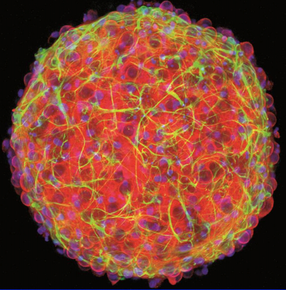
A bioengineering team at Brown University has grown 3-D “mini-brains” of neurons and supporting cells that form networks and are electrically active. This reconstruction of confocal images of a 21-day-in-vitro 3-D cortical neural spheroid shows β-III-butulin+ neurons in red, GFAP+ astrocytes in green, and DAPI-stained nuclei in blue. (credit: Hoffman-Kim lab/Brown University)

Brown University scientists have developed a “mini-brain” — an accessible method for making a working sphere of central nervous system tissue and providing an inexpensive, easy-to-make 3-D testbed for biomedical research such as drug testing, testing neural tissue transplants, or experimenting with how stem cells work. (No, they don’t think. Yet.)

Mini-brains (cortical neural spheroids) produce electrical signals and form their own synapses. “We think of this as a way to have a better in vitro [lab] model that can maybe reduce animal use,” said graduate student Molly Boutin, co-lead author of a paper on the research in the journal Tissue Engineering: Part C. “A lot of the work that’s done right now is in two-dimensional culture, but this is an alternative that is much more relevant to the in vivo [living] scenario.”

The mini-brains, about a third of a millimeter in diameter, are not the first or the most sophisticated working cell cultures of a central nervous system, the researchers acknowledge, but they require fewer steps to make and they use more readily available materials. Here’s the simple recipe:

  1. First, catch a rodent.
  2. Take a small sample of living tissue, which can make thousands of mini-brains from one rodent brain.
  3. Isolate and concentrate the desired cells with some centrifuge steps.
  4. Use that refined sample to seed the cell culture in medium in an agarose spherical mold.

The spheres of brain tissue in the study begin to form within a day after the cultures are seeded and have formed complex 3-D neural networks within two to three weeks.

Key properties

The researchers were interested in studying aspects of neural cell transplantation, which has been proposed to treat Parkinson’s disease, and in how adult neural stem cells develop. The method they developed yields mini-brains with several important properties:

  • Diverse cell types: The cultures contain both inhibitory and excitatory neurons and several varieties of essential neural support cells called glia.
  • Electrically active: the neurons fire and spike and form synaptic connections, producing complex networks.
  • 3-D: Cells connect and communicate within a realistic geometry, rather than merely across a flat plane as in a 2-D culture.
  • Natural density: Experiments showed that the mini-brains have a density of a few hundred thousand cells per cubic millimeter, which is similar to a natural rodent brain.
  • Physical structure: Cells in the mini-brain produce their own extracellular matrix, producing a tissue with the same mechanical properties (squishiness) as natural tissue. The cultures also don’t rely on foreign materials such as scaffolds of collagen.
  • Longevity: In testing, cultured tissues live for at least a month.
  • Cost: about $0.25.

Study senior author Diane Hoffman-Kim, associate professor of molecular pharmacology, physiology and biotechnology (also associate professor of engineering at Brown and affiliated with the Brown Institute for Brain Science and the Center for Biomedical Engineering) hopes the mini-brains might proliferate to many different labs, including those of researchers who have questions about neural tissue, but not necessarily the degree of neuroscience and cell culture equipment required of other methods.

The National Science Foundation, the National Institutes of Health, the Brown Institute for Brain Science, and the U.S. Department of Education funded the research.


Abstract of 3D Neural Spheroid Culture: An In Vitro Model for Cortical Studies

There is a high demand for in vitro models of the central nervous system to study neurological disorders, injuries, toxicity, and drug-efficacy. Three-dimensional (3D) in vitro models can bridge the gap between traditional 2D culture and animal models because they present an in vivo-like microenvironment in a tailorable experimental platform. Within the expanding variety of sophisticated 3D cultures, scaffold-free, self-assembled spheroid culture avoids the introduction of foreign materials and preserves the native cell populations and extracellular matrix types. In this study, we generated 3D spheroids with primary postnatal rat cortical cells using an accessible, size-controlled, reproducible, and cost-effective method. Neurons and glia formed laminin-containing 3D networks within the spheroids. The neurons were electrically active and formed circuitry via both excitatory and inhibitory synapses. The mechanical properties of the spheroids were in the range of brain tissue. These in vivo-like features of 3D cortical spheroids provide the potential for relevant and translatable investigations of the central nervous system in vitro.

NIH and Kavli Foundation invest $185 million for BRAIN Initiative research

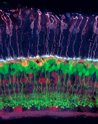
Scientists funded by the NIH BRAIN Initiative hope to diagram all of the circuits in the brain. One group will attempt to identify all of the connections among the retina’s ganglion cells (red), which transmit visual information from bipolar cells (green) and photoreceptors (purple) to the brain. (credit: Josh Morgan, Ph.D. and Rachel Wong, Ph.D./University of Washington)

The National Institutes of Health and the Kavli Foundation separately announced today (Oct. 1, 2015) commitments totaling $185 million in new funds supporting the BRAIN Initiative* — research aimed at deepening our understanding of the brain and brain-related disorders, such as traumatic brain injuries (TBI), Alzheimer’s disease, and Parkinson’s disease.

The NIH announced today its second wave of grants to support the goals of the Brain Research through Advancing Innovative Neurotechnologies (BRAIN) Initiative, bringing the total NIH investment to nearly $85 million in fiscal year 2015.

Sixty-seven new awards totaling more than $38 million will go to 131 investigators working at 125 institutions in the United States and eight other countries. These awards expand NIH’s efforts to develop new tools and technologies to understand neural circuit function and capture a dynamic view of the brain in action.

Projects include proposals to develop soft self-driving electrodes, ultrasound methods for measuring brain activity, and the use of deep brain stimulation to treat traumatic brain injuries.

NIH has also announced new funding opportunities.

Planning for the NIH component of the BRAIN initiative is guided by the long-term scientific plan, BRAIN 2025: A Scientific Vision, which details seven high priority research areas**.

Kavli Foundation and university partners commit $100 million to brain research

The Kavli Foundation and its university partners also announced today the commitment of more than $100 million in new funds to enable research that moves forward the BRAIN Initiative.

The majority of the funds will establish three new Kavli neuroscience institutes at the Johns Hopkins University (JHU), The Rockefeller University, and the University of California, San Francisco (UCSF). Each of the Institutes will receive a $20 million endowment supported equally by their universities and the Foundation, along with start-up funding. The three institutes will be joining an international network of seven Kavli Institutes carrying out fundamental research in neuroscience, and a broader network of 20 Kavli Institutes dedicated to astrophysics, nanoscience, neuroscience and theoretical physics.

The Foundation is also partnering with four other universities to build their Kavli Institute endowments further: at Columbia University; the University of California, San Diego; Yale University; and the Norwegian University of Science and Technology.

BRAIN Initiative event

A BRAIN Initiative event will be held October 20, 2015 at the Society for Neuroscience Annual Meeting in Chicago, featuring an interactive panel discussion/Q&A session with updates on activities and opportunities offered by the BRAIN Initiative. Representatives from federal agencies, private research foundations, and universities will be present.

* In 2014, President Obama launched the BRAIN Initiative as a large-scale effort to equip researchers with fundamental insights necessary for treating a wide variety of brain disorders like Alzheimer’s, schizophrenia, autism, epilepsy, and traumatic brain injury, with $46 million funding. These new tools and this deeper understanding will ultimately catalyze new treatments and cures for devastating brain disorders and diseases that are estimated by the World Health Organization to affect more than one billion people worldwide, according to NIH.

** From Brain Research through Advancing Innovative Neurotechnologies (BRAIN) Working Group Report to the Advisory Committee to the Director, NIH:

#1. Discovering diversity: Identify and provide experimental access to the different brain cell types to determine their roles in health and disease. It is within reach to characterize all cell types in the nervous system, and to develop tools to record, mark, and manipulate these precisely defined neurons in the living brain. We envision an integrated, systematic census of neuronal and glial cell types, and new genetic and non-genetic tools to deliver genes, proteins, and chemicals to cells of interest in non-human animals and in humans.

#2. Maps at multiple scales: Generate circuit diagrams that vary in resolution from synapses to the whole brain. It is increasingly possible to map connected neurons in local circuits and distributed brain systems, enabling an understanding of the relationship between neuronal structure and function. We envision improved technologies—faster, less expensive, scalable—for anatomic reconstruction of neural circuits at all scales, from non-invasive whole human brain imaging to dense reconstruction of synaptic inputs and outputs at the subcellular level.

#3. The brain in action: Produce a dynamic picture of the functioning brain by developing and applying improved methods for large-scale monitoring of neural activity. We should seize the challenge of recording dynamic neuronal activity from complete neural networks, over long periods, in all areas of the brain. There are promising opportunities both for improving existing technologies and for developing entirely new technologies for neuronal recording, including methods based on electrodes, optics, molecular genetics, and nanoscience, and encompassing different facets of brain activity.

#4. Demonstrating causality: Link brain activity to behavior with precise interventional tools that change neural circuit dynamics. By directly activating and inhibiting populations of neurons, neuroscience is progressing from observation to causation, and much more is possible. To enable the immense potential of circuit manipulation, a new generation of tools for optogenetics, chemogenetics, and biochemical and electromagnetic modulation should be developed for use in animals and eventually in human patients.

#5. Identifying fundamental principles: Produce conceptual foundations for understanding the biological basis of mental processes through development of new theoretical and data analysis tools. Rigorous theory, modeling, and statistics are advancing our understanding of complex, nonlinear brain functions where human intuition fails. New kinds of data are accruing at increasing rates, mandating new methods of data analysis and interpretation. To enable progress in theory and data analysis, we must foster collaborations between experimentalists and scientists from statistics, physics, mathematics, engineering, and computer science.

#6. Advancing human neuroscience: Develop innovative technologies to understand the human brain and treat its disorders; create and support integrated human brain research networks. Consenting humans who are undergoing diagnostic brain monitoring, or receiving neurotechnology for clinical applications, provide an extraordinary opportunity for scientific research. This setting enables research on human brain function, the mechanisms of human brain disorders, the effect of therapy, and the value of diagnostics. Meeting this opportunity requires closely integrated research teams performing according to the highest ethical standards of clinical care and research. New mechanisms are needed to maximize the collection of this priceless information and ensure that it benefits people with brain disorders.

#7. From BRAIN Initiative to the brain: Integrate new technological and conceptual approaches produced in Goals #1-6 to discover how dynamic patterns of neural activity are transformed into cognition, emotion, perception, and action in health and disease. The most important outcome of the BRAIN Initiative will be a comprehensive, mechanistic understanding of mental function that emerges from synergistic application of the new technologies and conceptual structures developed under the BRAIN Initiative.

The overarching vision of the BRAIN Initiative is best captured by Goal #7—combining these approaches into a single, integrated science of cells, circuits, brain, and behavior. For example, immense value is added if recordings are conducted from identified cell types whose anatomical connections are established in the same study. Such an experiment is currently an exceptional tour de force; with new technology, it could become routine. In another example, neuronal populations recorded during complex behavior might be immediately retested with circuit manipulation techniques to determine their causal role in generating the behavior. Theory and modeling should be woven into successive stages of ongoing experiments, enabling bridges to be built from single cells to connectivity, population dynamics, and behavior.

New prosthesis bypasses brain damage by re-encoding memories

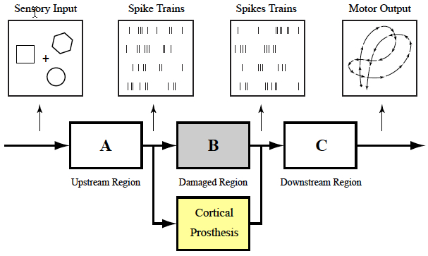
Cortical memory prosthesis uses internal brain signals (e.g., spike
trains) as inputs and outputs, bypassing damaged region (Dong Song et al.)

A brain prosthesis designed to help individuals suffering from memory loss has been developed by researchers at USC and Wake Forest Baptist Medical Center.

The prosthesis, which uses a small array of electrodes implanted into the brain, has performed well in laboratory testing in animals and is currently being evaluated in human patients.

The device builds on decades of research by Ted Berger and relies on a new algorithm created by Dong Song, both of the USC Viterbi School of Engineering. The development also builds on more than a decade of collaboration with Sam Deadwyler and Robert Hampson of the Department of Physiology & Pharmacology of Wake Forest Baptist, who have collected the neural data used to construct the models and algorithms.

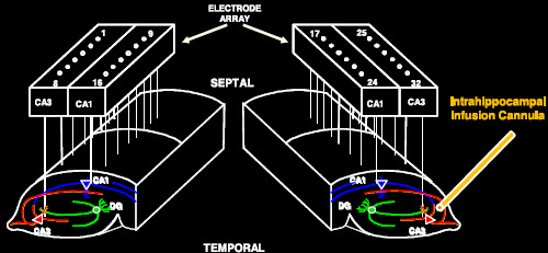
electrode_array

Electrode array for monitoring and duplicating hippocampus neuron activity (credit: T. Berger et al./Journal of Neural Engineering)

When your brain receives a sensory input, it creates a memory in the form of a complex electrical signal that travels through multiple regions of the hippocampus, the memory center of the brain. At each region, the signal is re-encoded until it reaches the final region as a wholly different signal that is sent off for long-term storage.

If there’s damage at any region that prevents this translation, there is the possibility that long-term memory will not be formed. That’s why an individual with hippocampal damage (for example, due to Alzheimer’s disease) can recall events from a long time ago — things that were already translated into long-term memories before the brain damage occurred — but have difficulty forming new long-term memories.

Bypassing a damaged hippocampal section

Song and Berger found a way to accurately mimic how a memory is translated from short-term memory into long-term memory, using data obtained by Deadwyler and Hampson, first from animals, and then from humans. Their prosthesis is designed to bypass a damaged hippocampal section and provide the next region with the correctly translated memory.

That’s despite the fact that there is currently no way of “reading” a memory just by looking at its electrical signal. “It’s like being able to translate from Spanish to French without being able to understand either language,” Berger said.

Their research was presented at the 37th Annual International Conference of the IEEE Engineering in Medicine and Biology Society in Milan on August 27, 2015.

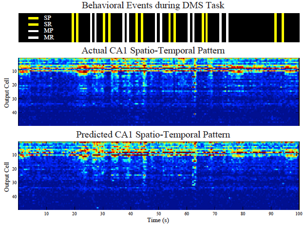
Predicting CA1 spatio-temporal patterns from CA3 spatio-temporal patterns with the sparse MIMO model. SP: sample presentation; SR: sample response; MP: match presentation; MR: match response. (Dong Song et al.)

The effectiveness of the model was tested by the USC and Wake Forest Baptist teams. With the permission of patients who had electrodes implanted in their hippocampi to treat chronic seizures, Hampson and Deadwyler read the electrical signals created during memory formation at two regions of the hippocampus, then sent that information to Song and Berger to construct the model.

The team then fed those signals into the model and read how the signals generated from the first region of the hippocampus were translated into signals generated by the second region of the hippocampus.

In hundreds of trials conducted with nine patients, the algorithm accurately predicted how the signals would be translated with about 90 percent accuracy.

“Being able to predict neural signals with the USC model suggests that it can be used to design a device to support or replace the function of a damaged part of the brain,” Hampson said.

Next, to try to bypass the damage and enable the formation of an accurate long-term memory, the team will attempt to send the translated signal back into the brain of a patient with damage at one of the regions.


Abstract of Sparse Generalized Volterra Model of Human Hippocampal Spike Train Transformation for Memory Prostheses

In order to build hippocampal prostheses for restoring memory functions, we build multi-input, multi-output (MIMO) nonlinear dynamical models of the human hippocampus. Spike trains are recorded from the hippocampal CA3 and CA1 regions of epileptic patients performing a memory-dependent delayed match-to-sample task. Using CA3 and CA1 spike trains as inputs and outputs respectively, second-order sparse generalized Laguerre-Volterra models are estimated with group lasso and local coordinate descent methods to capture the nonlinear dynamics underlying the spike train transformations. These models can accurately predict the CA1 spike trains based on the ongoing CA3 spike trains and thus will serve as the computational basis of the hippocampal memory prosthesis.

‘Designless’ nanoscale logic circuits resemble Darwinian evolution and neural networks

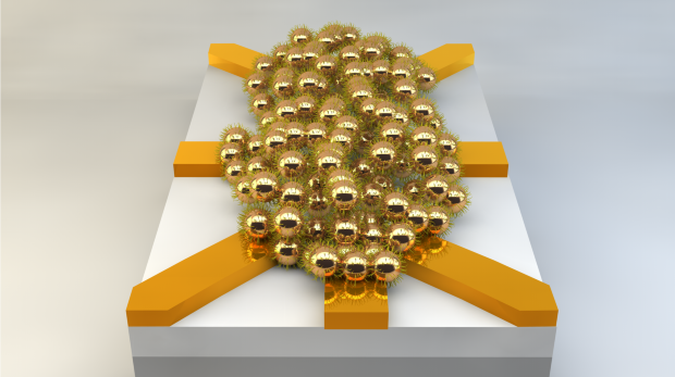
Illustration of a nanoparticle network (about 200 nanometers in diameter). By applying electrical signals at the electrodes (yellow), and using artificial evolution, this disordered network can be configured into useful logic circuits. (credit: University of Twente)

Researchers at the University of Twente in The Netherlands have designed and demonstrated working electronic logic circuits produced using methods that resemble Darwinian evolution and neural networks like the human brain.

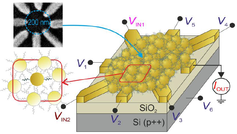
In a radical “designless” approach, the researchers used a 200-nanometer-wide cluster of 20-nanometer gold nanoparticles. They applied a series of voltages to eight electrodes and determined the resulting set of 16 different two-input Boolean logic gates.

Artificial evolution

Instead of designing logic circuits with specified functions, as with conventional transistors, this approach works around — or can even take advantage of — any material defects.

To do this, the researchers used an artificial evolution model — one that runs in less than an hour, rather than millions of years. “Natural evolution has led to powerful ‘computers’ like the human brain, which can solve complex problems in an energy-efficient way,” the researchers note. “Nature exploits complex networks that can execute many tasks in parallel.”

“This is the first time that scientists have succeeded in realizing robust electronics with small dimensions that can compete with commercial technology.”

Schematic device layout of the disordered network of gold nanoparticles, separated by ~1 nm 1-octanethiols, in between eight titanium-gold electrodes, as shown in the scanning electron micrograph (top inset). The gold nanoparticles act as single-electron transistors (SETs) at low temperature (<15 K). (credit: S. K. Bose, et al./Nature Nanotechnology)

Conventional transistors are limited to a handful of atoms. It would a major challenge to produce chips in which the millions of transistors required have the same characteristics, according to the researchers from the Twente MESA+ Institute for Nanotechnology and the CTIT Institute for ICT Research. Current transistor designs are also limited by their energy consumption, which is reaching unacceptable levels.

According to University of Twente prof. Wilfred van der Wiel, the logic circuits they discovered currently have limited computing power. “But with this research we have delivered a proof of principle. By scaling up the system, real added value will be produced in the future. This type of circuitry uses much less energy, both in production and use.” The researchers anticipate a wide range of applications — for example, in portable electronics and in the medical world.

“By choosing a smaller nanoparticle diameter, and scaling down the electrode geometry accordingly, our network would not only further reduce area, but room-temperature operation would come into sight as well,” the researchers note in a paper in the journal Nature Nanotechnology.

Mimicking brain-like systems

The researchers also contrast their “designless” reconfigurable approach with massively parallel (but still design-constrained) architectures such as IBM’s TrueNorth brain-inspired chip.

“An especially interesting avenue to explore is the suitability of this system for advanced functionality that is hard (or expensive) to realize in a conventional architecture, such as pattern recognition by mimicking brain-like systems, or simulations of complex physical systems,” the researchers note in the paper. “Our evolutionary approach works around device-to-device variations at the nanoscale and the accompanying uncertainties in performance, which is becoming more and more a bottleneck for the miniaturization of conventional electronic circuits.”


Abstract of Evolution of a designless nanoparticle network into reconfigurable Boolean logic

Natural computers exploit the emergent properties and massive parallelism of interconnected networks of locally active components. Evolution has resulted in systems that compute quickly and that use energy efficiently, utilizing whatever physical properties are exploitable. Man-made computers, on the other hand, are based on circuits of functional units that follow given design rules. Hence, potentially exploitable physical processes, such as capacitive crosstalk, to solve a problem are left out. Until now, designless nanoscale networks of inanimate matter that exhibit robust computational functionality had not been realized. Here we artificially evolve the electrical properties of a disordered nanomaterials system (by optimizing the values of control voltages using a genetic algorithm) to perform computational tasks reconfigurably. We exploit the rich behaviour that emerges from interconnected metal nanoparticles, which act as strongly nonlinear single-electron transistors, and find that this nanoscale architecture can be configured in situ into any Boolean logic gate. This universal, reconfigurable gate would require about ten transistors in a conventional circuit. Our system meets the criteria for the physical realization of (cellular) neural networks: universality (arbitrary Boolean functions), compactness, robustness and evolvability, which implies scalability to perform more advanced tasks. Our evolutionary approach works around device-to-device variations and the accompanying uncertainties in performance. Moreover, it bears a great potential for more energy-efficient computation, and for solving problems that are very hard to tackle in conventional architectures.

Brain-computer interface enables paralyzed man to walk without robotic support

A BCI system allows a man whose legs had been paralyzed to walk without robotic support (credit: courtesy of UCI’s Brain Computer Interface Lab)

A novel brain-computer-interface (BCI) technology created by University of California, Irvine researchers has allowed a paraplegic man to walk for a short distance, unaided by an exoskeleton or other types of robotic support.

The male participant, whose legs had been paralyzed for five years, walked along a 12-foot course using an electroencephalogram (EEG) brain-computer-interface system that lets the brain bypass the spinal cord to send messages to the legs.

It takes electrical signals from the subject’s brain, processes them through a computer algorithm, and fires them off to electrodes placed around the knees that trigger movement in the leg muscles.

“Even after years of paralysis, the brain can still generate robust brain waves that can be harnessed to enable basic walking,” said UCI biomedical engineer Zoran Nenadic, an associate professor of biomedical engineering.

“We showed that you can restore intuitive, brain-controlled walking after a complete spinal cord injury. This noninvasive system for leg muscle stimulation is a promising method and is an advance of our current brain-controlled systems that use virtual reality or a robotic exoskeleton.”

Study results of this preliminary proof-of-concept study appear in an open-access paper in the Journal of NeuroEngineering & Rehabilitation. The research was supported by a National Science Foundation grant.

Training and therapy process

Months of mental training to reactivate the brain’s walking ability and physical therapy were needed for the study participant to reach the stage where he could take steps. Wearing an EEG cap to read his brain waves, he was first asked to think about moving his legs. The brain waves this created were processed through a computer algorithm designed to isolate brain signals specifically related to leg movement.

The subject was first trained to control an avatar in a virtual reality environment, which validated the specific brain wave signals produced by the algorithm. This training process yielded a custom-made system, Nenadic said, so that when the participant sought to initiate leg movement, the computer algorithm could process the brain waves into signals that could stimulate his leg muscles.


UCI | Person with Paraplegia Uses a Brain-Computer Interface to Regain Overground Walking

To make this work, the subject required extensive physical therapy to recondition and strengthen his leg muscles. Then, with the EEG cap on, he practiced walking while suspended 5 centimeters above the floor, so he could freely move his legs without having to support himself. Finally, he translated these skills to the ground, wearing a body-weight support system and pausing to prevent falls.

Since this study involved a single patient, further research is needed to establish whether the results can be duplicated in a larger population of individuals with paraplegia, said An Do, a neurologist and an assistant clinical professor of neurology.

“Once we’ve confirmed the usability of this noninvasive system, we can look into invasive means, such as brain implants,” she said. “We hope that an implant could achieve an even greater level of prosthesis control because brain waves are recorded with higher quality. In addition, such an implant could deliver sensation back to the brain, enabling the user to feel his legs.”


Abstract of The feasibility of a brain-computer interface functional electrical stimulation system for the restoration of overground walking after paraplegia

Background: Direct brain control of overground walking in those with paraplegia due to spinal cord injury (SCI) has not been achieved. Invasive brain-computer interfaces (BCIs) may provide a permanent solution to this problem by directly linking the brain to lower extremity prostheses. To justify the pursuit of such invasive systems, the feasibility of BCI controlled overground walking should first be established in a noninvasive manner. To accomplish this goal, we developed an electroencephalogram (EEG)-based BCI to control a functional electrical stimulation (FES) system for overground walking and assessed its performance in an individual with paraplegia due to SCI.

Methods: An individual with SCI (T6 AIS B) was recruited for the study and was trained to operate an EEG-based BCI system using an attempted walking/idling control strategy. He also underwent muscle reconditioning to facilitate standing and overground walking with a commercial FES system. Subsequently, the BCI and FES systems were integrated and the participant engaged in several real-time walking tests using the BCI-FES system. This was done in both a suspended, off-the-ground condition, and an overground walking condition. BCI states, gyroscope, laser distance meter, and video recording data were used to assess the BCI performance.

Results: During the course of 19 weeks, the participant performed 30 real-time, BCI-FES controlled overground walking tests, and demonstrated the ability to purposefully operate the BCI-FES system by following verbal cues. Based on the comparison between the ground truth and decoded BCI states, he achieved information transfer rates >3 bit/s and correlations >0.9. No adverse events directly related to the study were observed.

Conclusion: This proof-of-concept study demonstrates for the first time that restoring brain-controlled overground walking after paraplegia due to SCI is feasible. Further studies are warranted to establish the generalizability of these results in a population of individuals with paraplegia due to SCI. If this noninvasive system is successfully tested in population studies, the pursuit of permanent, invasive BCI walking prostheses may be justified. In addition, a simplified version of the current system may be explored as a noninvasive neurorehabilitative therapy in those with incomplete motor SCI.

First brain-to-brain ‘telepathy’ communication via the Internet

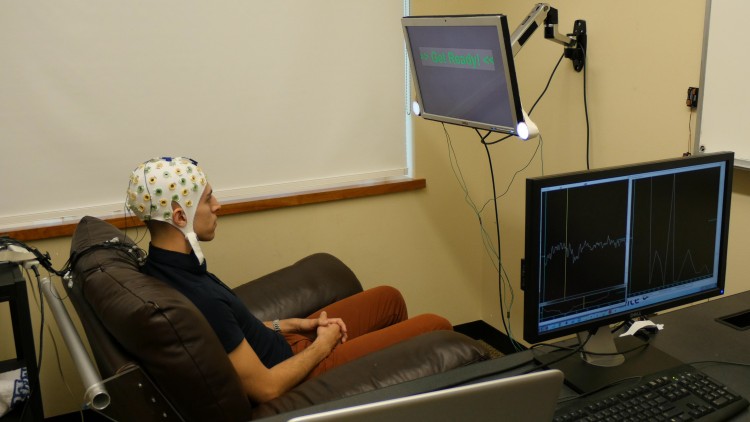
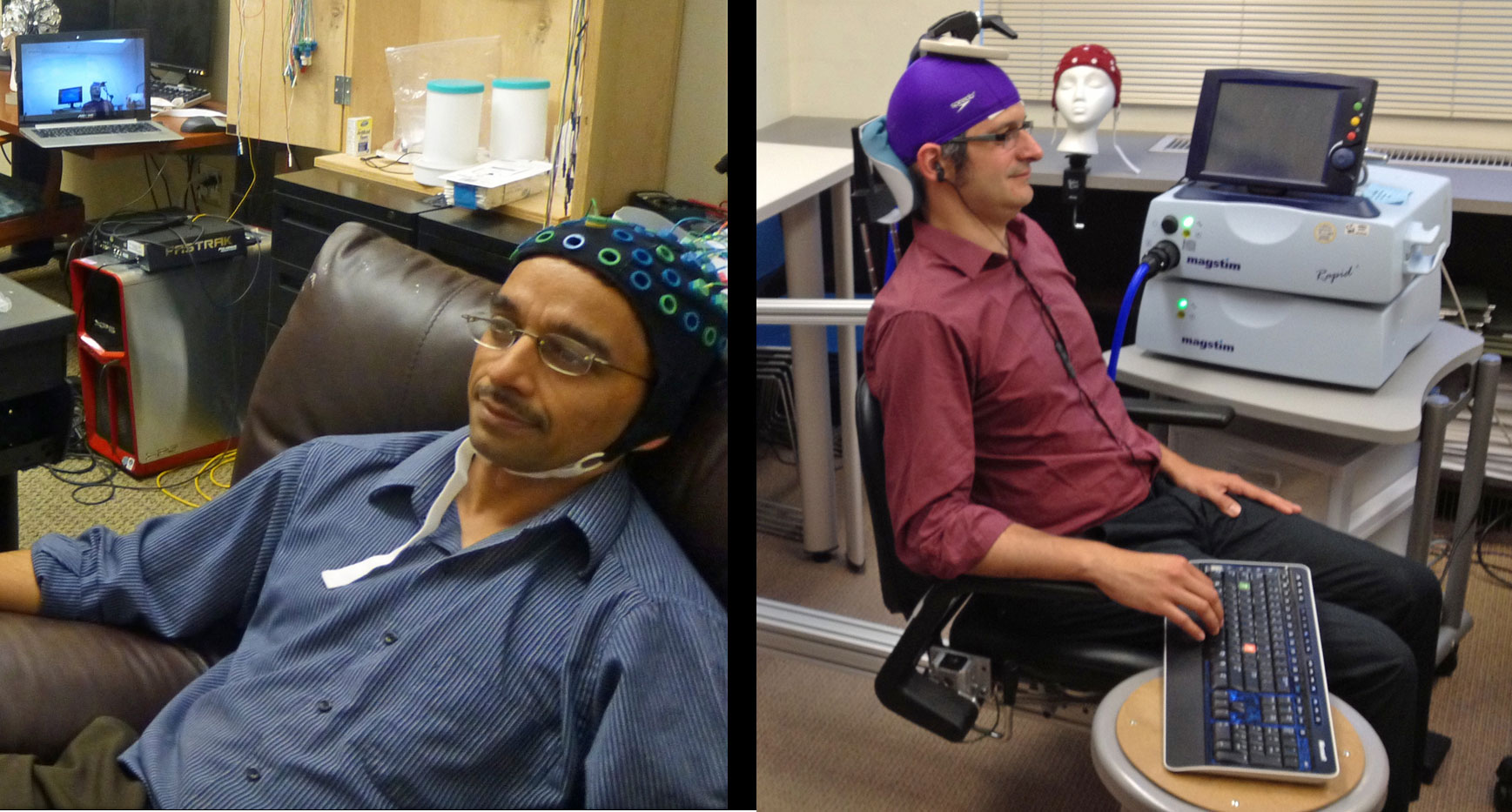
University of Washington graduate student Jose Ceballos wears an electroencephalography (EEG) cap that records brain activity and sends a response to a second participant over the Internet (credit: University of Washington)

The first brain-to-brain telepathy-like communication between two participants via the Internet has been performed by University of Washington researchers.*

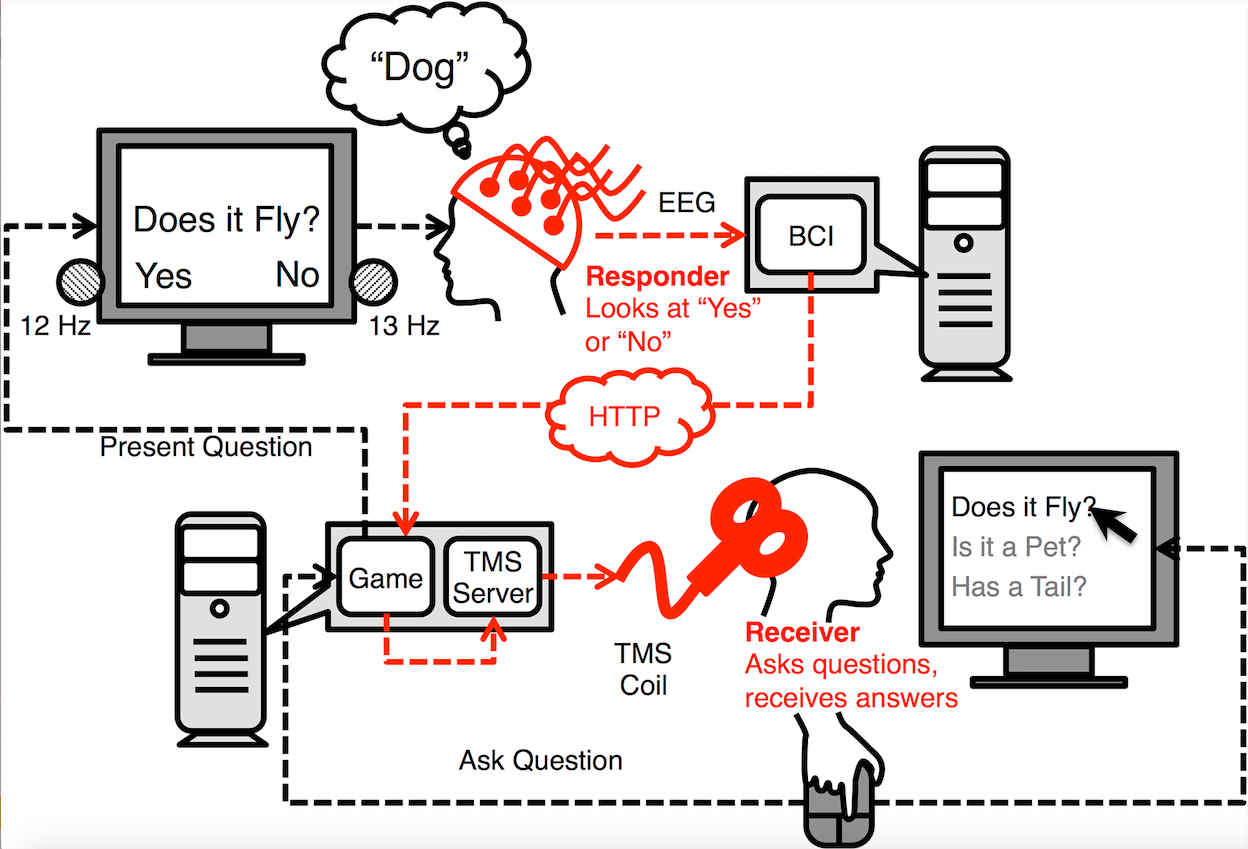
The experiment used a question-and-answer game. The goal is for the “inquirer” to determine which object the “respondent” is looking at from a list of possible objects. The inquirer sends a question (e.g., “Does it fly?) to the respondent, who answers “yes” or “no” by mentally focusing on one of two flashing LED lights attached to the monitor. The respondent is wearing an electroencephalography (EEG) helmet.

By focusing on the “yes” light, the EEG device generates send a signal to the inquirer via the Internet to activate a magnetic coil positioned behind the inquirer’s head, which stimulates the visual cortex and causes the inquirer to see a flash of light (known as a “phosphene”). A “no” signal works the same way, but is not strong enough to activate the coil.

Remote brain-to-brain communication process (credit: A. Stocco et al./PLoS ONE)

The experiment, detailed today in an open access paper in PLoS ONE, is the first to show that two brains can be directly linked to allow one person to guess what’s on another person’s mind. It is “the most complex brain-to-brain experiment, I think, that’s been done to date in humans,” said lead author Andrea Stocco, an assistant professor of psychology and researcher at UW’s Institute for Learning & Brain Sciences.

The experiment was carried out in dark rooms in two UW labs located almost a mile apart and involved five pairs of participants, who played 20 rounds of the question-and-answer game. Each game had eight objects and three questions. The sessions were a random mixture of 10 real games and 10 control games that were structured the same way.*

Participants were able to guess the correct object in 72 percent of the real games, compared with just 18 percent of the control rounds. Incorrect guesses in the real games could be caused by several factors, the most likely being uncertainty about whether a phosphene had appeared.

uw_brain2brain_interface_1

UW team’s initial experiment in 2013: University of Washington researcher Rajesh Rao, left, plays a computer game with his mind. Across campus, researcher Andrea Stocco, right, wears a magnetic stimulation coil over the left motor cortex region of his brain. Stocco’s right index finger moved involuntarily to hit the “fire” button as part of the first human brain-to-brain interface demonstration. (credit: University of Washington)

The study builds on the UW team’s initial experiment in 2013, which was the first to demonstrate a direct brain-to-brain connection between humans, using noninvasive technology to send a person’s brain signals over the Internet to control the hand motions of another person. Other scientists had previously connected the brains of rats and monkeys, and transmitted brain signals from a human to a rat, using electrodes inserted into animals’ brains.

The new experiment evolved out of research by co-author Rajesh Rao, a UW professor of computer science and engineering, on brain-computer interfaces that enable people to activate devices with their minds. In 2011, Rao began collaborating with Stocco and Prat to determine how to link two human brains together.


University of Washington | Team links two human brains for question-and-answer experiment

“Brain tutoring” next

In 2014, the researchers received a $1 million grant from the W.M. Keck Foundation that allowed them to broaden their experiments to decode more complex interactions and brain processes. They are now exploring the possibility of “brain tutoring,” transferring signals directly from healthy brains to ones that are developmentally impaired or impacted by external factors such as a stroke or accident, or simply to transfer knowledge from teacher to pupil.

The team is also working on transmitting brain states — for example, sending signals from an alert person to a sleepy one, or from a focused student to one who has attention deficit hyperactivity disorder, or ADHD.

“Imagine having someone with ADHD and a neurotypical student,” Prat said. “When the non-ADHD student is paying attention, the ADHD student’s brain gets put into a state of greater attention automatically.”

“Evolution has spent a colossal amount of time to find ways for us and other animals to take information out of our brains and communicate it to other animals in the forms of behavior, speech and so on,” Stocco said. “But it requires a translation. We can only communicate part of whatever our brain processes.

“What we are doing is kind of reversing the process a step at a time by opening up this box and taking signals from the brain and with minimal translation, putting them back in another person’s brain,” he said.

* “Telepathy-like” is KurzweilAI’s wording, meaning that no action by the subject outside of the brain were required in the communication. As noted above, the first experiment (known to KurzweilAI) to demonstrate a direct brain-to-brain connection between humans via the Internet, the UW team’s initial experiment in 2013, used involuntary finger movements on a keyboard. Proponents of “telepathy” or “psychic” experiments using the Internet as a link, if any, might counter this.

The researchers took steps to ensure participants couldn’t use clues other than direct brain communication to complete the game. Inquirers wore earplugs so they couldn’t hear the different sounds produced by the varying stimulation intensities of the “yes” and “no” responses. Since noise travels through the skull bone, the researchers also changed the stimulation intensities slightly from game to game and randomly used three different intensities each for “yes” and “no” answers to further reduce the chance that sound could provide clues.

The researchers also repositioned the coil on the inquirer’s head at the start of each game, but for the control games, added a plastic spacer undetectable to the participant that weakened the magnetic field enough to prevent the generation of phosphenes. Inquirers were not told whether they had correctly identified the items, and only the researcher on the respondent end knew whether each game was real or a control round.

UPDATE Sept. 9, 2015: Footnote expanded to clarify “telepathy-like.”