3D-printed silicone guide with chemical cues helps regenerate complex nerves after injury

3-D scans of a nerve from different angles are used to create a custom regeneration guide for complex nerves (credit: University of Minnesota)

A national team of researchers used a combination of 3-D imaging and 3-D printing techniques to create a custom silicone guide implanted with biochemical cues to help nerve regeneration after an injury.

Nerve regeneration is a complex process, which is why regrowth of nerves after injury or disease is very rare and often permanent, according to the Mayo Clinic.

As a test, the researchers used a 3-D scanner to reverse-engineer the structure of a rat’s sciatic nerve. They then used a specialized, custom-built 3-D printer to print a regeneration guide containing 3D-printed chemical cues to promote both motor and sensory nerve regeneration within the same structure. The guide was then implanted into the rat by surgically grafting it to the cut ends of the nerve. Within about 10 to 12 weeks, the rat’s ability to walk again was improved.

A 3D-printed complex nerve-regeneration pathway implanted in a rat helped to improve walking in 10 to 12 weeks after implantation (credit: University of Minnesota)

“Someday we hope that we could have a 3D scanner and printer right at the hospital to create custom nerve guides right on site to restore nerve function,” said University of Minnesota mechanical engineering professor Michael McAlpine, the study’s lead researcher.

Conventional nerve guidance channels are typically fabricated around cylindrical substrates, so the resulting guidance devices are limited to linear structures. This is is the first time a study has shown the creation of a custom guide for regrowth of a complex nerve like the Y-shaped sciatic nerve, which has both sensory and motor branches.

“The exciting next step would be to implant these guides in humans rather than rats,” McAlpine said. For cases where a patient’s nerve is unavailable for scanning, McAlpine said there could someday be a “library” of scanned nerves from other people or cadavers that hospitals could use to create closely matched 3D-printed guides for patients.

The study by researchers from the University of Minnesota, Virginia Tech, University of Maryland, Princeton University, and Johns Hopkins University was published Thursday (Sept. 17) in the journal Advanced Functional Materials.


UMN College of Science and Engineering | 3D printing of a nerve regeneration guide [no audio]


Abstract of 3D Printed Anatomical Nerve Regeneration Pathways

A 3D printing methodology for the design, optimization, and fabrication of a custom nerve repair technology for the regeneration of complex peripheral nerve injuries containing bifurcating sensory and motor nerve pathways is introduced. The custom scaffolds are deterministically fabricated via a microextrusion printing principle using 3D models, which are reverse engineered from patient anatomies by 3D scanning. The bifurcating pathways are augmented with 3D printed biomimetic physical cues (microgrooves) and path-specific biochemical cues (spatially controlled multicomponent gradients). In vitro studies reveal that 3D printed physical and biochemical cues provide axonal guidance and chemotractant/chemokinetic functionality. In vivo studies examining the regeneration of bifurcated injuries across a 10 mm complex nerve gap in rats showed that the 3D printed scaffolds achieved successful regeneration of complex nerve injuries, resulting in enhanced functional return of the regenerated nerve. This approach suggests the potential of 3D printing toward advancing tissue regeneration in terms of: (1) the customization of scaffold geometries to match inherent tissue anatomies; (2) the integration of biomanufacturing approaches with computational modeling for design, analysis, and optimization; and (3) the enhancement of device properties with spatially controlled physical and biochemical functionalities, all enabled by the same 3D printing process.

Minority Report, Limitless TV shows launch Monday, Tuesday

A sequel to Steven Spielberg’s epic movie, MINORITY REPORT is set in Washington, D.C., 10 years after the demise of Precrime, a law enforcement agency tasked with identifying and eliminating criminals … before their crimes were committed. Now, in 2065, crime-solving is different, and justice leans more on sophisticated and trusted technology than on the instincts of the precogs. Sept. 21 series premiere Mondays 9/8:00c

LIMITLESS, based on the feature film, is a fast-paced drama about Brian Finch, who discovers the brain-boosting power of the mysterious drug NZT and is coerced by the FBI into using his extraordinary cognitive abilities to solve complex cases for them. Sept. 22 series premiere Tuesdays 10/9c

Tunable brain cells that morph on demand

PV+ interneuron (credit: Nathalie Dehorter et al./Science)

King’s College London researchers have developed a new molecular “switch” that controls the properties of certain neurons in response to changes in the activity of their neural network — suggesting that these circuits in our brain are tuneable and could have implications that go far beyond basic neuroscience.

The researchers, from the MRC Centre for Developmental Neurobiology (MRC CDN) at the Institute of Psychiatry, Psychology & Neuroscience (IoPPN), led by Professor Oscar Marín, have discovered that some neurons in the cerebral cortex can adapt their properties in response to changes in network activity, such as learning a motor (muscle) task.

The authors studied two apparently different classes of fast-spiking interneurons but discovered they were actually looking at the same neuron — one with the ability to oscillate between two different ground states. The authors then identified the molecular factor responsible for tuning the properties of these cells: a transcription factor (a protein able to influence gene expression) known as Er81.

Neurons with “tremendous plasticity”

Fast-spiking interneurons, known as FS PV+, are members of a general class of neurons whose primary role is regulating the activity of pyramidal cells (the principal cells of the cerebral cortex). These PV+ interneurons play a prominent role in the regulation of plasticity and learning.

The researchers believe that PV+ interneurons take on properties based on how they adapt and respond to internal and external influences to encode information. “In other words, that our [brain's] ‘hardware’ is tuneable, at least to some extent,” said Nathalie Dehorter of the MRC CDN and first author of the study, published in the journal Science on Sept. 11, 2015.

“Our study demonstrates the tremendous plasticity of the brain, and how this relates to fundamental processes such as learning,” said Marín. “Understanding the mechanisms that regulate this plasticity, and why it tends to dissipate when we age, has enormous implications that go beyond fundamental neuroscience, from informing education policies to developing new therapies for neurological disorders such as epilepsy.”


Abstract of Tuning of fast-spiking interneuron properties by an activity-dependent transcriptional switch

The function of neural circuits depends on the generation of specific classes of neurons. Neural identity is typically established near the time when neurons exit the cell cycle to become postmitotic cells, and it is generally accepted that, once the identity of a neuron has been established, its fate is maintained throughout life. Here, we show that network activity dynamically modulates the properties of fast-spiking (FS) interneurons through the postmitotic expression of the transcriptional regulator Er81. In the adult cortex, Er81 protein levels define a spectrum of FS basket cells with different properties, whose relative proportions are, however, continuously adjusted in response to neuronal activity. Our findings therefore suggest that interneuron properties are malleable in the adult cortex, at least to a certain extent.

New optogenetics process could lead to neurological enhancements and treatments

Artist’s representation of a calcium ion channel affected by OptoSTIM1 (credit: Institute for Basic Science)

An advanced process for precision control of cellular calcium ion (Ca2+) channels in living organisms has been engineered by a research team at the Korea Advanced Institute of Science and Technology (KAIST) and the IBS Center for Cognition and Sociality.

Calcium ions are a crucial part of diverse cellular functions such as contraction, excitation, growth, differentiation and death. Severe Ca2+ deficiency is linked to cardiac arrhythmia, cognitive impairment, and ataxia.

The new process uses optogenetics, or control of cells by light. The researchers added a new light-sensitive, plant-human hybrid protein to cells to efficiently modulate calcium ion channels in cells by shining blue light on them.

The hybrid protein combines a photoreceptor protein called cryptochrome 2 (Cry2) from a small, flowering plant Arabidopsis thaliana with the STromal Interaction Molecule 1 (STIM1), a protein found in almost all animals that opens cellular Ca2+ channels.

They named the resultant hybrid molecule OptoSTIM1.

When they shined blue light on the OptoSTIM1-expressing cells, they were able to coax them to open their Ca2+ channels and allow an influx of 5 to 10 times more Ca2+ than in previous studies.

Increasing learning capacity in mice

Mouse with blue light apparatus attached (credit: Institute For Basic Science)

To test the functional effect of the Ca2+ influx, the IBS team introduced OptoSTIM1 to the hippocampus of a living mouse. They compared sets of light-illuminated mice to non-illuminated mice expressing OptoSTIM1 in an environment in which they introduced a conditioning cue followed by a fear stimulus.

In subsequent tests they observed that light-illuminated mice with the OptoSTIM1 expression had a nearly twofold increase in fear response when placed in the testing environment without the conditioning cue than the non-light-stimulated mice. That indicated that the OptoSTIM1 expression (and resultant Ca2+ uptake) was an effective method for memory enhancement.

Neurological enhancements and treatments

The researchers say this work opens the door for future research into optogenetically enhanced memory and learning studies and into treating neurological diseases that are a result of a dysfunction in Ca2+ regulation.

This may also be a step towards discovering applications for drugs as well as therapeutic Ca2+ modulation. According to Kyung, “There are diseases that result from dysfunction in cellular Ca2+ regulation, such as Alzheimer’s disease, so we can apply our system to those areas and hopefully in the near future help people to recover from those diseases.”

This may also allow for future non-invasive and non-drug treatments or may help to mitigate and eventually cure some neurological diseases.

Team is led by Won Do Heo, associate professor together with Professor Yong-Mahn Han and Professor Daesoo Kim.


Abstract of Optogenetic control of endogenous Ca2+ channels in vivo

Calcium (Ca2+) signals that are precisely modulated in space and time mediate a myriad of cellular processes, including contraction, excitation, growth, differentiation and apoptosis. However, study of Ca2+ responses has been hampered by technological limitations of existing Ca2+-modulating tools. Here we present OptoSTIM1, an optogenetic tool for manipulating intracellular Ca2+ levels through activation of Ca2+-selective endogenous Ca2+ release−activated Ca2+ (CRAC) channels. Using OptoSTIM1, which combines a plant photoreceptor and the CRAC channel regulator STIM1 (ref. 4), we quantitatively and qualitatively controlled intracellular Ca2+ levels in various biological systems, including zebrafish embryos and human embryonic stem cells. We demonstrate that activating OptoSTIM1 in the CA1 hippocampal region of mice selectively reinforced contextual memory formation. The broad utility of OptoSTIM1 will expand our mechanistic understanding of numerous Ca2+-associated processes and facilitate screening for drug candidates that antagonize Ca2+ signals.

Changing behavior with synapse engineering

The worm turns: injecting a transgenic nematode worm with tyramine induces the worm to switch from forward locomotion (dashed red line, starting at X) to backward locomotion (dashed blue line) (credit: Jennifer K. Pirri et al./PLOS Biology)

In 1963, Yale professor of physiology and psychiatry Dr. Jose Delgado implanted an stimulating electrode in the caudate nucleus of a fighting bull, bravely jumped into the bullring, and stopped the animal in its tracks by remotely activating the electrode. Now UMass Medical School scientists have taken neural control precision down to the synapse level, reversing a C. elegans (nematode) worm’s head position or locomotion direction by simply switching one of its synapses (neuron-to-neuron connections) from inhibitory to excitatory (“off” to “on”).

The research, published in an open-access paper in PLOS Biology, offers a new approach for studying the neural circuits that govern behavior and has implications for refining the connectome (neural roadmap), which is important for understanding how the 100 billion neurons and quadrillion (1015) synapse process information and control behavior.

Currently, the connectome doesn’t include information about the activity of specific neurons or the signals they transmit. The complexity of the human brain makes it almost impossible to address questions such as how stable are neural circuits in the brain and how their wiring constrains the flow of information or the behaviors they control.

So Mark Alkema, PhD, associate professor of neurobiology, turned to the nematode C. elegans. A tiny worm with only 302 neurons, it is the only animal whose neural roadmap has been completely defined.

Worm mind control

Alkema and colleagues wanted to determine if flipping the sign (inhibitory or excitatory) of a synapse in the worm’s brain was enough to reverse a behavior. To do this, they analyzed the touch response that allows C. elegans  to escape from carnivorous fungi that use threadlike nooses to catch nematodes. During this escape response, neurotransmitters in C. elegans are released that activate an inhibitory ion channel. This causes the worm to relax its head and quickly reverse direction away from the predator.

Synapse firing is determined by the charge of the ions that flow through channels. So they replaced the inhibitory ion channel with an excitatory version of the channel in a live nematode. “Surprisingly, the engineered channel does not affect the worms’ development and is properly incorporated into the neural circuits of the worm brain,” said Alkema. “Cells that are normally inhibited in the brain now get activated.”

“What was most striking is that we were able to completely reverse behavior by simply switching the sign of a synapse in the neural network,” explained Alkema. “Now the animal contracts its head and tends to move forward in response to touch. This suggests that the neural wiring diagram is remarkably stable and allows these types of changes.”

“Our studies indicate that switching the sign of a synapse not only provides a novel synthetic mechanism to flip behavioral output but could even be an evolutionary mechanism to change behavior,” said Alkema. “As we start to unravel the complexity and design of the neural network, it holds great promise as a novel mechanism to test circuit function or even design new neural circuits in vivo.”


Abstract of A Change in the Ion Selectivity of Ligand-Gated Ion Channels Provides a Mechanism to Switch Behavior

Behavioral output of neural networks depends on a delicate balance between excitatory and inhibitory synaptic connections. However, it is not known whether network formation and stability is constrained by the sign of synaptic connections between neurons within the network. Here we show that switching the sign of a synapse within a neural circuit can reverse the behavioral output. The inhibitory tyramine-gated chloride channel, LGC-55, induces head relaxation and inhibits forward locomotion during the Caenorhabditis elegans escape response. We switched the ion selectivity of an inhibitory LGC-55 anion channel to an excitatory LGC-55 cation channel. The engineered cation channel is properly trafficked in the native neural circuit and results in behavioral responses that are opposite to those produced by activation of the LGC-55 anion channel. Our findings indicate that switches in ion selectivity of ligand-gated ion channels (LGICs) do not affect network connectivity or stability and may provide an evolutionary and a synthetic mechanism to change behavior.

A breakthrough in creating transparent brains

A transparent 3-D view of amyloid beta plaques (green) and blood vessels (red) in a region of cerebral cortex from a 20-month-old AD model mouse (credit: RIKEN)

Researchers at the RIKEN Brain Science Institute in Japan have created a new technique for converting brain tissue into transparent tissue to reveal 3D brain anatomy at very high resolution.

The researchers say they have used the new technique, called ScaleS, to provide new insights into Alzheimer’s disease plaques and for large-scale connectomic mapping and 3D neural circuit reconstruction.

CLARITY_stained

Three-dimensional view of stained hippocampus showing fluorescent-expressing neurons (green), connecting interneurons (red) and supporting glia (blue), created with the Stanford CLARITY technique (credit: Deisseroth lab)

Previous techniques, such as Stanford’s CLARITY, for creating transparent brain samples — a process called “optical clearing — are useful for microscopy, but the transparency process itself can damage the structures under study, the researchers note. The structures are also not firm enough for electron microscopy, which is used to provides images at a finer level.

These limitations are now overcome with ScaleS, according to the researchers. The internal structures also maintain their original shape and are firm enough to permit the micron-thick slicing necessary for more detailed analyses.

Published in Nature Neuroscience, the new technique combines sorbitol in the right proportion with urea. “We could create transparent brains with minimal tissue damage,” said lead scientist Atsushi Miyawaki. The technique can handle “both florescent and immunohistochemical labeling techniques, and is even effective in older animals,” he added.

Miyawaki believes that the quality and preservation of cellular structures viewed by electron microscopy with ScaleS is unparalleled.

New Alzheimer’s disease findings

A 2-D version of a 3-D image of amyloid beta plaques in entire brain hemispheres of an Alzheimer’s disease model mouse created with ScaleS. The inset (bottom right) shows a high-magnification volume rendering of a representative senile plaque (credit: RIKEN)

The researchers were able to use ScaleS to visualize the mysterious “diffuse” plaques seen in the postmortem brains of Alzheimer’s disease patients that are typically undetectable using 2D imaging. They found that contrary to current assumptions, the diffuse plaques proved to be not isolated; they showed extensive associations with microglia — mobile cells that surround and protect neurons.

The researchers also examined 3-D images of active microglial cells and amyloid beta plaques. While some scientists suggest that active microglial cells are located near plaques, a detailed 3D reconstruction and analysis using ScaleS clearing showed that association with active microglial cells occurs early in plaque development, but not in later stages of the disease, after the plaques have accumulated.

“Clearing tissue with ScaleS followed by 3D microscopy has clear advantages over 2D stereology or immunohistochemistry,” states Miyawaki. “Our technique will [also be useful] for examining normal neural circuits and pinpointing structural changes that characterize other brain diseases.”


Abstract of ScaleS: an optical clearing palette for biological imaging

Optical clearing methods facilitate deep biological imaging by mitigating light scattering in situ. Multi-scale high-resolution imaging requires preservation of tissue integrity for accurate signal reconstruction. However, existing clearing reagents contain chemical components that could compromise tissue structure, preventing reproducible anatomical and fluorescence signal stability. We developed ScaleS, a sorbitol-based optical clearing method that provides stable tissue preservation for immunochemical labeling and three-dimensional (3D) signal rendering. ScaleS permitted optical reconstructions of aged and diseased brain in Alzheimer’s disease models, including mapping of 3D networks of amyloid plaques, neurons and microglia, and multi-scale tracking of single plaques by successive fluorescence and electron microscopy. Human clinical samples from Alzheimer’s disease patients analyzed via reversible optical re-sectioning illuminated plaque pathogenesis in the z axis. Comparative benchmarking of contemporary clearing agents showed superior signal and structure preservation by ScaleS. These findings suggest that ScaleS is a simple and reproducible method for accurate visualization of biological tissue.

Controlling brain cells with ultrasound

For the first time, sound waves are used to control brain cells. Salk scientists developed the new technique, dubbed sonogenetics, to selectively and noninvasively turn on groups of neurons in worms that could be a boon to science and medicine. (credit: Salk Institute)

Salk scientists have developed a new method, dubbed sonogenetics, to selectively activate brain, heart, muscle and other cells using ultrasonic waves (the same type of waves used in medical sonograms).

This new method may have advantages over the similar light-based approach known as optogenetics, particularly for human therapeutics. It is described today (Sept. 15, 2015) in the journal Nature Communications.

Experiment setup. (Left) Schematic of the computer-controlled imaging and ultrasound exposure system. (Right) Top-down view of petri dish with agar (food) plate with animals corralled into a small area by a copper barrier. (credit: Stuart Ibsen et al./Nature Communications)

Sreekanth Chalasani, an assistant professor in Salk’s Molecular Neurobiology Laboratory and senior author of the study, and his colleagues activated neurons in the nematode C. elegans that don’t usually react to ultrasound.

They found that microbubbles of gas outside of the worm were able to amplify the low-intensity ultrasound waves. “The microbubbles grow and shrink in [sync] with the ultrasound pressure waves,” Ibsen says. “These oscillations can then propagate noninvasively into the worm.”

They also found that when the ultrasound hits gas bubbles, they cause a membrane ion channel, TRP-4, to open up and activate the cell. Armed with that knowledge, the team tried adding the TRP-4 channel to neurons that don’t normally have it.

Possible human uses next

Worm exhibits reversals and omega bends upon ultrasound stimulus (single 10 ms pulse at 2.25 MHz with peak negative pressure of 0.9 MPa) in the presence of micro bubbles (credit: Stuart Ibsen et al./Nature Communications)

So far, sonogenetics has only been applied to C. elegans neurons. But TRP-4 could be added to any calcium-sensitive cell type in any organism including humans, Chalasani says. Here’s how it would work:

  1. The microbubbles are injected into the bloodstream, and are distributed throughout the body —- an approach already used in some human imaging techniques.
  2. Ultrasound could then noninvasively reach any tissue of interest, including the brain, be amplified by the microbubbles, and activate the cells of interest through TRP-4. (Many cells in the human body, he points out, can respond to the influxes of calcium caused by TRP-4.)

The researchers have already begun testing the approach in mice.

Both optogenetics and sonogenetics approaches, Chalasani says, hold promise in basic research by letting scientists study the effect of cell activation. And they also may be useful in therapeutics through the activation of cells affected by disease.

However, for either technique to be used in humans, researchers first need to develop safe ways to deliver the light or ultrasound-sensitive channels to target cells.

The work and the researchers involved were supported by a Salk Institute Pioneer Fund Postdoctoral Fellowship, a Salk Institute Innovation Grant, the Rita Allen Foundation, the W.M. Keck Foundation and the National Institutes of Health. A University of California, San Diego researcher was also involved in the research.


Abstract of Sonogenetics is a non-invasive approach to activating neurons in Caenorhabditis elegans

A major challenge in neuroscience is to reliably activate individual neurons, particularly those in  deeper brain regions. Current optogenetic approaches require invasive surgical procedures to  deliver light of specific wavelengths to target cells in order to activate or silence them. Here, we demonstrate the use of low-pressure ultrasound as a non-invasive trigger to activate specific ultrasonically-sensitized neurons in the nematode, Caenorhabditis elegans. We first show that wild-type animals are insensitive to low pressure ultrasound and require gas-filled microbubbles to transduce the ultrasound wave. We find that neuron-specific misexpression of TRP-4, the pore-forming subunit of a mechanotransduction channel, sensitizes neurons to ultrasound stimulus resulting in motor outputs. Furthermore, we use this approach to manipulate the  function of sensory neurons and interneurons and identify a role for the PVD sensory neurons in modifying locomotory behaviors. We suggest this method can be broadly applied to manipulate cellular functions in vivo.

Toyota invests $50 million in intelligent vehicle technology at Stanford, MIT AI research centers

MIT’s iconic Stata Center, which houses the Computer Science and Artificial Intelligence Laboratory (credit: MIT)

Toyota Motor Corporation (TMC) announced today (Fri. Sept. 4) that it will be investing approximately $50 million over the next five years to establish joint research centers at the Stanford Artificial Intelligence Lab (SAIL) and MIT’s Computer Science and Artificial Intelligence Laboratory (CSAIL).

Toyota also said Dr. Gill Pratt, former Program Manager at DARPA and leader of its recent Robotics Challenge, has joined Toyota to direct and accelerate these research activities and their application to intelligent vehicles and robotics.

Rather than fully autonomous vehicles (as in Google’s research), the program will initially focus on the acceleration of intelligent vehicle technology to help eliminate traffic casualties*, with the ultimate goal of helping improve quality of life through enhanced mobility and robotics, according to Kiyotaka Ise, who heads R&D at Toyota.

Specific research areas will include “improving the ability of intelligent vehicle technologies to recognize objects around the vehicle in diverse environments, provide elevated judgment of surrounding conditions, and safely collaborate with vehicle occupants, other vehicles, and pedestrians,” Pratt added. “The joint research will also look at applications of the same technology to human-interactive robotics and information service.”

“[The car] must ensure that it does no harm, not only some of the time, but almost all of the time,” said Pratt.

MIT research

Research at MIT, led by CSAIL director Professor Daniela Rus, will “develop advanced architectures that allow cars to better perceive and navigate their surroundings,” eventually developing a vehicle “incapable of getting into a collision.”

CSAIL researchers plan to explore an approach in which the human driver pays attention at all times, with an autonomous system that is there to jump in to save the driver in the event of an unavoidable accident. That will involve areas from computer vision and perception to planning and control to decision-making.

Rus envisions creating a system that could “prevent collisions and also provide drivers with assistance navigating tricky situations; support a tired driver by watching for unexpected dangers and diversions; and even offer helpful tips such as letting the driver know she is out of milk at home and planning a new route home that allows the driver to swing by the grocery store.”

Research at the new center will also include building new tools for collecting and analyzing navigation data, with the goal of learning from human driving; creating perception and decision-making systems for safe navigation; developing predictive models that can anticipate the behavior of humans, vehicles, and the larger environment; inventing state-of-the-art tools to handle congestion and high-speed driving in challenging situations including adverse weather; improving machine-vision algorithms used to detect and classify objects; and creating more intelligent user interfaces.

Stanford research

Led by Associate Professor Fei-Fei Li, the new SAIL-Toyota Center for AI Research will focus on teaching computers to see and make critical decisions about how to interact with the world.

Early on, the new effort will focus on AI-assisted driving to avoid automobile-related accidents. Li, a world-renowned expert in computer vision, said that Stanford will tackle the problem by addressing four main challenges of making a computer think like a person: perception, learning, reasoning, and interaction.

Stanford’s computer scientists will train computers to recognize objects and speech as well as data, and then use machine learning and statistical modeling to extract the meaningful data points — for instance, a swerving car versus a parked one. Other researchers will teach the AI platform to look at this critical data set and plot the safest driving action.

The first cars with AI technology will work as partners with the driver to make safe decisions, Li said, so devising ways to carefully and comfortably share control between the human and the computer will be instrumental in this technology gaining the public’s trust.

* The World Health Organization estimates that 3,400 people die each day from traffic-related accidents.

Completely paralyzed man voluntarily moves his legs, UCLA scientists report

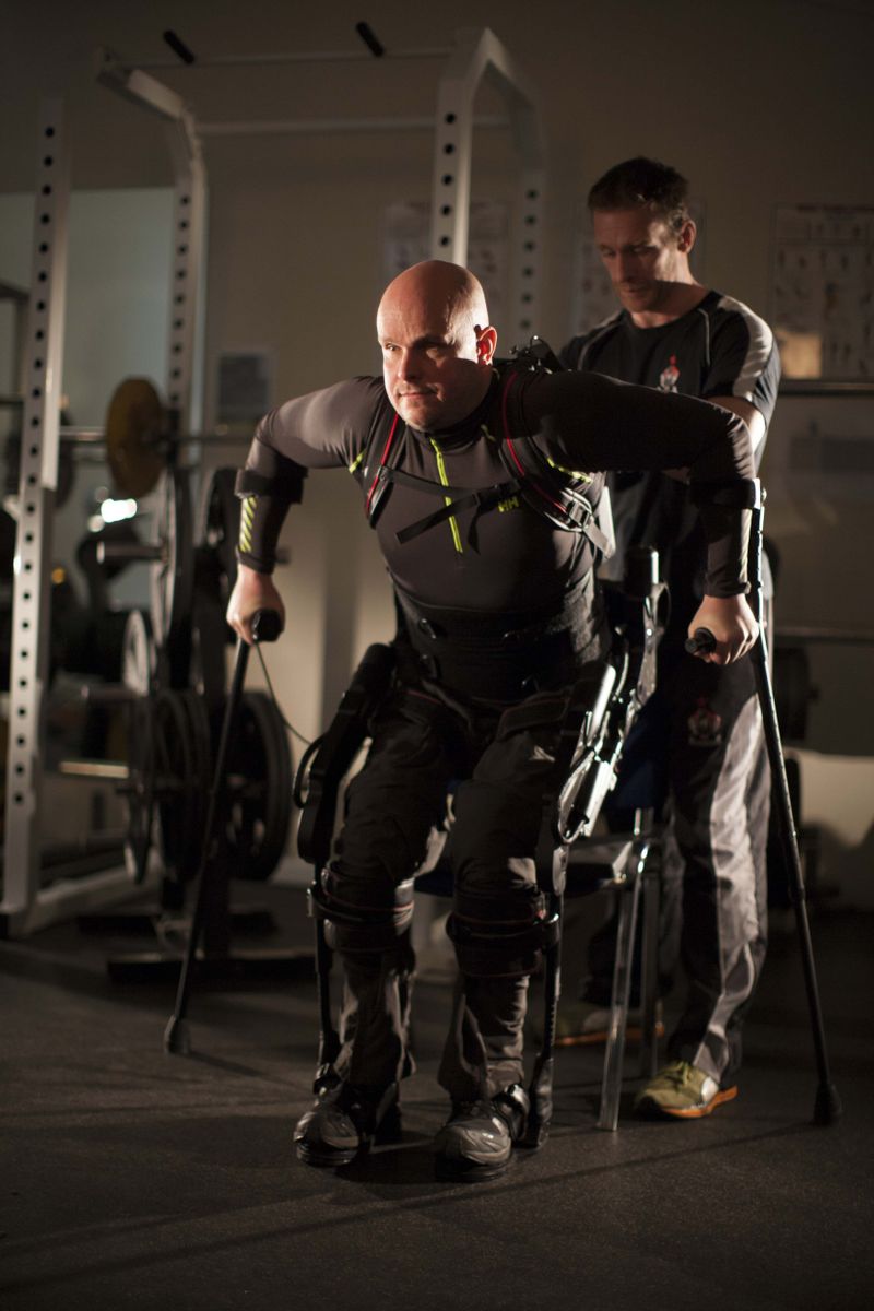
Mark Pollock and trainer Simon O’Donnell (credit: Mark Pollock)

A 39-year-old man who had been completely paralyzed for four years was able to voluntarily control his leg muscles and take thousands of steps in a “robotic exoskeleton” device during five days of training, and for two weeks afterward, UCLA scientists report.

This is the first time that a person with chronic, complete paralysis has regained enough voluntary control to actively work with a robotic device designed to enhance mobility.

In addition to the robotic device, the man was aided by a novel noninvasive spinal stimulation technique that does not require surgery. His leg movements also resulted in other health benefits, including improved cardiovascular function and muscle tone.

The new approach combines a battery-powered wearable bionic suit that enables people to move their legs in a step-like fashion, with a noninvasive procedure that the same researchers had previously used to enable five men who had been completely paralyzed to move their legs in a rhythmic motion.

That earlier achievement is believed to be the first time people who are completely paralyzed have been able to relearn voluntary leg movements without surgery. (The researchers do not describe the achievement as “walking” because no one who is completely paralyzed has independently walked in the absence of the robotic device and electrical stimulation of the spinal cord.)

Mountain racing blind? No problem. Paralyzed? “Iron ElectriRx” man is aceing that too

In the latest study, the researchers treated Mark Pollock, who lost his sight in 1998 and later became the first blind man to race to the South Pole. In 2010, Pollock fell from a second-story window and suffered a spinal cord injury that left him paralyzed from the waist down.

At UCLA, outfitted with the robotic exoskeleton, Pollock made substantial progress after receiving a few weeks of physical training without spinal stimulation and then just five days of spinal stimulation training in a one-week span, for about an hour a day.

“In the last few weeks of the trial, my heart rate hit 138 beats per minute,” Pollock said. “This is an aerobic training zone, a rate I haven’t even come close to since being paralyzed while walking in the robot alone, without these interventions. That was a very exciting, emotional moment for me, having spent my whole adult life before breaking my back as an athlete.”

Even in the years since he lost his sight, Pollock has competed in ultra-endurance races across deserts, mountains and the polar ice caps. He also won silver and bronze medals in rowing at the Commonwealth Games and launched a motivational speaking business.

“Stepping with the stimulation and having my heart rate increase, along with the awareness of my legs under me, was addictive. I wanted more,” he said.

The research was published by the IEEE Engineering in Medicine and Biology Society, the world’s largest society of biomedical engineers.

Expanding the clinical toolbox for the paralyzed

“It will be difficult to get people with complete paralysis to walk completely independently, but even if they don’t accomplish that, the fact they can assist themselves in walking will greatly improve their overall health and quality of life,” said V. Reggie Edgerton, senior author of the research and a UCLA distinguished professor of integrative biology and physiology, neurobiology and neurosurgery.

The procedure used a robotic device manufactured by Richmond, California-based Ekso Bionics that captures data that enables the research team to determine how much the subject is moving his own limbs, as opposed to being aided by the device.

“If the robot does all the work, the subject becomes passive and the nervous system shuts down,” Edgerton said.

The data showed that Pollock was actively flexing his left knee and raising his left leg and that during and after the electrical stimulation, he was able to voluntarily assist the robot during stepping; it wasn’t just the robotic device doing the work.

“For people who are severely injured but not completely paralyzed, there’s every reason to believe that they will have the opportunity to use these types of interventions to further improve their level of function. They’re likely to improve even more,” Edgerton said. “We need to expand the clinical toolbox available for people with spinal cord injury and other diseases.”


Edgerton Lab, University of California Los Angeles | Paralyzed subject Training in Ekso during spinal cord stimulation

The future of spinal-cord research

Edgerton and his research team have received many awards and honors for their research, including Popular Mechanics’ 2011 Breakthrough Award.

“Dr. Edgerton is a pioneer and we are encouraged by these findings to broaden our understanding of possible treatment options for paralysis,” said Peter Wilderotter, president and CEO of the Christopher and Dana Reeve Foundation, which helped fund the research. “Given the complexities of a spinal cord injury, there will be no one-size-fits-all cure but rather a combination of different interventions to achieve functional recovery.

“What we are seeing right now in the field of spinal cord research is a surge of momentum with new directions and approaches to remind the spine of its potential even years after an injury,” he said.

NeuroRecovery Technologies, a medical technology company Edgerton founded, designs and develops devices that help restore movement in patients with paralysis. The company provided the device used to stimulate the spinal cord in combination with the Ekso in this research.

Edgerton said although it likely will be years before the new approaches are widely available, he now believes it is possible to significantly improve quality of life for patients with severe spinal cord injuries, and to help them recover multiple body functions.

In addition to the Reeve foundation, the research was funded by the National Institutes of Health’s National Institute of Biomedical Imaging and Bioengineering, the F. M. Kirby Foundation, the Walkabout Foundation, the Dana and Albert R. Broccoli Foundation, Ekso Bionics, NeuroRecovery Technologies and the Mark Pollock Trust.

Almost 6 million Americans live with paralysis, including nearly 1.3 million with spinal cord injuries.


Abstract of Iron ‘ElectriRx’ Man: Overground Stepping in an Exoskeleton Combined with Noninvasive Spinal Cord Stimulation after Paralysis

We asked whether coordinated voluntary movement of the lower limbs could be regained in an individual having been completely paralyzed (>4 yr) and completely absent of vision (>15 yr) using a novel strategy – transcutaneous spinal cord stimulation at selected sites over the spinal vertebrae with just one week of training. We also asked whether this stimulation strategy could facilitate stepping assisted by an exoskeleton (EKSO, EKSO Bionics) that is designed so that the subject can voluntarily complement the work being performed by the exoskeleton. We found that spinal cord stimulation enhanced the level of effort that the subject could generate while stepping in the exoskeleton. In addition, stimulation improved the coordination patterns of the lower limb muscles resulting in a more continuous, smooth stepping motion in the exoskeleton. These stepping sessions in the presence of stimulation were accompanied by greater cardiac responses and sweating than could be attained without the stimulation. Based on the data from this case study it appears that there is considerable potential for positive synergistic effects after complete paralysis by combining the overground stepping in an exoskeleton, a novel transcutaneous spinal cord stimulation paradigm, and daily training.

First US patients treated with noninvasive focused ultrasound for Parkinson’s disease

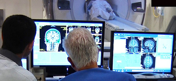
University of Maryland medical doctors monitor focused ultrasound treatment for essential tremor, guided by magnetic resonance imaging (MRI) (credit: University of Maryland School of Medicine)

Researchers at the University of Maryland have performed the first focused ultrasound treatments on a deep structure within the brain related to Parkinson’s disease* called the globus pallidus.

These treatments are part of international pilot studies of 40 patients assessing the feasibility, safety, and preliminary efficacy of focused ultrasound treatments for Parkinson’s disease, guided by magnetic resonance imaging (MRI).

The researchers are using MRI to help them guide ultrasound waves through the intact skin and skull to reach the globus pallidus part of the brain. If successful, focused ultrasound could offer an alternative approach for certain patients with Parkinson’s disease who have failed medical therapy or become disabled from medication-induced dyskinesia (tremor). To date, seven patients in Korea and one patient in Canada have been treated in studies.

The new Parkinson’s procedure

ExAblate Neuro system (credit: Insightec)

The non-invasive ultrasound and MRI imaging procedures are done on an outpatient basis in the Center for Metabolic Imaging and Image-Guided Therapeutics (CMIT) MRI suite, using the ExAblate Neuro system developed by Insightec.

During the Parkinson’s procedure, patients lie in an MRI scanner with a head-immobilizing frame fitted with a transducer helmet. Ultrasonic energy is targeted through the skull to the globus pallidus of the brain, and images acquired during the procedure give physicians a real-time map of the area being treated.

“We’re raising the temperature in a very restricted area of the brain to destroy tissue,” explained principal investigator Howard M. Eisenberg, MD, the Raymond K. Thompson Chair of Neurosurgery. “The ultrasound waves create a heat lesion that we can monitor through MRI.”

The entire procedure lasts two to four hours, and patients are awake and able to interact with the treatment team. This allows the physicians to monitor the immediate effects of treatment and make adjustments if necessary.

Researchers from the University of Virginia Health System reported in the New England Journal of Medicine in 2013 that 15 patients with essential tremor** — a related disorder — who received focused ultrasound saw “significant improvement” in their dominant hand tremor. Patients treated in the initial phase of the study at the University of Maryland experienced similar results.

The Michael J. Fox Foundation for Parkinson’s Research and the Focused Ultrasound Foundation are funding the new Parkinson’s study.

* As many as one million Americans have Parkinson’s disease, a chronic, degenerative disorder for which there is no cure. The second most common movement disorder, Parkinson’s results from the malfunction or loss of brain cells crucial for movement and coordination. Symptoms include motor difficulties such as tremor, rigidity and postural instability. People with Parkinson’s can also experience non-motor symptoms of cognitive impairment, depression, and anxiety, and autonomic dysfunction.

** Essential tremor, which is eight times more common than Parkinson’s disease, causes debilitating shaking that can be resistant to drug therapy. It mainly affects the hands, head and voice, making aspects of daily life like eating, drinking and writing extremely difficult.


Abstract of A Pilot Study of Focused Ultrasound Thalamotomy for Essential Tremor

Background

Recent advances have enabled delivery of high-intensity focused ultrasound through the intact human cranium with magnetic resonance imaging (MRI) guidance. This preliminary study investigates the use of transcranial MRI-guided focused ultrasound thalamotomy for the treatment of essential tremor.

Methods

From February 2011 through December 2011, in an open-label, uncontrolled study, we used transcranial MRI-guided focused ultrasound to target the unilateral ventral intermediate nucleus of the thalamus in 15 patients with severe, medication-refractory essential tremor. We recorded all safety data and measured the effectiveness of tremor suppression using the Clinical Rating Scale for Tremor to calculate the total score (ranging from 0 to 160), hand subscore (primary outcome, ranging from 0 to 32), and disability subscore (ranging from 0 to 32), with higher scores indicating worse tremor. We assessed the patients’ perceptions of treatment efficacy with the Quality of Life in Essential Tremor Questionnaire (ranging from 0 to 100%, with higher scores indicating greater perceived disability).

Results

Thermal ablation of the thalamic target occurred in all patients. Adverse effects of the procedure included transient sensory, cerebellar, motor, and speech abnormalities, with persistent paresthesias in four patients. Scores for hand tremor improved from 20.4 at baseline to 5.2 at 12 months (P=0.001). Total tremor scores improved from 54.9 to 24.3 (P=0.001). Disability scores improved from 18.2 to 2.8 (P=0.001). Quality-of-life scores improved from 37% to 11% (P=0.001).

Conclusions

In this pilot study, essential tremor improved in 15 patients treated with MRI-guided focused ultrasound thalamotomy. Large, randomized, controlled trials will be required to assess the procedure’s efficacy and safety. (Funded by the Focused Ultrasound Surgery Foundation; ClinicalTrials.gov number, NCT01304758.)