Study links aerobic fitness, thinner gray matter, and better math skills in kids

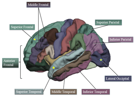
Cortical thickness regions of interest. Starred regions are areas in which higher-fit children showed decreased cortical thickness compared to lower-fit children. (credit: Laura Chaddock-Heyman et al./PLOS ONE)

A new study reveals that 9- and 10-year-old children who are aerobically fit tend to have significantly thinner gray matter than their “lower-fit” peers. Thinning of the outermost layer of brain cells in the cerebrum is associated with better mathematics performance, researchers report in an open-access paper in the journal PLOS ONE.

The study suggests, but does not prove, that cardiorespiratory fitness contributes to gray matter thinning — a normal process of child brain development. The study also offers the first evidence that fitness enhances math skills by aiding the development of brain structures that contribute to mathematics achievement.

“Gray-matter loss during child development is part of healthy maturation,” said University of Illinois postdoctoral researcher Laura Chaddock-Heyman, who led the research. “Gray-matter thinning is the sculpting of a fully formed, healthy brain. The theory is that the brain is pruning away unnecessary connections and strengthening useful connections.”

Previous studies have shown that gray-matter thinning is associated with better reasoning and thinking skills, Chaddock-Heyman said.

Role of aerobic fitness in math skills

“We show, for the first time, that aerobic fitness may play a role in this cortical thinning,” she said. “In particular, we find that higher-fit 9- and 10-year-olds show a decrease in gray-matter thickness in some areas known to change with development, specifically in the frontal, temporal and occipital lobes of the brain.”

The analysis included 48 children, all of whom had completed a maximal oxygen-uptake fitness test on a treadmill. Half of the children (the higher-fit kids) were at or above the 70th percentile for aerobic fitness, and half (the lower-fit kids) were at or below the 30th percentile. The researchers imaged the children’s brains using fMRI, and tested their math, reading, and spelling skills using the Wide Range Achievement Test-3, which correlates closely with academic achievement in these fields.

The team found differences in math skills and cortical brain structure between the higher-fit and lower-fit children: thinner gray matter corresponded to better math performance in the higher-fit kids. But they did not find significant fitness-associated differences in reading or spelling aptitude.

So why only math? “Successful mathematics problem solving is said to involve working memory, the ability to hold relevant information in mind for efficient and effective comprehension, as well as inhibition, the ability to ignore irrelevant information,” Chaddock-Heyman explained to KurzweilAI.

“Higher-fit children have shown superior performance on cognitive control tasks that challenge working memory and inhibitory control, relative to lower fit children. Other studies suggest superior performance on standardized tests of mathematics and reading in higher-fit children.”

“These findings arrive at an important time. Physical activity opportunities during the school day are being reduced or eliminated in response to mandates for increased academic time,” according to kinesiology and community health professor Charles H. Hillman. “Given that rates of physical inactivity are rising, there is an increased need to promote physical activity. Schools are the best institutions to implement such health behavior practices, due to the number of children they reach on a daily basis.”

“Future efforts should be directed toward determining whether these biomarkers predict performance on select academic subjects, as suggested in our study, or whether they serve as a more global index of overall school performance.”

The researchers next plan a longitudinal study of children participating in a physical activity training program. The goal is to establish additional neural biomarkers for scholastic success, based on a causal relationship between brain changes, changes in physical fitness, and changes in cognition, and to determine whether these biomarkers predict performance on select academic subjects (as in the current study), or overall school achievement .

The National Institute on Aging, the National Institute of Child Health and Human Development, and the National Institute of Diabetes and Digestive and Kidney Diseases at the National Institutes of Health supported this research. The National Institute of Food and Agriculture at the U.S. Department of Agriculture also provided funding.


Abstract of The Role of Aerobic Fitness in Cortical Thickness and Mathematics Achievement in Preadolescent Children

Growing evidence suggests that aerobic fitness benefits the brain and cognition during childhood. The present study is the first to explore cortical brain structure of higher fit and lower fit 9- and 10-year-old children, and how aerobic fitness and cortical thickness relate to academic achievement. We demonstrate that higher fit children (>70th percentile VO2max) showed decreased gray matter thickness in superior frontal cortex, superior temporal areas, and lateral occipital cortex, coupled with better mathematics achievement, compared to lower fit children (<30th percentile VO2max). Furthermore, cortical gray matter thinning in anterior and superior frontal areas was associated with superior arithmetic performance. Together, these data add to our knowledge of the biological markers of school achievement, particularly mathematics achievement, and raise the possibility that individual differences in aerobic fitness play an important role in cortical gray matter thinning during brain maturation. The establishment of predictors of academic performance is key to helping educators focus on interventions to maximize learning and success across the lifespan.

Biocompatible interfaces replace silicon and metal in neural prosthetic devices

Left: collagen; right: matrigel (credit: Wen Shen et al./Microsystems & Nanoengineering)

Researchers at the University of Georgia’s Regenerative Bioscience Center have developed a biocompatible implantable neural prosthetic device to replace silicon and noble metal in neural prosthetic devices. The goal is to avoid immune-system rejection, failures due to tissue strain, neurodegeneration, and decreased fidelity of recorded neural signals.

Implantable neural prosthetic devices in the brain have been around for almost two decades, helping people living with limb loss and spinal cord injury become more independent, for example. They are also used for deep brain stimulation and brain-controlled prosthetic devices. However, existing neural prosthetic devices suffer from immune-system rejection, and most are believed to eventually fail because of a mismatch between the soft brain tissue and the rigid devices.

The researchers used a combination of a two materials as structural support for neural electrodes.

Collagen. Its higher mechanical strength can support initial insertion while softening after implantation. Collagen is an extracellular matrix environment (ECM) protein that is critical in the formation of connective structures in tendons, organs, and basement membranes in the body and features long fibrils and 3D structures with high tensile strengths. The ECM is a collection of molecules secreted by cells that provides structural and biochemical support to surrounding cells.

Matrigel, a gelatinous ECM protein mixture resembling the complex extracellular neuronal environment, used to provide a more neuronal-compatible substrate.

A representative extracellular matrix-based implantable neural electrode device and an enlarged view of the electrode tip (credit: Wen Shen et al./Microsystems & Nanoengineering)

“This is not by any means the device that you’re going to implant into a patient,” said Karumbaiah, an assistant professor of animal and dairy science in the UGA College of Agricultural and Environmental Sciences. “This is proof of concept that extracellular matrix can be used to ensheathe a functioning electrode without the use of any other foreign or synthetic materials.”

The collaboration, led by Wen Shen and Mark Allen of the University of Pennsylvania, found that the extracellular matrix derived electrodes adapted to the mechanical properties of brain tissue and were capable of acquiring neural recordings from the brain cortex.

Currently, one out of every 190 Americans is living with limb loss, according to the National Institutes of Health. There is a significant burden in cost of care and quality of life for people suffering from this disability.

The research is described in an open-access paper in the journal Microsystems & Nanoengineering.


Abstract of Extracellular matrix-based intracortical microelectrodes: Toward a microfabricated neural interface based on natural materials

Extracellular matrix (ECM)-based implantable neural electrodes (NEs) were achieved using a microfabrication strategy on natural-substrate-based organic materials. The ECM-based design minimized the introduction of non-natural products into the brain. Further, it rendered the implants sufficiently rigid for penetration into the target brain region and allowed them subsequently to soften to match the elastic modulus of brain tissue upon exposure to physiological conditions, thereby reducing inflammatory strain fields in the tissue. Preliminary studies suggested that ECM-NEs produce a reduced inflammatory response compared with inorganic rigid and flexible approaches. In vivo intracortical recordings from the rat motor cortex illustrate one mode of use for these ECM-NEs.

How to create a genius mouse

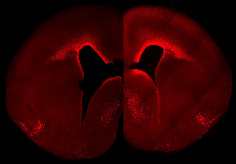
The left-brain hemisphere of a normal mouse shows the normal level and cellular distribution of the Pax6 gene expression in the developing neocortex. The right-brain hemisphere shows a sustained, primate-like Pax6 expression pattern in the neocortex of a double transgenic mouse embryo. These animals have more Pax6-positive progenitor cells and a higher Pax6 expression level in the germinal layer close to the ventricle in the right hemisphere. (credit: © MPI of Molecular Cell Biology & Genetics)

Researchers at the Max Planck Institute of Molecular Cell Biology and Genetics have created a transgenic mouse in which a gene called Pax6, during embryonic development, is highly expressed in a specific group of brain cortical cells called neural progenitor stem cells (the cells that generate all cells that make up the brain).

The resulting mouse brain generated more neurons than normal and exhibited primate-like features — notably those in the top layer, a characteristic feature of an expanded neocortex.

Mouse basal progenitors, in contrast to human, do not express Pax6. In humans, basal progenitors can undergo multiple rounds of cell division, thereby substantially increasing neuron number and ultimately the size of the neocortex.

“The evolutionary expansion of the neocortex is a hallmark of species with higher cognitive functions,” explains Wieland Huttner, the research group leader and director at the MPI-CBG. “Our findings contribute to our understanding of the molecular mechanisms underlying this expansion. While the findings demonstrate how altering the expression of a single key gene can make a big difference to brain development, a future challenge will be to obtain a comprehensive, integrated view of all the molecular changes that made our brains big.”

The study was published in an open-access paper in the journal PLOS Biology.

The paper, reassuringly, did not mention plans to create a transgenic genius cat in case the transgenic mouse gene escaped the laboratory.


Abstract of Sustained Pax6 Expression Generates Primate-like Basal Radial Glia in Developing Mouse Neocortex

The evolutionary expansion of the neocortex in mammals has been linked to enlargement of the subventricular zone (SVZ) and increased proliferative capacity of basal progenitors (BPs), notably basal radial glia (bRG). The transcription factor Pax6 is known to be highly expressed in primate, but not mouse, BPs. Here, we demonstrate that sustaining Pax6 expression selectively in BP-genic apical radial glia (aRG) and their BP progeny of embryonic mouse neocortex suffices to induce primate-like progenitor behaviour. Specifically, we conditionally expressed Pax6 by in utero electroporation using a novel, Tis21–CreERT2 mouse line. This expression altered aRG cleavage plane orientation to promote bRG generation, increased cell-cycle re-entry of BPs, and ultimately increased upper-layer neuron production. Upper-layer neuron production was also increased in double-transgenic mouse embryos with sustained Pax6 expression in the neurogenic lineage. Strikingly, increased BPs existed not only in the SVZ but also in the intermediate zone of the neocortex of these double-transgenic mouse embryos. In mutant mouse embryos lacking functional Pax6, the proportion of bRG among BPs was reduced. Our data identify specific Pax6 effects in BPs and imply that sustaining this Pax6 function in BPs could be a key aspect of SVZ enlargement and, consequently, the evolutionary expansion of the neocortex.

A precision brain-controlled prosthesis nearly as good as one-finger typing

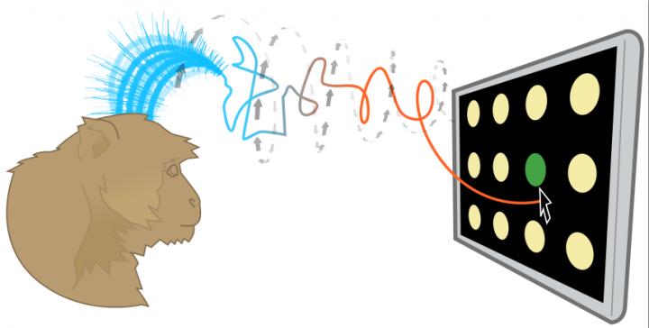
Brain-controlled prostheses sample a few hundred neurons to estimate motor commands that involve millions of neurons. So tiny sampling errors can reduce the precision and speed of thought-controlled keypads. A Stanford technique can analyze this sample and quickly make dozens of corrective adjustments to make thought control more precise. (credit: Jonathan Kao, Shenoy Lab)

An interdisciplinary team led by Stanford electrical engineer Krishna Shenoy has developed a technique to improve brain-controlled prostheses. These brain-computer-interface (BCI) devices, for people with neurological disease or spinal cord injury, deliver thought commands to devices such as virtual keypads, bypassing the damaged area.

The new technique addresses a problem with these brain-controlled prostheses: they currently access a sample of only a few hundred neurons, so tiny errors in the sample — neurons that fire too fast or too slow — reduce the precision and speed of thought-controlled keypads.

Understanding brain dynamics for arm movements

In essence the new prostheses analyze the neuron sample and quickly make dozens of corrective adjustments to the estimate of the brain’s electrical pattern.

Shenoy’s team tested a brain-controlled cursor meant to operate a virtual keyboard. The system is intended for people with paralysis and amyotrophic lateral sclerosis (ALS), also called Lou Gehrig’s disease, a condition that Stephen Hawking has. ALS degrades one’s ability to move.

The new corrective technique is based on a recently discovered understanding of how monkeys naturally perform arm movements. The researchers studied animals that were normal in every way. The monkeys used their arms, hands and fingers to reach for targets presented on a video screen. The researchers sought to learn, through hundreds of experiments, what the electrical patterns from the 100- to 200-neuron sample looked like during a normal reach — to understand the “brain dynamics” underlying reaching arm movements.

“These brain dynamics are analogous to rules that characterize the interactions of the millions of neurons that control motions,” said Jonathan Kao, a doctoral student in electrical engineering and first author of the open-access Nature Communications paper on the research. “They enable us to use a tiny sample more precisely.”

A decoding algorithm

In their current experiments, Shenoy’s team members distilled their understanding of brain dynamics into an algorithm that could decode (analyze) the measured electrical signals that their prosthetic device obtained from the sampled neurons. The algorithm tweaked these measured signals so that the sample’s dynamics were more like the baseline brain dynamics and thus more precise.

To test this algorithm, the Stanford researchers first trained two monkeys to choose targets on a simplified keypad. The keypad consisted of several rows and columns of blank circles. When a light flashed on a given circle the monkeys were trained to reach for that circle with their arms.

To set a performance baseline, the researchers measured how many targets the monkeys could tap with their fingers in 30 seconds. The monkeys averaged 29 correct finger taps in 30 seconds.

In the actual experiment, the researchers only scored virtual taps that came from the monkeys’ brain-controlled cursor. Although the monkey may still have moved his fingers, the researchers only counted a hit when the brain-controlled cursor, corrected by the algorithm, sent the virtual cursor to the target.

The prosthetic scored 26 thought-taps in 30 seconds, about 90 percent as quickly as a monkey’s finger. (See video of hand versus thought-controlled cursor taps.)

Thought-controlled keypads are not unique to Shenoy’s lab. Other brain-controlled prosthetics use different techniques to solve the problem of sampling error. But of several alternative techniques tested by the Stanford team, the closest resulted in 23 targets in 30 seconds.

Next steps

The goal of all this research is to get thought-controlled prosthetics to people with ALS. Today these people may use an eye-tracking system to direct cursors or a “head mouse” that tracks the movement of the head. Both are fatiguing to use. Neither provides the natural and intuitive control of readings taken directly from the brain.

“Brain-controlled prostheses will lead to a substantial improvement in quality of life,” Shenoy said. “The speed and accuracy demonstrated in this prosthesis results from years of basic neuroscience research and from combining these scientific discoveries with the principled design of mathematical control algorithms.”

The U.S. Food and Drug Administration recently gave Shenoy’s team the green light to conduct a pilot clinical trial of their thought-controlled cursor on people with spinal cord injuries.

“This is a fundamentally new approach that can be further refined and optimized to give brain-controlled prostheses greater performance, and therefore greater clinical viability,” Shenoy said.


Abstract of Single-trial dynamics of motor cortex and their applications to brain-machine interfaces

Increasing evidence suggests that neural population responses have their own internal drive, or dynamics, that describe how the neural population evolves through time. An important prediction of neural dynamical models is that previously observed neural activity is informative of noisy yet-to-be-observed activity on single-trials, and may thus have a denoising effect. To investigate this prediction, we built and characterized dynamical models of single-trial motor cortical activity. We find these models capture salient dynamical features of the neural population and are informative of future neural activity on single trials. To assess how neural dynamics may beneficially denoise single-trial neural activity, we incorporate neural dynamics into a brain–machine interface (BMI). In online experiments, we find that a neural dynamical BMI achieves substantially higher performance than its non-dynamical counterpart. These results provide evidence that neural dynamics beneficially inform the temporal evolution of neural activity on single trials and may directly impact the performance of BMIs.

Sleeping on your side may clear waste from your brain most effectively

The brain’s glymphatic pathway clears harmful wastes, especially during sleep. This lateral position could prove to be the best position for the brain-waste clearance process (credit: Stony Brook University)

Sleeping in the lateral, or side position, as compared to sleeping on one’s back or stomach, may more effectively remove brain waste, and could reduce the chances of developing Alzheimer’s, Parkinson’s and other neurological diseases, according to researchers at Stony Brook University.

Stony Brook University researchers discovered this in experiments with rodents by using dynamic contrast magnetic resonance imaging (MRI) to image the brain’s glymphatic pathway, a complex system that clears wastes and other harmful chemical solutes from the brain. They also used kinetic modeling to quantify the CSF-ISF exchange rates in anesthetized rodents’ brains in lateral, prone, and supine positions.

Colleagues at the University of Rochester used fluorescence microscopy and radioactive tracers to validate the MRI data and to assess the influence of body posture on the clearance of amyloid from the brains.

Their finding is published in the Journal of Neuroscience.

Most popular position in humans and animals

“It is interesting that the lateral sleep position is already the most popular in human and most animals —even in the wild — and it appears that we have adapted the lateral sleep position to most efficiently clear our brain of the metabolic waste products that built up while we are awake,” says Maiken Nedergaard, PhD, a co-author at the University of Rochester.

“The study therefore adds further support to the concept that sleep subserves a distinct biological function of sleep and that is to ‘clean up’ the mess that accumulates while we are awake. Many types of dementia are linked to sleep disturbances, including difficulties in falling asleep. It is increasing acknowledged that these sleep disturbances may accelerate memory loss in Alzheimer’s disease.”

The brain-waste clearing system

Cerebrospinal fluid (CSF) filters through the brain and exchanges with interstitial fluid (ISF) to clear waste in the glymphatic pathway, similar to the way the body’s lymphatic system clears waste from organs. The glymphatic pathway is most efficient during sleep. Brain waste includes amyloid β (amyloid) and tau proteins, chemicals that negatively affect brain processes if they build up.

Helene Benveniste, MD, PhD, Principal Investigator and a Professor in the Departments of Anesthesiology and Radiology at Stony Brook University School of Medicine, cautioned that further testing with MRI or other imaging methods in humans is necessary.

New York University Langone Medical Center was also involved in the research.


Abstract of The Effect of Body Posture on Brain Glymphatic Transport

The glymphatic pathway expedites clearance of waste, including soluble amyloidβ (Aβ) from the brain. Transport through this pathway is controlled by the brain’s arousal level because, during sleep or anesthesia, the brain’s interstitial space volume expands (compared with wakefulness), resulting in faster waste removal. Humans, as well as animals, exhibit different body postures during sleep, which may also affect waste removal. Therefore, not only the level of consciousness, but also body posture, might affect CSF–interstitial fluid (ISF) exchange efficiency. We used dynamic-contrast-enhanced MRI and kinetic modeling to quantify CSF-ISF exchange rates in anesthetized rodents” brains in supine, prone, or lateral positions. To validate the MRI data and to assess specifically the influence of body posture on clearance of Aβ, we used fluorescence microscopy and radioactive tracers, respectively. The analysis showed that glymphatic transport was most efficient in the lateral position compared with the supine or prone positions. In the prone position, in which the rat’s head was in the most upright position (mimicking posture during the awake state), transport was characterized by “retention” of the tracer, slower clearance, and more CSF efflux along larger caliber cervical vessels. The optical imaging and radiotracer studies confirmed that glymphatic transport and Aβ clearance were superior in the lateral and supine positions. We propose that the most popular sleep posture (lateral) has evolved to optimize waste removal during sleep and that posture must be considered in diagnostic imaging procedures developed in the future to assess CSF-ISF transport in humans.

An incredible nanoscale 3-D voyage through a tiny part of the mouse brain

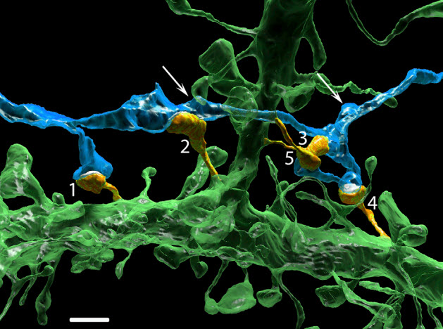
Multiple synapses of the same axon innervate multiple spines of the same postsynaptic cell. An extreme example in which one axon (blue) innervates five dendritic spines (orange, labeled 1–5) of a basal dendrite (green) is shown. Arrows point to other varicosities (swellings) of this axon that are innervating dendritic spines of other neurons. Scale bar, 2 mm. (credit: Narayanan Kasthuri et al./Cell)

Using an electron microscope, researchers have peered down inside the brain of an adult mouse at a scale previously unachievable, generating dramatic color images at 3-nm-pixel resolution. The research was published Thursday July 30 in an open-access paper in the journal Cell.

Focusing on a small area of the mouse brain that receives sensory information from mouse whiskers, the researchers built a system that automatically slices a subject brain into thousands of thin 29-nm coronal brain slices (each section 1 square millimeter) and sticks them to a conveyer belt. After staining the slices to differentiate different tissues, an automated electron microscope took pictures of each slice. A computer then assigned different colors to individual structures and combined the images to produce a 3-D map.

The scientists used a program called VAST, developed by co-author Daniel Berger of Harvard and MIT, to label and piece apart each individual “object” (e.g., neuron, glial cell, blood vessel cell, etc.) using different colors, as well as smaller structures such as dendrites and mitochondria. They also created an annotated inventory of 1700 synapses.

Synapses in contact with a dendrite (the large red object). The white dots are synaptic vesicles inside axons. (credit: N. Kasthuri et al./Cell)

“The complexity of the brain is much more than what we had ever imagined,” says study first author Narayanan “Bobby” Kasthuri, of the Boston University School of Medicine. “We had this clean idea of how there’s a really nice order to how neurons connect with each other, but if you actually look at the material it’s not like that. The connections are so messy that it’s hard to imagine a plan to it, but we checked and there’s clearly a pattern that cannot be explained by randomness.

“If we could make a map of a brain with schizophrenia and compare it to one without schizophrenia, we can look for inappropriate connections that may contribute to the disorder,” he said.

The cost and data storage demands for this type of research are still high, but the researchers expect expenses to drop over time (as has been the case with genome sequencing).

To allow for further inquiries and analyses in the high-resolution volume (80,000 cubic meters), scientists provide access to all of the image data via the Open Connectome Project, along with custom analytic software. They are also partnering with Argonne National Laboratory with the hopes of creating a national brain laboratory that neuroscientists around the world can access within the next few years.


Hauser Studio, Harvard University | Connections in a Cube/ Cell, July 30, 2015 (Vol. 162, Issue 3)


Abstract of Saturated Reconstruction of a Volume of Neocortex

We describe automated technologies to probe the structure of neural tissue at nanometer resolution and use them to generate a saturated reconstruction of a sub-volume of mouse neocortex in which all cellular objects (axons, dendrites, and glia) and many sub-cellular components (synapses, synaptic vesicles, spines, spine apparati, postsynaptic densities, and mitochondria) are rendered and itemized in a database. We explore these data to study physical properties of brain tissue. For example, by tracing the trajectories of all excitatory axons and noting their juxtapositions, both synaptic and non-synaptic, with every dendritic spine we refute the idea that physical proximity is sufficient to predict synaptic connectivity (the so-called Peters’ rule). This online minable database provides general access to the intrinsic complexity of the neocortex and enables further data-driven inquiries.

Memory problems? Go climb a tree.

(credit: iStock)

Climbing a tree or balancing on a beam can dramatically improve cognitive skills, according to a study recently conducted by researchers in the Department of Psychology at the University of North Florida.

The study is the first to show that proprioceptively dynamic activities like climbing a tree, done over a short period of time, have dramatic working memory benefits.

Working memory (the ability to process and recall information), is linked to performance in a wide variety of contexts from grades to sports. Proprioception (awareness of body positioning and orientation) is also associated with working memory.

The results of this research, led by Ross Alloway, a research associate, and Tracy Alloway, an associate professor, recently published in Perceptual and Motor Skills, suggest that working-memory improvements can be made in just a couple of hours with these physical exercises.

The aim of this study was to see if proprioceptive activities completed over a short period of time can enhance working memory performance, and whether an acute and highly intensive period of exercise would yield working memory gains.

The UNF researchers recruited adults ages 18 to 59 and tested their working memory. Next, they undertook proprioceptively dynamic activities, designed by the company Movnat, which required proprioception and at least one other element, such as locomotion or route planning.

Working memory capacity increase of 50 percent; better than yoga

In the study, such activities included climbing trees, walking and crawling on a beam approximately 3 inches wide, moving while paying attention to posture, running barefoot, navigating over, under and around obstacles, as well as lifting and carrying awkwardly weighted objects. After two hours, participants were tested again, and researchers found that their working memory capacity had increased by 50 percent, a dramatic improvement.

The researchers also tested two control groups. The first was a college class learning new information in a lecture setting to see if learning new information improved working memory. The second was a yoga class to see if static proprioceptive activities were cognitively beneficial. However, neither control group experienced working memory benefits.

Proprioceptively dynamic training may place a greater demand on working memory than either control condition because as environment and terrain changes, the individual recruits working memory to update information to adapt appropriately. Though the yoga control group engaged in proprioceptive activities that required awareness of body position, it was relatively static as they performed the yoga postures in a small space, which didn’t allow for locomotion or navigation.

“This research suggests that by doing activities that make us think, we can exercise our brains as well as our bodies,” said Alloway. “This research has wide-ranging implications for everyone from kids to adults. By taking a break to do activities that are unpredictable and require us to consciously adapt our movements, we can boost our working memory to perform better in the classroom and the boardroom.”


Abstract of  The working memory benefits of proprioceptively demanding training: A pilot study

The aim of this study was to investigate the effect of proprioception on working memory. It was also of interest whether an acute and highly intensive period of exercise would yield working memory gains. The training group completed a series of proprioceptively demanding exercises. There were also control classroom and yoga groups. Working memory was measured using a backward digit recall test. The data indicated that active, healthy adults who undertook acute, proprioceptively demanding training improved working memory scores compared to the classroom and yoga groups. One possible reason that the training yielded significant working memory gains could be that the training was proprioceptively dynamic, requiring proprioception and at least one other factor—such as locomotion or navigation—at the same time, which may have contributed to the improvements in working memory performance.

Non-surgical electrical/drug stimulation helps patients with paralysis to voluntarily move their legs — a first

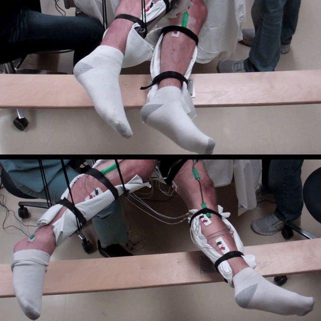
Range of voluntary movement prior to receiving stimulation compared to movement after receiving stimulation, physical conditioning, and the drug buspirone. The subject’s legs are supported so that they can move without resistance from gravity. The electrodes on the legs are used for recording muscle activity. (credit: Edgerton Lab/UCLA)

In a study conducted at UCLA, five men who had been completely paralyzed were able to move their legs in a rhythmic motion thanks to a new, noninvasive neuromodulation and pharmacological procedure that stimulates the spinal cord.

The researchers believe this to be the first time voluntary leg movements have ever been relearned in completely paralyzed patients without surgery. The results are reported in an open-access paper in the Journal of Neurotrauma.

“These findings tell us we have to look at spinal cord injury in a new way,” said V. Reggie Edgerton, senior author of the research and a UCLA distinguished professor of integrative biology and physiology, neurobiology and neurosurgery.

Edgerton said although it likely will be years before the new approaches are widely available, he now believes that it is possible to significantly improve quality of life for patients with severe spinal cord injuries, and to help them recover multiple body functions.

Earlier this year, a the researchers demonstrated that they could induce involuntary stepping movements in healthy, uninjured people using noninvasive stimulation. The finding led Edgerton to believe the same approach could be effective for people with complete paralysis.

Reawakening neural connections with electrical charges and a drug

In the new research, five men were given one 45-minute training session per week for 18 weeks. For four weeks, the men were also given twice daily doses of buspirone, a drug often used to treat anxiety disorders, as part of the treatment.

Researchers placed electrodes at strategic points on the skin, at the lower back and near the tailbone and then administered a unique pattern of noninvasive, painless transcutaneous (through the skin) electrical currents*. The electrical charges caused no discomfort to the patients, who were lying down.

“The fact that they regained voluntary control so quickly must mean that they had neural connections that were dormant, which we reawakened,” said Edgerton, who for nearly 40 years has conducted research on how the neural networks in the spinal cord regain control of standing, stepping and voluntary control of movements after paralysis. “It was remarkable.”

* The researchers used monopolar rectangular pulsed stimuli (30 Hz at T11 and 5 Hz at Co1 with 1 ms duration for each pulse) filled with a carrier frequency of 10 kHz and at an intensity ranging from 80 to 180 mA .


Edgerton Lab/UCLA | Non-invasive Neuromodulation to regain voluntary movements after paralysis

Edgerton said most experts, including himself, had assumed that people who were completely paralyzed would no longer have had neural connections across the area of the spinal cord injury.

The researchers do not know yet whether patients who are completely paralyzed can be trained to fully bear their weight and walk. But he and colleagues have now published data on nine people who have regained voluntary control of their legs —four with epidural implants and five in the latest study.

“Many people thought just a few years ago we might be able to achieve these results in perhaps one out of 100 subjects, but now we have nine of nine,” Edgerton said. “I think it’s a big deal, and when the subjects see their legs moving for the first time after paralysis, they say it’s a big deal.”

The men in the newest study ranged in age from 19 to 56; their injuries were suffered during athletic activities or, in one case, in an auto accident. All have been completely paralyzed for at least two years. Their identities are not being released.

The research was funded by the National Institutes of Health’s National Institute of Biomedical Imaging and Bioengineering (grants U01EB15521 and R01EB007615), the Christopher and Dana Reeve Foundation, the Walkabout Foundation and the Russian Scientific Fund.

“These encouraging results provide continued evidence that spinal cord injury may no longer mean a life-long sentence of paralysis and support the need for more research,” said Dr. Roderic Pettigrew, director of the National Institute of Biomedical Imaging and Bioengineering. “The potential to offer a life-changing therapy to patients without requiring surgery would be a major advance; it could greatly expand the number of individuals who might benefit from spinal stimulation. It’s a wonderful example of the power that comes from combining advances in basic biological research with technological innovation.”

Edgerton estimates that cost to patients of the new approach could be one-tenth the cost of treatment using the surgical epidural stimulator (which is also still experimental) — and, because no surgery is required, it would likely be more easily available to more patients.

The study’s co-authors were Gerasimenko, who conceived the new approach and is director of the laboratory of movement physiology at Russia’s Pavlov Institute and a researcher in the UCLA department of integrative biology and physiology, as well as Daniel Lu, associate professor of neurosurgery, researchers Morteza Modaber, Roland Roy and Dimitry Sayenko, research technician Sharon Zdunowski, research scientist Parag Gad, laboratory coordinator Erika Morikawa and research assistant Piia Haakana, all of UCLA; and Adam Ferguson, assistant professor of neurological surgery at UC San Francisco.

Edgerton and his research team also plan to study people who have severe, but not complete, paralysis. “They’re likely to improve even more,” he said.

The scientists can only work with a small number of patients, due to limited resources, but Edgerton is optimistic that the research can benefit many others. Almost 6 million Americans live with paralysis, including nearly 1.3 million with spinal cord injuries.

“A person can have hope, based on these results,” Edgerton said. “In my opinion, they should have hope.”


 Abstract of Noninvasive Reactivation of Motor Descending Control after Paralysis

The present prognosis for the recovery of voluntary control of movement in patients diagnosed as motor complete is generally poor. Herein we introduce a novel and noninvasive stimulation strategy of painless transcutaneous electrical enabling motor control and a pharmacological enabling motor control strategy to neuromodulate the physiological state of the spinal cord. This neuromodulation enabled the spinal locomotor networks of individuals with motor complete paralysis for 2-6 years (AIS B) to be reengaged and trained. We showed that locomotor-like stepping could be induced without voluntary effort within a single test session using electrical stimulation and training. We also observed significant facilitation of voluntary influence on the stepping movements in the presence of stimulation over a four-week period in each subject. Using these strategies we transformed brain-spinal neuronal networks from a dormant to a functional state sufficiently to enable recovery of voluntary movement in 5/5 subjects. Pharmacological intervention combined with stimulation and training resulted in further improvement in voluntary motor control of stepping-like movements in all subjects. We also observed on-command selective activation of the gastrocnemius and soleus muscles when attempting to plantarflex. At the end of 18 weeks of weekly interventions the mean changes in the amplitude of voluntarily controlled movement without stimulation was as high as occurred when combined with electrical stimulation. Additionally, spinally evoked motor potentials were readily modulated in the presence of voluntary effort, providing electrophysiological evidence of the re-establishment of functional connectivity among neural networks between the brain and the spinal cord.

A simulated robot with bacterial brain

Computational Simulation of microbiome-host interactions. (A) A basic gene circuit forms the core of all simulated gene network behavior. (B) Green fluorescent protein (GFP, shown as a green dot) from this circuit is conceptualized to be detected by an onboard miniature, epifluorescent microscope (EFM). (C) A computational simulation of microbiome GFP production based upon an analytical model for the circuit in (A). In a built system, this protein fluorescence signal would be the light detected by the EFM. (D) The conceptualized robot uses onboard electronics to convert the measured light signals into electrical (voltage) signals. (E) Voltage signals meeting specific criteria activate pre-programmed robot motion subroutines. (F) The resulting emergent behavior potentially leads a robot to a carbon fuel depot. Here, robot behavior resulting from a simulation of the circuit in (A) is shown. The robot was programmed with motion subroutines that activate to seek arabinose (synthesized from glucose, orange square) depots following receipt of lactose (cyan triangles). (credit: Keith C. Heyde & Warren C. Ruder/Scientific Reports)

Virginia Tech scientist Warren Ruder, an assistant professor of biological systems engineering, has created an in silico (computer-simulated) model of a biomimetic robot controlled by a bacterial brain.

The study was inspired by real-world experiments where the mating behavior of fruit flies was manipulated using bacteria, and in which mice exhibited signs of lower stress when implanted with probiotics (“healthy” bacteria).

A math model of microbiome-controlled behavior

The deeper motivation for the study was to understand how the microbiome (the bacteria in the human body, thought to number ten times more than human cells) might influence human behavior. For example, some studies show that the gut microbiome influences human eating behavior and dietary choices to favor the survival of the bacteria. (See Do gut bacteria control your mind? for example.)

As explained in an open-access paper published recently in Scientific Reports, Ruder’s study revealed unique decision-making behavior by a bacteria-robot system by coupling and computationally simulating equations that describe three distinct elements: engineered gene circuits in E. coli, microfluid bioreactors, and robot movement.

In the mathematical model, the theoretical robot was equipped with sensors and a miniature microscope to measure the color. The hypothetical robot was designed to read E. coli bacterial gene expression levels (how much protein is created by specific genes), using light sensors in miniature microscopes. The bacteria turned green or red, depending on what they ate.

Bacteria that act like tigers?

Interestingly, the bacteria in the model began to approach a fuel source with “stalk-pause-strike” behavior, characteristic of predators.

Ruder’s modeling study also demonstrates that these sorts of biosynthetic experiments could be done in the future with a minimal amount of funds, opening up the field to a much larger pool of researchers.

Understanding the biochemical sensing between organisms could have far reaching implications in ecology, biology, and robotics, Ruder suggests.

In agriculture, bacteria-robot model systems could enable robust studies that explore the interactions between soil bacteria and livestock. In healthcare, further understanding of bacteria’s role in controlling gut physiology could lead to bacteria-based prescriptions (probiotics) to treat mental and physical illnesses. Ruder also envisions droids that could execute tasks such as deploying bacteria to remediate oil spills.

Bacteria effects on behavior

The findings also add to the ever-growing body of research about bacteria in the human body that are thought to regulate health and mood, and especially the theory that bacteria also affect behavior.

“We hope to help democratize the field of synthetic biology for students and researchers all over the world with this model,” said Ruder. “In the future, rudimentary robots and E. coli that are already commonly used separately in classrooms could be linked with this model to teach students from elementary school through the Ph.D.-level about bacterial relationships with other organisms.”

Ruder plans next to create a real-world version of the experiment, creating mobile robots with bioreactors on board that harbor living colonies of bacteria that direct the robot’s behavior.

The Air Force Office of Scientific Research funded the mathematical modeling of gene circuitry in E. coli, and the Virginia Tech Student Engineers’ Council has provided funding to move these models and resulting mobile robots into the classroom as teaching tools.


Virginia Tech | Scientist shows bacteria could control robots


Abstract of Exploring Host-Microbiome Interactions using an in Silico Model of Biomimetic Robots and Engineered Living Cells

The microbiome’s underlying dynamics play an important role in regulating the behavior and health of its host. In order to explore the details of these interactions, we created anin silico model of a living microbiome, engineered with synthetic biology, that interfaces with a biomimetic, robotic host. By analytically modeling and computationally simulating engineered gene networks in these commensal communities, we reproduced complex behaviors in the host. We observed that robot movements depended upon programmed biochemical network dynamics within the microbiome. These results illustrate the model’s potential utility as a tool for exploring inter-kingdom ecological relationships. These systems could impact fields ranging from synthetic biology and ecology to biophysics and medicine.

The brain’s got rhythm

A snapshot illustration showing how the anterior (blue) and posterior (orange) regions of the frontal cortex sync up to communicate cognitive goals to one another  (credit: Bradley Voytek)

Like a jazz combo, the human brain improvises while its rhythm section keeps up a steady beat. But when it comes to taking on intellectually challenging tasks, groups of neurons tune in to one another for a fraction of a second and harmonize, then go back to improvising, according to new research led by UC Berkeley.

These findings, reported Monday (July 27) in the journal Nature Neuroscience, could pave the way for more targeted treatments for people with brain disorders marked by fast, slow, or chaotic brain waves (neural oscillations) — such as Parkinson’s disease, schizophrenia and autism, which are characterized in part by offbeat brain rhythms.

Keeping the beat

“The human brain has 86 billion or so neurons all trying to talk to each other in this incredibly messy, noisy and electrochemical soup,” said study lead author Bradley Voytek. “Our results help explain the mechanism for how brain networks quickly come together and break apart as needed.”

Working with cognitively healthy epilepsy patients, Voytek and fellow researchers at UC Berkeley’s Helen Wills Neuroscience Institute used electrocorticography (ECoG) — which places electrodes directly on the exposed surface of the brain — to measure neural oscillations as the patients performed cognitively challenging tasks.  This showed how the rhythms control communication between brain regions.

They found that as the mental exercises became more demanding, theta waves at 4–8 Hertz (cycles per second) synchronized within the brain’s frontal lobe, enabling it to connect with brain sub-regions, such as the motor cortex.

“In these brief moments of synchronization, quick communication occurs as the neurons between brain regions lock into these frequencies, and this measure is critical in a variety of disorders,” said Voytek, an assistant professor of cognitive science at UC San Diego who conducted the study as a postdoctoral fellow in neuroscience at UC Berkeley.

There are five types of brain wave frequencies — Gamma, Beta, Alpha, Theta and Delta — and each are thought to play a different role. For example, Theta waves help coordinate neurons as we move around our environment, and thus are key to processing spatial information.

Off-tempo 

In people with autism, the connection between Alpha waves and neural activity has been found to weaken when they process emotional images, according to Voytek. And people with Parkinson’s disease show abnormally strong Beta waves in the motor cortex, locking neurons into the wrong groove and inhibiting movement. Fortunately, electrical deep brain stimulation can disrupt abnormally strong Beta waves in Parkinson’s and alleviate symptoms,

For the study, epilepsy patients viewed shapes of increasing complexity on a computer screen and were tasked with using different fingers (index or middle) to push a button depending on the shape, color or texture of the shape. The exercise started out simply with participants hitting the button with, say, an index finger each time a square flashed on the screen. But it grew progressively more difficult as the shapes became more layered with colors and textures, and their fingers had to keep up.

As the tasks became more demanding, the oscillations kept up, coordinating more parts of the frontal lobe and synchronizing the information passing between those brain regions. “The results revealed a delicate coordination in the brain’s code,” Voytek said. “Our neural orchestra may need no conductor, just brain waves sweeping through to briefly excite neurons, like millions of fans in a stadium doing ‘The Wave.’”

Scientists at Brown University, the Department of Veterans Affairs, UCSF, Johns Hopkins University, and Stanford University were also involved in the research.

UPDATE July 29, 2015: lead author’s correction to UC Berkeley press release: “pre-frontal” in illustration caption changed to “frontal” and  “connect with other brain regions” changed to “connect with brain sub-regions” (H/T to “betaelements” for those catches)


Abstract of Oscillatory dynamics coordinating human frontal networks in support of goal maintenance

Humans have a capacity for hierarchical cognitive control—the ability to simultaneously control immediate actions while holding more abstract goals in mind. Neuropsychological and neuroimaging evidence suggests that hierarchical cognitive control emerges from a frontal architecture whereby prefrontal cortex coordinates neural activity in the motor cortices when abstract rules are needed to govern motor outcomes. We utilized the improved temporal resolution of human intracranial electrocorticography to investigate the mechanisms by which frontal cortical oscillatory networks communicate in support of hierarchical cognitive control. Responding according to progressively more abstract rules resulted in greater frontal network theta phase encoding (4–8 Hz) and increased prefrontal local neuronal population activity (high gamma amplitude, 80–150 Hz), which predicts trial-by-trial response times. Theta phase encoding coupled with high gamma amplitude during inter-regional information encoding, suggesting that inter-regional phase encoding is a mechanism for the dynamic instantiation of complex cognitive functions by frontal cortical subnetworks.