Tiny fibers open new windows into the brain

A multifunctional flexible fiber that enables viral delivery, optical stimulation, and recording with one-step surgery. (credit: Seongjun Park et al./Nature Neuroscience)

Imagine a single flexible polymer fiber 200 micrometers across — about the width of a human hair — that can deliver a combination of optical, electrical, and chemical signals between different brain regions, with the softness and flexibility of brain tissue — allowing neuroscientists to leave implants in place and have them retain their functions over much longer periods than is currently possible with typical stiff, metallic fibers.

That’s what a team of MIT scientists has reported in the journal Nature Neuroscience. (Previous research efforts in neuroscience have generally relied on separate devices: needles to inject viral vectors for optogenetics, optical fibers for light delivery, and arrays of electrodes for recording, adding complication and the need for tricky alignments among the different devices.)

Multifunctional

For example, in tests with lab mice, the researchers were able to inject viral vectors that carried genes called opsins (which sensitize neurons to light) through one of two fluid channels in the fiber. They waited for the opsins to take effect, then sent a pulse of light through the optical waveguide in the center, and recorded the resulting neuronal activity, using six electrodes to pinpoint specific reactions. All of this was done through a single flexible fiber.

“It can deliver the virus [containing the opsins] straight to the cell, and then stimulate the response and record the activity — and [the fiber] is sufficiently small and biocompatible so it can be kept in for a long time,” says Polina Anikeeva, a professor in the MIT Department of Materials Science and Engineering.

Since each fiber is so small, “potentially, we could use many of them to observe different regions of activity,” she says. In their initial tests, the researchers placed probes in two different brain regions at once, varying which regions they used from one experiment to the next, and measuring how long it took for responses to travel between them.

The key ingredient that made this multifunctional fiber possible was the development of conductive “wires” that maintained the needed flexibility while also carrying electrical signals well. The team engineered a composite of conductive polyethylene doped with graphite flakes. The polyethylene was initially formed into layers, sprinkled with graphite flakes, then compressed; then another pair of layers was added and compressed, and then another, and so on.

The team aims to reduce the width of the fibers further, to make their properties even closer to those of the neural tissue and use material that is even softer to match the adjacent tissue.

The research team included members of MIT’s Research Laboratory of Electronics, Department of Electrical Engineering and Computer Science, McGovern Institute for Brain Research, Department of Chemical Engineering, and Department of Mechanical Engineering, as well as researchers at Tohuku University in Japan and Virginia Polytechnic Institute. It was supported by the National Institute of Neurological Disorders and Stroke, the National Science Foundation, the MIT Center for Materials Science and Engineering, the Center for Sensorimotor Neural Engineering, and the McGovern Institute for Brain Research.


Abstract of One-step optogenetics with multifunctional flexible polymer fibers

Optogenetic interrogation of neural pathways relies on delivery of light-sensitive opsins into tissue and subsequent optical illumination and electrical recording from the regions of interest. Despite the recent development of multifunctional neural probes, integration of these modalities in a single biocompatible platform remains a challenge. We developed a device composed of an optical waveguide, six electrodes and two microfluidic channels produced via fiber drawing. Our probes facilitated injections of viral vectors carrying opsin genes while providing collocated neural recording and optical stimulation. The miniature (<200 μm) footprint and modest weight (<0.5 g) of these probes allowed for multiple implantations into the mouse brain, which enabled opto-electrophysiological investigation of projections from the basolateral amygdala to the medial prefrontal cortex and ventral hippocampus during behavioral experiments. Fabricated solely from polymers and polymer composites, these flexible probes minimized tissue response to achieve chronic multimodal interrogation of brain circuits with high fidelity.

Brain-imaging headband measures how our minds mirror a speaker when we communicate

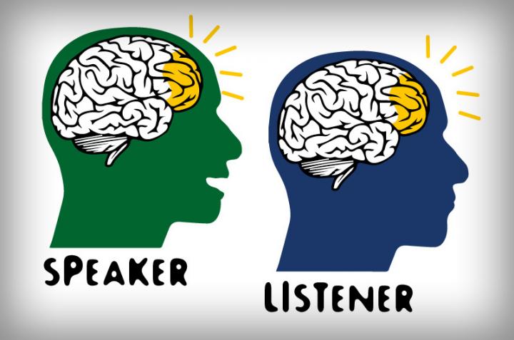
A cartoon image of brain “coupling” during communication (credit: Drexel University)

Drexel University biomedical engineers and Princeton University psychologists have used a wearable brain-imaging device called functional near-infrared spectroscopy (fNIRS) to measure brain synchronization when humans interact. fNIRS uses light to measure neural activity in the cortex of the brain (based on blood-oxygenation changes) during real-life situations and can be worn like a headband.

(KurzweilAI recently covered research with a fNIRS brain-computer interface that allows completely locked-in patients to communicate.)

A fNIRS headband (credit: Wyss Center for Bio and Neuroengineering)

Mirroring the speaker’s brain activity

The researchers found that a listener’s brain activity (in brain areas associated with speech comprehension) mirrors the speaker’s brain when he or she is telling a story about a real-life experience, with about a five-second delay. They also found that higher coupling is associated with better understanding.

The researchers believe the system can be used to offer important information about how to better communicate in many different environments, such as how people learn in classrooms and how to improve business meetings and doctor-patient communication. They also mentioned uses in analyzing political rallies and how people handle cable news.

“We now have a tool that can give us richer information about the brain during everyday tasks — such as person-to-person communication — that we could not receive in artificial lab settings or from single brain studies,” said Hasan Ayaz, PhD, an associate research professor in Drexel’s School of Biomedical Engineering, Science and Health Systems, who led the research team.

Traditional brain imaging methods like fMRI have limitations. In particular, fMRI requires subjects to lie down motionlessly in a noisy scanning environment. With this kind of setup, it’s not possible to simultaneously scan the brains of multiple individuals who are speaking face-to-face. Which is why the Drexel researchers turned to a portable fNIRS system, which could probe brain-to-brain coupling question in natural settings.

For their study, a native English speaker and two native Turkish speakers told an unrehearsed, real-life story in their native language. Their stories were recorded and their brains were scanned using fNIRS. Fifteen English speakers then listened to the recording, in addition to a story that was recorded at a live storytelling event.

The researchers targeted the prefrontal and parietal areas of the brain, which include cognitive and higher order areas that are involved in a person’s capacity to discern beliefs, desires, and goals of others. They hypothesized that a listener’s brain activity would correlate with the speaker’s only when listening to a story they understood (the English version). A second objective of the study was to compare the fNIRS results with data from a similar study that had used fMRI to compare the two methods.

They found that when the fNIRS measured the oxygenation and deoxygenation of blood cells in the test subject’s brains, the listeners’ brain activity matched only with the English speakers.* These results also correlated with the previous fMRI study.

The researchers believe the new research supports fNIRS as a viable future tool to study brain-to-brain coupling during social interaction. One can also imagine possible invasive uses in areas such as law enforcement and military interrogation.

The research was published in open-access Scientific Reports on Monday, Feb. 27.

* “During brain-to-brain coupling, activity in areas of prefrontal [in the speaker] and parietal cortex [in the listeners] previously reported to be involved in sentence comprehension were robustly correlated across subjects, as revealed in the inter-subject correlation analysis. As these are task-related (active listening) activation periods (not resting, etc.), the correlations reflect modulation of these regions by the time-varying content of the narratives, and comprise linguistic, conceptual and affective processing.” — Yichuan Liu et al./Scientific Reports)


Abstract of Measuring speaker–listener neural coupling with functional near infrared spectroscopy

The present study investigates brain-to-brain coupling, defined as inter-subject correlations in the hemodynamic response, during natural verbal communication. We used functional near-infrared spectroscopy (fNIRS) to record brain activity of 3 speakers telling stories and 15 listeners comprehending audio recordings of these stories. Listeners’ brain activity was significantly correlated with speakers’ with a delay. This between-brain correlation disappeared when verbal communication failed. We further compared the fNIRS and functional Magnetic Resonance Imaging (fMRI) recordings of listeners comprehending the same story and found a significant relationship between the fNIRS oxygenated-hemoglobin concentration changes and the fMRI BOLD in brain areas associated with speech comprehension. This correlation between fNIRS and fMRI was only present when data from the same story were compared between the two modalities and vanished when data from different stories were compared; this cross-modality consistency further highlights the reliability of the spatiotemporal brain activation pattern as a measure of story comprehension. Our findings suggest that fNIRS can be used for investigating brain-to-brain coupling during verbal communication in natural settings.

An ultra-low-power artificial synapse for neural-network computing

(Left) Illustration of a synapse in the brain connecting two neurons. (Right) Schematic of artificial synapse (ENODe), which functions as a transistor. It consists of two thin, flexible polymer films (black) with source, drain, and gate terminals, connected by an electrolyte of salty water that permits ions to cross. A voltage pulse applied to the “presynaptic” layer (top) alters the level of oxidation in the “postsynaptic layer” (bottom), triggering current flow between source and drain. (credit: Thomas Splettstoesser/CC and Yoeri van de Burgt et al./Nature Materials)

Stanford University and Sandia National Laboratories researchers have developed an organic artificial synapse based on a new memristor (resistive memory device) design that mimics the way synapses in the brain learn. The new artificial synapse could lead to computers that better recreate the way the human brain processes information. It could also one day directly interface with the human brain.

The new artificial synapse is an electrochemical neuromorphic organic device (dubbed “ENODe”) — a mixed ionic/electronic design that is fundamentally different from existing and other proposed resistive memory devices, which are limited by noise, required high write voltage, and other factors*, the researchers note in a paper published online Feb. 20 in Nature Materials.

Like a neural path in a brain being reinforced through learning, the artificial synapse is programmed by discharging and recharging it repeatedly. Through this training, the researchers have been able to predict within 1 percent of uncertainly what voltage will be required to get the synapse to a specific electrical state and, once there, remain at that state.

“The working mechanism of ENODes is reminiscent of that of natural synapses, where neurotransmitters diffuse through the cleft, inducing depolarization due to ion penetration in the postsynaptic neuron,” the researchers explain in the paper. “In contrast, other memristive devices switch by melting materials at relatively high temperatures (PCMs) or by voltage-induced breakdown/filament formation and ion diffusion in dense oxide layers (FFMOs).”

The ENODe achieves significant energy savings** in two ways:

  • Unlike a conventional computer, where you save your work to the hard drive before you turn it off, the artificial synapse can recall its programming without any additional actions or parts. Traditional computing requires separately processing information and then storing it into memory. Here, the processing creates the memory.
  • When we learn, electrical signals are sent between neurons in our brain. The most energy is needed the first time a synapse is traversed. Every time afterward, the connection requires less energy. This is how synapses efficiently facilitate both learning something new and remembering what we’ve learned. The artificial synapse, unlike most other versions of brain-like computing, also fulfills these two tasks simultaneously, and does so with substantial energy savings.

“More and more, the kinds of tasks that we expect our computing devices to do require computing that mimics the brain because using traditional computing to perform these tasks is becoming really power hungry,” said A. Alec Talin, distinguished member of technical staff at Sandia National Laboratories in Livermore, California, and co-senior author of the paper. “We’ve demonstrated a device that’s ideal for running these type of algorithms and that consumes a lot less power.”

A future brain-like computer with 500 states

Only one artificial synapse has been produced so far, but researchers at Sandia used 15,000 measurements to simulate how an array of them would work in a neural network. They tested the simulated network’s ability to recognize handwriting of digits 0 through 9. Tested on three datasets, the simulated array was able to identify the handwritten digits with an accuracy between 93 to 97 percent.

This artificial synapse may one day be part of a brain-like computer, which could be especially useful for processing visual and auditory signals, as in voice-controlled interfaces and driverless cars, but without energy-consuming computer hardware.

This device is also well suited for the kind of signal identification and classification that traditional computers struggle to perform. Whereas digital transistors can be in only two states, such as 0 and 1, the researchers successfully programmed 500 states in the artificial synapse, which is useful for neuron-type computation models. In switching from one state to another they used about one-tenth as much energy as a state-of-the-art computing system needs to move data from the processing unit to the memory.

However, this is still about 10,000 times as much energy as the minimum a biological synapse needs in order to fire**. The researchers hope to attain neuron-level energy efficiency once they test the artificial synapse in smaller devices.

Linking to live organic neurons

This new artificial synapse may one day be part of a brain-like computer, which could be especially beneficial for computing that works with visual and auditory signals. Examples of this are seen in voice-controlled interfaces and driverless cars. Past efforts in this field have produced high-performance neural networks supported by artificially intelligent algorithms but these depend on energy-consuming traditional computer hardware.

Every part of the device is made of inexpensive organic materials. These aren’t found in nature but they are largely composed of hydrogen and carbon and are compatible with the brain’s chemistry. Cells have been grown on these materials and they have even been used to make artificial pumps for neural transmitters. The switching voltages applied to train the artificial synapse (about 0.5 mV) are also the same as those that move through human neurons — about 1,000 times lower than the “write” voltage for a typical memristor.

That means it’s possible that the artificial synapse could communicate with live neurons, leading to improved brain-machine interfaces. The softness and flexibility of the device also lends itself to being used in biological environments.

This research was funded by the National Science Foundation, the Keck Faculty Scholar Funds, the Neurofab at Stanford, the Stanford Graduate Fellowship, Sandia’s Laboratory-Directed Research and Development Program, the U.S. Department of Energy, the Holland Scholarship, the University of Groningen Scholarship for Excellent Students, the Hendrik Muller National Fund, the Schuurman Schimmel-van Outeren Foundation, the Foundation of Renswoude (The Hague and Delft), the Marco Polo Fund, the Instituto Nacional de Ciência e Tecnologia/Instituto Nacional de Eletrônica Orgânica in Brazil, the Fundação de Amparo à Pesquisa do Estado de São Paulo and the Brazilian National Council.

* “A resistive memory device has not yet been demonstrated with adequate electrical characteristics to fully realize the efficiency and performance gains of a neural architecture. State-of-the-art memristors suffer from excessive write noise, write non-linearities, and high write voltages and currents.  Reducing the noise and lowering the switching voltage significantly below 0.3 V (~10 kT) in a two-terminal device without compromising long-term data retention has proven difficult.” … Organic memristive devices have been recently proposed, but are limited by “the slow kinetics of ion diffusion through a polymer to retain their states or on charge storage in metal nanoparticles, which inherently limits performance and stability.” — Yoeri van de Burgt et al., Nature Materials

** ENODe switches at low voltage and energy (< 10 pJ for 1000-square-micrometer devices), compared to an estimated ∼ 1–100 fJ per synaptic event for the human brain.
 

Abstract of A non-volatile organic electrochemical device as a low-voltage artificial synapse for neuromorphic computing

The brain is capable of massively parallel information processing while consuming only ~1–100 fJ per synaptic event. Inspired by the efficiency of the brain, CMOS-based neural architectures and memristors are being developed for pattern recognition and machine learning. However, the volatility, design complexity and high supply voltages for CMOS architectures, and the stochastic and energy-costly switching of memristors complicate the path to achieve the interconnectivity, information density, and energy efficiency of the brain using either approach. Here we describe an electrochemical neuromorphic organic device (ENODe) operating with a fundamentally different mechanism from existing memristors. ENODe switches at low voltage and energy (<10 pJ for 103 μm2 devices), displays >500 distinct, non-volatile conductance states within a ~1 V range, and achieves high classification accuracy when implemented in neural network simulations. Plastic ENODes are also fabricated on flexible substrates enabling the integration of neuromorphic functionality in stretchable electronic systems. Mechanical flexibility makes ENODes compatible with three-dimensional architectures, opening a path towards extreme interconnectivity comparable to the human brain.

Brain-computer interface advance allows paralyzed people to type almost as fast as some smartphone users

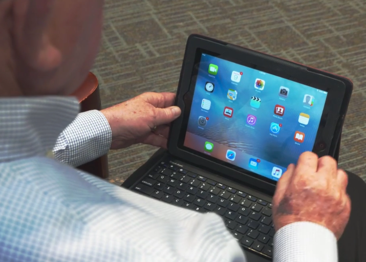
Typing with your mind. You are paralyzed. But now, tiny electrodes have been surgically implanted in your brain to record signals from your motor cortex, the brain region controlling muscle movement. As you think of mousing over to a letter (or clicking to choose it), those electrical brain signals are transmitted via a cable to a computer (replacing your spinal cord and muscles). There, advanced algorithms decode the complex electrical brain signals, converting them instantly into screen actions. (credit: Chethan Pandarinath et al./eLife)

Stanford University researchers have developed a brain-computer interface (BCI) system that can enable people with paralysis* to type (using an on-screen cursor) at speeds and accuracy levels of about three times faster than reported to date.

Simply by imagining their own hand movements, one participant was able to type 39 correct characters per minute (about eight words per minute); the other two participants averaged 6.3 and 2.7 words per minute, respectively — all without auto-complete assistance (so it could be much faster).

Those are communication rates that people with arm and hand paralysis would also find useful, the researchers suggest. “We’re approaching the speed at which you can type text on your cellphone,” said Krishna Shenoy, PhD, professor of electrical engineering, a co-senior author of the study, which was published in an open-access paper online Feb. 21 in eLife.

Braingate and beyond

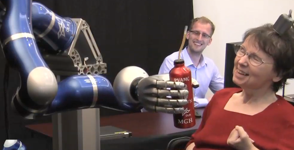
The three study participants used a brain-computer interface called the “BrainGate Neural Interface System.” On KurzweilAI, we first discussed Braingate in 2011, followed by a 2012 clinical trial that allowed a paralyzed patient to control a robot.

Braingate in 2012 (credit: Brown University)

The new research, led by Stanford, takes the Braingate technology way further**. Participants can now move a cursor (by just thinking about a hand movement) on a computer screen that displays the letters of the alphabet, and they can “point and click” on letters, computer-mouse-style, to type letters and sentences.

The new BCI uses a tiny silicon chip, just over one-sixth of an inch square, with 100 electrodes that penetrate the brain to about the thickness of a quarter and tap into the electrical activity of individual nerve cells in the motor cortex.

As the participant thinks of a specific hand-to-mouse movement (pointing at or clicking on a letter), neural electrical activity is recorded using 96-channel silicon microelectrode arrays implanted in the hand area of the motor cortex. These signals are then filtered to extract multiunit spiking activity and high-frequency field potentials, then decoded (using two algorithms) to provide “point-and-click” control of a computer cursor.

What’s next

The team next plans is to adapt the system so that brain-computer interfaces can control commercial computers, phones and tablets — perhaps extending out to the internet.

Beyond that, Shenoy predicted that a self-calibrating, fully implanted wireless BCI system with no required caregiver assistance and no “cosmetic impact” would be available in five to 10 years from now (“closer to five”).

Perhaps a future wireless, noninvasive version could let anyone simply think to select letters, words, ideas, and images — replacing the mouse and finger touch — along the lines of Elon Musk’s neural lace concept?

* Millions of people with paralysis reside in the U.S.

** The study’s results are the culmination of the long-running multi-institutional BrainGate consortium, which includes scientists at Massachusetts General Hospital, Brown University, Case Western University, and the VA Rehabilitation Research and Development Center for Neurorestoration and Neurotechnology in Providence, Rhode Island. The study was funded by the National Institutes of Health, the Stanford Office of Postdoctoral Affairs, the Craig H. Neilsen Foundation, the Stanford Medical Scientist Training Program, Stanford BioX-NeuroVentures, the Stanford Institute for Neuro-Innovation and Translational Neuroscience, the Stanford Neuroscience Institute, Larry and Pamela Garlick, Samuel and Betsy Reeves, the Howard Hughes Medical Institute, the U.S. Department of Veterans Affairs, the MGH-Dean Institute for Integrated Research on Atrial Fibrillation and Stroke and Massachusetts General Hospital.


Stanford | Stanford researchers develop brain-controlled typing for people with paralysis


Abstract of High performance communication by people with paralysis using an intracortical brain-computer interface

Brain-computer interfaces (BCIs) have the potential to restore communication for people with tetraplegia and anarthria by translating neural activity into control signals for assistive communication devices. While previous pre-clinical and clinical studies have demonstrated promising proofs-of-concept (Serruya et al., 2002; Simeral et al., 2011; Bacher et al., 2015; Nuyujukian et al., 2015; Aflalo et al., 2015; Gilja et al., 2015; Jarosiewicz et al., 2015; Wolpaw et al., 1998; Hwang et al., 2012; Spüler et al., 2012; Leuthardt et al., 2004; Taylor et al., 2002; Schalk et al., 2008; Moran, 2010; Brunner et al., 2011; Wang et al., 2013; Townsend and Platsko, 2016; Vansteensel et al., 2016; Nuyujukian et al., 2016; Carmena et al., 2003; Musallam et al., 2004; Santhanam et al., 2006; Hochberg et al., 2006; Ganguly et al., 2011; O’Doherty et al., 2011; Gilja et al., 2012), the performance of human clinical BCI systems is not yet high enough to support widespread adoption by people with physical limitations of speech. Here we report a high-performance intracortical BCI (iBCI) for communication, which was tested by three clinical trial participants with paralysis. The system leveraged advances in decoder design developed in prior pre-clinical and clinical studies (Gilja et al., 2015; Kao et al., 2016; Gilja et al., 2012). For all three participants, performance exceeded previous iBCIs (Bacher et al., 2015; Jarosiewicz et al., 2015) as measured by typing rate (by a factor of 1.4–4.2) and information throughput (by a factor of 2.2–4.0). This high level of performance demonstrates the potential utility of iBCIs as powerful assistive communication devices for people with limited motor function.

Brain-computer interface enables completely locked-in patients to communicate for the first time

NIRS/EEG brain computer interface system applied to a model (credit: Wyss Center for Bio and Neuroengineering)

Four advanced ALS (amyotrophic lateral sclerosis) patients who were “completely locked in” (totally unable to communicate) for years have suddenly broken through in a lab at the Wyss Center for Bio and Neuroengineering in Geneva, Switzerland — communicating a “yes” or “no” by simply thinking the answers.

The brain–computer interface (BCI) system achieved this remarkable breakthrough by using functional near-infrared spectroscopy (fNIRS) to measure changes in blood oxygen levels in the frontal lobes of the brain.

Patients suffering from ALS paralysis, but with preserved awareness, cognition, eye movements and blinking, are classified as having “locked-in syndrome” and can communicate via a BCI by looking at a computer screen, for example. But when the disorder progresses until the patient loses control of the last muscular response, usually the eye muscles, the condition is known as “completely locked-in state” (CLIS), with no possibility of communication. For most of us: a nightmare world.

“Are you happy?”

But surprisingly, when the researchers asked the question, “Are you happy?,” the answer from all four was consistently “yes,” repeated over weeks of questioning. In response to the researchers’ statement, “I love to live,” three of the four replied yes. The researchers asked other personal questions that required “yes” or “no” answers, such as: “Your husband’s name is Joachim?”

They found the questions elicited correct responses in 70% of the trials. In one case, a family requested that the researchers asked the patient whether he would agree for his daughter to marry her boyfriend, Mario. The answer: “no” nine times out of ten.

Overturning previous theories, the research was published in an open-access paper January 31 in PLoS Biology. It was conducted by a multinational team led by Professor Niels Birbaumer, affiliated with the University of Tübingen in Germany; Ospedale San Camillo, IRCCS, Venice, Italy; and the Wyss Center for Bio and Neuroengineering. “If we could make this technique widely clinically available, it could have a huge impact on the day-to-day life of people with completely locked-in syndrome,” said Birbaumer.

“Restoring communication for completely locked-in patients is a crucial first step in the challenge to regain movement,” said Professor John Donoghue, Director of the Wyss Center. “The Wyss Center plans to build on the results of this study to develop clinically useful technology that will be available to people with paralysis resulting from ALS, stroke, or spinal cord injury. The technology used in the study also has broader applications that we believe could be further developed to treat and monitor people with a wide range of neuro-disorders.”

How fNIRS detected “yes” and “no”

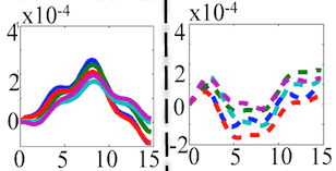
One measurement on one channel of relative changes in oxygenated hemoglobin levels (vertical) vs. seconds (horizontal) for “yes” (left) and “no” (right) questions. (credit: Ujwal Chaudhary et al./PLos Biology)

The brain-computer interface in the study was based on functional near-infrared spectroscopy (fNIRS), which measures blood oxygenation (O2Hb). While other brain-computer interfaces have previously enabled some paralyzed patients to communicate, near-infrared spectroscopy is, so far, the only successful approach to restore communication to patients suffering from completely locked-in syndrome, according to the researchers.

After training a classifier separating “yes” from “no” answers for several days, the patients were given feedback of their affirmative or negative response to questions with known answers and open questions over a period of weeks. To measure relative change in oxygenated hemoglobin in the blood (indicating neural changes), the researchers used a NIRSport functional near-infrared spectroscopy system, which provides eight near-infrared sources and eight detectors, and placed these “optodes” over the frontocentral brain region.

To measure changes in O2Hb, the fNIRS system shined two wavelengths (760 nm and 850 nm) of pulsed near-infrared light. The blood component hemoglobin scatters light, and the ratio of infrared light absorbed to light scattered depends on the amount of hemoglobin binding with oxygen. NIRS measures the change of this ratio and infers the change in O2Hb concentration from that change. The NIRS device can reach to about 3 centimeters in the brain, with a resolution on the order of 5–10 mm, according to NIRX.

The work was supported by Deutsche Forschungsgemeinschaft; Stiftung Volkswagenwerk; German Ministry of Education and Research; Baden-Wuerttemberg Stiftung EMOIO from the Federal Ministry of Education and Research; Eva and Horst Koehler-Stiftung; National Natural Science Foundation of China; EU grant LUMINOUS; San Camillo hospital; and NINDS, NIH.


Brain-computer interface allows completely locked-in people to communicate

Mayo Clinic researchers find mentally stimulating activities after age 70 associated with lower new cognitive-impairment risk

Mentally stimulating activities after age 70 found associated with decreased risk of new-onset mild cognitive impairment (credit: Mayo Clinic)

Mayo Clinic researchers have found that engaging in mentally stimulating activities, even after age 70, was associated with decreased risk of new-onset mild cognitive impairment (the intermediate stage between normal cognitive aging and dementia) over an average study period of 4 years.

The study discovered that for cognitively normal people 70 or older, the risk of new-onset mild cognitive impairment decreased by 30 percent with computer use, 28 percent with craft activities, 23 percent with social activities, and 22 percent with playing games — at least one to two times per week.*

The researchers found that persons who performed these activities had less cognitive decline than those who engaged in the same activities only two to three times per month or less,” says Yonas Geda, M.D., psychiatrist and behavioral neurologist at Mayo Clinic’s Arizona campus and senior author of the study.**

“Even for a person who is at genetic risk for cognitive decline***, engaging in some activities was beneficial,” says Janina Krell-Roesch, Ph.D., the first author of the study and a postdoctoral researcher in Dr. Geda’s Translational Neuroscience and Aging Program (TAP).

The results are published in an open-access article in the Jan. 30 edition of JAMA Neurology. Video.

* The researchers followed 1,929 cognitively normal participants of the population-based Mayo Clinic Study of Aging in Olmsted County, Minn., for an average duration of four years. The researchers conducted a neurocognitive assessment at the time of enrollment in the study, with evaluations every 15 months. They adjusted for sex, age and educational level. Following the assessment, an independent expert consensus panel at the Alzheimer Disease Research Center at Mayo Clinic made the classification of normal cognition or mild cognitive impairment for each study participant, based on published criteria. 

** The researchers note in the paper that a limitation of the study “pertains to potential recall bias that stems from the questionnaire on self-reported mentally stimulating activities. Also, we did not control for mentally stimulating activities performed in early life or mid-life. We can assume that individuals who engaged in mentally stimulating activities in early life or mid-life are more likely to engage in these activities in late life compared with persons who did not engage in these activities during the life span. Furthermore, an observational study like ours allows investigating associations but does not permit drawing conclusions about cause and effect, which can only be done by interventional (experimental) studies. Therefore, we cannot exclude a ‘reverse causality’ explanation (i.e., it is possible that participants who are at higher risk for MCI are less likely to engage in mentally stimulating activities).”

*** The benefits of being cognitively engaged were even seen among apolipoprotein E (APOE) ε4 carriers. APOE ε4 is a genetic risk factor for mild cognitive impairment and Alzheimer’s dementia. However, for APOE ε4 carriers, only computer use and social activities were associated with a decreased risk of mild cognitive impairment.


Abstract of Association Between Mentally Stimulating Activities in Late Life and the Outcome of Incident Mild Cognitive Impairment, With an Analysis of the APOE ε4 Genotype

Importance  Cross-sectional associations between engagement in mentally stimulating activities and decreased odds of having mild cognitive impairment (MCI) or Alzheimer disease have been reported. However, little is known about the longitudinal outcome of incident MCI as predicted by late-life (aged ≥70 years) mentally stimulating activities.

Objectives  To test the hypothesis of an association between mentally stimulating activities in late life and the risk of incident MCI and to evaluate the influence of the apolipoprotein E (APOE) ε4 genotype.

Design, Setting, and Participants  This investigation was a prospective, population-based cohort study of participants in the Mayo Clinic Study of Aging in Olmsted County, Minnesota. Participants 70 years or older who were cognitively normal at baseline were followed up to the outcome of incident MCI. The study dates were April 2006 to June 2016.

Main Outcomes and Measures  At baseline, participants provided information about mentally stimulating activities within 1 year before enrollment into the study. Neurocognitive assessment was conducted at baseline, with evaluations at 15-month intervals. Cognitive diagnosis was made by an expert consensus panel based on published criteria. Hazard ratios (HRs) and 95% CIs were calculated using Cox proportional hazards regression models after adjusting for sex, age, and educational level.

Results  The final cohort consisted of 1929 cognitively normal persons (median age at baseline, 77 years [interquartile range, 74-82 years]; 50.4% [n = 973] female) who were followed up to the outcome of incident MCI. During a median follow-up period of 4.0 years, it was observed that playing games (HR, 0.78; 95% CI, 0.65-0.95) and engaging in craft activities (HR, 0.72; 95% CI, 0.57-0.90), computer use (HR, 0.70; 95% CI, 0.57-0.85), and social activities (HR, 0.77; 95% CI, 0.63-0.94) were associated with a decreased risk of incident MCI. In a stratified analysis by APOE ε4 carrier status, the data point toward the lowest risk of incident MCI for APOE ɛ4 noncarriers who engage in mentally stimulating activities (eg, computer use: HR, 0.73; 95% CI, 0.58-0.92) and toward the highest risk of incident MCI for APOE ɛ4 carriers who do not engage in mentally stimulating activities (eg, no computer use: HR, 1.74; 95% CI, 1.33-2.27).

Conclusions and Relevance  Cognitively normal elderly individuals who engage in specific mentally stimulating activities even in late life have a decreased risk of incident MCI. The associations may vary by APOE ε4 carrier status.

Magnetic brain stimulation improves a precise type of memory

An individual receiving noninvasive rTMS brain stimulation via electromagnetic coil (credit: Northwestern University)

Non-invasive magnetic brain stimulation can be used to precisely improve a specific type of memory — remembering highly precise contextual and spatial information — Northwestern Medicine scientists shown for the first time.

The new research could help in developing new treatments for people with brain injuries or dementia, said Joel Voss, assistant professor of medical social sciences at Northwestern University Feinberg School of Medicine, senior author of a paper published Jan. 19 in Current Biology.

Blue indicates where electromagnetic pulses were applied to modify the specific “core” (red) of a precise memory network in the hippocampus.  (credit: Northwestern University)

Precise memory (known as “recollection precision”), rather than general memory, is critical for knowing details such as the specific color, shape, and location of a building you are looking for, rather than simply knowing the part of town it’s in. This type of memory is crucial for normal functioning, and it is often lost in people with serious memory disorders.

Improving the spatial-precision-memory part of the brain

“We show that it is possible to target the portion of the brain responsible for this type of memory and to improve it,” said Voss. “People with brain injuries have problems with precise memory as do individuals with dementia, and so our findings could be useful in  for these conditions.”

The scientists first used MRI to identify brain networks related to spatial precision memory. Then they stimulated the specific brain network responsible for spatial memory* with repetitive transcranial magnetic stimulation (rTMS), using powerful electromagnets. That allowed the scientists to improve the precision of people’s memory for identifying locations. This benefit lasted a full 24 hours after receiving stimulation and corresponded to changes in brain activity.

Most previous studies using noninvasive magnetic brain stimulation have found only very general and short-lived effects on thinking abilities, rather than highly specific and long-lasting effects on an ability such as precise memory. The scientists used detailed memory tests to show that this procedure actually improved spatial-precision memory; they used EEG to show that these memory improvements corresponded to indicators of improved brain network function.

The research was supported by grants from the National Institute of Mental Health, the National Institute on Aging and the National Institute of Neurological Disorders and Stroke, all of the National Institutes of Health.

* The “hippocampal posterior-medial (HPM) network” (for which evidence has previously been mostly indirect, derived from correlative measures such as EEG neural-activity recordings).


Abstract of Stimulation of the Posterior Cortical-Hippocampal Network Enhances Precision of Memory Recollection

Episodic memory is thought to critically depend on interaction of the hippocampus with distributed brain regions [ 1–3 ]. Specific contributions of distinct networks have been hypothesized, with the hippocampal posterior-medial (HPM) network implicated in the recollection of highly precise contextual and spatial information [ 3–6 ]. Current evidence for HPM specialization is mostly indirect, derived from correlative measures such as neural activity recordings. Here we tested the causal role of the HPM network in recollection using network-targeted noninvasive brain stimulation in humans, which has previously been shown to increase functional connectivity within the HPM network [ 7 ]. Effects of multiple-day electromagnetic stimulation were assessed using an object-location memory task that segregated recollection precision from general recollection success. HPM network-targeted stimulation produced lasting (∼24 hr) enhancement of recollection precision, without effects on general success. Canonical neural correlates of recollection [ 8–10 ] were also modulated by stimulation. Late-positive evoked potential amplitude and theta-alpha oscillatory power were reduced, suggesting that stimulation can improve memory through enhanced reactivation of detailed visuospatial information at retrieval. The HPM network was thus specifically implicated in the processing of fine-grained memory detail, supporting functional specialization of hippocampal-cortical networks. These findings demonstrate that brain networks can be causally linked to distinct and specific neurocognitive functions and suggest mechanisms for long-lasting changes in memory due to network-targeted stimulation.

MRI breakthroughs include ultra-sensitive MRI magnetic field sensing, more-sensitive monitoring without chemical or radioactive labels

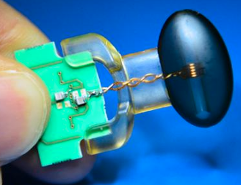
Highly sensitive magnetic field sensor (credit: ETH Zurich/Peter Rüegg)

Swiss researchers have succeeded in measuring changes in strong magnetic fields with unprecedented precision, they report in the open-access journal Nature Communications. The finding may find widespread use in medicine and other areas.

In their experiments, the researchers at the Institute for Biomedical Engineering, which is operated jointly by ETH Zurich and the University of Zurich, magnetized a water droplet inside a magnetic resonance imaging (MRI) scanner, a device used for medical imaging. The researchers were able to detect even the tiniest variations of the magnetic field strength within the droplet. These changes were up to 10-12 (1 trillion) times smaller than the 7 tesla field strength of the MRI scanner used in the experiment.

“Until now, it was possible only to measure such small variations in weak magnetic fields,” says Klaas Prüssmann, Professor of Bioimaging at ETH Zurich and the University of Zurich. An example of a weak magnetic field is that of the Earth, where the field strength is just a few dozen microtesla. For fields of this kind, highly sensitive measurement methods are already able to detect variations of about a trillionth of the field strength, says Prüssmann. “Now, we have a similarly sensitive method for strong fields of more than one tesla, such as those used … in medical imaging.”

The scientists based the sensing technique on the principle of nuclear magnetic resonance (NMR), which also serves as the basis for magnetic resonance imaging and the spectroscopic methods that biologists use to elucidate the 3D structure of molecules, but with 1000 times greater sensitivity than current NMR methods.

Ultra-sensitive recordings of heart contractions in an MRI machine
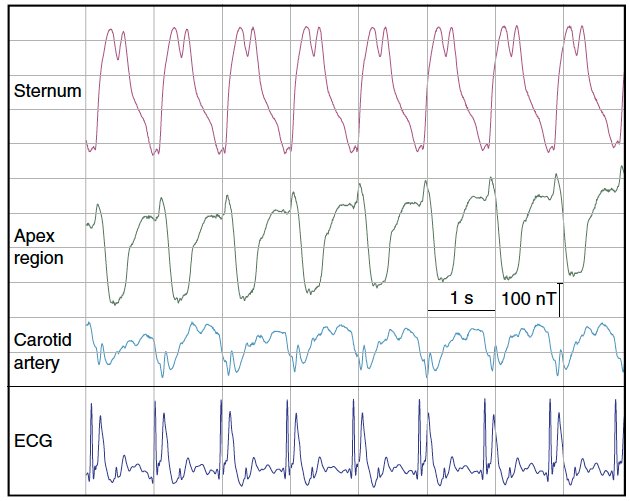
Real-time magnetic field recordings of cardiac activity. Magnetic field dynamics generated by the beating human heart in a background of 7 tesla, recorded at three different positions on the chest and neck, along with simultaneous electrocardiogram (ECG). (credit: Simon Gross et al./Nature Communications)

The scientists carried out an experiment in which they positioned their sensor in front of the chest of a volunteer test subject inside an MRI scanner. They were able to detect periodic changes in the magnetic field, which pulsated in time with the heartbeat. The measurement curve is similar to an electrocardiogram (ECG), but measures a mechanical process (the contraction of the heart) rather than electrical conduction.

“We are in the process of analyzing and refining our magnetometer measurement technique in collaboration with cardiologists and signal processing experts,” says Prüssmann. “Ultimately, we hope that our sensor will be able to provide information on heart disease — and do so non-invasively and in real time.”

The new measurement technique could also be used in the development of new contrast agents for magnetic resonance imaging and improved nuclear magnetic resonance (NMR) spectroscopy for applications in biological and chemical research.

A radiation-free approach to imaging molecules in the brain
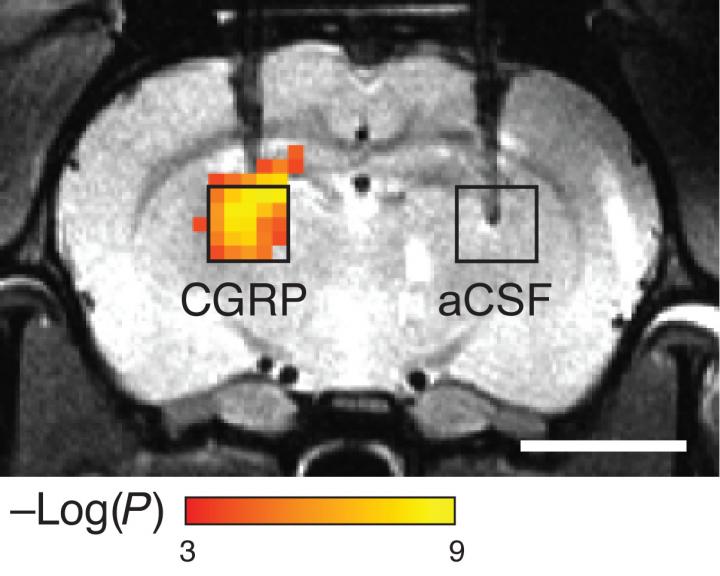
Scientists hoping to see molecules that control brain activity have devised a probe that lets them image such molecules without using chemical or radioactive labels. The sensors consist of proteins that detect a particular target, which causes them to dilate blood vessels, producing a change in blood flow that can be imaged with magnetic resonance imaging (MRI) or other techniques. (credit: Mitul Desai et al./ Nature Communications)

In a related development, MIT scientists hoping to get a glimpse of molecules that control brain activity have devised a new sensor that allows them to image these molecules without using any chemical or radioactive labels (which feature low resolution and can’t be easily used to watch dynamic events).

The new sensors consist of enzymes called proteases designed to detect a particular target, which causes them to dilate blood vessels in the immediate area. This produces a change in blood flow that can be imaged with magnetic resonance imaging (MRI) or other imaging techniques.*

A peptide called calcitonin gene-related peptide (CGRP) acts on a receptor in smooth muscle cells (left) to induce cAMP production, resulting in relaxation of vascular smooth muscle cells and consequent vasodilation (middle). That induces haemodynamic effects visible by MRI and other imaging methods (right). (credit: Mitul Desai et al./ Nature Communications)

“This is an idea that enables us to detect molecules that are in the brain at biologically low levels, and to do that with these imaging agents or contrast agents that can ultimately be used in humans,” says Alan Jasanoff, an MIT professor of biological engineering and brain and cognitive sciences. “We can also turn them on and off, and that’s really key to trying to detect dynamic processes in the brain.”

Monitoring neurotransmitters at 100 times lower levels

In a paper appearing in the Dec. 2 issue of open-access Nature Communications, Jasanoff and his colleagues explain that they used proteases (sometimes used as biomarkers to diagnose diseases such as cancer and Alzheimer’s disease) to demonstrate the validity of their approach. But now they’re working on adapting these imaging agents to monitor neurotransmitters, such as dopamine and serotonin, which are critical to cognition and processing emotions.

“What we want to be able to do is detect levels of neurotransmitter that are 100-fold lower than what we’ve seen so far. We also want to be able to use far less of these molecular imaging agents in organisms. That’s one of the key hurdles to trying to bring this approach into people,” Jasanoff says.

“Many behaviors involve turning on genes, and you could use this kind of approach to measure where and when the genes are turned on in different parts of the brain,” Jasanoff says.

His lab is also working on ways to deliver the peptides without injecting them, which would require finding a way to get them to pass through the blood-brain barrier. This barrier separates the brain from circulating blood and prevents large molecules from entering the brain.

Jeff Bulte, a professor of radiology and radiological science at the Johns Hopkins School of Medicine, described the technique as “original and innovative,” while adding that its safety and long-term physiological effects will require more study.

“It’s interesting that they have designed a reporter without using any kind of metal probe or contrast agent,” says Bulte, who was not involved in the research. “An MRI reporter that works really well is the holy grail in the field of molecular and cellular imaging.”

The research was funded by the National Institutes of Health BRAIN Initiative, the MIT Simons Center for the Social Brain, and fellowships from the Boehringer Ingelheim Fonds and the Friends of the McGovern Institute.

* To make their probes, the researchers modified a naturally occurring peptide called calcitonin gene-related peptide (CGRP), which is active primarily during migraines or inflammation. The researchers engineered the peptides so that they are trapped within a protein cage that keeps them from interacting with blood vessels. When the peptides encounter proteases in the brain, the proteases cut the cages open and the CGRP causes nearby blood vessels to dilate. Imaging this dilation with MRI allows the researchers to determine where the proteases were detected.

Another possible application for this type of imaging is to engineer cells so that the gene for CGRP is turned on at the same time that a gene of interest is turned on. That way, scientists could use the CGRP-induced changes in blood flow to track which cells are expressing the target gene, which could help them determine the roles of those cells and genes in different behaviors. Jasanoff’s team demonstrated the feasibility of this approach by showing that implanted cells expressing CGRP could be recognized by imaging.


Abstract of Dynamic nuclear magnetic resonance field sensing with part-per-trillion resolution

High-field magnets of up to tens of teslas in strength advance applications in physics, chemistry and the life sciences. However, progress in generating such high fields has not been matched by corresponding advances in magnetic field measurement. Based mostly on nuclear magnetic resonance, dynamic high-field magnetometry is currently limited to resolutions in the nanotesla range. Here we report a concerted approach involving tailored materials, magnetostatics and detection electronics to enhance the resolution of nuclear magnetic resonance sensing by three orders of magnitude. The relative sensitivity thus achieved amounts to 1 part per trillion (10−12). To exemplify this capability we demonstrate the direct detection and relaxometry of nuclear polarization and real-time recording of dynamic susceptibility effects related to human heart function. Enhanced high-field magnetometry will generally permit a fresh look at magnetic phenomena that scale with field strength. It also promises to facilitate the development and operation of high-field magnets.


Abstract of Molecular imaging with engineered physiology

In vivo imaging techniques are powerful tools for evaluating biological systems. Relating image signals to precise molecular phenomena can be challenging, however, due to limitations of the existing optical, magnetic and radioactive imaging probe mechanisms. Here we demonstrate a concept for molecular imaging which bypasses the need for conventional imaging agents by perturbing the endogenous multimodal contrast provided by the vasculature. Variants of the calcitonin gene-related peptide artificially activate vasodilation pathways in rat brain and induce contrast changes that are readily measured by optical and magnetic resonance imaging. CGRP-based agents induce effects at nanomolar concentrations in deep tissue and can be engineered into switchable analyte-dependent forms and genetically encoded reporters suitable for molecular imaging or cell tracking. Such artificially engineered physiological changes, therefore, provide a highly versatile means for sensitive analysis of molecular events in living organisms.

Immune cells in covering of brain discovered; may play critical role in battling neurological diseases

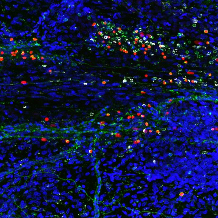
A composite image showing newly discovered immune cells in the brain (credit: Sachin Gadani | University of Virginia School of Medicine)

University of Virginia School of Medicine researchers have discovered a rare and powerful type of immune cell in the meninges (protective covering) of the brain that are activated in response to central nervous system injury — suggesting that these cells may play a critical role in battling Alzheimer’s, multiple sclerosis, meningitis, and other neurological diseases, and in supporting healthy mental functioning.

By harnessing the power of the cells, known as “type 2 innate lymphocytes” (ILC2s), doctors may be able to develop new treatments for neurological diseases, traumatic brain injury, and spinal cord injuries, as well as migraines, the researchers suggest. They also suspect the cells may be the missing link connecting the brain and the microbiota in our guts, a relationship that has been shown to be important in the development of Parkinson’s disease.

Important immune roles

ILC2 cells have previously been found in the gut, lung, and skin, the body’s barriers to disease. Their discovery by UVA researcher Jonathan Kipnis, PhD, in the meninges, the membranes surrounding the brain, comes as a surprise. They were found along the same vessels discovered by the Kipnis lab last year, which showed that the brain and the immune system are directly connected.

“This all comes down to immune system and brain interaction,” said Kipnis, chairman of UVA’s Department of Neuroscience. These where previously believed to be not communicating, but not only are these [immune] cells present in the areas near the brain, they are integral to its function, Kipnis said.

Immune cells play several important roles within the body, including guarding against pathogens, triggering allergic reactions, and responding to spinal cord injuries. But its their role in the gut that makes Kipnis suspect they may also be serving as a vital communicator between the brain’s immune response and our microbiomes (microbes in the body). That could be very important, because our intestinal flora is critical for maintaining our health and well being.

“These cells are potentially the mediator between the gut and the brain. They are the main responder to microbiota changes in the gut,” Kipnis said. “They may go from the gut to the brain, or they may just produce something that will impact those cells. We know the brain responds to things happening in the gut. Is it logical that these will be the cells that connect the two? Potentially.”

The findings have been published online by the Journal of Experimental Medicine. The work was supported by a National Institutes of Health grant.


Abstract of Characterization of meningeal type 2 innate lymphocytes and their response to CNS injury

The meningeal space is occupied by a diverse repertoire of immune cells. Central nervous system (CNS) injury elicits a rapid immune response that affects neuronal survival and recovery, but the role of meningeal inflammation remains poorly understood. Here, we describe type 2 innate lymphocytes (ILC2s) as a novel cell type resident in the healthy meninges that are activated after CNS injury. ILC2s are present throughout the naive mouse meninges, though are concentrated around the dural sinuses, and have a unique transcriptional profile. After spinal cord injury (SCI), meningeal ILC2s are activated in an IL-33–dependent manner, producing type 2 cytokines. Using RNAseq, we characterized the gene programs that underlie the ILC2 activation state. Finally, addition of wild-type lung-derived ILC2s into the meningeal space of IL-33R−/− animals partially improves recovery after SCI. These data characterize ILC2s as a novel meningeal cell type that responds to SCI and could lead to new therapeutic insights for neuroinflammatory conditions.

Method discovered to remove damaging amyloid plaques found in Alzheimer’s disease

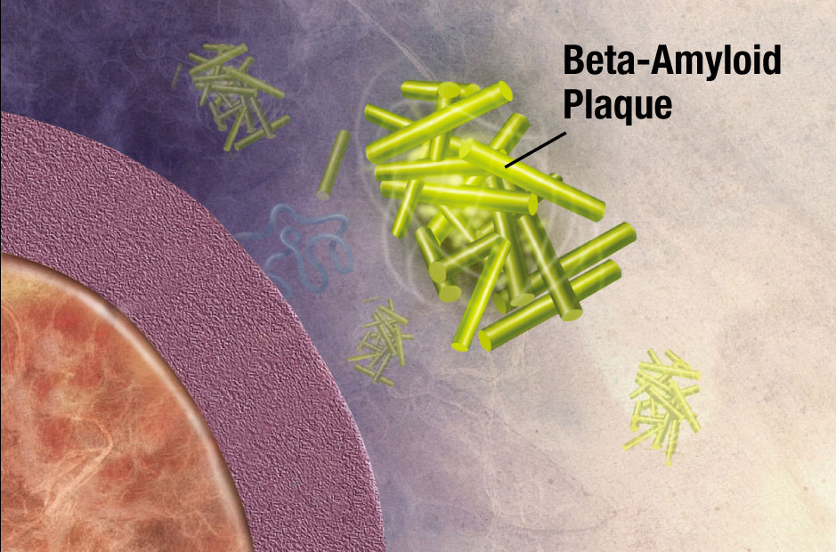
Illustration of formation of beta-amyloid plaque. Enzymes act on the APP (amyloid precursor protein) and cut it into fragments. The beta-amyloid fragment is crucial in the formation of senile plaques in Alzheimer’s disease. (credit: National Institute on Aging/NIH)

German scientists have discovered a strategy for removing amyloid plaques — newly forming clumps in a brain with Alzheimer’s disease that are created by misfolded proteins that clump together and damage nerve cells.

The scientists from the German Center for Neurodegenerative Diseases (DZNE) in Munich and the Ludwig Maximilians University (LMU) Munich took aged microglia cells (the  scavenger cells of the brain’s immune system) from a mouse model of Alzheimer’s disease and co-cultured them with microglia tissue from younger mouse brains. Surprisingly, within a few days, they found that the older amyloid plaques started to rejuvenate, resume cell division, and clear the brain from plaques by engulfing them. The amyloid plaque clearance potential of aged Alzheimer’s disease microglia could be fully restored, despite the continuous loss of neurons and astrocytes.

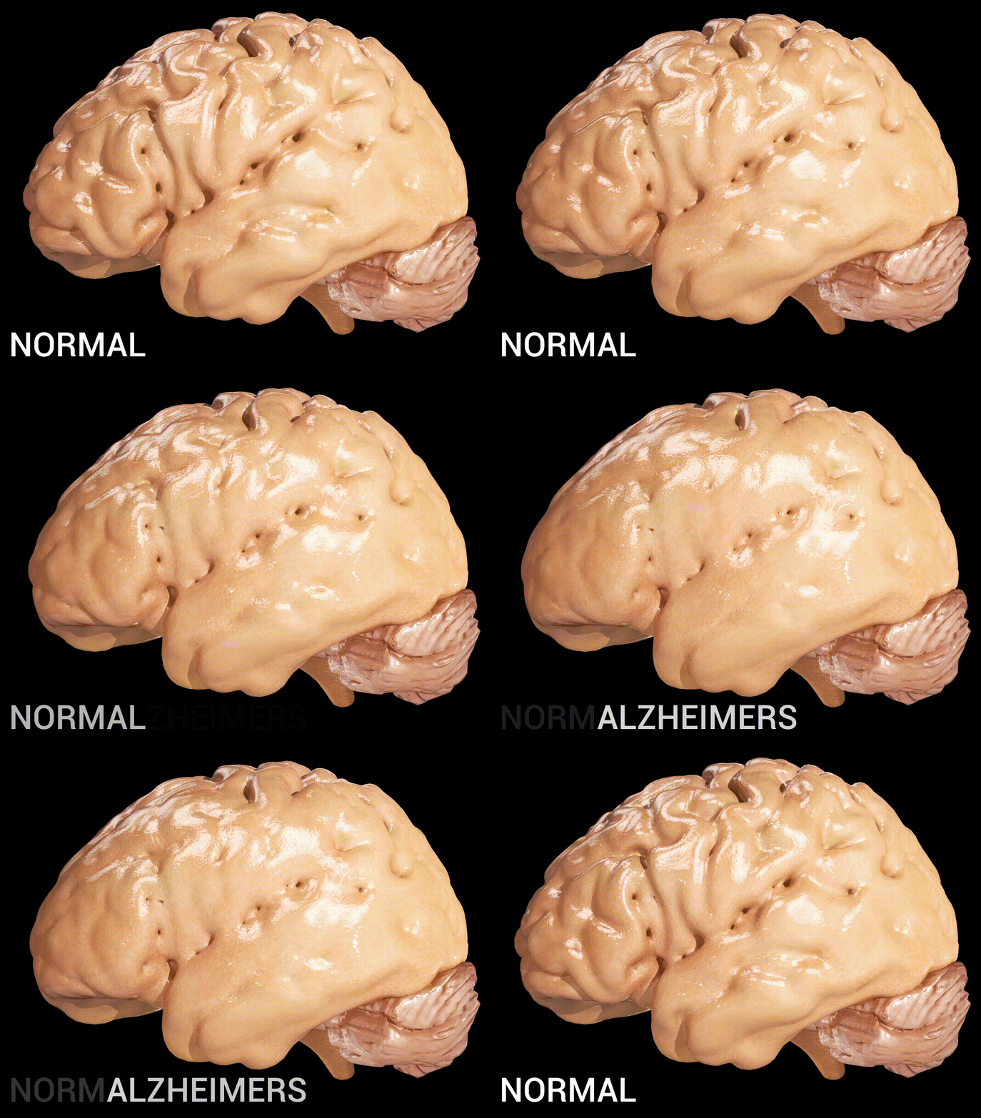
Cortical atrophy in Alzheimer’s Disease, associated with loss of gyri and sulci in the temporal lobe and parietal lobe, and parts of the frontal cortex and cingulate gyrus (credit: Doctor Jana/CC via Wikipedia)

They also found that the young microglia cells were secreting factors that helped the old microglia rejuvenate — especially a protein called “granulocyte-macrophage colony stimulating factor” (GM-CSF)* — which alone could do the job. Depletion of either old or young microglial cells prevented amyloid plaque clearance, indicating a synergistic effect of both populations.

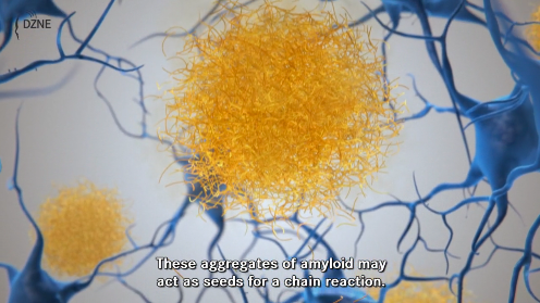
Click the image to view a DZNE video that describes the role of dysfunctional proteins in creating amyloid plaque in early Alzheimer’s disease, starting at 2:25. (credit: DZNE)

GM-CSF has previously been reported to reduce plaques and improve cognition in a mouse model of Alzheimer’s disease, but it’s not yet known if GM-CSF could potentially work as a new drug for Alzheimer’s disease in humans. Also, microglia secrete small proteins that induce inflammatory reactions and may harm neurons, the researchers say.

The researchers suggest that the new model system can be explored further to search for additional factors that enhance the clearance of amyloid plaques.

The work was published this week in The EMBO Journal.


National Institute on Aging | Inside the Brain: Unraveling the Mystery of Alzheimer’s Disease

* GM-CSF is manufactured using recombinant DNA technology. It is marketed as a protein therapeutic alled molgramostim or, when the protein is expressed in yeast cells, sargramostim. It is used as a medication to stimulate the production of white blood cells and thus prevent neutropenia  (loss of neutrophils, a type of white blood cell) following chemotherapy. (Patients with neutropenia are more susceptible to bacterial infections and, without prompt medical attention, the condition may become life-threatening.)


Abstract of Young microglia restore amyloid plaque clearance of aged microglia

Alzheimer′s disease (AD) is characterized by deposition of amyloid plaques, neurofibrillary tangles, and neuroinflammation. In order to study microglial contribution to amyloid plaque phagocytosis, we developed a novel ex vivo model by co‐culturing organotypic brain slices from up to 20‐month‐old, amyloid‐bearing AD mouse model (APPPS1) and young, neonatal wild‐type (WT) mice. Surprisingly, co‐culturing resulted in proliferation, recruitment, and clustering of old microglial cells around amyloid plaques and clearance of the plaque halo. Depletion of either old or young microglial cells prevented amyloid plaque clearance, indicating a synergistic effect of both populations. Exposing old microglial cells to conditioned media of young microglia or addition of granulocyte‐macrophage colony‐stimulating factor (GM‐CSF) was sufficient to induce microglial proliferation and reduce amyloid plaque size. Our data suggest that microglial dysfunction in AD may be reversible and their phagocytic ability can be modulated to limit amyloid accumulation. This novel ex vivo model provides a valuable system for identification, screening, and testing of compounds aimed to therapeutically reinforce microglial phagocytosis.