Metalens with artificial muscle simulates (and goes way beyond) human-eye and camera optical functions

A silicon-based metalens just 30 micrometers thick is mounted on a transparent, stretchy polymer film. The colored iridescence is produced by the large number of nanostructures within the metalens. (credit:Harvard SEAS)

Researchers at the Harvard John A. Paulson School of Engineering and Applied Sciences (SEAS) have developed a breakthrough electronically controlled artificial eye. The thin, flat, adaptive silicon nanostructure (“metalens”) can simultaneously control focus, astigmatism, and image shift (three of the major contributors to blurry images) in real time, which the human eye (and eyeglasses) cannot do.

The 30-micrometers-thick metalens makes changes laterally to achieve optical zoom, autofocus, and image stabilization — making it possible to replace bulky lens systems in future optical systems used in eyeglasses, cameras, cell phones, and augmented and virtual reality devices.

The research is described in an open-access paper in Science Advances. In another paper recently published in Optics Express, the researchers demonstrated the design and fabrication of metalenses up to centimeters or more in diameter.* That makes it possible to unify two industries: semiconductor manufacturing and lens-making. So the same technology used to make computer chips will be used to make metasurface-based optical components, such as lenses.

The adaptive metalens (right) focuses light rays onto an image sensor (left), such as one in a camera. An electrical signal controls the shape of the metalens to produce the desired optical wavefront patterns (shown in red), resulting in improved images. In the future, adaptive metalenses will be built into imaging systems, such as cell phone cameras and microscopes, enabling flat, compact autofocus as well as the capability for simultaneously correcting optical aberrations and performing optical image stabilization, all in a single plane of control. (credit: Second Bay Studios/Harvard SEAS)

Simulating the human eye’s lens and ciliary muscles

In the human eye, the lens is surrounded by ciliary muscle, which stretches or compresses the lens, changing its shape to adjust its focal length. To achieve that function, the researchers adhered a metalens to a thin, transparent dielectric elastomer actuator (“artificial muscle”). The researchers chose a dielectic elastomer with low loss — meaning light travels through the material with little scattering — to attach to the lens.

(Top) Schematic of metasurface and dielectric elastomer actuators (“artificial muscles”), showing how the new artificial muscles change focus, similar to how the ciliary muscle in the eye work. An applied voltage supplies transparent, stretchable electrode layers (gray), made up of single-wall carbon-nanotube nanopillars, with electrical charges (acting as a capacitor). The resulting electrostatic attraction compresses (red arrows) the dielectric elastomer actuators (artificial muscles) in the thickness direction and expands (black arrows) the elastomers in the lateral direction. The silicon metasurface (in the center), applied by photolithography, can simultaneously focus, control aberrations caused by astigmatisms, and perform image shift. (Bottom) Photo of actual device. (credit: Alan She et al./Sci. Adv.)

Next, the researchers aim to further improve the functionality of the lens and decrease the voltage required to control it.

The research was performed at the Harvard John A. Paulson School of Engineering and Applied Sciences, supported in part by the Air Force Office of Scientific Research and by the National Science Foundation. This work was performed in part at the Center for Nanoscale Systems (CNS), which is supported by the National Science Foundation. The Harvard Office of Technology Development is exploring commercialization opportunities.

* To build the artificial eye with a larger (more functional) metalens, the researchers had to develop a new algorithm to shrink the file size to make it compatible with the technology currently used to fabricate integrated circuits.

** “All optical systems with multiple components — from cameras to microscopes and telescopes — have slight misalignments or mechanical stresses on their components, depending on the way they were built and their current environment, that will always cause small amounts of astigmatism and other aberrations, which could be corrected by an adaptive optical element,” said Alan She, a graduate student at SEAS and first author of the paper. “Because the adaptive metalens is flat, you can correct those aberrations and integrate different optical capabilities onto a single plane of control. Our results demonstrate the feasibility of embedded autofocus, optical zoom, image stabilization, and adaptive optics, which are expected to become essential for future chip-scale image sensors and  Furthermore, the device’s flat construction and inherently lateral actuation without the need for motorized parts allow for highly stackable systems such as those found in stretchable electronic eye camera sensors, providing possibilities for new kinds of imaging systems.”


Abstract of Adaptive metalenses with simultaneous electrical control of focal length, astigmatism, and shift

Focal adjustment and zooming are universal features of cameras and advanced optical systems. Such tuning is usually performed longitudinally along the optical axis by mechanical or electrical control of focal length. However, the recent advent of ultrathin planar lenses based on metasurfaces (metalenses), which opens the door to future drastic miniaturization of mobile devices such as cell phones and wearable displays, mandates fundamentally different forms of tuning based on lateral motion rather than longitudinal motion. Theory shows that the strain field of a metalens substrate can be directly mapped into the outgoing optical wavefront to achieve large diffraction-limited focal length tuning and control of aberrations. We demonstrate electrically tunable large-area metalenses controlled by artificial muscles capable of simultaneously performing focal length tuning (>100%) as well as on-the-fly astigmatism and image shift corrections, which until now were only possible in electron optics. The device thickness is only 30 μm. Our results demonstrate the possibility of future optical microscopes that fully operate electronically, as well as compact optical systems that use the principles of adaptive optics to correct many orders of aberrations simultaneously.


Abstract of Large area metalenses: design, characterization, and mass manufacturing

Optical components, such as lenses, have traditionally been made in the bulk form by shaping glass or other transparent materials. Recent advances in metasurfaces provide a new basis for recasting optical components into thin, planar elements, having similar or better performance using arrays of subwavelength-spaced optical phase-shifters. The technology required to mass produce them dates back to the mid-1990s, when the feature sizes of semiconductor manufacturing became considerably denser than the wavelength of light, advancing in stride with Moore’s law. This provides the possibility of unifying two industries: semiconductor manufacturing and lens-making, whereby the same technology used to make computer chips is used to make optical components, such as lenses, based on metasurfaces. Using a scalable metasurface layout compression algorithm that exponentially reduces design file sizes (by 3 orders of magnitude for a centimeter diameter lens) and stepper photolithography, we show the design and fabrication of metasurface lenses (metalenses) with extremely large areas, up to centimeters in diameter and beyond. Using a single two-centimeter diameter near-infrared metalens less than a micron thick fabricated in this way, we experimentally implement the ideal thin lens equation, while demonstrating high-quality imaging and diffraction-limited focusing.

Metalens with artificial muscle simulates (and goes way beyond) human-eye and camera optical functions

A silicon-based metalens just 30 micrometers thick is mounted on a transparent, stretchy polymer film. The colored iridescence is produced by the large number of nanostructures within the metalens. (credit:Harvard SEAS)

Researchers at the Harvard John A. Paulson School of Engineering and Applied Sciences (SEAS) have developed a breakthrough electronically controlled artificial eye. The thin, flat, adaptive silicon nanostructure (“metalens”) can simultaneously control focus, astigmatism, and image shift (three of the major contributors to blurry images) in real time, which the human eye (and eyeglasses) cannot do.

The 30-micrometers-thick metalens makes changes laterally to achieve optical zoom, autofocus, and image stabilization — making it possible to replace bulky lens systems in future optical systems used in eyeglasses, cameras, cell phones, and augmented and virtual reality devices.

The research is described in an open-access paper in Science Advances. In another paper recently published in Optics Express, the researchers demonstrated the design and fabrication of metalenses up to centimeters or more in diameter.* That makes it possible to unify two industries: semiconductor manufacturing and lens-making. So the same technology used to make computer chips will be used to make metasurface-based optical components, such as lenses.

The adaptive metalens (right) focuses light rays onto an image sensor (left), such as one in a camera. An electrical signal controls the shape of the metalens to produce the desired optical wavefront patterns (shown in red), resulting in improved images. In the future, adaptive metalenses will be built into imaging systems, such as cell phone cameras and microscopes, enabling flat, compact autofocus as well as the capability for simultaneously correcting optical aberrations and performing optical image stabilization, all in a single plane of control. (credit: Second Bay Studios/Harvard SEAS)

Simulating the human eye’s lens and ciliary muscles

In the human eye, the lens is surrounded by ciliary muscle, which stretches or compresses the lens, changing its shape to adjust its focal length. To achieve that function, the researchers adhered a metalens to a thin, transparent dielectric elastomer actuator (“artificial muscle”). The researchers chose a dielectic elastomer with low loss — meaning light travels through the material with little scattering — to attach to the lens.

(Top) Schematic of metasurface and dielectric elastomer actuators (“artificial muscles”), showing how the new artificial muscles change focus, similar to how the ciliary muscle in the eye work. An applied voltage supplies transparent, stretchable electrode layers (gray), made up of single-wall carbon-nanotube nanopillars, with electrical charges (acting as a capacitor). The resulting electrostatic attraction compresses (red arrows) the dielectric elastomer actuators (artificial muscles) in the thickness direction and expands (black arrows) the elastomers in the lateral direction. The silicon metasurface (in the center), applied by photolithography, can simultaneously focus, control aberrations caused by astigmatisms, and perform image shift. (Bottom) actual device. (credit: She et al./Sci. Adv.)

Next, the researchers aim to further improve the functionality of the lens and decrease the voltage required to control it.

The research was performed at the Harvard John A. Paulson School of Engineering and Applied Sciences, supported in part by the Air Force Office of Scientific Research and by the National Science Foundation. This work was performed in part at the Center for Nanoscale Systems (CNS), which is supported by the National Science Foundation. The Harvard Office of Technology Development is exploring commercialization opportunities.

* To build the artificial eye with a larger (more functional) metalens, the researchers had to develop a new algorithm to shrink the file size to make it compatible with the technology currently used to fabricate integrated circuits.

** “All optical systems with multiple components — from cameras to microscopes and telescopes — have slight misalignments or mechanical stresses on their components, depending on the way they were built and their current environment, that will always cause small amounts of astigmatism and other aberrations, which could be corrected by an adaptive optical element,” said Alan She, a graduate student at SEAS and first author of the paper. “Because the adaptive metalens is flat, you can correct those aberrations and integrate different optical capabilities onto a single plane of control. Our results demonstrate the feasibility of embedded autofocus, optical zoom, image stabilization, and adaptive optics, which are expected to become essential for future chip-scale image sensors and  Furthermore, the device’s flat construction and inherently lateral actuation without the need for motorized parts allow for highly stackable systems such as those found in stretchable electronic eye camera sensors, providing possibilities for new kinds of imaging systems.”


Abstract of Adaptive metalenses with simultaneous electrical control of focal length, astigmatism, and shift

Focal adjustment and zooming are universal features of cameras and advanced optical systems. Such tuning is usually performed longitudinally along the optical axis by mechanical or electrical control of focal length. However, the recent advent of ultrathin planar lenses based on metasurfaces (metalenses), which opens the door to future drastic miniaturization of mobile devices such as cell phones and wearable displays, mandates fundamentally different forms of tuning based on lateral motion rather than longitudinal motion. Theory shows that the strain field of a metalens substrate can be directly mapped into the outgoing optical wavefront to achieve large diffraction-limited focal length tuning and control of aberrations. We demonstrate electrically tunable large-area metalenses controlled by artificial muscles capable of simultaneously performing focal length tuning (>100%) as well as on-the-fly astigmatism and image shift corrections, which until now were only possible in electron optics. The device thickness is only 30 μm. Our results demonstrate the possibility of future optical microscopes that fully operate electronically, as well as compact optical systems that use the principles of adaptive optics to correct many orders of aberrations simultaneously.


Abstract of Large area metalenses: design, characterization, and mass manufacturing

Optical components, such as lenses, have traditionally been made in the bulk form by shaping glass or other transparent materials. Recent advances in metasurfaces provide a new basis for recasting optical components into thin, planar elements, having similar or better performance using arrays of subwavelength-spaced optical phase-shifters. The technology required to mass produce them dates back to the mid-1990s, when the feature sizes of semiconductor manufacturing became considerably denser than the wavelength of light, advancing in stride with Moore’s law. This provides the possibility of unifying two industries: semiconductor manufacturing and lens-making, whereby the same technology used to make computer chips is used to make optical components, such as lenses, based on metasurfaces. Using a scalable metasurface layout compression algorithm that exponentially reduces design file sizes (by 3 orders of magnitude for a centimeter diameter lens) and stepper photolithography, we show the design and fabrication of metasurface lenses (metalenses) with extremely large areas, up to centimeters in diameter and beyond. Using a single two-centimeter diameter near-infrared metalens less than a micron thick fabricated in this way, we experimentally implement the ideal thin lens equation, while demonstrating high-quality imaging and diffraction-limited focusing.

Two new wearable sensors may replace traditional medical diagnostic devices

Throat-motion sensor monitors stroke effects more effectively

A radical new type of stretchable, wearable sensor that measures vocal-cord movements could be a “game changer” for stroke rehabilitation, according to Northwestern University scientists. The sensors can also measure swallowing ability (which may be affected by stroke), heart function, muscle activity, and sleep quality. Developed in the lab of engineering professor John A. Rogers, Ph.D., in partnership with Shirley Ryan AbilityLab in Chicago, the new sensors have been deployed to tens of patients.

“One of the biggest problems we face with stroke patients is that their gains tend to drop off when they leave the hospital,” said Arun Jayaraman, Ph.D., research scientist at the Shirley Ryan AbilityLab and a wearable-technology expert. “With the home monitoring enabled by these sensors, we can intervene at the right time, which could lead to better, faster recoveries for patients.”

(credit: Elliott Abel/ Shirley Ryan AbilityLab)

Monitoring movements, not sounds. The new band-aid-like stretchable throat sensor (two are applied) measures speech patterns by detecting throat movements to improve diagnosis and treatment of aphasia, a communication disorder associated with stroke.

Speech-language pathologists currently use microphones to monitor patients’ speech functions, which can’t distinguish between patients’ voices and ambient noise.

(credit: Elliott Abel/ Shirley Ryan AbilityLab)

Full-body kinematics. AbilityLab also uses similar electronic biosensors (developed in Rogers’ lab) on the legs, arms and chest to monitor stroke patients’ recovery progress. The sensors stream data wirelessly to clinicians’ phones and computers, providing a quantitative, full-body picture of patients’ advanced physical and physiological responses in real time.

Patients can wear them even after they leave the hospital, allowing doctors to understand how their patients are functioning in the real world.


(credit: Elliott Abel/ Shirley Ryan AbilityLab)

Mobile displays. Data from the sensors will be presented in a simple iPad-like display that is easy for both clinicians and patients to understand. It will send alerts when patients are under-performing on a certain metric and allow them to set and track progress toward their goals. A smartphone app can also help patients make corrections.

The researchers plan to test the sensors on patients with other conditions, such as Parkinson’s disease.


(credit: Elliott Abel/ Shirley Ryan AbilityLab)

Body-chemicals sensor. Another patch developed by the Rogers Lab does colorimetric analysis — determining the concentration of a chemical — for measuring sweat rate/loss and electrolyte loss. The Rogers Lab has a contract with Gatorade, and is testing this technology with the U.S. Air Force, the Seattle Mariners, and other unnamed sports teams.

Phone apps will also be available to capture precise colors and for data extraction, using algorithms.

A wearable electrocardiogram

Electrocardiogram on a prototype skin sensor (credit: 2018 Takao Someya Research Group)

Wearing your heart of your sleeve. Imagine looking at a electrocardiogram displayed on your wrist, using a simple skin sensor (replacing the usual complex array of EKG body electrodes), linked wirelessly to a smartphone or the cloud.

That’s the concept for a new wearable device developed by a team headed by Professor Takao Someya at the University of Tokyo’s Graduate School of Engineering and Dai Nippon Printing (DNP). It’s designed to provide continuous, non-invasive health monitoring.


The soft, flexible skin display is about 1 millimeter thick. (credit: 2018 Takao Someya Research Group.)

Stretchable nanomesh. The device uses a lightweight sensor made from a nanomesh electrode and a display made from a 16 x 24 array of micro LEDs and stretchable wiring, mounted on a rubber sheet. It’s stretchable by up 45 percent of its original length and can be worn on the skin continuously for a week without causing inflammation.

The sensor can also measure temperature, pressure, and the electrical properties of muscle, and can display messages on skin.

DNP hopes to bring the integrated skin display to market within three years.

Are you a cyborg?

Bioprinting a brain

Cryogenic 3D-printing soft hydrogels. Top: the bioprinting process. Bottom: SEM image of general microstructure (scale bar: 100 µm). (credit: Z. Tan/Scientific Reports)

A new bioprinting technique combines cryogenics (freezing) and 3D printing to create geometrical structures that are as soft (and complex) as the most delicate body tissues — mimicking the mechanical properties of organs such as the brain and lungs.

The idea: “Seed” porous scaffolds that can act as a template for tissue regeneration (from neuronal cells, for example), where damaged tissues are encouraged to regrow — allowing the body to heal without tissue rejection or other problems. Using “pluripotent” stem cells that can change into different types of cells is also a possibility.

Smoothy. Solid carbon dioxide (dry ice) in an isopropanol bath is used to rapidly cool hydrogel ink (a rapid liquid-to-solid phase change) as it’s extruded, yogurt-smoothy-style. Once thawed, the gel is as soft as body tissues, but doesn’t collapse under its own weight — a previous problem.

Current structures produced with this technique are “organoids” a few centimeters in size. But the researchers hope to create replicas of actual body parts with complex geometrical structures — even whole organs. That could allow scientists to carry out experiments not possible on live subjects, or for use in medical training, replacing animal bodies for surgical training and simulations. Then on to mechanobiology and tissue engineering.

Source: Imperial College London, Scientific Reports (open-access).

How to generate electricity with your body

Bending a finger generates electricity in this prototype device. (credit: Guofeng Song et al./Nano Energy)

A new triboelectric nanogenerator (TENG) design, using a gold tab attached to your skin, will convert mechanical energy into electrical energy for future wearables and self-powered electronics. Just bend your finger or take a step.

Triboelectric charging occurs when certain materials become electrically charged after coming into contact with a different material. In this new design by University of Buffalo and Chinese scientists, when a stretched layer of gold is released, it crumples, creating what looks like a miniature mountain range. An applied force leads to friction between the gold layers and an interior PDMS layer, causing electrons to flow between the gold layers.

More power to you. Previous TENG designs have been difficult to manufacture (requiring complex lithography) or too expensive. The new 1.5-centimeters-long prototype generates a maximum of 124 volts but at only 10 microamps. It has a power density of 0.22 millwatts per square centimeter. The team plans larger pieces of gold to deliver more electricity and a portable battery.

Source: Nano Energy. Support: U.S. National Science Foundation, the National Basic Research Program of China, National Natural Science Foundation of China, Beijing Science and Technology Projects, Key Research Projects of the Frontier Science of the Chinese Academy of Sciences ,and National Key Research and Development Plan.

This artificial electrical eel may power your implants

How the eel’s electrical organs generate electricity by moving sodium (Na) and potassium (K) ions across a selective membrane. (credit: Caitlin Monney)

Taking it a giant (and a bit scary) step further, an artificial electric organ, inspired by the electric eel, could one day power your implanted implantable sensors, prosthetic devices, medication dispensers, augmented-reality contact lenses, and countless other gadgets. Unlike typical toxic batteries that need to be recharged, these systems are soft, flexible, transparent, and potentially biocompatible.

Doubles as a defibrillator? The system mimicks eels’ electrical organs, which use thousands of alternating compartments with excess potassium or sodium ions, separated by selective membranes. To create a jolt of electricity (600 volts at 1 ampere), an eel’s membranes allow the ions to flow together. The researchers built a similar system, but using sodium and chloride ions dissolved in a water-based hydrogel. It generates more than 100 volts, but at safe low current — just enough to power a small medical device like a pacemaker.

The researchers say the technology could also lead to using naturally occurring processes inside the body to generate electricity, a truly radical step.

Source: Nature, University of Fribourg, University of Michigan, University of California-San Diego. Funding: Air Force Office of Scientific Research, National Institutes of Health.

E-skin for Terminator wannabes

A section of “e-skin” (credit: Jianliang Xiao / University of Colorado Boulder)

A new type of thin, self-healing, translucent “electronic skin” (“e-skin,” which mimicks the properties of natural skin) has applications ranging from robotics and prosthetic development to better biomedical devices and human-computer interfaces.

Ready for a Terminator-style robot baby nurse? What makes this e-skin different and interesting is its embedded sensors, which can measure pressure, temperature, humidity and air flow. That makes it sensitive enough to let a robot take care of a baby, the University of Colorado mechanical engineers and chemists assure us. The skin is also rapidly self-healing (by reheating), as in The Terminator, using a mix of three commercially available compounds in ethanol.

The secret ingredient: A novel network polymer known as polyimine, which is fully recyclable at room temperature. Laced with silver nanoparticles, it can provide better mechanical strength, chemical stability and electrical conductivity. It’s also malleable, so by applying moderate heat and pressure, it can be easily conformed to complex, curved surfaces like human arms and robotic hands.

Source: University of Colorado, Science Advances (open-access). Funded in part by the National Science Foundation.

Altered Carbon

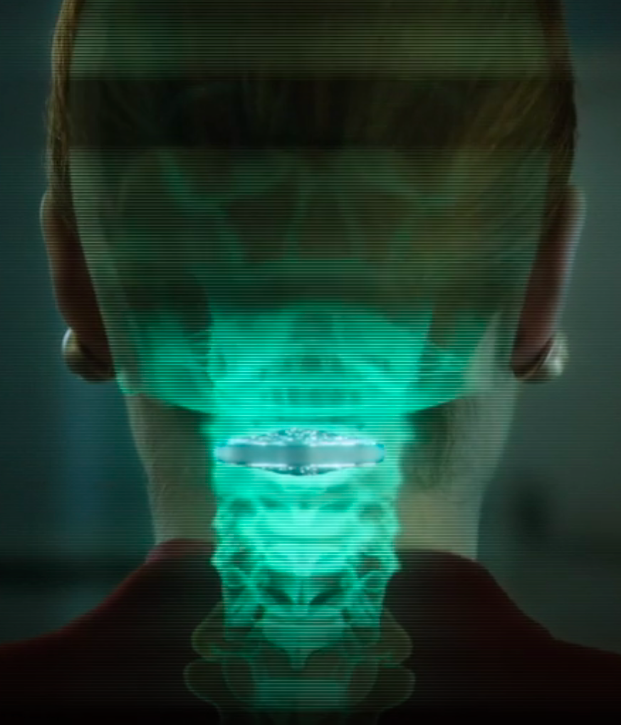
Vertebral cortical stack (credit: Netflix)

Altered Carbon takes place in the 25th century, when humankind has spread throughout the galaxy. After 250 years in cryonic suspension, a prisoner returns to life in a new body with one chance to win his freedom: by solving a mind-bending murder.

Resleeve your stack. Human consciousness can be digitized and downloaded into different bodies. A person’s memories have been encapsulated into “cortical stack” storage devices surgically inserted into the vertebrae at the back of the neck. Disposable physical bodies called “sleeves” can accept any stack.

But only the wealthy can acquire replacement bodies on a continual basis. The long-lived are called Meths, as in the Biblical figure Methuselah. The uber rich are also able to keep copies of their minds in remote storage, which they back up regularly, ensuring that even if their stack is destroyed, the stack can be resleeved (except for periods of time not backed up — as in the hack-murder).

Source: Netflix. Premiered on February 2, 2018. Based on the 2002 novel of the same title by Richard K. Morgan.


Penn researchers create first optical transistor comparable to an electronic transistor

By precisely controlling the mixing of optical signals, Ritesh Agarwal’s research team says they have taken an important step toward photonic (optical) computing. (credit: Sajal Dhara)

In an open-access paper published in Nature Communications, Ritesh Agarwal, a professor the University of Pennsylvania School of Engineering and Applied Science, and his colleagues say that they have made significant progress in photonic (optical) computing by creating a prototype of a working optical transistor with properties similar to those of a conventional electronic transistor.*

Optical transistors, using photons instead of electrons, promise to one day be more powerful than the electronic transistors currently used in computers.

Agarwal’s research on photonic computing has been focused on finding the right combination and physical configuration of nonlinear materials that can amplify and mix light waves in ways that are analogous to electronic transistors. “One of the hurdles in doing this with light is that materials that are able to mix optical signals also tend to have very strong background signals as well. That background signal would drastically reduce the contrast and on/off ratios leading to errors in the output,” Agarwal explained.

How the new optical transistor works

Schematic of a cadmium sulfide nanobelt device with source (S) and drain (D) electrodes. The fundamental wave at the frequency of ω, which is normally incident upon the belt, excites the second-harmonic (twice the frequency) wave at 2ω, which is back-scattered. (credit: Ming-Liang Ren et al./Nature Communications)

To address this issue, Agarwal’s research group started by creating a system with no disruptive optical background signal. To do that, they used a “nanobelt”* made out of cadmium sulfide. Then, by applying an electrical field across the nanobelt, the researchers were able to introduce optical nonlinearities (similar to the nonlinearities in electronic transistors), which enabled a signal mixing output that was otherwise zero.

“Our system turns on from zero to extremely large values,” Agarwal said.** “For the first time, we have an optical device with output that truly resembles an electronic transistor.”

The next steps toward a fully functioning photonic computer will involve integrating optical circuits with optical interconnects, modulators, and detectors to achieve actual on-chip integrated photonic computation.

The research was supported by the US Army Research Office and the National Science Foundation.

* “Made of semiconducting metal oxides, nanobelts are extremely thin and flat structures. They are chemically pure, structurally uniform, largely defect-free, with clean surfaces that do not require protection against oxidation. Each is made up of a single crystal with specific surface planes and shape.” — Reade International Corp.

** That is, the system was capable of precisely controlling the mixing of optical signals via controlled electric fields, with outputs with near-perfect contrast and extremely large on/off ratios. “Our study demonstrates a new way to dynamically control nonlinear optical signals in nanoscale materials with ultrahigh signal contrast and signal saturation, which can enable the development of nonlinear optical transistors and modulators for on-chip photonic devices with high-performance metrics and small-form factors, which can be further enhanced by integrating with nanoscale optical cavities,” the researchers note in the paper.


Abstract of Strong modulation of second-harmonic generation with very large contrast in semiconducting CdS via high-field domain

Dynamic control of nonlinear signals is critical for a wide variety of optoelectronic applications, such as signal processing for optical computing. However, controlling nonlinear optical signals with large modulation strengths and near-perfect contrast remains a challenging problem due to intrinsic second-order nonlinear coefficients via bulk or surface contributions. Here, via electrical control, we turn on and tune second-order nonlinear coefficients in semiconducting CdS nanobelts from zero to up to 151 pm V−1, a value higher than other intrinsic nonlinear coefficients in CdS. We also observe ultrahigh ON/OFF ratio of >104 and modulation strengths ~200% V−1 of the nonlinear signal. The unusual nonlinear behavior, including super-quadratic voltage and power dependence, is ascribed to the high-field domain, which can be further controlled by near-infrared optical excitation and electrical gating. The ability to electrically control nonlinear optical signals in nanostructures can enable optoelectronic devices such as optical transistors and modulators for on-chip integrated photonics.

Ultra-thin ‘atomistor’ synapse-like memory storage device paves way for faster, smaller, smarter computer chips

Illustration of single-atom-layer “atomristors” — the thinnest-ever memory-storage device (credit: Cockrell School of Engineering, The University of Texas at Austin)

A team of electrical engineers at The University of Texas at Austin and scientists at Peking University has developed a one-atom-thick 2D “atomristor” memory storage device that may lead to faster, smaller, smarter computer chips.

The atomristor (atomic memristor) improves upon memristor (memory resistor) memory storage technology by using atomically thin nanomaterials (atomic sheets). (Combining memory and logic functions, similar to the synapses of biological brains, memristors “remember” their previous state after being turned off.)

Schematic of atomristor memory sandwich based on molybdenum sulfide (MoS2) in a form of a single-layer atomic sheet grown on gold foil. (Blue: Mo; yellow: S) (credit: Ruijing Ge et al./Nano Letters)

Memory storage and transistors have, to date, been separate components on a microchip. Atomristors combine both functions on a single, more-efficient device. They use metallic atomic sheets (such as graphene or gold) as electrodes and semiconducting atomic sheets (such as molybdenum sulfide) as the active layer. The entire memory cell is a two-layer sandwich only ~1.5 nanometers thick.

“The sheer density of memory storage that can be made possible by layering these synthetic atomic sheets onto each other, coupled with integrated transistor design, means we can potentially make computers that learn and remember the same way our brains do,” said Deji Akinwande, associate professor in the Cockrell School of Engineering’s Department of Electrical and Computer Engineering.

“This discovery has real commercialization value, as it won’t disrupt existing technologies,” Akinwande said. “Rather, it has been designed to complement and integrate with the silicon chips already in use in modern tech devices.”

The research is described in an open-access paper in the January American Chemical Society journal Nano Letters.

Longer battery life in cell phones

For nonvolatile operation (preserving data after power is turned off), the new design also “offers a substantial advantage over conventional flash memory, which occupies far larger space. In addition, the thinness allows for faster and more efficient electric current flow,” the researchers note in the paper.

The research team also discovered another unique application for the atomristor technology: Atomristors are the smallest radio-frequency (RF) memory switches to be demonstrated, with no DC battery consumption, which could ultimately lead to longer battery life for cell phones and other battery-powered devices.*

Funding for the UT Austin team’s work was provided by the National Science Foundation and the Presidential Early Career Award for Scientists and Engineers, awarded to Akinwande in 2015.

* “Contemporary switches are realized with transistor or microelectromechanical devices, both of which are volatile, with the latter also requiring large switching voltages [which are not ideal] for mobile technologies,” the researchers note in the paper. Atomristors instead allow for nonvolatile low-power radio-frequency (RF) switches with “low voltage operation, small form-factor, fast switching speed, and low-temperature integration compatible with silicon or flexible substrates.”


Abstract of Atomristor: Nonvolatile Resistance Switching in Atomic Sheets of Transition Metal Dichalcogenides

Recently, two-dimensional (2D) atomic sheets have inspired new ideas in nanoscience including topologically protected charge transport,1,2 spatially separated excitons,3 and strongly anisotropic heat transport.4 Here, we report the intriguing observation of stable nonvolatile resistance switching (NVRS) in single-layer atomic sheets sandwiched between metal electrodes. NVRS is observed in the prototypical semiconducting (MX2, M = Mo, W; and X = S, Se) transitional metal dichalcogenides (TMDs),5 which alludes to the universality of this phenomenon in TMD monolayers and offers forming-free switching. This observation of NVRS phenomenon, widely attributed to ionic diffusion, filament, and interfacial redox in bulk oxides and electrolytes,6−9 inspires new studies on defects, ion transport, and energetics at the sharp interfaces between atomically thin sheets and conducting electrodes. Our findings overturn the contemporary thinking that nonvolatile switching is not scalable to subnanometre owing to leakage currents.10 Emerging device concepts in nonvolatile flexible memory fabrics, and brain-inspired (neuromorphic) computing could benefit substantially from the wide 2D materials design space. A new major application, zero-static power radio frequency (RF) switching, is demonstrated with a monolayer switch operating to 50 GHz.

An artificial synapse for future miniaturized portable ‘brain-on-a-chip’ devices

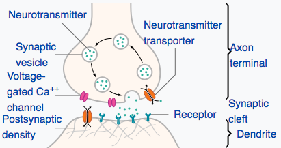
Biological synapse structure (credit: Thomas Splettstoesser/CC)

MIT engineers have designed a new artificial synapse made from silicon germanium that can precisely control the strength of an electric current flowing across it.

In simulations, the researchers found that the chip and its synapses could be used to recognize samples of handwriting with 95 percent accuracy. The engineers say the new design, published today (Jan. 22) in the journal Nature Materials, is a major step toward building portable, low-power neuromorphic chips for use in pattern recognition and other machine-learning tasks.

Controlling the flow of ions: the challenge

Researchers in the emerging field of “neuromorphic computing” have attempted to design computer chips that work like the human brain. The idea is to apply a voltage across layers that would cause ions (electrically charged atoms) to move in a switching medium (synapse-like space) to create conductive filaments in a manner that’s similar to how the “weight” (connection strength) of a synapse changes.

There are more than 100 trillion synapses (in a typical human brain) that mediate neuron signaling in the brain, strengthening some neural connections while pruning (weakening) others — a process that enables the brain to recognize patterns, remember facts, and carry out other learning tasks, all at lightning speeds.

Instead of carrying out computations based on binary, on/off signaling, like current digital chips, the elements of a “brain on a chip” would work in an analog fashion, exchanging a gradient of signals, or “weights” — much like neurons that activate in various ways (depending on the type and number of ions that flow across a synapse).

But it’s been difficult to control the flow of ions in existing synapse designs. These have multiple paths that make it difficult to predict where ions will make it through, according to research team leader Jeehwan Kim, PhD, an assistant professor in the departments of Mechanical Engineering and Materials Science and Engineering, a principal investigator in MIT’s Research Laboratory of Electronics and Microsystems Technology Laboratories.

“Once you apply some voltage to represent some data with your artificial neuron, you have to erase and be able to write it again in the exact same way,” Kim says. “But in an amorphous solid, when you write again, the ions go in different directions because there are lots of defects. This stream is changing, and it’s hard to control. That’s the biggest problem — nonuniformity of the artificial synapse.”

Epitaxial random access memory (epiRAM)

(Left) Cross-sectional transmission electron microscope image of 60 nm silicon-germanium (SiGe) crystal grown on a silicon substrate (diagonal white lines represent candidate dislocations). Scale bar: 25 nm. (Right) Cross-sectional scanning electron microscope image of an epiRAM device with titanium (Ti)–gold (Au) and silver (Ag)–palladium (Pd) layers. Scale bar: 100 nm. (credit: Shinhyun Choi et al./Nature Materials)

So instead of using amorphous materials as an artificial synapse, Kim and his colleagues created an new “epitaxial random access memory” (epiRAM) design.

They started with a wafer of silicon. They then grew a similar pattern of silicon germanium — a material used commonly in transistors — on top of the silicon wafer. Silicon germanium’s lattice is slightly larger than that of silicon, and Kim found that together, the two perfectly mismatched materials could form a funnel-like dislocation, creating a single path through which ions can predictably flow.*

This is the most uniform device we could achieve, which is the key to demonstrating artificial neural networks,” Kim says.

Testing the ability to recognize samples of handwriting

As a test, Kim and his team explored how the epiRAM device would perform if it were to carry out an actual learning task: recognizing samples of handwriting — which researchers consider to be a practical test for neuromorphic chips. Such chips would consist of artificial “neurons” connected to other “neurons” via filament-based artificial “synapses.”

Image-recognition simulation. (Left) A 3-layer multilayer-perception neural network with black and white input signal for each layer in algorithm level. The inner product (summation) of input neuron signal vector and first synapse array vector is transferred after activation and binarization as input vectors of second synapse arrays. (Right) Circuit block diagram of hardware implementation showing a synapse layer composed of epiRAM crossbar arrays and the peripheral circuit. (credit: Shinhyun Choi et al./Nature Materials)

They ran a computer simulation of an artificial neural network consisting of three sheets of neural layers connected via two layers of artificial synapses, based on measurements from their actual neuromorphic chip. They fed into their simulation tens of thousands of samples from the MNIST handwritten recognition dataset**, commonly used by neuromorphic designers.

They found that their neural network device recognized handwritten samples 95.1 percent of the time — close to the 97 percent accuracy of existing software algorithms running on large computers.

A chip to replace a supercomputer

The team is now in the process of fabricating a real working neuromorphic chip that can carry out handwriting-recognition tasks. Looking beyond handwriting, Kim says the team’s artificial synapse design will enable much smaller, portable neural network devices that can perform complex computations that are currently only possible with large supercomputers.

“Ultimately, we want a chip as big as a fingernail to replace one big supercomputer,” Kim says. “This opens a stepping stone to produce real artificial intelligence hardware.”

This research was supported in part by the National Science Foundation. Co-authors included researchers at Arizona State University.

* They applied voltage to each synapse and found that all synapses exhibited about the same current, or flow of ions, with about a 4 percent variation between synapses — a much more uniform performance compared with synapses made from amorphous material. They also tested a single synapse over multiple trials, applying the same voltage over 700 cycles, and found the synapse exhibited the same current, with just 1 percent variation from cycle to cycle.

** The MNIST (Modified National Institute of Standards and Technology database) is a large database of handwritten digits that is commonly used for training various image processing systems and for training and testing in the field of machine learning. It contains 60,000 training images and 10,000 testing images. 


Abstract of SiGe epitaxial memory for neuromorphic computing with reproducible high performance based on engineered dislocations

Although several types of architecture combining memory cells and transistors have been used to demonstrate artificial synaptic arrays, they usually present limited scalability and high power consumption. Transistor-free analog switching devices may overcome these limitations, yet the typical switching process they rely on—formation of filaments in an amorphous medium—is not easily controlled and hence hampers the spatial and temporal reproducibility of the performance. Here, we demonstrate analog resistive switching devices that possess desired characteristics for neuromorphic computing networks with minimal performance variations using a single-crystalline SiGe layer epitaxially grown on Si as a switching medium. Such epitaxial random access memories utilize threading dislocations in SiGe to confine metal filaments in a defined, one-dimensional channel. This confinement results in drastically enhanced switching uniformity and long retention/high endurance with a high analog on/off ratio. Simulations using the MNIST handwritten recognition data set prove that epitaxial random access memories can operate with an online learning accuracy of 95.1%.

Remote-controlled DNA nanorobots could lead to the first nanorobotic production factory

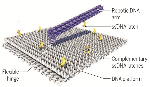
German researchers created a 55-nm-by-55-nm DNA-based molecular platform with a 25-nm-long robotic arm that can be actuated with externally applied electrical fields, under computer control. (credit: Enzo Kopperger et al./Science)

By powering a self-assembling DNA nanorobotic arm with electric fields, German scientists have achieved precise nanoscale movement that is at least five orders of magnitude (hundreds of thousands times) faster than previously reported DNA-driven robotic systems, they suggest today (Jan. 19) in the journal Science.

DNA origami has emerged as a powerful tool to build precise structures. But now, “Kopperger et al. make an impressive stride in this direction by creating a dynamic DNA origami structure that they can directly control from the macroscale with easily tunable electric fields—similar to a remote-controlled robot,” notes Björn Högberg of Karolinska Institutet in a related Perspective in Science, (p. 279).

The nanorobotic arm resembles the gearshift lever of a car. Controlled by an electric field (comparable to the car driver), short, single-stranded DNA serves as “latches” (yellow) to momentarily grab and lock the 25-nanometer-long arm into predefined “gear” positions. (credit: Enzo Kopperger et al./Science)

The new biohybrid nanorobotic systems could even act as a molecular mechanical memory (a sort of nanoscale version of the Babbage Analytical Engine), he notes. “With the capability to form long filaments with multiple DNA robot arms, the systems could also serve as a platform for new inventions in digital memory, nanoscale cargo transfer, and 3D printing of molecules.”

“The robot-arm system may be scaled up and integrated into larger hybrid systems by a combination of lithographic and self-assembly techniques,” according to the researchers. “Electrically clocked synthesis of molecules with a large number of robot arms in parallel could then be the first step toward the realization of a genuine nanorobotic production factory.”


Taking a different approach to a nanofactory, this “Productive Nanosystems: from Molecules to Superproducts” film — a collaborative project of animator and engineer John Burch and pioneer nanotechnologist K. Eric Drexler in 2005 — demonstrated key steps in a hypothetical process that converts simple molecules into a billion-CPU laptop computer. More here.


Abstract of A self-assembled nanoscale robotic arm controlled by electric fields

The use of dynamic, self-assembled DNA nanostructures in the context of nanorobotics requires fast and reliable actuation mechanisms. We therefore created a 55-nanometer–by–55-nanometer DNA-based molecular platform with an integrated robotic arm of length 25 nanometers, which can be extended to more than 400 nanometers and actuated with externally applied electrical fields. Precise, computer-controlled switching of the arm between arbitrary positions on the platform can be achieved within milliseconds, as demonstrated with single-pair Förster resonance energy transfer experiments and fluorescence microscopy. The arm can be used for electrically driven transport of molecules or nanoparticles over tens of nanometers, which is useful for the control of photonic and plasmonic processes. Application of piconewton forces by the robot arm is demonstrated in force-induced DNA duplex melting experiments.

A breakthrough low-light image sensor for photography, life sciences, security

A sample photo (right) taken with the one-megapixel low-light Quanta Image Sensor operating at 1,040 frames per second. It is a binary single-photon image, so if the pixel was hit by one or more photons, it is white; if not, it is black. The photo was created by summing up eight frames of binary images taken continuously. A de-noising algorithm was applied to the final image. (credit: Jiaju Ma, adapted by KurzweilAI)

Engineers from Dartmouth’s Thayer School of Engineering have created a radical new imaging technology called “Quanta Image Sensor” (QIS) that may revolutionize a wide variety of imaging applications that require high quality at low light.

These include security, photography, cinematography, and medical and life sciences research.

Low-light photography (at night with only moonlight, for example) currently requires photographers to use time exposure (keeping the shutter open for seconds or minutes), making it impossible to photograph moving images.

Capturing single photons at room temperature

The new QIS technology can capture or count at the lowest possible level of light (single photons) with a resolution as high as one megapixel* (one million pixels) — scalable for higher resolution up to hundreds of megapixels per chip** — and as fast as thousands of frames*** per second (required for “bullet time” cinematography in “The Matrix”).

The QIS works at room temperature, using existing mainstream CMOS image sensor technology. Current lab-research technology may require cooling to very low temperatures, such as 4 kelvin, and is limited to low pixel count.

Quanta Image Sensor applications (credit: Gigajot)

For astrophysicists, the QIS will allow for detecting and capturing signals from distant objects in space at higher quality. For life-science researchers, it will provide improved visualization of cells under a microscope, which is critical for determining the effectiveness of therapies.

The QIS technology is commercially accessible, inexpensive, and compatible with mass-production manufacturing, according to inventor Eric R. Fossum, professor of engineering at Dartmouth. Fossum is senior author of an open-access paper on QIS in the Dec. 20 issue of The Optical Society’s (OSA) Optica. He invented the CMOS image sensor found in nearly all smartphones and cameras in the world today.

The research was performed in cooperation with Rambus, Inc. and the Taiwan Semiconductor Manufacturing Corporation and was funded by Rambus and the Defense Advanced Research Projects Agency (DARPA). The low-light capability promises to allow for improved security uses. Fossum and associates have co-founded the startup company Gigajot Technology to further develop and apply the technology to promising applications.

* By comparison, the iPhone 8 can capture 12 megapixels (but is not usable in low light).

** The technology is based on what the researchers call “jots,” which function like miniature pixels. Each jot can collect one photon, enabling the extreme low-light capability and high resolution.

*** By comparison, the iPhone 8 can record 24 to 60 frames per second.


Abstract of Photon-number-resolving megapixel image sensor at room temperature without avalanche gain

In several emerging fields of study such as encryption in optical communications, determination of the number of photons in an optical pulse is of great importance. Typically, such photon-number-resolving sensors require operation at very low temperature (e.g., 4 K for superconducting-based detectors) and are limited to low pixel count (e.g., hundreds). In this paper, a CMOS-based photon-counting image sensor is presented with photon-number-resolving capability that operates at room temperature with resolution of 1 megapixel. Termed a quanta image sensor, the device is implemented in a commercial stacked (3D) backside-illuminated CMOS image sensor process. Without the use of avalanche multiplication, the 1.1 μm pixel-pitch device achieves 0.21e−  rms0.21e−  rms average read noise with average dark count rate per pixel less than 0.2e−/s0.2e−/s, and 1040 fps readout rate. This novel platform technology fits the needs of high-speed, high-resolution, and accurate photon-counting imaging for scientific, space, security, and low-light imaging as well as a broader range of other applications.

A new low-cost, simple way to measure medical vital signs with radio waves

A radio-frequency identification (RFID) tag, used to monitor vital signs, can go into your pocket or be woven into a shirt (credit: Cornell)

Replacing devices based on 19th-century technology* and still in use, Cornell University engineers have developed a simple method for gathering blood pressure, heart rate, and breath rate from multiple patients simultaneously. It uses low-power radio-frequency signals and low-cost microchip radio-frequency identification (RFID) “tags” — similar to the ubiquitous anti-theft tags used in department stores.

The RFID tags measure internal body motion, such as a heart as it beats or blood as it pulses under skin. Powered remotely by electromagnetic energy supplied by a central reader, the tags use a new concept called “near-field coherent sensing.” Mechanical motions (heartbeat, etc.) in the body modulate (modify) radio waves that are bounced off the body and internal organs by passive (no battery required) RFID tags.

The modulated signals detected by the tag then bounce back to an electronic reader, located elsewhere in the room, that gathers the data. Each tag has a unique identification code that it transmits with its signal, allowing up to 200 people to be monitored simultaneously.

Electromagnetic simulations of monitoring vital signs via radio transmission, showing heartbeat sensing (left) and pulse sensing (right) (credit: Xiaonan Hui and Edwin C. Kan/Nature Electronics)

“If this is an emergency room, everybody that comes in can wear these tags or can simply put tags in their front pockets, and everybody’s vital signs can be monitored at the same time. I’ll know exactly which person each of the vital signs belongs to,” said Edwin Kan, a Cornell professor of electrical and computer engineering.

The signal is as accurate as an electrocardiogram or a blood-pressure cuff, according to Kan, who believes the technology could also be used to measure bowel movement, eye movement, and many other internal mechanical motions produced by the body.

The researchers envision embedding the RFID chips in clothing to monitor health in real time, with little or no effort required by the user. They have also developed a method for embroidering the tags directly onto clothing using fibers coated with nanoparticles. A cellphone could read (and display) your vital signs and also transmit them for remote medical monitoring.

The system is detailed in the open-access paper “Monitoring Vital Signs Over Multiplexed Radio by Near-Field Coherent Sensing,” published online Nov. 27 in the journal Nature Electronics. “Current approaches to monitoring vital signs are based on body electrodes, optical absorption, pressure or strain gauges, stethoscope, and ultrasound or radiofrequency (RF) backscattering, each of which suffers particular drawbacks during application,” the paper notes.

* The sphygmomanometer was invented by Samuel Siegfried Karl Ritter von Basch in 1881. Devices based on its basic pressure principle are still in use. (credit: Wellcome Trustees)


Abstract of Monitoring vital signs over multiplexed radio by near-field coherent sensing

Monitoring the heart rate, blood pressure, respiration rate and breath effort of a patient is critical to managing their care, but current approaches are limited in terms of sensing capabilities and sampling rates. The measurement process can also be uncomfortable due to the need for direct skin contact, which can disrupt the circadian rhythm and restrict the motion of the patient. Here we show that the external and internal mechanical motion of a person can be directly modulated onto multiplexed radiofrequency signals integrated with unique digital identification using near-field coherent sensing. The approach, which does not require direct skin contact, offers two possible implementations: passive and active radiofrequency identification tags. To minimize deployment and maintenance cost, passive tags can be integrated into garments at the chest and wrist areas, where the two multiplexed far-field backscattering waveforms are collected at the reader to retrieve the heart rate, blood pressure, respiration rate and breath effort. To maximize reading range and immunity to multipath interference caused by indoor occupant motion, active tags could be placed in the front pocket and in the wrist cuff to measure the antenna reflection due to near-field coherent sensing and then the vital signals sampled and transmitted entirely in digital format. Our system is capable of monitoring multiple people simultaneously and could lead to the cost-effective automation of vital sign monitoring in care facilities.