Manipulating silicon atoms to create future ultra-fast, ultra-low-power chip technology

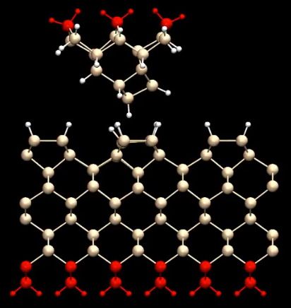
Model showing interactions between atomic-force microscope tip (top) and silicon surface (hydrogen: white; silicon: tan and red), using a new technique for coating the tip with hydrogen — part of a study to create future electronic circuits at the atomic level. (credit: Wolkow Lab)

Imagine a hybrid silicon-molecular computer that uses one thousand times less energy or a cell phone battery that lasts weeks at a time.

University of Alberta scientists, headed by University of Alberta physics professor Robert Wolkow, have taken a major step in that direction by visualizing and geometrically patterning silicon at the atomic level — using an innovative  atomic-force microscopy* (AFM) technique. The goal: chip technology that performs dramatically better than today’s CMOS architecture.

(Left) Ball-and-stick theoretical model of the pentacene molecule. (Right) AFM image of pentacene molecule showing the pattern of the bonds in the model. The five hexagonal carbon rings are resolved clearly and even the carbon-hydrogen bonds (white in the model) are imaged. Scale bar: 5 angstroms (0.5 nanometer) (credit: IBM Zurich)

Visualizing bonds in atoms at atomic resolution was first achieved by IBM Zurich scientists in 2009, when they imaged the pentacene molecule on copper. But imaging silicon is a problem: the sharp tip damages the fragile silicon molecules, the researchers note in an open-access paper published in the February 13, 2017 issue of Nature Communications.

To avoid damaging the silicon surface, the researchers created the first hydrogen-covered AFM tip, making it possible to manipulate silicon atoms. It was “a bit like Goldilocks,” PhD student and co-author Taleana Huff explained to KurzweilAI. “There is a sweet-spot region where you are probing the surface without interacting with it. Getting close enough to the surface with just the right parameters allows you to see these bonds materialize.
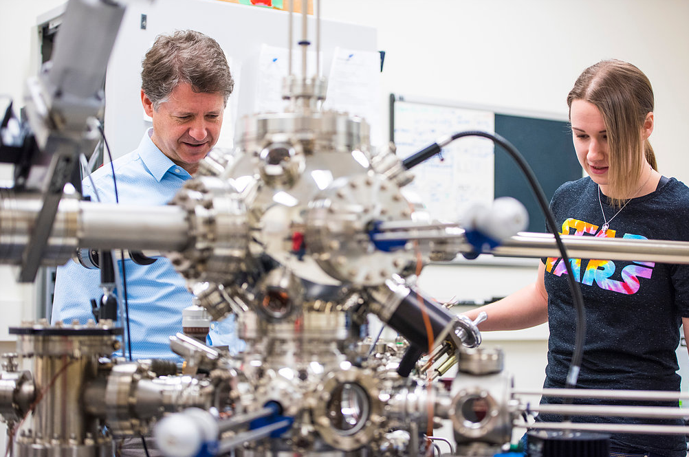
Bob Wolkow and Taleana Huff patterning and imaging electronic circuits at the atomic level (credit: Wolkow Lab)

“If you get too close though, you end up transferring atoms to the surface or, conversely, to the tip, ruining the experiment. A lot of tech and knowledge goes into getting all these settings just right, including a powerful new computational approach that analyzes and verifies the identity of the atoms and bonds.”

Hydrogen-terminated silicon for ultra-fast, ultra-low-power technology

“We see hydrogen-terminated silicon as the platform for a whole new paradigm of efficient and fast silicon-based electronics,” Huff said. “Now that we understand the surface intimately and have these powerful tools and the experience, the next step is to start using the AFM to look at computational elements made using quantum dots [nanoscale semiconductor particles], which we create by removing hydrogen atoms from the silicon surface. When we cleverly pattern them geometrically, these atomic silicon quantum dots can be used to make very fast and incredibly low-power computational patterns.”

The long-term goal is making ultra-fast and ultra-low-power silicon-based circuits that potentially consume one thousand times less power than what is currently on the market, according to the researchers, along with novel quantum applications.

* Typical atomic force microscope (AFM) setup
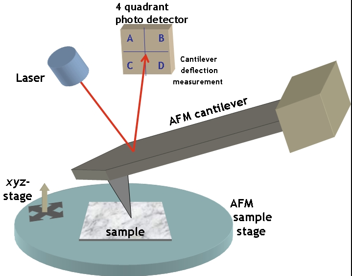
To image a surface, an AFM sharp tip scans across the sample to detect irregularities in the surface, which cause deflection of the tip and the connected cantilever and generating a topological map of the sample surface. The deflection is measured by reflecting a laser beam off the backside of the cantilever. (credit: CC/Opensource Handbook of Nanoscience and Nanotechnology)


Wolkow Lab | An animation illustrating patterning and imagining electronic circuits at the atomic level. It shows the tip and surface atoms’ relaxation during calculations of a part of the image simulation at small tip-surface distance. The bending and rotation of bonds is visible, giving a sense of the interactions and atomic relaxations involved.


UAlbertaScience | Less is more for atomic-scale manufacturing

This animation represents an electrical current being switched on and off. Remarkably, the current is confined to a channel that is just one atom wide. Also, the switch is made of just one atom. When the atom in the center feels an electric field tugging at it, it loses its electron. Once that electron is lost, the many electrons in the body of the silicon (to the left) have a clear passage to flow through. When the electric field is removed, an electron gets trapped in the central atom, switching the current off. This represents the latest work out of Robert Wolkow’s lab at the University of Alberta.


Abstract of Indications of chemical bond contrast in AFM images of a hydrogen-terminated silicon surface

The origin of bond-resolved atomic force microscope images remains controversial. Moreover, most work to date has involved planar, conjugated hydrocarbon molecules on a metal substrate thereby limiting knowledge of the generality of findings made about the imaging mechanism. Here we report the study of a very different sample; a hydrogen-terminated silicon surface. A procedure to obtain a passivated hydrogen-functionalized tip is defined and evolution of atomic force microscopy images at different tip elevations are shown. At relatively large tip-sample distances, the topmost atoms appear as distinct protrusions. However, on decreasing the tip-sample distance, features consistent with the silicon covalent bonds of the surface emerge. Using a density functional tight-binding-based method to simulate atomic force microscopy images, we reproduce the experimental results. The role of the tip flexibility and the nature of bonds and false bond-like features are discussed.

How to build your own bio-bot

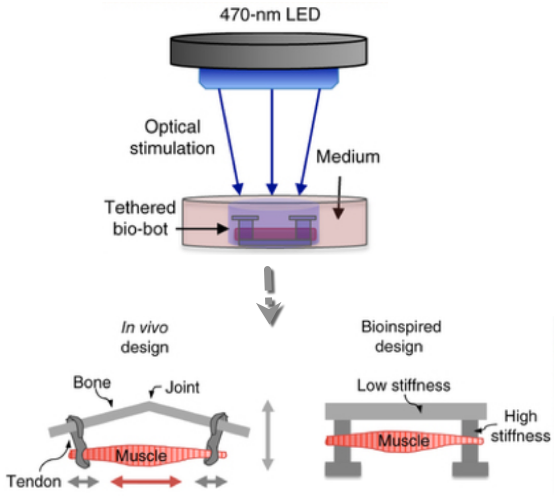
Bio-bot design inspired by the muscle-tendon-bone complex found in the human body, with 3D-printed flexible skeleton. Optical stimulation of the muscle tissue (orange), which is genetically engineered to contract in response to blue light, makes the bio-bot walk across a surface in the direction of the light. (credit: Ritu Raman et al./Nature Protocols)

For the past several years, researchers at the University of Illinois at Urbana-Champaign have reverse-engineered native biological tissues and organs — creating tiny walking “bio-bots” powered by muscle cells and controlled with electrical and optical pulses.

Now, in an open-access cover paper in Nature Protocols, the researchers are sharing a protocol with engineering details for their current generation of millimeter-scale soft robotic bio-bots*.

Using 3D-printed skeletons, these devices would be coupled to tissue-engineered skeletal muscle actuators to drive locomotion across 2D surfaces, and could one day be used for studies of muscle development and disease, high-throughput drug testing, and dynamic implants, among other applications.

In a new design, the researchers worked with MIT optogenetics experts to genetically engineer a light-responsive skeletal muscle cell line that could be stimulated to contract by pulses of blue light. (credit: Ritu Raman et al./Nature Protocols)

The future of bio-bots

The researchers envision future generations of bio-bots as biological building blocks that lead to the machines of the future. The bio-bots would integrate multiple cell and tissue types, including neuronal networks for sensing and processing, and vascular networks for delivery of nutrients and other biochemical factors. They might also have some of the higher-order properties of biological materials, such as self-organization and self-healing.

“These next iterations of biohybrid machines could, for example, be designed to sense chemical toxins, locomote toward them, and neutralize them through cell-secreted factors. Such a functionality could have broad relevance in medical diagnostics and targeted therapeutics in vivo, or even be extended to environmental use as a method of cleaning pathogens from public water supplies,” the research note in the paper.

“This protocol is essentially intended to be a one-stop reference for any scientist around the world who wants to replicate the results we showed in our PNAS 2016 and PNAS 2014 papers, and give them a framework for building their own bio-bots for a variety of applications,” said Bioengineering Professor Rashid Bashir**, who heads the bio-bots research group.

Bashir’s group has been a pioneer in designing and building bio-bots, less than a centimeter in size, made of flexible 3D printed hydrogels and living cells. In 2012, the group demonstrated bio-bots that could “walk” on their own, powered by beating heart cells from rats. In 2014, they switched to muscle cells controlled with electrical pulses, giving researchers unprecedented command over their function.

* Not to be confused with swimming biobots and rescue biobots using remotely controlled cockroaches.

** Bashir is also Grainger Distinguished Chair in Engineering and head of the Department of Bioengineering. Work on the bio-bots was conducted at the Micro + Nanotechnology Lab at Illinois.


NewsAtIllinois | Light illuminates the way for bio-bots


Abstract of A modular approach to the design, fabrication, and characterization of muscle-powered biological machines

Biological machines consisting of cells and biomaterials have the potential to dynamically sense, process, respond, and adapt to environmental signals in real time. As a first step toward the realization of such machines, which will require biological actuators that can generate force and perform mechanical work, we have developed a method of manufacturing modular skeletal muscle actuators that can generate up to 1.7 mN (3.2 kPa) of passive tension force and 300 μN (0.56 kPa) of active tension force in response to external stimulation. Such millimeter-scale biological actuators can be coupled to a wide variety of 3D-printed skeletons to power complex output behaviors such as controllable locomotion. This article provides a comprehensive protocol for forward engineering of biological actuators and 3D-printed skeletons for any design application. 3D printing of the injection molds and skeletons requires 3 h, seeding the muscle actuators takes 2 h, and differentiating the muscle takes 7 d.

Terahertz wireless could lead to fiber-optics speed in-flight and mobile metropolitan internet

Terahertz wireless links to spaceborne satellites could one day make gigabit-per-second connection speeds available to anyone, anytime, anywhere on the face of the earth, on the ground or in flight (credit: Fujishima et al./Hiroshima University)

Hiroshima University researchers and associates have developed a terahertz* (THz) transmitter capable of transmitting digital data over a single channel at a speed of 105 gigabits per second (Gbps), and demonstrated the technology at the International Solid-State Circuits Conference (ISSCC) 2017 conference last week.

For perspective, that’s more than 100 times faster than the fastest (1 Gbps) internet connection in the U.S. or more than 3,000 times faster than the 31 Mbps available to the average U.S. household in 2014, according to an FCC report. It’s also ten times or more faster than the fastest rate expected to be offered by fifth-generation mobile networks (5G) for metropolitan areas around 2020.

Major uses: faster in-flight and metropolitan internet, high-frequency trading

Applications of this forthcoming THz technology include higher-speed in-flight network connection speeds via satellite, fast download of videos and other large files for mobile devices, and ultrafast wireless links between base stations, according to Hiroshima University professor Minoru Fujishima.

An important business application is faster high-frequency trading, which requires minimal latency (delay). Recently, the time it takes to execute these trades has gone from milliseconds (thousandths of a second) to microseconds (millionths of a second), as KurzweilAI has explained. However, long-distance fiber optics connections (currently used for long-distance trading) have significant latency because light (as radio waves) travels 50%  faster in a vacuum than through glass fiber, while microwaves traveling in air have a less than a 1% speed reduction.

The National Institute of Information and Communications Technology and Panasonic Corporation are also partners in this research.

* Terahertz, a frequency range that is 1,000 times higher than gigahertz, or 1012 Hz, actually starts at 100 GHz or .1 THz. The researchers transmitted in this unregulated THz band — a vast new frequency resource expected to be used for future ultrahigh-speed wireless communications — using the frequency range from 290 GHz to 315 GHz. The full range of frequencies in the THz band (275 GHz to 450 GHz) are currently unallocated, but are expected to be discussed at the World Radiocommunication Conference 2019.


Abstract of A 105Gb/s 300GHz CMOS Transmitter

“High speed” in communications often means “high data-rate” and fiber-optic technologies have long been ahead of wireless technologies in that regard. However, an often overlooked definite advantage of wireless links over fiber-optic links is that waves travel at the speed of light c, which is about 50% faster than in optical fibers as shown in Fig. 17.9.1 (top left). This “minimum latency” is crucial for applications requiring real-time responses over a long distance, including high-frequency trading [1]. Further opportunities and new applications might be created if the absolute minimum latency and fiber-optic data-rates are put together. (Sub-)THz frequencies have an extremely broad atmospheric transmission window with manageable losses as shown in Fig. 17.9.1 (top right) and will be ideal for building light-speed links supporting fiber-optic data-rates. This paper presents a 105Gb/s 300GHz transmitter (TX) fabricated using a 40nm CMOS process.


Abstract of A 300GHz 40nm CMOS transmitter with 32-QAM 17.5Gb/s/ch capability over 6 channels

The vast unallocated frequency band lying above 275GHz offers enormous potential for ultrahigh-speed wireless communication. An overall bandwidth that could be allocated for multi-channel communication can easily be several times the 60GHz unlicensed bandwidth of 9GHz. We present a 300GHz transmitter (TX) in 40nm CMOS, capable of 32-quadrature amplitude modulation (QAM) 17.5Gb/s/ch signal transmission. It can cover the frequency range from 275 to 305GHz with 6 channels as shown at the top of Fig. 20.1.1. Figure 20.1.1 also lists possible THz TX architectures, based on recently reported above-200GHz TXs. The choice of architecture depends very much on the transistor unity-power-gain frequency fmax. If the fmax is sufficiently higher than the carrier frequency, the ordinary power amplifier (PA)-last architecture (Fig. 20.1.1, top row of the table) is possible and preferable [1-3], although the presence of a PA is, of course, not a requirement [4,5]. If, on the other hand, the fmax is comparable to or lower than the carrier frequency as in our case, a PA-less architecture must be adopted. A typical such architecture is the frequency multiplier-last architecture (Fig. 20.1.1, middle row of the table). For example, a 260GHz quadrupler-last on-off keying (OOK) TX [6] and a 434GHz tripler-last amplitude-shift keying (ASK) TX [7] were reported. A drawback of this architecture is the inefficient bandwidth utilization due to signal bandwidth spreading. Another drawback is that the use of multibit digital modulation is very difficult, if not impossible. An exception to this is the combination of quadrature phase-shift keying (QPSK) and frequency tripling. When a QPSK-modulated intermediate frequency (IF) signal undergoes frequency tripling, the resulting signal constellation remains that of QPSK with some symbol permutation. Such a tripler-last 240GHz QPSK TX was reported [8]. However, a 16-QAM constellation, for example, would suffer severe distortion by frequency tripling. If the 300GHz band is to be seriously considered for a platform for ultrahigh-speed wireless communication, QAM-capability will be a requisite.

These may be the last glasses you will ever need to buy

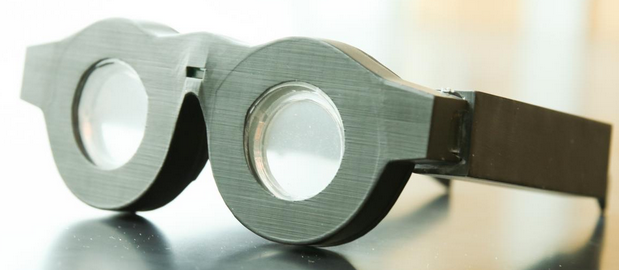
Early prototype of “smart glasses” with liquid-based lenses that can automatically adjust the focus on what a person is seeing, whether it’s far away or close up. The battery-powered frames can automatically adjust the focal length. Researchers expect to have smaller, lighter frames and packaged technology within three years. (credit: Dan Hixson/University of Utah College of Engineering)

Don’t throw away your bifocals or multiple glasses yet, but those days might soon be over. A team led by University of Utah engineers has created “smart glasses” with liquid-based lenses that can automatically adjust the focus on what you’re seeing, at any distance.

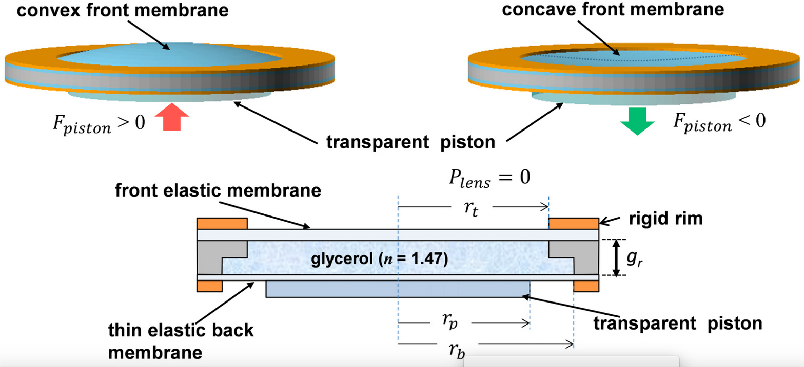
They’ve created eyeglass lenses made of glycerin, a thick colorless liquid, enclosed by flexible rubber-like membranes in the front and back. The rear membrane in each lens is connected to a series of three mechanical actuators that push the membrane back and forth like a transparent piston, changing the curvature of the liquid lens and therefore the focal length between the lens and the eye.

Simplified schematic of soft-membrane liquid lens (excluding actuators). The lens optical power is adjusted by vertically displacing the fluid with a transparent piston, deflecting the top membrane and changing its curvature. (credit: Nazmul Hasan et al./Optics Express)

In the bridge of the glasses is a distance meter that measures the distance from the glasses to an object via pulses of near-infrared light. When the wearer looks at an object, the meter instantly measures the distance and tells the actuators how to curve the lenses. If the user then sees another object that’s closer, the distance meter readjusts and tells the actuators to reshape the lens for farsightedness.

The lenses can change focus from one object to another in 14 milliseconds (faster than human reaction time). A rechargeable battery in the frames could last more than 24 hours per charge, according to electrical and computer engineering professor Carlos Mastrangelo, senior author of an open-access paper in a special edition of the journal Optics Express.

Before putting them on for the first time, users would input their eyeglasses prescription into an accompanying smartphone app, which then calibrates the lenses automatically via Bluetooth. Users only need to do that once, except for when their prescription changes over time. Theoretically, eyeglass wearers will never have to buy another pair again since these glasses would constantly adjust to their eyesight.

A startup company, Sharpeyes LLC, has been created to commercialize the glasses. The project was funded with a grant from the National Institutes of Health and the National Institute of Biomedical Imaging and Bioengineering.


University of Utah | Smart glasses that automatically focus on whatever you look at


Abstract of Tunable-focus lens for adaptive eyeglasses

We demonstrate the implementation of a compact tunable-focus liquid lens suitable for adaptive eyeglass application. The lens has an aperture diameter of 32 mm, optical power range of 5.6 diopter, and electrical power consumption less than 20 mW. The lens inclusive of its piezoelectric actuation mechanism is 8.4 mm thick and weighs 14.4 gm. The measured lens RMS wavefront aberration error was between 0.73 µm and 0.956 µm.

A 3D bioprinter that prints fully functional human skin

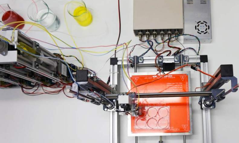
Skin-producing bioprinter (credit: Universidad Carlos III de Madrid)

A prototype 3D bioprinter that can create totally functional human skin has been developed by scientists from Universidad Carlos III de Madrid (UC3M) and BioDan Group in Spain. The skin has been used to treat burns as well as traumatic and surgical wounds in a large number of patients in Spain, according to the scientists.

The system provides two processes.

Autologous skin (from the patient’s own cells to generate human collagen) for therapeutic use, such as in the treatment of severe burns, instead of the animal collagen used in other methods.  The researchers have applied for approval by various European regulatory agencies to guarantee that the skin that is produced is adequate for use in transplants on burn patients and on those with other skin problems.

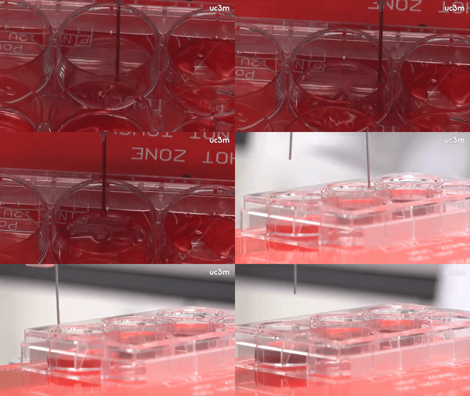
3D skin bioprinter in operation (credit: Universidad Carlos III de Madrid)

The 3D-printed skin replicates human bilayered skin, using “bioinks” (biological components) containing human plasma as well as primary human fibroblasts and keratinocytes obtained from skin biopsies. These are controlled by a computer, which deposits them on a print bed in an orderly manner to then produce the skin.

The researchers were able to generate 100 cm2 of printed skin in less than 35 minutes (including the 30 min required for fibrin gelation).

Allogeneic skin (from a stock of cells), done on a large scale for industrial processes. This skin can be used to test pharmaceutical products, cosmetics, and consumer chemical products where current regulations require testing that does not use animals.

“This method of bioprinting allows skin to be generated in a standardized, automated way, and the process is less expensive than manual production,” says Alfredo Brisac, CEO of BioDan Group, the Spanish bioengineering firm specializing in regenerative medicine that is collaborating on this research and commercializing this technology.

The research was published online in the journal Biofabrication.


UC3M | Científicos españoles crean una bioimpresora 3D de piel humana


Abstract of 3D bioprinting of functional human skin: production and in vivo analysis

Significant progress has been made over the past 25 years in the development of in vitro-engineered substitutes that mimic human skin, either to be used as grafts for the replacement of lost skin, or for the establishment of in vitro human skin models. In this sense, laboratory-grown skin substitutes containing dermal and epidermal components offer a promising approach to skin engineering. In particular, a human plasma-based bilayered skin generated by our group, has been applied successfully to treat burns as well as traumatic and surgical wounds in a large number of patients in Spain. There are some aspects requiring improvements in the production process of this skin; for example, the relatively long time (three weeks) needed to produce the surface required to cover an extensive burn or a large wound, and the necessity to automatize and standardize a process currently performed manually. 3D bioprinting has emerged as a flexible tool in regenerative medicine and it provides a platform to address these challenges. In the present study, we have used this technique to print a human bilayered skin using bioinks containing human plasma as well as primary human fibroblasts and keratinocytes that were obtained from skin biopsies. We were able to generate 100 cm2, a standard P100 tissue culture plate, of printed skin in less than 35 min (including the 30 min required for fibrin gelation). We have analysed the structure and function of the printed skin using histological and immunohistochemical methods, both in 3D in vitro cultures and after long-term transplantation to immunodeficient mice. In both cases, the generated skin was very similar to human skin and, furthermore, it was indistinguishable from bilayered dermo-epidermal equivalents, handmade in our laboratories. These results demonstrate that 3D bioprinting is a suitable technology to generate bioengineered skin for therapeutical and industrial applications in an automatized manner.

Microbiologists make big leap in developing ‘green’ electronics


An artist’s rendition of Geobacter expressing electrically conductive nanowires. Microbiologists at UMass Amherst have discovered a new type of natural wire produced by bacteria that could greatly accelerate the development of sustainable “green” conducting materials for the electronics industry. (credit: UMass Amherst)

UMass Amherst research finds microbe yields better electronic material

Microbiologists at the University of Massachusetts Amherst report that they have discovered a new type of microbial nanowire produced by bacteria that could greatly accelerate the development of sustainable “green” conducting materials for the electronics industry.

The study by Derek Lovley and colleagues appears this week in an open-access paper in mBio, the American Society of Microbiology’s premier journal.

A bacterium known as Geobacter sulfurreducens uses the protein filaments naturally to make electrical connections with other microbes or minerals.

As Lovley explains, “Microbial nanowires are a revolutionary electronic material with substantial advantages over man-made materials. Chemically synthesizing nanowires in the lab requires toxic chemicals, high temperatures and/or expensive metals. The energy requirements are enormous. By contrast, natural microbial nanowires can be mass-produced at room temperature from inexpensive renewable feedstocks in bioreactors with much lower energy inputs. And the final product is free of toxic components.”

Confocal scanning laser micrographs of G. sulfurreducens anode biofilms harvested on day 10. Bar, 25 µm. (credit: Yang Tan et al./mBio)

The Microbial nanowires offer an unprecedented potential for developing novel electronic devices and sensors for diverse applications with a new environmentally friendly technology, Lovely says. “This is an important advance in microbial nanowire technology. The approach we outline in this paper demonstrates a rapid method for prospecting in nature to find better electronic materials.”

When his lab began looking at the protein filaments of other Geobacter species, they were surprised to find a wide range in conductivities. For example, one species recovered from uranium-contaminated soil produced poorly conductive filaments. However, another species, Geobacter metallireducens produced nanowires 5,000 times more conductive than the G. sulfurreducens wires. Lovley recalls, “I isolated metallireducens from mud in the Potomac River 30 years ago, and every couple of years it gives us a new surprise.”

In their new study supported by the U.S. Office of Naval Research, they did not study the G. metallireducens strain directly. Instead, they took the gene for the protein that assembles into microbial nanowires from it and inserted this into G. sulfurreducens. The result is a genetically modified G. sulfurreducens that expresses the G. metallireducens protein, making nanowires much more conductive than G. sulfurreducens would naturally produce.

Further, Lovley says, “We have found that G. sulfurreducens will express filament genes from many different types of bacteria. This makes it simple to produce a diversity of filaments in the same microorganism and to study their properties under similar conditions.”

The high conductivity of the G. metallireducens nanowires suggests that they may be an attractive material for the construction of conductive materials, electronic devices ,and sensors for medical or environmental applications. The authors say discovering more about the mechanisms of nanowire conductivity “provides important insight into how we might make even better wires with genes that we design ourselves.”


Abstract of Expressing the Geobacter metallireducens PilA in Geobacter sulfurreducens Yields Pili with Exceptional Conductivity

The electrically conductive pili (e-pili) of Geobacter sulfurreducens serve as a model for a novel strategy for long-range extracellular electron transfer. e-pili are also a new class of bioelectronic materials. However, the only other Geobacter pili previously studied, which were from G. uraniireducens, were poorly conductive. In order to obtain more information on the range of pili conductivities in Geobacter species, the pili of G. metallireducens were investigated. Heterologously expressing the PilA gene of G. metallireducens in G. sulfurreducens yielded a G. sulfurreducens strain, designated strain MP, that produced abundant pili. Strain MP exhibited phenotypes consistent with the presence of e-pili, such as high rates of Fe(III) oxide reduction and high current densities on graphite anodes. Individual pili prepared at physiologically relevant pH 7 had conductivities of 277 ± 18.9 S/cm (mean ± standard deviation), which is 5,000-fold higher than the conductivity of G. sulfurreducens pili at pH 7 and nearly 1 million-fold higher than the conductivity of G. uraniireducens pili at the same pH. A potential explanation for the higher conductivity of the G. metallireducens pili is their greater density of aromatic amino acids, which are known to be important components in electron transport along the length of the pilus. The G. metallireducens pili represent the most highly conductive pili found to date and suggest strategies for designing synthetic pili with even higher conductivities.

Wearable sensors can alert you when you are getting sick, Stanford study shows

Current versions of three of the devices used for heart-rate and peripheral capillary oxygen saturation measurements in the study (credits left to right: Scanadu, iHealth, and Masimo)

Fitness monitors and other wearable biosensors can tell when your heart rate, activity, skin temperature, and other measures are abnormal, suggesting possible illness, including the onset of infection, inflammation, and even insulin resistance, according to a study by researchers at the Stanford University School of Medicine.

The team collected nearly 2 billion measurements from 60 people, including continuous data from each participant’s wearable biosensor devices* and periodic data from laboratory tests of their blood chemistry, gene expression, and other measures, and related this data to a range of normal (baseline) values for each person in the study compared to when they were ill.**

Participants wore between one and seven commercially available activity monitors and other monitors that collected more than 250,000 measurements a day. The team collected data on weight; heart rate; oxygen in the blood; skin temperature; activity, including sleep, steps, walking, biking and running; calories expended; acceleration; and even exposure to gamma rays and X-rays.

There were three participant groups: Participant #1 wore seven portable devices for large segments of this study; 43 individuals wore Intel’s Basis to measure activity (steps), heart rate, sleep, and skin temperature, with data securely uploaded to the cloud; and 16 individuals wore either iHealth or Masimo finger devices for sensing heart rate and SpO2 (peripheral capillary oxygen saturation).

Identifying health problems in advance

Wearable devices used by participant 1 (credit: Xiao Li/PLOS Biology)

The study, led by Michael Snyder, PhD, professor and chair of genetics, and senior author of the study, was published online Jan. 12 in open-access PLOS Biology. It demonstrated that, given a baseline range of values for each person, it is possible to monitor deviations from normal and associate those deviations with environmental conditions, illness, or other factors that affect health. Distinctive patterns of deviation from normal seem to correlate with particular health problems. Algorithms designed to pick up on these patterns of change could potentially contribute to clinical diagnostics and research.

The results of the current study raise the possibility of identifying inflammatory disease in individuals who may not even know they are getting sick.

For example, in several participants, higher-than-normal readings for heart rate and skin temperature correlated with increased levels of C reactive protein in blood tests. C reactive protein is an immune system marker for inflammation and often indicative of infection, autoimmune diseases, developing cardiovascular disease or even cancer. Snyder’s own data revealed four separate bouts of illness and inflammation, including a Lyme disease infection and another that he was unaware of until he saw his sensor data and an increased level of C reactive protein.

The wearable devices could also help distinguish participants with insulin resistance, a precursor for Type 2 diabetes. Of 20 participants who received glucose tests, 12 were insulin-resistant. The team designed and tested an algorithm combining participants’ daily steps, daytime heart rate and the difference between daytime and nighttime heart rate. The algorithm was able to process the data from just these few simple measures to predict which individuals in the study were likely to be insulin-resistant.

The study also revealed that declines in blood-oxygen levels during airplane flights were correlated with fatigue. Fortunately, the study showed that people tend to adapt on long flights; oxygen levels in their blood go back up, and they generally feel less fatigued as the hours go by.

The future of wearable devices: monitoring human health continuously

During a visit to the doctor, patients normally have their blood pressure and body temperature measured, but such data is typically collected only every year or two and often ignored unless the results are outside of normal range for entire populations. But biomedical researchers envisage a future in which human health is monitored continuously.

“We have more sensors on our cars than we have on human beings,” said Snyder. In the future, he said, he expects the situation will be reversed and people will have more sensors than cars do. Already, consumers have purchased millions of wearable devices, including more than 50 million smart watches and 20 million other fitness monitors. Most monitors are used to track activity, but they could easily be adjusted to more directly track health measures, Snyder said.

The work is an example of Stanford Medicine’s focus on “precision health,” whose goal is to anticipate and prevent disease in the healthy and to precisely diagnose and treat disease in the ill. With a precision health approach, every person could know his or her normal baseline for dozens of measures. Automatic data analysis could spot patterns of outlier data points and flag the onset of ill health, providing an opportunity for intervention, prevention or cure.

Researcher Elizabeth Colbert, of the Veterans Affairs Palo Alto Health Care System, is also a co-author. This research was funded by the National Institutes of Health, a gift from Bert and Candace Forbes, and Stanford’s Department of Genetics.

* “After evaluating more than 400 available wearable devices at the beginning of the study, we selected [seven] for participants to use. The criteria for selection [were] (1) ability to access the raw data from the manufacturer, (2) cost, (3) overlap in measurement of at least one component with another device to assist in reproducibility, and (4) ease of use, reasonable accuracy, and had a direct interface for raw data. These devices collectively measure (a) three physiological parameters, including heart rate, peripheral capillary oxygen saturation, and skin temperature, (b) six activity-related parameters, including sleep, steps, walking, biking, running, calories, and acceleration forces caused by movement, (c) weight, and (d) total gamma and X-ray radiation exposure.” — PLOS Biology paper authors

** “In this work, we investigate the use of portable devices to (1) easily and accurately record physiological measurements in individuals in real time (or at high frequency), (2) quantify daily patterns and reveal interesting physiological responses to different circadian cycles and environmental conditions, (3) identify personalized baseline norms and differences among individuals, (4) detect differences in health states among individuals (e.g., people with diabetes versus people without diabetes), and (5) detect inflammatory responses and assist in medical diagnosis at the early phase of disease development, thereby potentially impacting medical care.” — PLOS Biology paper authors


Abstract of Digital Health: Tracking Physiomes and Activity Using Wearable Biosensors Reveals Useful Health-Related Information

A new wave of portable biosensors allows frequent measurement of health-related physiology. We investigated the use of these devices to monitor human physiological changes during various activities and their role in managing health and diagnosing and analyzing disease. By recording over 250,000 daily measurements for up to 43 individuals, we found personalized circadian differences in physiological parameters, replicating previous physiological findings. Interestingly, we found striking changes in particular environments, such as airline flights (decreased peripheral capillary oxygen saturation [SpO2] and increased radiation exposure). These events are associated with physiological macro-phenotypes such as fatigue, providing a strong association between reduced pressure/oxygen and fatigue on high-altitude flights. Importantly, we combined biosensor information with frequent medical measurements and made two important observations: First, wearable devices were useful in identification of early signs of Lyme disease and inflammatory responses; we used this information to develop a personalized, activity-based normalization framework to identify abnormal physiological signals from longitudinal data for facile disease detection. Second, wearables distinguish physiological differences between insulin-sensitive and -resistant individuals. Overall, these results indicate that portable biosensors provide useful information for monitoring personal activities and physiology and are likely to play an important role in managing health and enabling affordable health care access to groups traditionally limited by socioeconomic class or remote geography.

Nanowire ‘inks’ enable low-cost paper- or plastic-based printable electronics

Duke University chemists have found that silver nanowire films like these conduct electricity well enough to form functioning circuits without applying high temperatures, enabling printable electronics on materials like paper or plastic. (credit: Ian Stewart and Benjamin Wiley)

By suspending tiny metal nanoparticles in liquids, Duke University scientists can use conductive ink-jet-printed conductive “inks” to print inexpensive, customizable RFID and other electronic circuit patterns on just about any surface — even on paper and plastics.

Printed electronics, which are already being used widely in devices such as the anti-theft radio frequency identification (RFID) tags you might find on the back of new DVDs, currently have one major drawback: for the circuits to work, they first have to be heated to 200° C (392°F) to melt all the nanoparticles together into a single conductive wire.

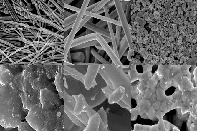
But Duke researchers have now found that electrons are conducted through films made of silver nanowires much more easily than with films made from other shapes (like nanospheres or microflakes). And the nanowire films can now function in printed circuits without the need to melt them first — heating at only 70° C (158°F) is required — and that means the circuits can be printed on cheaper plastics or paper.

“The nanowires [in their research] had a 4,000 times higher conductivity than the more commonly used silver nanoparticles that you would find in printed antennas for RFID tags,” said Benjamin Wiley, assistant professor of chemistry at Duke.

The technology could also be used to make lower-cost solar cells, printed displays, LEDs, touchscreens, amplifiers, batteries, and even some implantable bio-electronic devices. The results appeared online Dec. 16 in ACS Applied Materials and Interfaces.

The team is now experimenting with using aerosol jets to print silver nanowire inks in usable circuits. Wiley says they also want to explore whether silver-coated copper nanowires, which are significantly cheaper to produce than pure silver nanowires, will give the same effect.

This research was supported by funding from the National Science Foundation and a GAANN Fellowship through the Duke Chemistry Department.


Abstract of Effect of Morphology on the Electrical Resistivity of Silver Nanostructure Films

The relatively high temperatures (>200 °C) required to sinter silver nanoparticle inks have limited the development of printed electronic devices on low-cost, heat-sensitive paper and plastic substrates. This article explores the change in morphology and resistivity that occurs upon heating thick films of silver nanowires (of two different lengths; Ag NWs), nanoparticles (Ag NPs), and microflakes (Ag MFs) at temperatures between 70 and 400 °C. After heating at 70 °C, films of long Ag NWs exhibited a resistivity of 1.8 × 10⁻⁵ Ω cm, 4000 times more conductive than films made from Ag NPs. This result indicates the resistivity of thick films of silver nanostructures is dominated by the contact resistance between particles before sintering. After sintering at 300 °C, the resistivity of short Ag NWs, long Ag NWs, and Ag NPs converge to a value of (2–3) × 10–5 Ω cm, while films of Ag MFs remain ∼10× less conductive (4.06 × 10⁻⁴ Ω cm). Thus, films of long Ag NW films heated at 70 °C are more conductive than Ag NP films sintered at 300 °C. Adding 10 wt % nanowires to a film of nanoparticles results in a 400-fold improvement in resistivity.

How to form the world’s smallest self-assembling nanowires — just 3 atoms wide

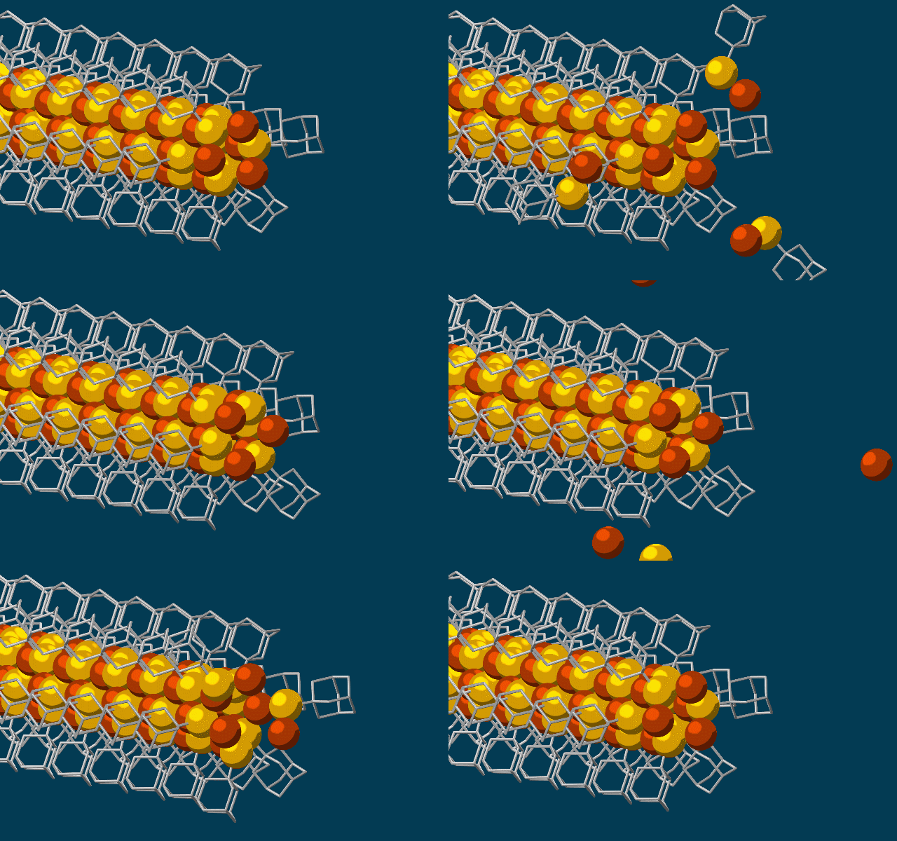
This animation shows molecular building blocks joining the tip of a growing self-assembling nanowire. Each block consists of a diamondoid — the smallest possible bit of diamond — attached to sulfur and copper atoms (yellow and brown spheres). Like LEGO blocks, they only fit together in certain ways that are determined by their size and shape. The copper and sulfur atoms form a conductive wire in the middle, and the diamondoids form an insulating outer shell. (credit: SLAC National Accelerator Laboratory)

Scientists at Stanford University and the Department of Energy’s SLAC National Accelerator Laboratory have discovered a way to use diamondoids* — the smallest possible bits of diamond — to self-assemble atoms, LEGO-style, into the thinnest possible electrical wires, just three atoms wide.

The new technique could potentially be used to build tiny wires for a wide range of applications, including fabrics that generate electricity, optoelectronic devices that employ both electricity and light, and superconducting materials that conduct electricity without any loss. The scientists reported their results last week in Nature Materials.

The researchers started with the smallest possible diamondoids —interlocking cages of carbon and hydrogen — and attached a sulfur atom to each. Floating in a solution, each sulfur atom bonded with a single copper ion — creating a semiconducting combination of copper and sulfur known as a chalcogenide.

A conventional insulated electrical copper wire (credit: Alibaba)

That created the basic nanowire building blocks, which then drifted toward each other, drawn by “unusually strong” van der Waals attraction between the diamondoids, and attached themselves to the growing tip of the nanowire. The attached diamondoids formed an insulating shell — creating the nanoscale equivalent of a conventional insulated electrical wire.

Although there are other ways to get materials to self-assemble, this is the first one shown to make a nanowire with a solid, crystalline core that has good electronic properties, said study co-author Nicholas Melosh, an associate professor at SLAC and Stanford and investigator with SIMES, the Stanford Institute for Materials and Energy Sciences at SLAC.

The team also included researchers from Lawrence Berkeley National Laboratory, the National Autonomous University of Mexico (UNAM) and Justus-Liebig University in Germany. The work was funded by the DOE Office of Science and the German Research Foundation.

* Found naturally in petroleum fluids, they are extracted and separated by size and geometry in a SLAC laboratory.


Citation: Yan et al., Nature Materials, 26 December 2016 (10.1038/nmat4823)

Press Office Contact: Andrew Gordon, agordon@slac.stanford.edu, (650) 926-2282


Abstract of Hybrid metal–organic chalcogenide nanowires with electrically conductive inorganic core through diamondoid-directed assembly

Controlling inorganic structure and dimensionality through structure-directing agents is a versatile approach for new materials synthesis that has been used extensively for metal–organic frameworks and coordination polymers. However, the lack of ‘solid’ inorganic cores requires charge transport through single-atom chains and/or organic groups, limiting their electronic properties. Here, we report that strongly interacting diamondoid structure-directing agents guide the growth of hybrid metal–organic chalcogenide nanowires with solid inorganic cores having three-atom cross-sections, representing the smallest possible nanowires. The strong van der Waals attraction between diamondoids overcomes steric repulsion leading to a cis configuration at the active growth front, enabling face-on addition of precursors for nanowire elongation. These nanowires have band-like electronic properties, low effective carrier masses and three orders-of-magnitude conductivity modulation by hole doping. This discovery highlights a previously unexplored regime of structure-directing agents compared with traditional surfactant, block copolymer or metal–organic framework linkers.

A robotic hand with a human’s delicate sense of touch

A soft, sensitive robotic hand mounted on a robotic arm (credit: Cornell University)

Cornell University engineers have invented a new kind of robotic hand with a human’s delicate sense of touch.

Scenario: you lost part of your arm in a car accident. That artificial arm and hand you got from the hospital lets you feel and pick up things — even type on a keyboard. But not with the same sensitivity as a real hand. Now, an artificial prosthesis can even let you feel which one of three tomatoes is ripe (as shown in the video below).

The engineers’ trick was to use soft, stretchable optoelectronic (light + electronics) sensors in the fingers to detect shape and texture. (The sensors in existing prosthetic and robot hands use cruder tactile, or touch, sensors with bulky, rigid motors to measure strain.) The new prosthetic hand is a lot more sensitive. It can measure softness or hardness, how much the material stretches when touched, and how much force needs to be supplied to make the material deform.

How to make an (almost) human hand

A Cornell group led by Robert Shepherd, assistant professor of mechanical and aerospace engineering and principal investigator of Organic Robotics Lab, has published a paper describing this research in the debut edition of the journal Science Robotics (open access until Jan. 31).

Schematic of prosthetic hand structure and components (credit: Cornell University)

Unlike existing prosthetic and robot sensors, these sensors are integrated within the hand (instead of on the surface), so they can actually detect forces being transmitted through the thickness of the robot hand — simulating how a human hand feels. The more the prosthetic hand deforms (as it touches an object), the more light is lost. That variable loss of light, as detected by the photodiode, is what allows the prosthetic hand to “sense” its surroundings with high sensitivity and discrimination.*

This work was supported by a grant from Air Force Office of Scientific Research, and made use of the Cornell NanoScale Science and Technology Facility and the Cornell Center for Materials Research, both of which are supported by the National Science Foundation.

* The optoelectronic sensors are based on novel elastomeric optical waveguides, using a 3D-printed mold and a soft-lithography process to create a fluidically powered, stretchable material. Each high-precision waveguide incorporates an LED to generate light and a photodiode to measure the amount of light lost (which depends dynamically on the curvature, elongation, and force of the prosthetic hand).

To make the hand, Shepherd’s group used a four-step soft lithography process to produce the inside of the hand (core) and the cladding (outer surface of the waveguide), which also houses the LED (light-emitting diode) and the photodiode (light detector).


CNBC International | Scientists build a robotic hand with a soft touch | CNBC International


Abstract of Optoelectronically innervated soft prosthetic hand via stretchable optical waveguides

Because of their continuous and natural motion, fluidically powered soft actuators have shown potential in a range of robotic applications, including prosthetics and orthotics. Despite these advantages, robots using these actuators require stretchable sensors that can be embedded in their bodies for sophisticated functions. Presently, stretchable sensors usually rely on the electrical properties of materials and composites for measuring a signal; many of these sensors suffer from hysteresis, fabrication complexity, chemical safety and environmental instability, and material incompatibility with soft actuators. Many of these issues are solved if the optical properties of materials are used for signal transduction. We report the use of stretchable optical waveguides for strain sensing in a prosthetic hand. These optoelectronic strain sensors are easy to fabricate, are chemically inert, and demonstrate low hysteresis and high precision in their output signals. As a demonstration of their potential, the photonic strain sensors were used as curvature, elongation, and force sensors integrated into a fiber-reinforced soft prosthetic hand. The optoelectronically innervated prosthetic hand was used to conduct various active sensation experiments inspired by the capabilities of a real hand. Our final demonstration used the prosthesis to feel the shape and softness of three tomatoes and select the ripe one.