Should we use CRISPR to domesticate wild plants, creating ‘biologically inspired organisms’?

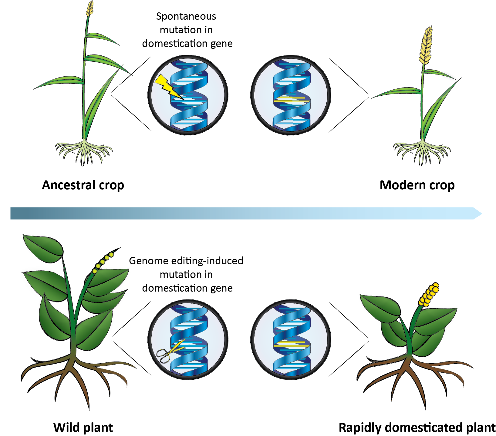
Accelerating the domestication of wild plants. During the domestication of ancestral crops, plants carrying spontaneous mutations in domestication genes were selected for. The same genes can be targeted in wild plants by genome editing, resulting in a rapidly domesticated plant.  (credit: Cell)

Here’s a radical new idea for creating new GMO (genetically modified organism) plants that may appeal to staunch organic-food consumers/farmers and even #NonGMOProjectVerified advocates: don’t insert a foreign gene in today’s domestic plants — delete already existing genes in semi-domesticated or even wild plants to make those plants more domestic, and reducing pesticide use in the process.

“All of the plants we eat today are mutants, but the crops we have now were selected for over thousands of years, and their mutations … such as reduced bitterness and those that facilitate easy harvest … arose by chance,” says Michael Palmgren, a botanist who heads an interdisciplinary think tank* called “Plants for a Changing World” at the University of Copenhagen. “With gene editing, we can create ‘biologically inspired organisms’ in that we don’t want to improve nature, we want to benefit from what nature has already created.”

Palmgen is senior author of an open-access review published March 2 in the journal Trends in Plant Science.

How to turn nitrogen in the atmosphere into fertilizer, reducing environmental damage

This strategy could also address problems from pesticide use and the damaging impact of large-scale agriculture on the environment. For example, runoff from excess nitrogen in fertilizers is a common pollutant; however, wild legumes, through symbiosis with bacteria, can turn nitrogen available in the atmosphere into their own fertilizer, he suggests.

Future logo? (credit: KurzweilAI)

Out of the more than 300,000 plant species in existence, fewer than 200 are commercially important, and only three species — rice, wheat, and maize — account for most of the plant matter that humans consume, partly because in the history of agriculture, mutations arose that made these crops the easiest to harvest, the reseachers note.

But with CRISPR technology, we don’t have to wait for nature to help us domesticate plants, argue the researchers. Instead, gene editing could make, for example, wild legumes, quinoa, or amaranth, which are already sustainable and nutritious, more farmable.

The approach has already been successful in accelerating domestication of undervalued crops using less precise gene-editing methods. For example, researchers used chemical mutagenesis to induce random mutations in weeping rice grass, an Australian wild relative of domestic rice, to make it more likely to hold onto its seeds after ripening. And in wild field cress, a type of weedy grass, scientists silenced genes with RNA interference involved with fatty acid synthesis, resulting in improved seed oil quality.

Palmgren’s group published a related open-access paper two years ago on using gene editing to make domesticated plants more “wild” and thus hardier for organic farmers.

While we’re at it, what about pharming (creating pharmaceuticals from plants) — using genetically modified wild plants?

* Supported by the University of Copenhagen Excellence Programme for Interdisciplinary Research.


Abstract of Accelerating the Domestication of New Crops: Feasibility and Approaches

The domestication of new crops would promote agricultural diversity and could provide a solution to many of the problems associated with intensive agriculture. We suggest here that genome editing can be used as a new tool by breeders to accelerate the domestication of semi-domesticated or even wild plants, building a more varied foundation for the sustainable provision of food and fodder in the future. We examine the feasibility of such plants from biological, social, ethical, economic, and legal perspectives.

Caltech scientists use bacterial protein to merge silicon and carbon and create new organosilicon compounds

Artist rendering of organosilicon-based life (credit: Lei Chen and Yan Liang (BeautyOfScience.com) for Caltech)

Scientists at Caltech have “bred” a bacterial protein with the ability to make silicon-carbon bonds, with applications in several industries — something only chemists could do before. The research was published in the Nov. 24 issue of the journal Science.

Molecules with silicon-carbon (organosilicon) compounds are found in pharmaceuticals and many other products, including agricultural chemicals, paints, semiconductors, and computer and TV screens. Currently, these products are made synthetically, since silicon-carbon bonds are not found in nature.

The new research demonstrates that biology can be used to manufacture these bonds in ways that are more environmentally friendly and potentially much less expensive, according to the researchers.


Caltech | Bringing Silicon to Life: Scientists Persuade Nature to Make Silicon-Carbon Bonds

Directed evolution

The key to this research involves deliberate messing with nature: a method called directed evolution* pioneered in the early 1990s by Frances Arnold, Caltech’s Dick and Barbara Dickinson Professor of Chemical Engineering, Bioengineering and Biochemistry, and principal investigator of this project.

An example of directed evolution with comparison to natural evolution. The inner cycle indicates the 3 stages of the directed evolution cycle with the natural process being mimicked in parentheses. The outer circle demonstrates steps a typical experiment. The red symbols indicate functional variants, the pale symbols indicate variants with reduced function. (credit: Thomas Shafee CC)

Directed evolution has been used for years to make enzymes for household products, like detergents; and for “green” sustainable routes to making pharmaceuticals, agricultural chemicals, and fuels.

In directed evolution, new and better enzymes are created in labs by artificial selection, similar to the way that breeders modify corn, cows, or cats. Enzymes are a class of proteins that catalyze, or facilitate, chemical reactions. The directed evolution process begins with an enzyme that scientists want to enhance. The DNA coding for the enzyme is mutated in more-or-less random ways, and the resulting enzymes are tested for a desired trait. The top-performing enzyme is then mutated again, and the process is repeated until an enzyme that performs much better than the original is created.

Going where no enzyme has gone before

In the new study, the goal was not just to improve an enzyme’s biological function but to actually persuade it to do something that it had not done before. The researchers’ first step was to find a suitable candidate, an enzyme showing potential for making the silicon-carbon bonds.

“It’s like breeding a racehorse,” says Arnold, who is also the director of the Donna and Benjamin M. Rosen Bioengineering Center at Caltech. “A good breeder recognizes the inherent ability of a horse to become a racer and has to bring that out in successive generations. We just do it with proteins.”

An Icelandic hot spring (credit: Nordic Visitor)

The ideal candidate turned out to be a protein from a bacterium, Rhodothermus marinus, that grows in hot springs in Iceland. That protein, called cytochrome c, normally shuttles electrons to other proteins, but the researchers found that it also happens to act like an enzyme to create silicon-carbon bonds at low levels. The scientists then mutated the DNA coding for that protein within a region that specifies an iron-containing portion of the protein thought to be responsible for its silicon-carbon bond-forming activity. Next, they tested these mutant enzymes for their ability to make organosilicon compounds better than the original.

cytochrome c (credit: Caltech)

After only three rounds, they had created an enzyme that can selectively make silicon-carbon bonds 15 times more efficiently than the best catalyst invented by chemists. Furthermore, the enzyme is highly selective, which means that it makes fewer unwanted byproducts that have to be chemically separated out.

“This iron-based, genetically encoded catalyst is nontoxic, cheaper, and easier to modify compared to other catalysts used in chemical synthesis,” says Jennifer Kan, a postdoctoral scholar in Arnold’s lab and lead author of the new study. “The new reaction can also be done at room temperature and in water.”

The synthetic process for making silicon-carbon bonds often uses precious metals and toxic solvents, and requires extra processing to remove unwanted byproducts, all of which add to the cost of making these compounds.

Could life on Earth (or elsewhere) have evolved based on silicon-carbon?

The study is the first to show that nature can adapt to incorporate silicon into carbon-based molecules, the building blocks of life.

Carbon and silicon are chemically very similar, and silicon is the second most abundant element in Earth’s crust. They can both form bonds to four atoms simultaneously, making them well suited to form the long chains of molecules found in life, such as proteins and DNA. Science-fiction authors have imagined alien worlds with silicon-based life, like the lumpy Horta creatures portrayed in an episode of the 1960s TV series Star Trek.

“This study shows how quickly nature can adapt to new challenges,” says Arnold. “The DNA-encoded catalytic machinery of the cell can rapidly learn to promote new chemical reactions when we provide new reagents and the appropriate incentive in the form of artificial selection.”

However, no living organism is known [yet] to put silicon-carbon bonds together, even though silicon is so abundant, all around us, in rocks and all over the beach,” says Kan.

What about other planets (Mars has both silicon and carbon, for example) and asteroids? And could alien life have evolved silicon-carbon semiconductor brains? It would also be interesting to see if such a lifeform could be invented on Earth.

This research is funded by the National Science Foundation, the Caltech Innovation Initiative program, and the Jacobs Institute for Molecular Engineering for Medicine at Caltech.

* Not to be confused with a transhumanist concept for controlling human evolution.


Abstract of Directed evolution of cytochrome c for carbon–silicon bond formation: Bringing silicon to life

Enzymes that catalyze carbon–silicon bond formation are unknown in nature, despite the natural abundance of both elements. Such enzymes would expand the catalytic repertoire of biology, enabling living systems to access chemical space previously only open to synthetic chemistry. We have discovered that heme proteins catalyze the formation of organosilicon compounds under physiological conditions via carbene insertion into silicon–hydrogen bonds. The reaction proceeds both in vitro and in vivo, accommodating a broad range of substrates with high chemo- and enantioselectivity. Using directed evolution, we enhanced the catalytic function of cytochrome c from Rhodothermus marinus to achieve more than 15-fold higher turnover than state-of-the-art synthetic catalysts. This carbon–silicon bond-forming biocatalyst offers an environmentally friendly and highly efficient route to producing enantiopure organosilicon molecules.

How birds unlock their ultraviolet vision super-sense

Some birds have been found to be as intelligent as mammals. And some that can see ultraviolet (UV) light live in a super-sensory world apart, able to transmit and receive signals between each other in a way that is invisible to many other species.

Now the ability of finches, sparrows, and many other birds to see ultraviolet (UV) light is explained in a study published in the journal eLife by scientists at the Washington University School of Medicine in St. Louis.

Carotenoid pigments determine bird’s UV perception or not

The study reveals two essential adaptions that enable birds to expand their vision into the UV range: chemical changes in light-filtering pigments called carotenoids (such as carotene, found in carrots, associated with vitamin A) and the tuning of light-sensitive proteins called opsins (light-sensitive proteins), some of which are also used in optogenetics research.

Birds acquire carotenoids through their diets and process them in a variety of ways to shift their light absorption toward longer (visible light) or shorter (UV) wavelengths.

“There are two types of light-sensitive cells, called photoreceptors, in the eye: rods and cones. Cone photoreceptors are responsible for color vision. While humans have blue, green, and red-sensitive cones only, birds have a fourth cone type which is either violet or UV-sensitive, depending on the species,” says senior author Joseph Corbo, MD, PhD, Associate Professor of Pathology and Immunology.

UV vs visual perception by cones in the eye is fine-tuned by evolution

“Our approach showed that blue-cone sensitivity is fine-tuned through a change in the chemical structure of carotenoid pigments within the photoreceptor, allowing both violet and UV-sighted birds to maximize how many colors they can see.”

The study also revealed that sensitivity of the violet/UV cone and the blue cone in birds must move in sync to allow for optimum vision. Among bird species, there is a strong relationship between the light sensitivity of opsins within the violet/UV cone and mechanisms within the blue cone, which coordinate to ensure even UV vision.

Taken together, these results suggest that both blue and violet cone cells have adapted during evolution to enhance color vision in birds.

Birds have achieved UV vision by use of a specialized optical organelle, the pigmented cone oil droplet. These oil droplets are located in the path of light through the receptor and act as cutoff filters matched to the visual pigment sensitivity of each cone subtype.

Spectral filtering in bird cones. a) A flat-mounted chicken retina under brightfield illumination that shows the distinctive pigmentation of the cone oil droplets. (b) A diagram of the avian single cone photoreceptors showing the relative position of the oil droplet within the cells (top) and a representation of the spectral filtering cutoff effects of the droplet (bottom). (credit: Matthew B Toomey et al./eLife)

“The majority of bird species rely on vision as their primary sense, and color discrimination plays a crucial role in their essential behaviors, such as choosing mates and foraging for food. This explains why birds have evolved one of the most richly endowed color vision systems among vertebrates,” says first author Matthew Toomey, a postdoctoral fellow at the Washington University School of Medicine.

“The precise coordination of sensitivity and filtering in the visual system may, for example, help female birds discriminate very fine differences in the elaborate coloration of their suitors and choose the fittest mates. This refinement of visual sensitivity could also facilitate the search for hidden seeds, fruits, and other food items in the environment.”

The team now plans to investigate the underlying molecular mechanisms that help modify the carotenoid pigments and light-sensitive protein tuning in a wide range of bird species, to gather further insights into the evolution of UV vision.


Abstract of Complementary shifts in photoreceptor spectral tuning unlock the full adaptive potential of ultraviolet vision in birds

Color vision in birds is mediated by four types of cone photoreceptors whose maximal sensitivities (λmax) are evenly spaced across the light spectrum. In the course of avian evolution, the λmax of the most shortwave-sensitive cone, SWS1, has switched between violet (λmax > 400 nm) and ultraviolet (λmax < 380 nm) multiple times. This shift of the SWS1 opsin is accompanied by a corresponding short-wavelength shift in the spectrally adjacent SWS2 cone. Here, we show that SWS2 cone spectral tuning is mediated by modulating the ratio of two apocarotenoids, galloxanthin and 11’,12’-dihydrogalloxanthin, which act as intracellular spectral filters in this cell type. We propose an enzymatic pathway that mediates the differential production of these apocarotenoids in the avian retina, and we use color vision modeling to demonstrate how correlated evolution of spectral tuning is necessary to achieve even sampling of the light spectrum and thereby maintain near-optimal color discrimination.

Why evolution may be intelligent, based on deep learning

Moth Orchid flower (credit: Christian Kneidinger)

A computer scientist and biologist propose to unify the theory of evolution with learning theories to explain the “amazing, apparently intelligent designs that evolution produces.”

The scientists — University of Southampton School of Electronics and Computer Science professor Richard Watson* and Eötvös Loránd University (Budapest) professor of biology Eörs Szathmáry* — say they’ve found that it’s possible for evolution to exhibit some of the same intelligent behaviors as learning systems — including neural networks.

Writing in an opinion paper published in the journal Trends in Ecology and Evolution, they use “formal analogies” and transfer specific models and results between the two theories in an attempt to solve several evolutionary puzzles.

The authors cite work by Pavlicev and colleagues** showing that selection on relational alleles (gene variants) increases phenotypic (organism trait) correlation if the traits are selected together and decreases correlation if they are selected antagonistically, which is characteristic of Hebbian learning, they note.

“This simple step from evolving traits to evolving correlations between traits is crucial; it moves the object of natural selection from fit phenotypes (which ultimately removes phenotypic variability altogether) to the control of phenotypic variability,” the researchers say.

Why evolution is not blind

“Learning theory is not just a different way of describing what Darwin already told us,” said Watson. “It expands what we think evolution is capable of. It shows that natural selection is sufficient to produce significant features of intelligent problem-solving.”

Conventionally, evolution, which depends on random variation, has been considered blind, or at least myopic, he notes. “But showing that evolving systems can learn from past experience means that evolution has the potential to anticipate what is needed to adapt to future environments in the same way that learning systems do.

“A system exhibits learning if its performance at some task improves with experience,” the authors note in the paper. “Reusing behaviors that have been successful in the past (reinforcement learning) is intuitively similar to the way selection increases the proportion of fit phenotypes [an organism's observable characteristics or traits] in a population. In fact, evolutionary processes and simple learning processes are formally equivalent.

“In particular, learning can be implemented by incrementally adjusting a probability distribution over behaviors (e.g., Bayesian learning or Bayesian updating). Or, if a behavior is represented by a vector of features or components, by adjusting the probability of using each individual component in proportion to its average reward in past behaviors (e.g., Multiplicative Weights Update Algorithm, MWUA).”

The evolution of connections in a Recurrent Gene Regulation Network (GRN) shows associative learning behaviors. When a Hopfield network is trained on a set of patterns with Hebbian learning, it forms an associative memory of the patterns in the training set. When subsequently stimulated with random excitation patterns, the activation dynamics of the trained network will spontaneously recall the patterns from the training set or generate new patterns that are generalizations of the training patterns. (A–D) A GRN is evolved to produce first one phenotype (set of characteristics or traits — Charles Darwin in this example) and then another (Donald Hebb) in an alternating manner. The resulting phenotype is not merely an average of the two phenotypic patterns that were selected in the past. Rather, different embryonic phenotypes (e.g., random initial conditions C and D) developed into different adult phenotypes (with this evolved GRN) and match either A or B. These two phenotypes can be produced from genotypes (DNA sequences) that are a single mutation apart. In a separate experiment, selection iterates over a set of target phenotypes (E–H). In addition to developing phenotypes that match patterns selected in the past (e.g., I), this GRN also generalizes to produce new phenotypes that were not selected for in the past but belong to a structurally similar class, for example, by creating novel combinations of evolved modules (e.g., developmental attractors exist for a phenotype with all four “loops” (J). This demonstrates a capability for evolution to exhibit phenotypic novelty in exactly the same sense that learning neural networks can generalize from past experience. (credit: Richard A. Watson and Eörs Szathmáry/Trends in Ecology and Evolution)

Unsupervised learning

An even more interesting process in evolution is unsupervised learning, where mechanisms do not depend on an external reward signal, the authors say in the paper:

By reinforcing correlations that are frequent, regardless of whether they are good, unsupervised correlation learning can produce system-level behaviors without system-level rewards. This can be implemented without centralized learning mechanisms. (Recent theoretical work shows that selection acting only to maximize individual growth rate, when applied to interspecific competition coefficients within an ecological community, produces unsupervised learning at the system level.)

This is an exciting possibility because it means that, despite not being a unit of selection, an ecological community might exhibit organizations that confer coordinated collective behaviors — for example, a distributed ecological memory that can recall multiple past ecological states. …

Taken together, correlation learning, unsupervised correlation learning, and deep correlation learning thus provide a formal way to understand how variation, selection, and inheritance, respectively, might be transformed over evolutionary time.

The authors’ new approach also offers an alternative to “intelligent design” (ID), which negates natural selection as an explanation for apparently intelligent features of nature. (The leading proponents of ID are associated with the Discovery Institute. See Are We Spiritual Machines? Ray Kurzweil vs. the Critics of Strong A.I.*** — a debate between Kurzweil and several Discovery Institute fellows.)

So if evolutionary theory can learn from the principles of cognitive science and deep learning, can cognitive science and deep learning learn from evolutionary theory?

* The authors are also affiliated with the Parmenides Foundation in Munich.

** Watson, R.A. et al. (2014) The evolution of phenotypic correlations and ‘developmental memory.’ Evolution 68, 1124–1138 and Pavlicev, M.et al. (2011) Evolution of adaptive phenotypic variation patterns by direct selection for evolvability. Proc. R. Soc. B Biol. Sci. 278, 1903–1912

*** This book is available free on KurzweilAI, as noted.


Abstract of How Can Evolution Learn?
The theory of evolution links random variation and selection to incremental adaptation. In a different intellectual domain, learning theory links incremental adaptation (e.g., from positive and/or negative reinforcement) to intelligent behaviour. Specifically, learning theory explains how incremental adaptation can acquire knowledge from past experience and use it to direct future behaviours toward favourable outcomes. Until recently such cognitive learning seemed irrelevant to the ‘uninformed’ process of evolution. In our opinion, however, new results formally linking evolutionary processes to the principles of learning might provide solutions to several evolutionary puzzles – the evolution of evolvability, the evolution of ecological organisation, and evolutionary transitions in individuality. If so, the ability for evolution to learn might explain how it produces such apparently intelligent designs.

Biologists induce flatworms to grow heads and brains of other species

Tufts biologists induced one species of flatworm —- G. dorotocephala, top left — to grow heads and brains characteristic of other species of flatworm, top row, without altering genomic sequence. Examples of the outcomes can be seen in the bottom row of the image. (credit: Center for Regenerative and Developmental Biology, School of Arts and Sciences, Tufts University.)

Tufts University biologists have electrically modified flatworms to grow heads and brains characteristic of another species of flatworm — without altering their genomic sequence. This suggests bioelectrical networks as a new kind of epigenetics (information existing outside of a genomic sequence) to determine large-scale anatomy.

Besides the overall shape of the head, the changes included the shape of the brain and the distribution of the worm’s adult stem cells.

The discovery could help improve understanding of birth defects and regeneration by revealing a new pathway for controlling complex pattern formation similar to how neural networks exploit bioelectric synapses to store and re-write information in the brain.

The findings are detailed in the open-access cover story of the November 2015 edition of the International Journal of Molecular Sciences, appearing online Nov. 24.

“These findings raise significant questions about how genes and bioelectric networks interact to build complex body structures,” said the paper’s senior author Michael Levin, Ph.D., who holds the Vannevar Bush Chair in biology and directs the Center for Regenerative and Developmental Biology in the School of Arts and Sciences at Tufts. Knowing how shape is determined and how to influence it is important because biologists could use that knowledge, for example, to fix birth defects or cause new biological structures to grow after an injury, he explained.

How they did it

The researchers worked with Girardia dorotocephala — free-living planarian flatworms, which have remarkable regenerative capacity. They induced the development of different species-specific head shapes by interrupting gap junctions, which are protein channels that enable cells to communicate with each other by passing electrical signals back and forth.

A conceptual model of shape change driven by physiological network dynamics. Planaria regeneration (B) parallels classical neural network behavior (A); both can be described in terms of free energy landscapes with multiple attractor states. (credit: Maya Emmons-Bell et al./Int. J. Mol. Sci.)

The ease with which a particular shape could be coaxed from a G. dorotocephala worm was proportional to the proximity of the target worm on the evolutionary timeline. The closer the two species were related, the easier it was to effect the change. This observation strengthens the connection to evolutionary history, suggesting that modulation of physiological circuits may be one more tool exploited by evolution to alter animal body plans.

However, this shape change was only temporary. Weeks after the planaria completed regeneration to the other species’ head shapes, the worms once again began remodeling and re-acquired their original head morphology. Additional research is needed to determine how this occurs. The authors also presented a computational model that explains how changes in cell-to-cell communication can give rise to the diverse shape types.

The interdisciplinary research involved U.S.- and Canada-based biologists and European mathematicians.


Abstract of Gap Junctional Blockade Stochastically Induces Different Species-Specific Head Anatomies in Genetically Wild-Type Girardia dorotocephala Flatworms

The shape of an animal body plan is constructed from protein components encoded by the genome. However, bioelectric networks composed of many cell types have their own intrinsic dynamics, and can drive distinct morphological outcomes during embryogenesis and regeneration. Planarian flatworms are a popular system for exploring body plan patterning due to their regenerative capacity, but despite considerable molecular information regarding stem cell differentiation and basic axial patterning, very little is known about how distinct head shapes are produced. Here, we show that after decapitation in G. dorotocephala, a transient perturbation of physiological connectivity among cells (using the gap junction blocker octanol) can result in regenerated heads with quite different shapes, stochastically matching other known species of planaria (S. mediterraneaD. japonica, and P. felina). We use morphometric analysis to quantify the ability of physiological network perturbations to induce different species-specific head shapes from the same genome. Moreover, we present a computational agent-based model of cell and physical dynamics during regeneration that quantitatively reproduces the observed shape changes. Morphological alterations induced in a genomically wild-type G. dorotocephala during regeneration include not only the shape of the head but also the morphology of the brain, the characteristic distribution of adult stem cells (neoblasts), and the bioelectric gradients of resting potential within the anterior tissues. Interestingly, the shape change is not permanent; after regeneration is complete, intact animals remodel back to G. dorotocephala-appropriate head shape within several weeks in a secondary phase of remodeling following initial complete regeneration. We present a conceptual model to guide future work to delineate the molecular mechanisms by which bioelectric networks stochastically select among a small set of discrete head morphologies. Taken together, these data and analyses shed light on important physiological modifiers of morphological information in dictating species-specific shape, and reveal them to be a novel instructive input into head patterning in regenerating planaria.

Most Earth-like worlds have yet to be born, says new NASA study

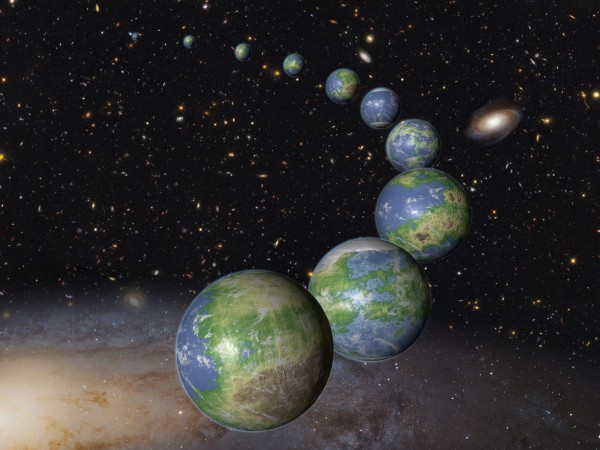
This is an artist’s impression of innumerable Earth-like planets that have yet to be born over the next trillion years in the evolving universe (credit: NASA, ESA, and G. Bacon (STScI); Science: NASA, ESA, P. Behroozi and M. Peeples (STScI))

When our solar system was born 4.6 billion years ago, only eight percent of the potentially habitable planets that will ever form in the universe existed, according to an assessment of data collected by NASA’s Hubble Space Telescope and Kepler space observatory and published today (Oct. 20) in an open-access paper in the Monthly Notices of the Royal Astronomical Society.


In related news, UCLA geochemists have found evidence that life probably existed on Earth at least 4.1 billion years ago, which is 300 million years earlier than previous research suggested. The research suggests life in the universe could be abundant, said Mark Harrison, co-author of the research and a professor of geochemistry at UCLA. The research was published Monday Oct. 19 in the online early edition of the journal Proceedings of the National Academy of Sciences.


The data show that the universe was making stars at a fast rate 10 billion years ago, but the fraction of the universe’s hydrogen and helium gas that was involved was very low. Today, star birth is happening at a much slower rate than long ago, but there is so much leftover gas available after the big bang that the universe will keep making stars and planets for a very long time to come.

A billion Earth-sized worlds

Based on the survey, scientists predict that there should already be 1 billion Earth-sized worlds in the Milky Way galaxy. That estimate skyrockets when you include the other 100 billion galaxies in the observable universe.

Kepler’s planet survey indicates that Earth-sized planets in a star’s habitable zone — the perfect distance that could allow water to pool on the surface — are ubiquitous in our galaxy. This leaves plenty of opportunity for untold more Earth-sized planets in the habitable zone to arise in the future — the last star isn’t expected to burn out until 100 trillion years from now.

The researchers say that future Earths are more likely to appear inside giant galaxy clusters and also in dwarf galaxies, which have yet to use up all their gas for building stars and accompanying planetary systems. By contrast, our Milky Way galaxy has used up much more of the gas available for future star formation.

A big advantage to our civilization arising early in the evolution of the universe is our being able to use powerful telescopes like Hubble to trace our lineage from the big bang through the early evolution of galaxies.

Regrettably, the observational evidence for the big bang and cosmic evolution, encoded in light and other electromagnetic radiation, will be all but erased away 1 trillion years from now, due to the runaway expansion of space. Any far-future civilizations that might arise will be largely clueless as to how or if the universe began and evolved.


Abstract of On The History and Future of Cosmic Planet Formation

We combine constraints on galaxy formation histories with planet formation models, yielding the Earth-like and giant planet formation histories of the Milky Way and the Universe as a whole. In the Hubble volume (1013 Mpc3), we expect there to be ∼1020 Earth-like and ∼1020giant planets; our own galaxy is expected to host ∼109 and ∼1010 Earth-like and giant planets, respectively. Proposed metallicity thresholds for planet formation do not significantly affect these numbers. However, the metallicity dependence for giant planets results in later typical formation times and larger host galaxies than for Earth-like planets. The Solar system formed at the median age for existing giant planets in the Milky Way, and consistent with past estimates, formed after 80 per cent of Earth-like planets. However, if existing gas within virialized dark matter haloes continues to collapse and form stars and planets, the Universe will form over 10 times more planets than currently exist. We show that this would imply at least a 92 per cent chance that we are not the only civilization the Universe will ever have, independent of arguments involving the Drake equation.


Abstract of Potentially biogenic carbon preserved in a 4.1 billion-year-old zircon

Evidence for carbon cycling or biologic activity can be derived from carbon isotopes, because a high12C/13C ratio is characteristic of biogenic carbon due to the large isotopic fractionation associated with enzymatic carbon fixation. The earliest materials measured for carbon isotopes at 3.8 Ga are isotopically light, and thus potentially biogenic. Because Earth’s known rock record extends only to ∼4 Ga, earlier periods of history are accessible only through mineral grains deposited in later sediments. We report 12C/13C of graphite preserved in 4.1-Ga zircon. Its complete encasement in crack-free, undisturbed zircon demonstrates that it is not contamination from more recent geologic processes. Its 12C-rich isotopic signature may be evidence for the origin of life on Earth by 4.1 Ga.

‘Tree of life’ for 2.3 million species released

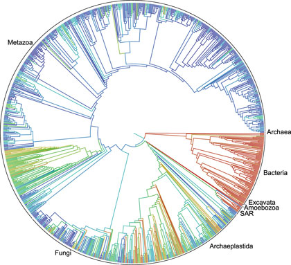
This circular family tree of Earth’s lifeforms is considered a first draft of the 3.5-billion-year history of how life evolved and diverged (credit: Duke University)

A first draft of the “tree of life” for the roughly 2.3 million named species of animals, plants, fungi and microbes — from platypuses to puffballs — has been released.

A collaborative effort among eleven institutions, the tree depicts the relationships among living things as they diverged from one another over time, tracing back to the beginning of life on Earth more than 3.5 billion years ago.

Tens of thousands of smaller trees have been published over the years for select branches of the tree of life — some containing upwards of 100,000 species — but this is the first time those results have been combined into a single tree that encompasses all of life.

“This is the first real attempt to connect the dots and put it all together,” said principal investigator Karen Cranston of Duke University. “Think of it as Version 1.0.” The current version of the tree — along with the underlying data and source code — is available to browse, edit, and download free at https://tree.opentreeoflife.org — a sort of “Wikipedia” for the evolutionary trees.

It is also described in an open-access article appearing Sept. 18 in the Proceedings of the National Academy of Sciences.

Uses of evolutionary trees

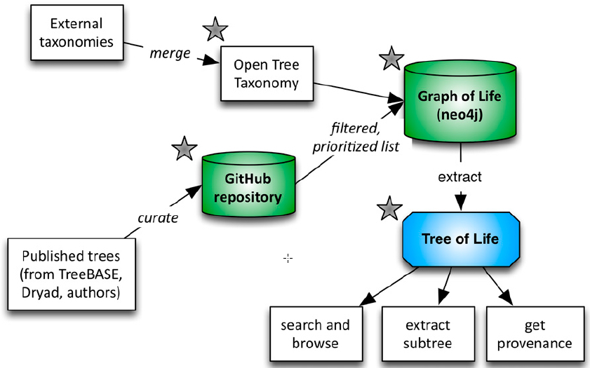
Open Tree of Life workflow (credit: Cody E. Hinchliff et al./PNAS)

Understanding how the millions of species on Earth are related to one another helps scientists discover new drugs, increase crop and livestock yields, and trace the origins and spread of infectious diseases such as HIV, Ebola and influenza, the scientists say.

The researchers pieced it together by compiling thousands of smaller chunks that had already been published online and merging them together into a gigantic “supertree” that encompasses all named species. The initial draft is based on nearly 500 smaller trees from previously published studies.

To map trees from different sources to the branches and twigs of a single supertree, one of the biggest challenges was simply accounting for the name changes, alternate names, common misspellings and abbreviations for each species. The eastern red bat, for example, is often listed under two scientific names, Lasiurus borealis and Nycteris borealis. Spiny anteaters once shared their scientific name with a group of moray eels.

“Although a massive undertaking in its own right, this draft tree of life represents only a first step,” the researchers wrote. For one, only a tiny fraction of published trees are digitally available.

A survey of more than 7,500 phylogenetic studies published between 2000 and 2012 in more than 100 journals found that only one out of six studies had deposited their data in a digital, downloadable format that the researchers could use.

The vast majority of evolutionary trees are published as PDFs and other image files that are impossible to enter into a database or merge with other trees.

As a result, the relationships depicted in some parts of the tree, such as the branches representing the pea and sunflower families, don’t always agree with expert opinion.

Other parts of the tree, particularly insects and microbes, remain elusive. That’s because even the most popular online archive of raw genetic sequences — from which many evolutionary trees are built — contains DNA data for less than five percent of the tens of millions species estimated to exist on Earth.

“As important as showing what we do know about relationships, this first tree of life is also important in revealing what we don’t know,” said co-author Douglas Soltis of the University of Florida.

To help fill in the gaps, the team is also developing software that will enable researchers to log on and update and revise the tree as new data come in for the millions of species still being named or discovered.

“It’s by no means finished,” Cranston said. “It’s critically important to share data for already-published and newly-published work if we want to improve the tree.”

“Twenty five years ago people said this goal of huge trees was impossible,” Soltis said. “The Open Tree of Life is an important starting point that other investigators can now refine and improve for decades to come.”


Abstract of Synthesis of phylogeny and taxonomy into a comprehensive tree of life

Reconstructing the phylogenetic relationships that unite all lineages (the tree of life) is a grand challenge. The paucity of homologous character data across disparately related lineages currently renders direct phylogenetic inference untenable. To reconstruct a comprehensive tree of life, we therefore synthesized published phylogenies, together with taxonomic classifications for taxa never incorporated into a phylogeny. We present a draft tree containing 2.3 million tips—the Open Tree of Life. Realization of this tree required the assembly of two additional community resources: (i) a comprehensive global reference taxonomy and (ii) a database of published phylogenetic trees mapped to this taxonomy. Our open source framework facilitates community comment and contribution, enabling the tree to be continuously updated when new phylogenetic and taxonomic data become digitally available. Although data coverage and phylogenetic conflict across the Open Tree of Life illuminate gaps in both the underlying data available for phylogenetic reconstruction and the publication of trees as digital objects, the tree provides a compelling starting point for community contribution. This comprehensive tree will fuel fundamental research on the nature of biological diversity, ultimately providing up-to-date phylogenies for downstream applications in comparative biology, ecology, conservation biology, climate change, agriculture, and genomics.

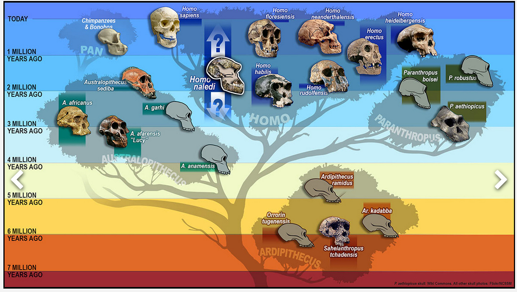
Discovery of Homo naledi adds a new branch to the human family tree

Skeletal fossil of the hand of Homo naledi (photo credit: John Hawks, UW–Madison)

An international research team has discovered a new species of a human relative, Homo naledi, uncovered in a cave outside of Johannesburg, South Africa. The discovery in late 2013 may shed light on the diversity of our genus and possibly its origin.

The team’s findings, which are published in two papers in the open-access journal eLife, were announced by South Africa’s University of the Witwatersrand, the National Geographic Society, and the South African National Research Foundation.

Homo naledi: A New Species on the Human Family Tree (illustration credit: S.V. Medaris/UW-Madison)

The authors describe Homo naledi as being similar in size and weight to a small modern human, with human-like hands and feet. The skull had a small braincase “similar in size to other early hominin species that lived between four million and two million years ago.”

The discovery indicates that H. naledi intentionally deposited bodies of its dead in a remote cave chamber — behaviors previously thought limited to humans.

This figure includes the 737 partial or complete anatomical elements of Homo naledi found (credit: Lee R. Berger et al./eLife)

Lee Berger, a research professor in the Evolutionary Studies Institute at the University of the Witwatersrand and a National Geographic Explorer-in-Residence, led the expeditions that recovered the fossils — more than 1,500 bones belonging to at least 15 individuals.

The National Geographic article “This Face Changes the Human Story. But How?” offers striking art and photography on this discovery.


University of Wisconsin-Madison | Meet our newest ancestor: Homo naledi


Abstract of Homo naledi, a new species of the genus Homo from the Dinaledi Chamber, South Africa
Homo naledi is a previously-unknown species of extinct hominin discovered within the Dinaledi Chamber of the Rising Star cave system, Cradle of Humankind, South Africa. This species is characterized by body mass and stature similar to small-bodied human populations but a small endocranial volume similar to australopiths. Cranial morphology of H. naledi is unique, but most similar to early Homo species including Homo erectusHomo habilis orHomo rudolfensis. While primitive, the dentition is generally small and simple in occlusal morphology. H. naledi has humanlike manipulatory adaptations of the hand and wrist. It also exhibits a humanlike foot and lower limb. These humanlike aspects are contrasted in the postcrania with a more primitive or australopith-like trunk, shoulder, pelvis and proximal femur. Representing at least 15 individuals with most skeletal elements repeated multiple times, this is the largest assemblage of a single species of hominins yet discovered in Africa.


Abstract of Geological and taphonomic context for the new hominin species Homo naledi from the Dinaledi Chamber, South Africa

We describe the physical context of the Dinaledi Chamber within the Rising Star cave, South Africa, which contains the fossils of Homo naledi. Approximately 1550 specimens of hominin remains have been recovered from at least 15 individuals, representing a small portion of the total fossil content. Macro-vertebrate fossils are exclusively H. naledi, and occur within clay-rich sediments derived from in situ weathering, and exogenous clay and silt, which entered the chamber through fractures that prevented passage of coarser-grained material. The chamber was always in the dark zone, and not accessible to non-hominins. Bone taphonomy indicates that hominin individuals reached the chamber complete, with disarticulation occurring during/after deposition. Hominins accumulated over time as older laminated mudstone units and sediment along the cave floor were eroded. Preliminary evidence is consistent with deliberate body disposal in a single location, by a hominin species other than Homo sapiens, at an as-yet unknown date.

How mass extinctions can accelerate robot evolution

At the start of the simulation, a biped robot controlled by a computationally evolved brain stands upright on a 16 meter by 16 meter surface. The simulation proceeds until the robot falls or until 15 seconds have elapsed. (credit: Joel Lehman)

Robots evolve more quickly and efficiently after a virtual mass extinction modeled after real-life disasters, such as the one that killed off the dinosaurs, computer scientists at The University of Texas at Austin have found.

Mass extinctions speed up evolution by unleashing new creativity in adaptations.

Computer scientists Risto Miikkulainen and Joel Lehman co-authored the study published in an open-access paper in the journal PLOS One.

“Focused destruction can lead to surprising outcomes,” said Miikkulainen, a professor of computer science at UT Austin. “Sometimes you have to develop something that seems objectively worse in order to develop the tools you need to get better.”

Survival of the evolvable

In biology, mass extinctions are known for being highly destructive, erasing a lot of genetic material from the tree of life. But some evolutionary biologists hypothesize that extinction events actually accelerate evolution by promoting those lineages that are the most evolvable, meaning ones that can quickly create useful new features and abilities.

Miikkulainen and Lehman found that, at least with robots, this is the case.

For years, computer scientists have used computer algorithms inspired by evolution to train simulated robot brains, called neural networks, to improve at a task from one generation to the next. But could mass destruction speed things up?

To find out, they connected neural networks to simulated robotic legs with the goal of evolving a robot that could walk smoothly and stably. As with real evolution, random mutations were introduced through the computational evolution process. The scientists created many different niches so that a wide range of novel features and abilities would come about.

Pruning to achieve super-robots

After hundreds of generations, a wide range of robotic behaviors had evolved to fill these niches, many of which were not directly useful for walking. Then the researchers randomly killed off the robots in 90 percent of the niches, mimicking a mass extinction.

After several such cycles of evolution and extinction, they discovered that the lineages that survived were the most evolvable and, therefore, had the greatest potential to produce new behaviors. Not only that, but overall, better solutions to the task of walking were evolved in simulations with mass extinctions, compared with simulations without them.

Practical applications of the research could include the development of robots that can better overcome obstacles (such as robots searching for survivors in earthquake rubble, exploring Mars or navigating a minefield) and human-like game agents.

“This is a good example of how evolution produces great things in indirect, meandering ways,” explains Lehman, a former postdoctoral researcher in Miikkulainen’s lab, now at the IT University of Copenhagen. He and a former student of Miikkulainen’s at UT Austin, Kenneth Stanley, recently published a popular science book about evolutionary meandering, The Myth of the Objective: Why Greatness Cannot Be Planned. “Even destruction can be leveraged for evolutionary creativity,” Lehman says.

This research was funded by the National Science Foundation (NSF), National Institutes of Health and UT Austin’s Freshman Research Initiative. Funding from NSF was provided through grants to BEACON, a multi-university center established to study evolution in action in natural and virtual settings. The University of Texas at Austin is a member of BEACON. Evolutionary biologists in BEACON assisted Miikkulainen and Lehman in designing the research project and interpreting the results.


Abstract of Extinction Events Can Accelerate Evolution

Extinction events impact the trajectory of biological evolution significantly. They are often viewed as upheavals to the evolutionary process. In contrast, this paper supports the hypothesis that although they are unpredictably destructive, extinction events may in the long term accelerate evolution by increasing evolvability. In particular, if extinction events extinguish indiscriminately many ways of life, indirectly they may select for the ability to expand rapidly through vacated niches. Lineages with such an ability are more likely to persist through multiple extinctions. Lending computational support for this hypothesis, this paper shows how increased evolvability will result from simulated extinction events in two computational models of evolved behavior. The conclusion is that although they are destructive in the short term, extinction events may make evolution more prolific in the long term.