New microscope creates near-real-time videos of nanoscale processes


MIT | Microscope creates near-real-time videos of nanoscale processes

MIT engineers have designed an atomic force microscope (AFM) that scans images 2,000 times faster than existing commercial models. Operating at near-real-time-video speed, it can capture structures as small as a fraction of a nanometer from single strands of DNA down to individual hydrogen bonds.

Existing AFMs have similar spatial resolution but function at slow speeds.

In one dramatic demonstration of the instrument’s capabilities (see video), the researchers scanned a 70- by-70-micrometers sample of calcite as it was first immersed in deionized water and later exposed to sulfuric acid. Over a period of several seconds, the team observed the acid eating away at the calcite, expanding existing nanometer-sized pits in the material that quickly merged and led to a layer-by-layer removal of calcite along the material’s crystal pattern.
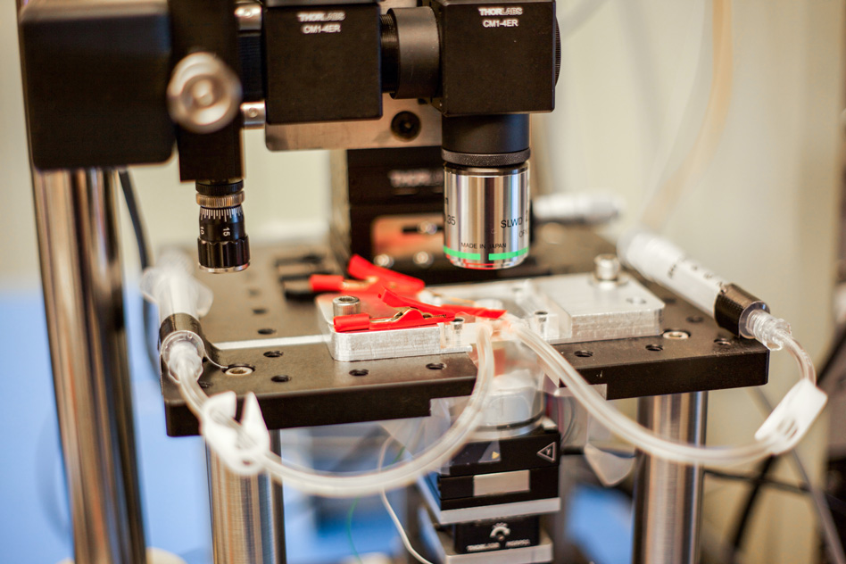
The new MIT high-speed microscope produces images of chemical processes taking place at the nanoscale at a rate that is close to real-time video. This closeup shot of the microscope shows transparent tubes used to inject various liquids into the imaging environment. This liquid can be water, acid, buffer solution for live bacteria, cells, or electrolytes in an electrochemical process. Researchers use one as an inlet and the other as an outlet to circulate and refresh the solutions throughout the experiment. (credit: Jose-Luis Olivares/MIT)

Kamal Youcef-Toumi, a professor of mechanical engineering at MIT, says the instrument’s sensitivity and speed will enable scientists to watch atomic-sized processes play out as high-resolution “movies” for the first time.

“People can see, for example, condensation, nucleation, dissolution, or deposition of material, and how these happen in real-time — things that people have never seen before,” Youcef-Toumi says. “This is fantastic to see these details emerging. And it will open great opportunities to explore all of this world that is at the nanoscale.”
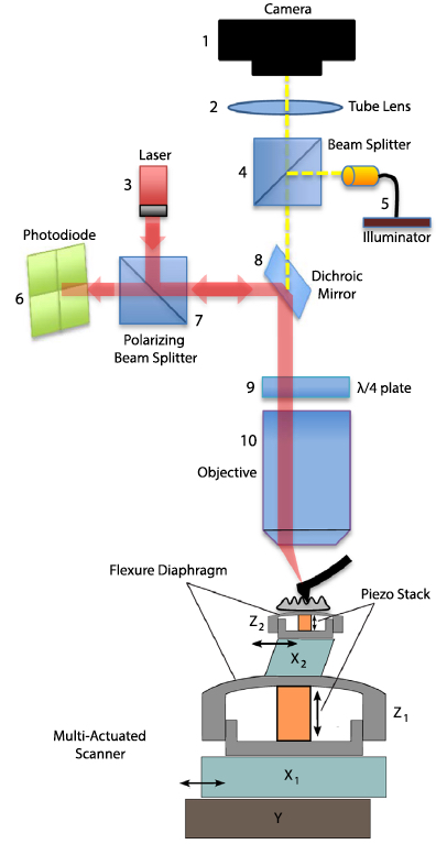
A schematic of the AFM (credit: I. Soltani Bozchalooi et al./Ultramicroscopy)

The group’s design and images, which are based on the PhD work of Iman Soltani Bozchalooi, now a postdoc in the Department of Mechanical Engineering, are published in the journal Ultramicroscopy.

Atomic force microscopes typically scan samples using an ultrafine probe, or needle, that skims along the surface of a sample, tracing its topography, similarly to how a blind person reads Braille. Samples sit on a movable platform, or scanner, that moves the sample laterally and vertically beneath the probe.

Because AFMs scan incredibly small structures, the instruments have to work slowly, line by line, to avoid any sudden movements that could alter the sample or blur the image. Such conventional microscopes typically scan only about one to two lines per second.

To speed up the scanning process, scientists have built platforms that scan samples more quickly, but over a smaller area, and the platforms don’t allow scientists to zoom out to see a wider view or study larger features.

Synchronized Scanners

The main innovation of the new design is a multi-actuated scanner. The sample platform incorporates both a smaller, speedier scanner and a larger, slower scanner for every direction, which work together as one system to scan a wide 3-D region at high speed.

The microscope operates at about eight to 10 frames per second and can scan across hundreds of microns and image features that are several microns high.

“We want to go to real video, which is at least 30 frames per second,” Youcef-Toumi says. “Hopefully we can work on improving the instrument and controls so that we can do video-rate imaging while maintaining its large range and keeping it user-friendly.”


Abstract of Design and control of multi-actuated atomic force microscope for large-range and high-speed imaging
This paper presents the design and control of a high-speed and large-range atomic force microscopy (AFM). A multi-actuation scheme is proposed where several nano-positioners cooperate to achieve the range and speed requirements. A simple data-based control design methodology is presented to effectively operate the AFM scanner components. The proposed controllers compensate for the coupled dynamics and divide the positioning responsibilities between the scanner components. As a result, the multi-actuated scanner behavior is equivalent to that of a single XYZ positioner with large range and high speed. The scanner of the designed AFM is composed of five nano-positioners, features 6 μm out-of-plane and 120 μm lateral ranges and is capable of high-speed operation. The presented AFM has a modular design with laser spot size of 3.5 μm suitable for small cantilever, an optical view of the sample and probe, a conveniently large waterproof sample stage and a 20 MHz data throughput for high resolution image acquisition at high imaging speeds. This AFM is used to visualize etching of calcite in a solution of sulfuric acid. Layer-by-layer dissolution and pit formation along the crystalline lines in a low pH environment is observed in real time.


Abstract of Multi-actuation and PI control: A simple recipe for high-speed and large-range atomic force microscopy

High speed atomic force microscopy enables observation of dynamic nano-scale processes. However, maintaining a minimal interaction force between the sample and the probe is challenging at high speed specially when using conventional piezo-tubes. While rigid AFM scanners are operational at high speeds with the drawback of reduced tracking range, multi-actuation schemes have shown potential for high-speed and large-range imaging. Here we present a method to seamlessly incorporate additional actuators into conventional AFMs. The equivalent behavior of the resulting multi-actuated setup resembles that of a single high-speed and large-range actuator with maximally flat frequency response. To achieve this, the dynamics of the individual actuators and their couplings are treated through a simple control scheme. Upon the implementation of the proposed technique, commonly used PI controllers are able to meet the requirements of high-speed imaging. This forms an ideal platform for retroactive enhancement of existing AFMs with minimal cost and without compromise on the tracking range. A conventional AFM with tube scanner is retroactively enhanced through the proposed method and shows an order of magnitude improvement in closed loop bandwidth performance while maintaining large range. The effectiveness of the method is demonstrated on various types of samples imaged in contact and tapping modes, in air and in liquid.

Will this DNA molecular switch replace conventional transistors?

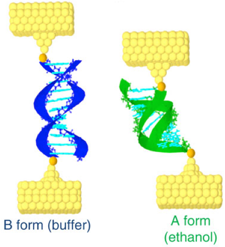
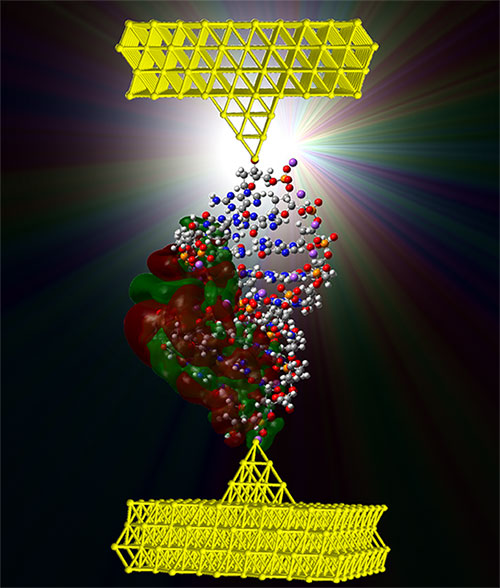
A model of one form of double-stranded DNA attached to two electrodes (credit: UC Davis)

What do you call a DNA molecule that changes between high and low electrical conductance (amount of current flow)?

Answer: a molecular switch (transistor) for nanoscale computing. That’s what a team of researchers from the University of California, Davis and the University of Washington have documented in a paper published in Nature Communications Dec. 9.

“As electronics get smaller they are becoming more difficult and expensive to manufacture, but DNA-based devices could be designed from the bottom-up using directed self-assembly techniques such as ‘DNA origami’,” said Josh Hihath, assistant professor of electrical and computer engineering at UC Davis and senior author on the paper.

DNA origami is the folding of DNA to create two- and three-dimensional shapes at the nanoscale level (see DNA origami articles on KurzweilAI).

Hihath suggests that DNA-based devices may also improve the energy efficiency of electronic circuits, compared to traditional transistors, where power density on-chip has increased as transistors have become miniaturized, limiting further miniaturization.

This illustration shows double-stranded DNA in two configurations, B-form (blue) and A-form (green), bound to gold electrodes (yellow). The linkers to the electrodes (either amines or thiols) are shown in orange. (credit: Juan Manuel Artés et al./Nature Communications)

To develop DNA into a reversible switch, the scientists focused on switching between two stable conformations of DNA, known as the A-form and the B-form. In DNA, the B-form is the conventional DNA duplex molecule. The A-form is a more compact version with different spacing and tilting between the base pairs. Exposure to ethanol forces the DNA into the A-form conformation, resulting in increased conductance. Removing the ethanol causes the DNA to switch back to the B-form and return to its original reduced conductance value.

But the authors advise that to develop this finding into a technologically viable platform for electronics will require a great deal of work to overcome two major hurdles: billions of active DNA molecular devices must be integrated into the same circuit, as is done currently in conventional electronics; and scientists must be able to gate specific devices individually in such a large system.


Abstract of Conformational gating of DNA conductance

DNA is a promising molecule for applications in molecular electronics because of its unique electronic and self-assembly properties. Here we report that the conductance of DNA duplexes increases by approximately one order of magnitude when its conformation is changed from the B-form to the A-form. This large conductance increase is fully reversible, and by controlling the chemical environment, the conductance can be repeatedly switched between the two values. The conductance of the two conformations displays weak length dependencies, as is expected for guanine-rich sequences, and can be fit with a coherence-corrected hopping model. These results are supported by ab initio electronic structure calculations that indicate that the highest occupied molecular orbital is more disperse in the A-form DNA case. These results demonstrate that DNA can behave as a promising molecular switch for molecular electronics applications and also provide additional insights into the huge dispersion of DNA conductance values found in the literature.

Shaking out the nanomaterials: a new method to purify water

Extracting one- and two-dimensional nanomaterials from contaminated water (credit: Michigan Tech)

A new study published in the American Chemical Society’s journal Applied Materials and Interfaces has found a novel—and very simple—way to remove nearly 100 percent of nanomaterials from water.

Water and oil don’t mix, of course, but shaking them together is what makes salad dressing so great. Only instead of emulsifying and capturing bits of shitake or basil in tiny olive oil bubbles, this mixture grabs nanomaterials.

Dongyan Zhang, a research professor of physics at Michigan Tech, led the experiments, which covered tests on carbon nanotubes, graphene, boron nitride nanotubes, boron nitride nanosheets and zinc oxide nanowires. Those are used in everything from carbon fiber golf clubs to sunscreen.

“These materials are very, very tiny, and that means if you try to remove them and clean them out of contaminated water, that it’s quite difficult,” Zhang says, adding that techniques like filter paper or meshes often don’t work.

What makes shaking work is the shape of one- and two-dimensional nanomaterials. As the oil and water separate after some rigorous shaking, the wires, tubes and sheets settle at the bottom of the oil, just above the water. The oils trap them. However, zero-dimensional nanomaterials, such as nanospheres do not get trapped.

Green Nanotechnology

We don’t have to wait until the final vote is in on whether nanomaterials have a positive or negative impact on people’s health and environmental health. With the simplicity of this technique, and how prolific nanomaterials are becoming, removing nanomaterials makes sense. Also, finding ways to effectively remove nanomaterials sooner rather than later could improve the technology’s market potential.

“Ideally for a new technology to be successfully implemented, it needs to be shown that the technology does not cause adverse effects to the environment,” the authors write. “Therefore, unless the potential risks of introducing nanomaterials into the environment are properly addressed, it will hinder the industrialization of products incorporating nanotechnology.”

Purifying water and greening nanotechnology could be as simple as shaking a vial of water and oil.


Michigan Technological University | Shaking the Nanomaterials Out: New Method to Purify Water


Abstract of A Simple and Universal Technique To Extract One- and Two-Dimensional Nanomaterials from Contaminated Water

We demonstrate a universal approach to extract one- and two-dimensional nanomaterials from contaminated water, which is based on a microscopic oil–water interface trapping mechanism. Results indicate that carbon nanotubes, graphene, boron nitride nanotubes, boron nitride nanosheets, and zinc oxide nanowires can be successfully extracted from contaminated water at a successful rate of nearly 100%. The effects of surfactants, particle shape, and type of organic extraction fluids are evaluated. The proposed extraction mechanism is also supported by in situ monitoring of the extraction process. We believe that this extraction approach will prove important for the purification of water contaminated by nanoparticles and will support the widespread adoption of nanomaterial applications.

Skyscraper-style carbon-nanotube chip design ‘boosts electronic performance by factor of a thousand’

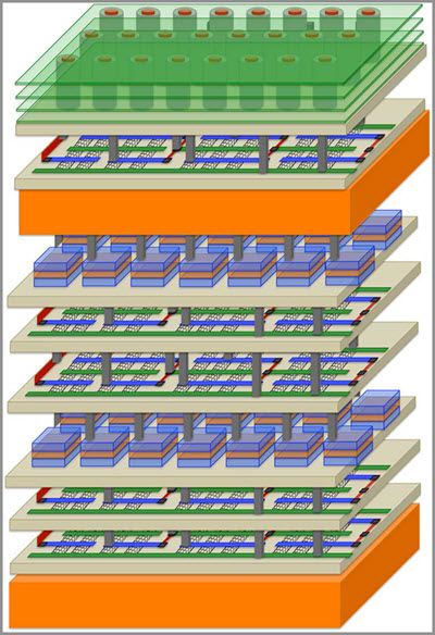
A new revolutionary high-rise architecture for computing (credit: Stanford University)

Researchers at Stanford and three other universities are creating a revolutionary new skyscraper-like high-rise architecture for computing based on carbon nanotube materials instead of silicon.

In Rebooting Computing, a special issue (in press) of the IEEE Computer journal, the team describes its new approach as “Nano-Engineered Computing Systems Technology,” or N3XT.

Suburban-style chip layouts create long commutes and regular traffic jams in electronic circuits, wasting time and energy, they note.

N3XT will break data bottlenecks by integrating processors and memory-like floors in a skyscraper and by connecting these components with millions of “vias,” which play the role of tiny electronic elevators.

The N3XT high-rise approach will move more data, much faster, using far less energy, than would be possible using low-rise circuits, according to the researchers.

Stanford researchers including Associate Professor Subhasish Mitra and Professor H.-S. Philip Wong have “assembled a group of top thinkers and advanced technologies to create a platform that can meet the computing demands of the future,” Mitra says.

“When you combine higher speed with lower energy use, N3XT systems outperform conventional approaches by a factor of a thousand,” Wong claims.

Carbon nanotube transistors

Engineers have previously tried to stack silicon chips but with limited success, the researchers suggest. Fabricating a silicon chip requires temperatures close to 1,800 degrees Fahrenheit, making it extremely challenging to build a silicon chip atop another without damaging the first layer. The current approach to what are called 3-D, or stacked, chips is to construct two silicon chips separately, then stack them and connect them with a few thousand wires.

But conventional 3-D silicon chips are still prone to traffic jams and it takes a lot of energy to push data through what are a relatively few connecting wires.

The N3XT team is taking a radically different approach: building layers of processors and memory directly atop one another, connected by millions of vias that can move more data over shorter distances that traditional wire, using less energy, and immersing computation and memory storage into an electronic super-device.

The key is the use of non-silicon materials that can be fabricated at much lower temperatures than silicon, so that processors can be built on top of memory without the new layer damaging the layer below. As in IBM’s recent chip breakthrough (see “Method to replace silicon with carbon nanotubes developed by IBM Research“), N3XT chips are based on carbon nanotube transistors.

Transistors are fundamental units of a computer processor, the tiny on-off switches that create digital zeroes and ones. CNTs are faster and more energy-efficient than silicon processors, and much thinner. Moreover, in the N3XT architecture, they can be fabricated and placed over and below other layers of memory.

Among the N3XT scholars working at this nexus of computation and memory are Christos Kozyrakis and Eric Pop of Stanford, Jeffrey Bokor and Jan Rabaey of the University of California, Berkeley, Igor Markov of the University of Michigan, and Franz Franchetti and Larry Pileggi of Carnegie Mellon University.

New storage technologies 

Team members also envision using data storage technologies that rely on materials other than silicon. This would allow for the new materials to be manufactured on top of CNTs, using low-temperature fabrication processes.

One such data storage technology is called resistive random-access memory, or RRAM (see “‘Memristors’ based on transparent electronics offer technology of the future“). Resistance slows down electrons, creating a zero, while conductivity allows electrons to flow, creating a one. Tiny jolts of electricity switch RRAM memory cells between these two digital states. N3XT team members are also experimenting with a variety of nanoscale magnetic storage materials.

Just as skyscrapers have ventilation systems, N3XT high-rise chip designs incorporate thermal cooling layers. This work, led by Stanford mechanical engineers Kenneth Goodson and Mehdi Asheghi, ensures that the heat rising from the stacked layers of electronics does not degrade overall system performance.

Mitra and Wong have already demonstrated a working prototype of a high-rise chip. At the International Electron Devices Meeting in December 2014 they unveiled a four-layered chip made up of two layers of RRAM memory sandwiched between two layers of CNTs (see “Stanford engineers invent radical ‘high-rise’ 3D chips“).

In their N3XT paper they ran simulations showing how their high-rise approach was a thousand times more efficient in carrying out many important and highly demanding industrial software applications.


These are the thinnest, strongest plates that can be picked up by hand

Even though they are less than 100 nanometers thick, the researchers’ plates are strong enough to be picked up by hand and retain their shape after being bent and squeezed. (credit: University of Pennsylvania)

Researchers at the University of Pennsylvania have created the thinnest plates that can be picked up and manipulated by hand, using corrugated plates of aluminum oxide. They are thousands of times thinner than a sheet of paper and hundreds of times thinner than household cling wrap, but they spring back to their original shape after being bent and twisted.

Like cling wrap, comparably thin materials immediately curl up on themselves and get stuck in deformed shapes if they are not stretched on a frame or backed by another material. Graphene is even thinner, but it also curls up.

Being able to stay in shape without additional support would allow this material, and others designed on its principles, to be used in aviation and other structural applications where low weight is at a premium.

The plates’ corrugation allow them to remain stiff and stable without the addition of a heavy frame or backing. (credit: University of Pennsylvania)

Mechanical metamaterials

The innovation was to use corrugation (like corrugated cardboard) instead of a frame to keep the material rigid and freestanding. The researchers’ plates are between 25 and 100 nanometers thick and are made of aluminum oxide, which is deposited one atomic layer at a time to achieve precise control of thickness and their distinctive honeycomb shape.

“Aluminum oxide is actually a ceramic, so something that is ordinarily pretty brittle,”  said Igor Bargatin, Assistant Professor of Mechanical Engineering and Applied Mechanics. “You would expect it, from daily experience, to crack very easily. But the plates bend, twist, deform and recover their shape in such a way that you would think they are made out of plastic. The first time we saw it, I could hardly believe it.”

The hexagonal corrugation of the plates is responsible for their stiffness and strength. (credit: University of Pennsylvania)

The plates’ corrugation provides enhanced stiffness. When held from one end, similarly thin films would readily bend or sag, while the honeycomb plates remain rigid. This guards against the common flaw in un-patterned thin films, where they curl up on themselves.

This ease of deformation is tied to another behavior that makes ultra-thin films hard to use outside controlled conditions: they have the tendency to conform to the shape of any surface and stick to it due to Van der Waals forces. Once stuck, they are hard to remove without damaging them. Totally flat films are also particularly susceptible to tears or cracks, which can quickly propagate across the entire material.

The corrugated pattern of the plates is an example of a relatively new field of research: mechanical metamaterials. Like their electromagnetic counterparts, mechanical metamaterials achieve otherwise impossible properties from the careful arrangement of nanoscale features. With mechanical metamaterials, these properties are things like stiffness and strength, rather than their ability to manipulate electromagnetic waves.

A Solara 50 solar-powered drone by Google-owned Titan Aerospace (credit: Titan Aerospace)

That combination of traits could be used to make wings for insect-inspired flying robots or solar-powered drones for beaming the Internet down to Earth that are being explored by Google and Facebook.

“The wings of insects are a few microns thick, and can’t thinner because they’re made of cells,” Bargatin said. “The thinnest man-made wing material I know of is made by depositing a Mylar film on a frame, and it’s about half a micron thick. Our plates can be ten or more times thinner than that, and don’t need a frame at all. As a result, they weigh as little as than a tenth of a gram per square meter.”

The research was published in an open-access paper in the journal Nature Communications.


Abstract of Ultralight shape-recovering plate mechanical metamaterials

Unusual mechanical properties of mechanical metamaterials are determined by their carefully designed and tightly controlled geometry at the macro- or nanoscale. We introduce a class of nanoscale mechanical metamaterials created by forming continuous corrugated plates out of ultrathin films. Using a periodic three-dimensional architecture characteristic of mechanical metamaterials, we fabricate free-standing plates up to 2 cm in size out of aluminium oxide films as thin as 25 nm. The plates are formed by atomic layer deposition of ultrathin alumina films on a lithographically patterned silicon wafer, followed by complete removal of the silicon substrate. Unlike unpatterned ultrathin films, which tend to warp or even roll up because of residual stress gradients, our plate metamaterials can be engineered to be extremely flat. They weigh as little as 0.1 g cm−2 and have the ability to ‘pop-back’ to their original shape without damage even after undergoing multiple sharp bends of more than 90°.

Recyclable, sustainable petroleum-free bioplastics

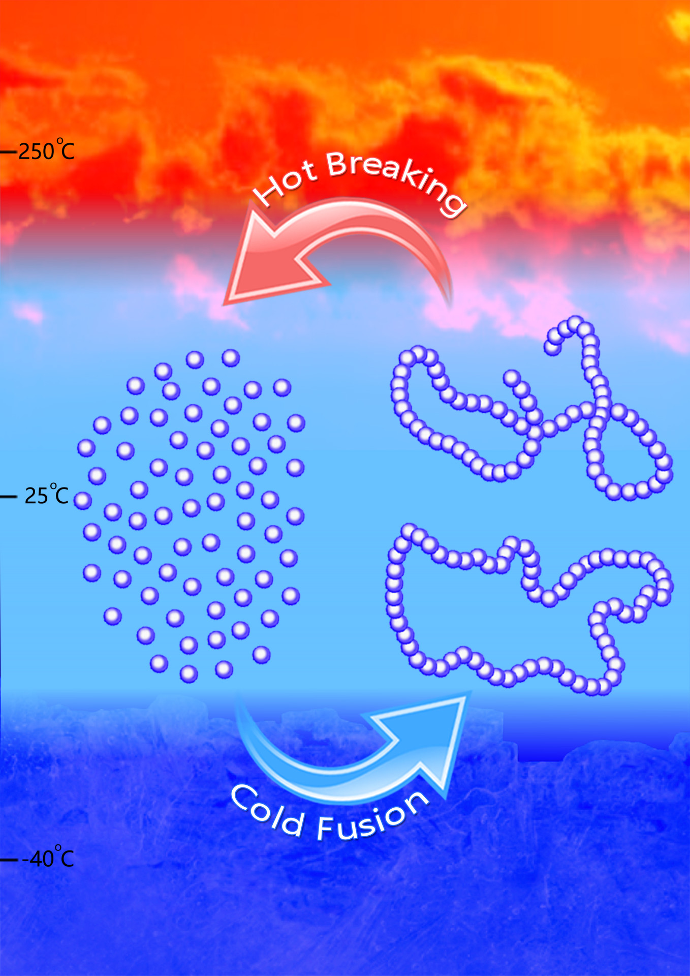
The new polymer synthesis process. The single molecules (monomers) are cooled to polymerize; to cycle back, heat is applied. (credit: Jing Tang/Chen lab)

The textbooks and journals (and especially the oil companies) said making a completely recyclable, biodegradable, petroleum-free polymer couldn’t be done.

But Colorado State University chemists have done it — paving a potential new road to truly sustainable, petroleum-free plastics. Just reheat is for an hour and it converts back to its original molecular state, ready for reuse.

Their starting feedstock: a biorenewable monomer that textbooks and journal papers had declared non-polymerizable, meaning it could not be bonded into the large molecules (polymers) typically required for use as a material.

Renewable plastics

Plastics are the most common type of manmade polymer, which is the chemical term for a long chain of repeating small molecules, or monomers. Petroleum-based polymers like polyethylene and polystyrene have come under fire for piling up in landfills and even in oceans.

“More than 200 pounds of synthetic polymers are consumed per person each year — plastics probably the most in terms of production volume. And most of these polymers are not biorenewable,” said Colorado State professor of chemistry Eugene Chen. “The big drive now is to produce biorenewable and biodegradable polymers or plastics. That is, however, only one part of the solution, as biodegradable polymers are not necessarily recyclable, in terms of feedstock recycling.”

There are several biodegradable plastics on the market today, chief among them a starch-based material made from polylactic acid, or PLA. Compostable cups, cutlery and packaging in dining halls are made from PLA. They’re biodegradable, but they’re not truly recyclable — once made, they can’t be completely reconstituted into their original monomeric states without forming other, unwanted byproducts.

Sustainable biomass source

The researchers’ starting monomer is gamma-butyrolactone, or GBL. It is a colorless liquid and common chemical reagent, derived from a biomass compound best suited to replace petrochemicals, according to the Department of Energy. Textbooks and scientific literature had described these small molecules as thermally stable in their monomeric chemical states could not polymerize.

But Chen and Hong figured out how to get this material to take different shapes, such as linear or cyclic, based on the catalysts and conditions they selected. They used both metal-based and metal-free catalysts to synthesize the polymer, called poly(GBL), which is chemically equivalent to a commercial biomaterial called poly(4-hydroxybutyrate), or P4HB.

To convert the polymer back into the original monomer, demonstrating the thermal recyclability of the polymer, they employed specifically designed reaction conditions, including low temperature, to make the polymer, along with heat between 220–300 degrees Celsius.

P4HB is derived from bacteria, which is a more expensive, complex process than how most plastics are made. Instead, by starting with the readily available GBL and ending up with a replacement material for P4HB, Chen’s discovery has promising market potential, and a provisional patent has been filed with the help of CSU Ventures.


Abstract of Completely recyclable biopolymers with linear and cyclic topologies via ring-opening polymerization of γ-butyrolactone

Ring-opening polymerization (ROP) is a powerful synthetic methodology for the chemical synthesis of technologically important biodegradable aliphatic polyesters from cyclic esters or lactones. However, the bioderived five-membered γ-butyrolactone (γ-BL) is commonly referred as ‘non-polymerizable’ because of its low strain energy. The chemical synthesis of poly(γ-butyrolactone) (PγBL) through the ROP process has been realized only under ultrahigh pressure (20,000 atm, 160 °C) and only produces oligomers. Here we report that the ROP of γ-BL can, with a suitable catalyst, proceed smoothly to high conversions (90%) under ambient pressure to produce PγBL materials with a number-average molecular weight up to 30 kg mol–1 and with controlled linear and/or cyclic topologies. Remarkably, both linear and cyclic PγBLs can be recycled back into the monomer in quantitative yield by simply heating the bulk materials at 220 °C (linear polymer) or 300 °C (cyclic polymer) for one hour, which thereby demonstrates the complete recyclability of PγBL.

White graphene + graphene –> super-thin, cooler, more flexible electronics

Growth and transfer of 2-D material such as hexagonal boron nitride and graphene was performed by a team that included Yijing Stehle of Oak Ridge National Laboratory. (credit: ORNL)

A new era of electronics and even quantum devices could be ushered in with the fabrication of a virtually perfect single layer of “white graphene,” according to researchers at the Department of Energy’s Oak Ridge National Laboratory.

The material is technically known as hexagonal boron nitride (see “New inventions track greenhouse gas, remediate oil spills“). It is an insulator (instead of a conductor of electricity as with graphene), so it could serve as a 2-D dielectric (insulating material) in electronic devices such as thin-film capacitors.

It also has even better transparency than graphene, making it useful as a substrate and the foundation for the electronics in cell phones, laptops, tablets and many other devices.

“As thin as a piece of paper”

ORNL’s Yijing Stehle, postdoctoral associate and lead author of a paper published in Chemistry of Materials and colleagues are working on combining graphene and boron nitride in a 2-D capacitor and fuel cell prototype that are “super thin” and also transparent.

With their recipe for white graphene, ORNL researchers hope to unleash the full potential of graphene as a conductor. By combining it with white graphene as a substrate, researchers believe they can make thinner, more-flexible multilayer electronic devices.

“Imagine batteries, capacitors, solar cells, video screens and fuel cells as thin as a piece of paper,” she said.

For its part, graphene on a white-graphene substrate also has several thousand times higher electron mobility than using graphene on other substrates. That feature could enable data transfers that are much faster than what is available today.

Cool electronics

A recent theoretical study led by Rice University proposed the use of white graphene to cool electronics (see “Why ‘white graphene’ structures are cool“). Stehle and colleagues have made high-quality layers of hexagonal boron nitride that support that study; they believe the material can be cost-effectively scaled up to large production volumes.

The Rice process consists of standard atmospheric pressure chemical vapor deposition with a similar furnace, temperature and time. But Stehle describes “a more gentle, controllable way to release the reactant into the furnace and figuring out how to take advantage of inner furnace conditions.”

New Mexico State University researchers were also involved in the study, which was supported by the DOE’s Office of Science.


Abstract of Synthesis of Hexagonal Boron Nitride Monolayer: Control of Nucleation and Crystal Morphology

Monolayer hexagonal boron nitride (hBN) attracts significant attention due to the potential to be used as a complementary two-dimensional dielectric in fabrication of functional 2D heterostructures. Here we investigate the growth stages of the hBN single crystals and show that hBN crystals change their shape from triangular to truncated triangular and further to hexagonal depending on copper substrate distance from the precursor. We suggest that the observed hBN crystal shape variation is affected by the ratio of boron to nitrogen active species concentrations on the copper surface inside the CVD reactor. Strong temperature dependence reveals the activation energies for the hBN nucleation process of ∼5 eV and crystal growth of ∼3.5 eV. We also show that the resulting h-BN film morphology is strongly affected by the heating method of borazane precursor and the buffer gas. Elucidation of these details facilitated synthesis of high quality large area monolayer hexagonal boron nitride by atmospheric pressure chemical vapor deposition on copper using borazane as a precursor.

How to make diamond objects with a laser at room temperature

A scanning electron microscopy image of microdiamonds made using the new technique (credit: Jagdish Narayan, Anagh Bhaumik/APL Materials)

Researchers from North Carolina State University have discovered a new phase of solid carbon, called Q-carbon, that is distinct from the known phases of graphite and diamond. They have also developed a technique for using Q-carbon to make diamond-related structures at room temperature and at ambient atmospheric pressure in air.*

Phases are distinct forms of the same material. Graphite is one of the solid phases of carbon; diamond is another.

“We’ve now created a third solid phase of carbon,” says Jay Narayan, the John C. Fan Distinguished Chair Professor of Materials Science and Engineering at NC State and lead author of three papers describing the work. “The only place it may be found in the natural world would be possibly in the core of some planets.”

Q-carbon has some unusual characteristics:

  • It’s ferromagnetic, which other solid forms of carbon are not.
  • It’s harder than diamond, and glows when exposed to even low levels of energy. “Q-carbon’s strength and low work-function — its willingness to release electrons — make it very promising for developing new electronic display technologies,” Narayan says.
  • It can be used to create a variety of single-crystal diamond objects. “We can create diamond nanoneedles or microneedles, nanodots, or large-area diamond films, with applications for drug delivery, industrial processes and for creating high-temperature switches and power electronics,” Narayan says. “These diamond objects have a single-crystalline structure, making them stronger than polycrystalline materials. And it is all done at room temperature and at ambient atmosphere — we’re basically using a laser like the ones used for laser eye surgery. So, not only does this allow us to develop new applications, but the process itself is relatively inexpensive.”

Coming soon: Q-carbon nanodots

“We can make Q-carbon films, and we’re learning its properties, but we are still in the early stages of understanding how to manipulate it,” Narayan says. “We know a lot about diamond, so we can make diamond nanodots. We don’t yet know how to make Q-carbon nanodots or microneedles. That’s something we’re working on.”

NC State has filed two provisional patents on the Q-carbon and diamond creation techniques.

The work is described in two papers, one to be published online Nov. 30 in the Journal of Applied Physics and another was published in an open-access paper, Oct. 7 in the journal APL Materials. The work was supported in part by the National Science Foundation.

* Researchers start with a substrate, such as such as sapphire, glass or a plastic polymer. The substrate is then coated with amorphous carbon — elemental carbon that, unlike graphite or diamond, does not have a regular, well-defined crystalline structure. The carbon is then hit with a single laser pulse lasting approximately 200 nanoseconds. During this pulse, the temperature of the carbon is raised to 4,000 Kelvin (or around 3,727 degrees Celsius) and then rapidly cooled. This operation takes place at one atmosphere — the same pressure as the surrounding air.

The end result is a film of Q-carbon, and researchers can control the process to make films between 20 nanometers and 500 nanometers thick. By using different substrates and changing the duration of the laser pulse, the researchers can also control how quickly the carbon cools. By changing the rate of cooling, they are able to create diamond structures within the Q-carbon.


Abstract of Research Update: Direct conversion of amorphous carbon into diamond at ambient pressures and temperatures in air

We report on fundamental discovery of conversion of amorphous carbon into diamond by irradiating amorphous carbon films with nanosecond lasers at room-temperature in air at atmospheric pressure. We can create diamond in the form of nanodiamond (size range <100 nm) and microdiamond (>100 nm). Nanosecond laser pulses are used to melt amorphousdiamondlike carbon and create a highly undercooled state, from which various forms ofdiamond can be formed upon cooling. The quenching from the super undercooled state results in nucleation of nanodiamond. It is found that microdiamonds grow out of highly undercooled state of carbon, with nanodiamond acting as seed crystals.


Abstract of Novel Phase of Carbon, Ferromagnetism and Conversion into Diamond

We report the discovery of new phase of carbon (referred to as Q-carbon) and address fundamental issues related to direct conversion of carbon into diamond at ambient temperatures and pressures in air without any need for catalyst and presence of hydrogen. The Q-carbon is formed as result of quenching from super undercooled state by using high-power nanosecond laser pulses. We discuss the equilibrium phase diagram (P vs. T) of carbon, and show that by rapid quenching kinetics can shift thermodynamic graphite/diamond/ liquid carbon triple point from 5000K/12GPa to super undercooled carbon at atmospheric pressure in air. It is shown that nanosecond laser heating of diamondlike amorphous carbon on sapphire, glass and polymer substrates can be confined to melt carbon in a super undercooled state. By quenching the carbon from the super undercooled state, we have created a new state of carbon (Q-carbon) from which nanodiamond, microdiamond, microneedles and thin films are formed depending upon the nucleation and growth times allowed for diamond formation. The Q-carbon quenched from liquid is a new state of solid carbon with a higher mass density than amorphous carbon and a mixture of mostly four-fold sp3 (75-85%) and the rest three-fold sp2 bonded carbon (with distinct entropy). It is expected to have new and improved mechanical hardness, electrical conductivity, chemical and physical properties, including room-temperature ferromagnetism (RTFM) and enhanced field emission. Here we present interesting results on RTFM, enhanced electrical conductivity, and surface potential of Q-carbon to emphasize its unique properties. The Q-carbon exhibits robust bulk ferromagnetism with estimated Curie temperature of about 500K and saturation magnetization value of 20 emu g-1. From the Q-carbon, diamond phase is nucleated and a variety of micro- and nanostructures, and large-area single-crystal diamond sheets are grown by allowing growth times as needed. Subsequent laser pulses can be used to grow nanodiamond into microdiamond and nucleate other nanostructures of diamond on the top of existing microdiamond and create novel nanostructured materials. The microstructural details provide insights into the mechanism of nanodiamond and microdiamond formation. This process allows carbon to diamond conversion and formation of useful nanostructures and microstructures at ambient temperatures in air at atmospheric pressure on practical and heat-sensitive substrates in a controlled way without need for any catalysts and hydrogen to stabilize sp3 bonding for diamond formation.

New inventions track greenhouse gas, remediate oil spills

Camera test at Foljesjon, a lake in a research area west of Vanersborg, Sweden (credit: Linkoping University)

A new camera that can image methane in the air, allowing for precision monitoring of a greenhouse gas, has been developed by a team of researchers from Linköping and Stockholm Universities.

The new camera should help us better understand the rapid but irregular increase of methane in the atmosphere (which has puzzled researchers) and identify the sources and sinks of methane in the landscape. It may also suggest ways to reduce emissions.

”The camera is very sensitive, which means that the methane is both visible and measureable close to ground level, with much higher resolution [less than a square meter and at ambient levels (~1.8 ppmv, or parts per million volume)] than previously. Being able to measure on a small scale is crucial,” says Magnus Gålfalk, Assistant Professor at Tema Environmental Change, Linköping University who led the study.

An image of methane gas from the hyperspectral infrared camera, visualized in purple (credit: Linköping University)

The advanced hyperspectral (across the spectrum) thermal infrared camera weighs 30 kilos and measures 50 x 45 x 25 centimeters. It is optimized to measure the same portion of the solar radiation spectrum that methane absorbs and which makes methane such a powerful greenhouse gas.

The camera can be used to measure emissions from many environments including sewage sludge deposits, combustion processes, animal husbandry, and lakes.

For each pixel in the image (320 x 256 pixels), the camera records a precise spectrum range (in the 7.7 microns thermal IR region), which makes it possible to quantify the methane separately from the other gases.

The camera was developed by a team with expertise in astronomy, biogeochemistry, engineering. and environmental sciences. “We’re working to make it airborne for more large-scale methane mapping,” says principal investigator David Bastviken, professor at Tema Environmental Change, Linköping University.

The research was recently published in Nature Climate Change.

Super-absorbent material to soak up oil spills

Boron nitride material supported by a plant spike, demonstrating its light weight (credit: Weiwei Lei et al./Nature Communications)

In hopes of limiting the disastrous environmental effects of massive oil spills, materials scientists from Drexel University and Deakin University (Australia) have teamed up to manufacture and test a new “boron nitride nanosheet” material that can absorb oils and organic solvents up to 33 times its weight. That could make it possible to quickly mitigate these costly, environmentally damaging accidents.

The material, which literally absorbs oil like a sponge, is now ready to be tested by industry after two years of refinement in the laboratory at Deakin’s Institute for Frontier Materials (IFM).

Deakin Professor Ying (Ian) Chen, PhD, the lead author of the project’s research paper, recently published in Nature Communications, said the material is the most exciting advancement in oil spill remediation technology in decades.

“Oil spills are a global problem and wreak havoc on our aquatic ecosystems, not to mention they cost billions of dollars in damage,” Chen said. “Everyone remembers the Gulf Coast disaster, but here in Australia they are a regular problem, and not just in our waters. Oil spills from trucks and other vehicles can close freeways for an entire day, again amounting to large economic losses,” Chen said.

The nanosheet is made up of flakes just several nanometers (one billionth of a meter) in thickness with tiny holes. This strecture enables the nanosheet to increase its effective surface area to 273 square meters (3000 square feet) per gram.

Researchers from Drexel’s College of Engineering helped to study and functionalize the material, which started as boron nitride powder, commonly called “white graphite.” By forming the powder into atomically thin sheets, the material could be made into a sponge.

“The mechanochemical technique developed meant it was possible to produce high-concentration stable aqueous colloidal solutions of boron nitride sheets, which could then be transformed into the ultralight porous aerogels and membranes for oil clean-up,” said Vadym Mochalin, PhD, a co-author of the paper, who was a research associate professor at Drexel while working on the project, and is now an associate professor at Missouri University of Science and Technology.

The Drexel team used computational modeling to help understand the intimate details of how the material was formed. In the process, the team learned that the boron nitride nanosheets are flame resistant — which means they could also find applications in electrical and heat insulation.

The nanotechnology team at Deakin’s Institute for Frontier Materials has been working on boron nitride nanomaterials for two decades and has been internationally recognized for its work in the development of boron nitride nanotubes and nanosheets. This project is the next step in the IFM’s continued research to discover new uses for the material.


Abstract of Making methane visible

Methane (CH4) is one of the most important greenhouse gases, and an important energy carrier in biogas and natural gas. Its large-scale emission patterns have been unpredictable and the source and sink distributions are poorly constrained. Remote assessment of CH4 with high sensitivity at a m2 spatial resolution would allow detailed mapping of the near-ground distribution and anthropogenic sources in landscapes but has hitherto not been possible. Here we show that CH4 gradients can be imaged on the <m2scale at ambient levels (~1.8 ppm) and filmed using optimized infrared (IR) hyperspectral imaging. Our approach allows both spectroscopic confirmation and quantification for all pixels in an imaged scene simultaneously. It also has the ability to map fluxes for dynamic scenes. This approach to mapping boundary layer CH4 offers a unique potential way to improve knowledge about greenhouse gases in landscapes and a step towards resolving source–sink attribution and scaling issues.


Abstract of Boron nitride colloidal solutions, ultralight aerogels and freestanding membranes through one-step exfoliation and functionalization

Manufacturing of aerogels and membranes from hexagonal boron nitride (h-BN) is much more difficult than from graphene or graphene oxides because of the poor dispersibility of h-BN in water, which limits its exfoliation and preparation of colloidal solutions. Here, a simple, one-step mechano-chemical process to exfoliate and functionalize h-BN into highly water-dispersible, few-layer h-BN containing amino groups is presented. The colloidal solutions of few-layer h-BN can have unprecedentedly high concentrations, up to 30 mg ml−1, and are stable for up to several months. They can be used to produce ultralight aerogels with a density of 1.4 mg cm−3, which is ~1,500 times less than bulk h-BN, and freestanding membranes simply by cryodrying and filtration, respectively. The material shows strong blue light emission under ultraviolet excitation, in both dispersed and dry state.

A molecular light-driven nanosubmarine

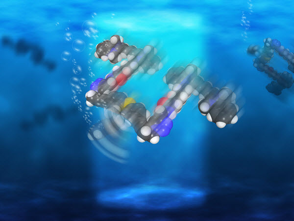
Rice University scientists have created light-driven, single-molecule submersibles that contain just 244 atoms (credit: Loïc Samuel/Rice University)

The Rice University lab of chemist James Tour has created single-molecule, 244-atom submersibles with motors powered by ultraviolet light, as they reported this month in the American Chemical Society journal Nano Letters.

With each full revolution, the motor’s tail-like propeller moves the sub forward 18 nanometers, but with the motors running at more than a million RPM, that translates into almost 1 inch per second — a breakneck pace on the molecular scale, says Tour. “These are the fastest-moving molecules ever seen in solution,” he said.

This chemical schematic shows the design of single-molecule nanosubmersibles created at Rice University. The nanosub’s fluorescent pontoons are blue; the motor is red. (credit: Victor García-López/Rice University)

While they can’t be steered yet, the study proves molecular motors are powerful enough to drive the sub-10-nanometer subs through solutions of moving molecules of about the same size. “This is akin to a person walking across a basketball court with 1,000 people throwing basketballs at him,” Tour said.

In 2006, Tour’s lab introduced the world to nanocars, single-molecule cars with four wheels, axles, and independent suspensions that could be “driven” across a surface (see “Rice scientists attach motor to single-molecule car“).

Tour said many scientists have created microscopic machines with motors over the years, but most have either used or generated toxic chemicals. He said a motor that was conceived in 2009 by a group in the Netherlands proved suitable for Rice’s submersibles, which were produced in a 20-step chemical synthesis.

“These motors are well-known and used for different things,” said lead author and Rice graduate student Victor García-López. “But we were the first ones to propose they can be used to propel nanocars and now submersibles.”

Operate like a bacteria’s flagellum

A nanosubmarine found in nature: this whip-like flagellum powers a bacterium’s swimming (credit: LadyofHats/CC)

The motors, which operate more like a bacteria’s flagellum than a propeller, complete each revolution in four steps. When excited by light, the double bond that holds the rotor to the body becomes a single bond, allowing it to rotate a quarter step. As the motor seeks to return to a lower energy state, it jumps adjacent atoms for another quarter turn. The process repeats as long as the light is on.

For comparison tests, the lab also made submersibles with no motors, slow motors, and motors that paddle back and forth. All versions of the submersibles have pontoons that fluoresce red when excited by a laser, according to the researchers.

Once built, the sub’s performance was independently confirmed by Gufeng Wang at North Carolina State University.

Rice’s researchers hope future nanosubs will be able to carry cargoes for medical and other purposes. “There’s a path forward,” García-López said. “This is the first step, and we’ve proven the concept. Now we need to explore opportunities and potential applications.”

Wang is an assistant professor of analytical chemistry at North Carolina State. Tour is the T.T. and W.F. Chao Chair in Chemistry as well as a professor of computer science and of materials science and nanoengineering.

The National Science Foundation, the National Institutes of Health, the Welch Foundation, and North Carolina State supported the research.


Abstract of Unimolecular Submersible Nanomachines. Synthesis, Actuation, and Monitoring

Unimolecular submersible nanomachines (USNs) bearing light-driven motors and fluorophores are synthesized. NMR experiments demonstrate that the rotation of the motor is not quenched by the fluorophore and that the motor behaves in the same manner as the corresponding motor without attached fluorophores. No photo or thermal decomposition is observed. Through careful design of control molecules with no motor and with a slow motor, we found using single molecule fluorescence correlation spectroscopy that only the molecules with fast rotating speed (MHz range) show an enhancement in diffusion by 26% when the motor is fully activated by UV light. This suggests that the USN molecules give ∼9 nm steps upon each motor actuation. A non-unidirectional rotating motor also results in a smaller, 10%, increase in diffusion. This study gives new insight into the light actuation of motorized molecules in solution.