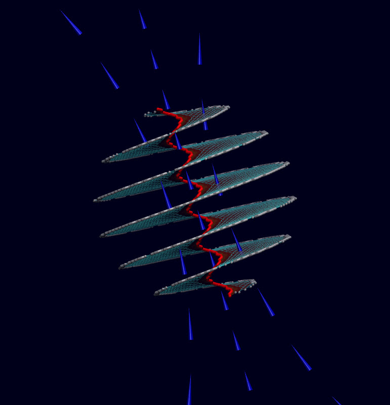
A new 3-​​D printing method for creating patient-​​specific medical devices

The 3D magnetic printing process systematically aligns and selectively polymerizes groupings of voxels (volume “pixels”) programmed to have specific reinforcement orientation within each layer of printed material based upon a shifting field. The 3-D printer build plate peels after a layer is complete to print additional layers. (credit: Joshua J. Martin et al./Nature Communications)

Northeastern University engineers have devel­oped a 3-D printing process that uses mag­netic fields to shape com­posite materials (mixes of plas­tics and ceramics) into patient-specific biomedical devices, such as catheters.

The devices are intended to be stronger and lighter than cur­rent models and the cus­tomized design could ensure an appro­priate fit, said Ran­dall Erb, assis­tant pro­fessor in the Depart­ment of Mechan­ical and Indus­trial Engi­neering.

The magnetic field enables the engineers to con­trol how the ceramic fibers are arranged, allowing for con­trol of the mechan­ical prop­er­ties of the mate­rial. That con­trol is crit­ical if you’re crafting devices with com­plex archi­tec­tures, such as cus­tomized minia­ture bio­med­ical devices. Within a single patient-specific device, the cor­ners, the curves, and the holes must all be rein­forced by ceramic fibers arranged in just the right con­fig­u­ra­tion to make the device durable.

This is the strategy taken by many nat­ural com­pos­ites from bones to trees. Fibers of cal­cium phos­phate, the min­eral com­po­nent of bone, are nat­u­rally ori­ented precisely around the holes for blood ves­sels to ensure the bone’s strength and sta­bility to enable, say, your femur to with­stand a daily jog.

Aligning fibers with magnets

The 3D magnetic-printer setup. A digital light processor (DLP) photo-polymerizes resin with UV while a magnetic field is simultaneously applied via electromagnetic solenoids. (credit: Joshua J. Martin et al./Nature Communications)

Erb ini­tially described the role of magnets in the composite-making process in a 2012 paper in the journal Sci­ence. First the researchers “mag­ne­tize” the ceramic fibers by dusting them very lightly with iron oxide, which has been FDA-approved for drug-delivery appli­ca­tions.

They then apply ultra-low mag­netic fields to indi­vidual sec­tions of the com­posite material — the ceramic fibers immersed in liquid plastic — to align the fibers according to the exacting spec­i­fi­ca­tions dic­tated by the product they are printing.

In a video accom­pa­nying the Sci­ence article, you can see the fibers spring to atten­tion when the mag­netic field is turned on. “Mag­netic fields are very easy to apply,” says Erb. “They’re safe, and they pen­e­trate not only our bodies but many other materials.”

Finally, in a process called “stere­olith­o­g­raphy,” they build the product, layer by layer, using a computer-controlled laser beam that hardens the plastic. Each six-by-six inch layer takes a minute to complete.

Using mag­nets, the new printing method aligns each minus­cule fiber in the direc­tion that con­forms pre­cisely to the geom­etry of the item being printed.

“If you can print a catheter whose geom­etry is spe­cific to the indi­vidual patient, you can insert it up to a cer­tain crit­ical spot, you can avoid punc­turing veins, and you can expe­dite delivery of the contents.”

The engineers’ open-access paper on the new tech­nology appears in the Oct. 23 issue of Nature Com­mu­ni­ca­tions.

Custom-designing neonatal catheters

Erb has received a $225,000 Small Busi­ness Tech­nology Transfer grant from the National Institutes of Health to develop neonatal catheters with a local com­pany. “Another of our goals is to use cal­cium phos­phate fibers and bio­com­pat­ible plas­tics to design sur­gical implants.”

Neonatal preemie with catheters (credit: March of Dimes Foundation)

The new technology is especially valuable for prema­ture babies (“preemies”) in neonatal care units, some weighing just a bit over a pound, with plastic tubes snaking through their nose or mouth, or dis­ap­pearing into veins or other parts of the body. Those tubes, or “catheters,” are how the babies get the nec­es­sary oxygen, nutri­ents, fluid, and med­ica­tions to stay alive.

The problem is, today’s catheters only come in stan­dard sizes and shapes, which means they cannot accom­mo­date the needs of all pre­ma­ture babies. “With neonatal care, each baby is a dif­ferent size, each baby has a dif­ferent set of prob­lems,” says Erb.

Worldwide, “15 million babies are born too soon every year” and of those, “1 million children die each year due to complications of preterm birth,” according to a report by the World Health Organization. This data was cited in the “March of Dimes Premature Birth Report Card,” issued today (Nov. 5) by March of Dimes. “Babies who survive an early birth often face serious and lifelong health problems, including breathing problems, jaundice, vision loss, cerebral palsy, and intellectual delays,” the March of Dimes report noted.

The report provides rates and grades for major cities or counties in each U.S. state and Puerto Rico. It also provides preterm birth rates by race and ethnicity. The U.S. preterm birth rate ranks among the worst of high-resource countries, the March of Dimes says.


Abstract of Designing bioinspired composite reinforcement architectures via 3D magnetic printing

Discontinuous fibre composites represent a class of materials that are strong, lightweight and have remarkable fracture toughness. These advantages partially explain the abundance and variety of discontinuous fibre composites that have evolved in the natural world. Many natural structures out-perform the conventional synthetic counterparts due, in part, to the more elaborate reinforcement architectures that occur in natural composites. Here we present an additive manufacturing approach that combines real-time colloidal assembly with existing additive manufacturing technologies to create highly programmable discontinuous fibre composites. This technology, termed as ‘3D magnetic printing’, has enabled us to recreate complex bioinspired reinforcement architectures that deliver enhanced material performance compared with monolithic structures. Further, we demonstrate that we can now design and evolve elaborate reinforcement architectures that are not found in nature, demonstrating a high level of possible customization in discontinuous fibre composites with arbitrary geometries.

Controlling acoustic properties with algorithms and computational methods

A “zoolophone” with animal shapes automatically created using a computer algorithm. The tone of each key is comparable to those of professionally made instruments as a demonstration of an algorithm for computationally designing an object’s vibrational properties and sounds. (Changxi Zheng/Columbia Engineering)

Computer scientists at Columbia Engineering, Harvard, and MIT have demonstrated that acoustic properties — both sound and vibration — can be controlled by 3D-printing specific shapes.

They designed an optimization algorithm and used computational methods and digital fabrication to alter the shape of 2D and 3D objects, creating what looks to be a simple children’s musical instrument — a xylophone with keys in the shape of zoo animals.

Practical uses

“Our discovery could lead to a wealth of possibilities that go well beyond musical instruments,” says Changxi Zheng, assistant professor of computer science at Columbia Engineering, who led the research team.

“Our algorithm could lead to ways to build less noisy computer fans and bridges that don’t amplify vibrations under stress, and advance the construction of micro-electro-mechanical resonators whose vibration modes are of great importance.”

Zheng, who works in the area of dynamic, physics-based computational sound for immersive environments, wanted to see if he could use computation and digital fabrication to actively control the acoustical property, or vibration, of an object.

Zheng’s team decided to focus on simplifying the slow, complicated, manual process of designing “idiophones” — musical instruments that produce sounds through vibrations in the instrument itself, not through strings or reeds.

The surface vibration and resulting sounds depend on the idiophone’s shape in a complex way, so designing the shapes to obtain desired sound characteristics is not straightforward, and their forms have so far been limited to well-understood designs such as bars that are tuned by careful drilling of dimples on the underside of the instrument.

Optimizing sound properties

To demonstrate their new technique, the team settled on building a “zoolophone,” a metallophone with playful animal shapes (a metallophone is an idiophone made of tuned metal bars that can be struck to make sound, such as a glockenspiel).

Their algorithm optimized and 3D-printed the instrument’s keys in the shape of colorful lions, turtles, elephants, giraffes, and more, modelling the geometry to achieve the desired pitch and amplitude of each part.

“Our zoolophone’s keys are automatically tuned to play notes on a scale with overtones and frequency of a professionally produced xylophone,” says Zheng, whose team spent nearly two years on developing new computational methods while borrowing concepts from computer graphics, acoustic modeling, mechanical engineering, and 3D printing.

“By automatically optimizing the shape of 2D and 3D objects through deformation and perforation, we were able to produce such professional sounds that our technique will enable even novices to design metallophones with unique sound and appearance.”

3D metallophone cups automatically created by computers (credit: Changxi Zheng/Columbia Engineering)

The zoolophone represents fundamental research into understanding the complex relationships between an object’s geometry and its material properties, and the vibrations and sounds it produces when struck.

While previous algorithms attempted to optimize either amplitude (loudness) or frequency, the zoolophone required optimizing both simultaneously to fully control its acoustic properties. Creating realistic musical sounds required more work to add in overtones, secondary frequencies higher than the main one that contribute to the timbre associated with notes played on a professionally produced instrument.

Looking for the most optimal shape that produces the desired sound when struck proved to be the core computational difficulty: the search space for optimizing both amplitude and frequency is immense. To increase the chances of finding the most optimal shape, Zheng and his colleagues developed a new, fast stochastic optimization method, which they called Latin Complement Sampling (LCS).

They input shape and user-specified frequency and amplitude spectra (for instance, users can specify which shapes produce which note) and, from that information, optimized the shape of the objects through deformation and perforation to produce the wanted sounds. LCS outperformed all other alternative optimizations and can be used in a variety of other problems.

“Acoustic design of objects today remains slow and expensive,” Zheng notes. “We would like to explore computational design algorithms to improve the process for better controlling an object’s acoustic properties, whether to achieve desired sound spectra or to reduce undesired noise. This project underscores our first step toward this exciting direction in helping us design objects in a new way.”

Zheng, whose previous work in computer graphics includes synthesizing realistic sounds that are automatically synchronized to simulated motions, has already been contacted by researchers interested in applying his approach to micro-electro-mechanical systems (MEMS), in which vibrations filter RF signals.

Their work—“Computational Design of Metallophone Contact Sounds”—will be presented at SIGGRAPH Asia on November 4 in Kobe, Japan.

The work at Columbia Engineering was supported in part by the National Science Foundation (NSF) and Intel, at Harvard and MIT by NSF, Air Force Research Laboratory, and DARPA.


Longer-lasting, lighter lithium-ion batteries from silicon anodes

Schematic of electrode process design. (a) Components mixing under ultrasonic irradiation, (b) an optical image of the as-fabricated electrode made of silicon nanoparticles (SiNP), sulpher-doped graphene (SG), and polyacrylonitrile (PAN), (c) the electrode after sluggish heat treatment (SHT), (d) Schematic of the atomic-scale structure of the electrode. (credit: Fathy M. Hassan et al./Nature Communications)

Zhongwei Chen, a chemical engineering professor at the University of Waterloo, and a team of graduate students have created a new low-cost battery design using silicon instead of graphite, boosting the performance and life of lithium-ion batteries.

Waterloo’s silicon battery technology promises a 40 to 60 per cent increase in energy density (energy storage per unit volume), which is important for consumers with smartphones, smart homes, and smart wearables. It also means an electric car could be driven up to 500 kilometers (311 miles) between charges while reducing its overall weight.

The graphite bottleneck

The Waterloo engineers found that silicon anode materials are capable of producing batteries that store almost 10 times more energy than with graphite.

“As batteries improve, graphite is slowly becoming a performance bottleneck because of the limited amount of energy that it can store,” said Chen, the Canada Research Chair in Advanced Materials for Clean Energy and a member of the Waterloo Institute for Nanotechnology and the Waterloo Institute for Sustainable Energy.

The most critical challenge the Waterloo researchers faced in the new design was the loss of energy that occurs when silicon contracts and then expands by as much as 300 per cent with each charge cycle. The resulting increase and decrease in silicon volume forms cracks that reduce battery performance, create short circuits, and eventually cause the battery to stop operating.

To overcome this problem, Chen’s team along with the General Motors Global Research and Development Centre developed a flash heat treatment for fabricated silicon-based lithium-ion electrodes that minimizes volume expansion while boosting the performance and cycle capability of lithium-ion batteries.

“The economical flash heat treatment creates uniquely structured silicon anode materials that deliver extended cycle life to more than 2000 cycles with increased energy capacity of the battery,” said Chen.

Chen expects to see new batteries based on the design on the market next year.

Their findings are published in an open-access paper in the latest issue of Nature Communications.


Abstract of Evidence of covalent synergy in silicon–sulfur–graphene yielding highly efficient and long-life lithium-ion batteries

Silicon has the potential to revolutionize the energy storage capacities of lithium-ion batteries to meet the ever increasing power demands of next generation technologies. To avoid the operational stability problems of silicon-based anodes, we propose synergistic physicochemical alteration of electrode structures during their design. This capitalizes on covalent interaction of Si nanoparticles with sulfur-doped graphene and with cyclized polyacrylonitrile to provide a robust nanoarchitecture. This hierarchical structure stabilized the solid electrolyte interphase leading to superior reversible capacity of over 1,000 mAh g−1 for 2,275 cycles at 2 A g−1. Furthermore, the nanoarchitectured design lowered the contact of the electrolyte to the electrode leading to not only high coulombic efficiency of 99.9% but also maintaining high stability even with high electrode loading associated with 3.4 mAh cm−2. The excellent performance combined with the simplistic, scalable and non-hazardous approach render the process as a very promising candidate for Li-ion battery technology.

Cobalt atoms on graphene: a low-cost catalyst for producing hydrogen from water

A new catalyst just 15 microns thick has proven nearly as effective as platinum-based catalysts but at a much lower cost, according to scientists at Rice University. The catalyst is made of nitrogen-doped graphene with individual cobalt atoms that activate the process. (credit: Tour Group/Rice University)

Graphene doped with nitrogen and augmented with cobalt atoms has proven to be an effective, durable catalyst for the production of hydrogen from water, according to scientists at Rice University.

The Rice University lab of chemist James Tour and colleagues has developed a robust, solid-state catalyst that shows promise to replace expensive platinum for hydrogen generation. (Catalysts can split water into its constituent hydrogen and oxygen atoms, a process required for fuel cells.)

The latest discovery, detailed in Nature Communications, is a significant step toward lower-cost catalysts for energy production, according to the researchers.

Disordered graphitic carbon doped with nitrogen and augmented with cobalt atoms serves as an efficient, robust catalyst for hydrogen separation from water. The material discovered at Rice University could challenge more expensive platinum-based catalysts. (credit: Tour Group/Rice University)

Cost-effective replacement for platinum

“What’s unique about this paper is that we show … the use of atoms,” Tour said, instead of the conventional use of metal particles or nanoparticles. “The particles doing this chemistry are as small as you can possibly get.”

Even particles on the nanoscale work only at the surface, he explained. “There are so many atoms inside the nanoparticle that never do anything. But in our process, the atoms driving catalysis have no metal atoms next to them. We’re getting away with very little cobalt to make a catalyst that nearly matches the best platinum catalysts.” He said that in comparison tests, the new material nearly matched platinum’s efficiency to begin reacting at a low onset voltage (the amount of electricity it needs to begin separating water into hydrogen and oxygen).

The researchers discovered that heat-treating graphene oxide and small amounts of cobalt salts in a gaseous environment forced individual cobalt atoms to bind to the material. Electron microscope images showed cobalt atoms widely dispersed throughout the samples. They also tested nitrogen-doped graphene on its own and found it lacked the ability to kick the catalytic process into gear. But adding cobalt in very small amounts significantly increased its ability to split acidic or basic water.

The new catalyst is mixed as a solution and can be reduced to a paper-like material or used as a surface coating. Tour said single-atom catalysts have been realized in liquids, but rarely on a surface. “This way we can build electrodes out of it,” he said. “It should be easy to integrate into devices.”

Cobalt atoms shine in an electron microscope image of a new catalyst for hydrogen production invented at Rice University. The widely separated cobalt atoms are bound to a sheet of nitrogen-doped graphene. (credit: Tour Group/Rice University)

“This is an extremely high-performance material,” Tour added. He noted platinum-carbon catalysts still boast the lowest onset voltage. “No question, they’re the best. But this is very close to it and much easier to produce and hundreds of times less expensive.”

Atom-thick graphene is the ideal substrate, Tour said, because of its high surface area, stability in harsh operating conditions, and high conductivity. Samples of the new catalyst showed a negligible decrease in activity after 10 hours of accelerated degradation studies in the lab.

Rice colleagues at the Chinese Academy of Sciences, the University of Texas at San Antonio, and the University of Houston were also involved in the research.


Rice University | H2 evolution


Abstract of Atomic cobalt on nitrogen-doped graphene for hydrogen generation

Reduction of water to hydrogen through electrocatalysis holds great promise for clean energy, but its large-scale application relies on the development of inexpensive and efficient catalysts to replace precious platinum catalysts. Here we report an electrocatalyst for hydrogen generation based on very small amounts of cobalt dispersed as individual atoms on nitrogen-doped graphene. This catalyst is robust and highly active in aqueous media with very low overpotentials (30 mV). A variety of analytical techniques and electrochemical measurements suggest that the catalytically active sites are associated with the metal centres coordinated to nitrogen. This unusual atomic constitution of supported metals is suggestive of a new approach to preparing extremely efficient single-atom catalysts.

How to 3-D print a heart

Coronary artery structure being 3-D bioprinted (credit: Carnegie Mellon University College of Engineering)

Carnegie Mellon scientists are creating cutting-edge technology that could one day solve the shortage of heart transplants, which are currently needed to repair damaged organs.

“We’ve been able to take MRI images of coronary arteries and 3-D images of embryonic hearts and 3-D bioprint them with unprecedented resolution and quality out of very soft materials like collagens, alginates and fibrins,” said Adam Feinberg, an associate professor of Materials Science and Engineering and Biomedical Engineering at Carnegie Mellon University.

Feinberg leads the Regenerative Biomaterials and Therapeutics Group, and the group’s study was published in an open-access paper today (Oct. 23) in the journal Science Advances.


College of Engineering, Carnegie Mellon University | Adam Feinberg Demonstrates 3-D Bioprinting Process

“The challenge with soft materials is that they collapse under their own weight when 3-D printed in air,” explained Feinberg. “So we developed a method of printing these soft materials inside a support bath material. Essentially, we print one gel inside of another gel, which allows us to accurately position the soft material as it’s being printed, layer-by-layer.”

A FRESH idea

A schematic of the FRESH process showing the hydrogel (green) — representing an artery — being added to the gelatin slurry support bath (yellow). The 3D object is built layer by layer and, when completed, is released by heating to 37°C and melting the gelatin. (credit: Thomas J. Hinton et al./Science Advances)

With this new FRESH (Freeform Reversible Embedding of Suspended Hydrogels) technique, after printing, the support gel can be easily melted away and removed by heating to body temperature, which does not damage the delicate biological molecules or living cells that were bioprinted.

(Left) A model of a section of a human right coronary arterial tree created from a 3D MRI image is processed at full scale into machine code for FRESH printing. (Right) An example of the arterial tree printed in alginate (black) and embedded in the gelatin slurry support bath. Scale bar: 10 mm. (credit: Thomas J. Hinton et al./Science Advances)

As a next step, the group is working toward incorporating real heart cells into these 3-D printed tissue structures, providing a scaffold to help form contractile muscle.

Accessible bioprinters

Most 3-D bioprinters cost more than $100,000 and/or require specialized expertise to operate, limiting wider-spread adoption. Feinberg’s group, however, has been able to implement their technique on a range of consumer-level 3-D printers, which cost less than $1,000 and use open-source hardware and software.

“Not only is the cost low, but by using open-source software, we have access to fine-tune the print parameters, optimize what we’re doing, and maximize the quality of what we’re printing,” Feinberg said.

More than 4,000 Americans are currently on the waiting list to receive a heart transplant. With failing hearts, these patients have no other options; heart tissue, unlike other parts of the body, is unable to heal itself once it is damaged.


Abstract of Three-dimensional printing of complex biological structures by freeform reversible embedding of suspended hydrogels

We demonstrate the additive manufacturing of complex three-dimensional (3D) biological structures using soft protein and polysaccharide hydrogels that are challenging or impossible to create using traditional fabrication approaches. These structures are built by embedding the printed hydrogel within a secondary hydrogel that serves as a temporary, thermoreversible, and biocompatible support. This process, termed freeform reversible embedding of suspended hydrogels, enables 3D printing of hydrated materials with an elastic modulus <500 kPa including alginate, collagen, and fibrin. Computer-aided design models of 3D optical, computed tomography, and magnetic resonance imaging data were 3D printed at a resolution of ~200 μm and at low cost by leveraging open-source hardware and software tools. Proof-of-concept structures based on femurs, branched coronary arteries, trabeculated embryonic hearts, and human brains were mechanically robust and recreated complex 3D internal and external anatomical architectures.

Custom 3-D printed ear models help surgeons carve new ears

Children with under-formed or missing ears can undergo surgeries to fashion a new ear from rib cartilage, as shown in the above photo. But aspiring surgeons lack lifelike practice models. (credit: University of Washington)

A University of Washington (UW) otolaryngology resident and a bioengineering student have used 3-D printing to create a low-cost pediatric rib cartilage model that more closely resembles the feel of real cartilage, which is used in an operation called auricular reconstruction (ear replacement).

The innovation could make it possible for aspiring surgeons to become proficient in the sought-after but challenging procedure. And because the UW models are printed from a CT scan, they mimic an individual’s specific unique anatomy. That offers the opportunity for even an experienced surgeon to practice a particular tricky surgery ahead of time on a patient-specific rib model.

As part of the study, three experienced surgeons practiced carving, bending, and suturing the UW team’s silicone models, which were produced from a 3-D printed mold modeled from a CT scan of an 8-year-old patient. They compared their firmness, feel, and suturing quality to real rib cartilage, and to a more expensive material made out of dental impression material. They preferred the 3-D printed versions.

The UW team used a 3-D printer to create a negative mold of a patient’s ribs from a CT scan. Surgeons take pieces of those ribs and “carve” them into a new ear. (credit: University of Washington)

Co-author Sharon Newman, who graduated from the UW with a bioengineering degree in June, teamed up with lead author Angelique Berens, a UW School of Medicine otolaryngologist, while they both worked in the UW BioRobotics Lab under electrical engineering professor Blake Hannaford.

Newman figured out how to upload and process a CT scan through a series of free, open-source modeling and imaging programs, and ultimately use a 3-D printer to print a negative mold of a patient’s ribs.

Newman had previously tested different combinations of silicone, corn starch, mineral oil and glycerin to replicate human tissue that the lab’s surgical robot could manipulate. She poured them into the molds and let them cure to see which mixture most closely resembled rib cartilage.

The team’s next steps are to get the models into the hands of surgeons and surgeons-in-training, and hopefully to demonstrate that more lifelike practice models can elevate their skills and abilities.

“With one 3-D printed mold, you can make a billion of these models for next to nothing,” said Berens. “What this research shows is that we can move forward with one of these models and start using it.”

Long waiting list

Kathleen Sie, a UW Medicine professor of otolaryngology – head and neck surgery and director of the Childhood Communication Center at Seattle Children’s, said the lack of adequate training models makes it difficult for surgeons to become comfortable performing the delicate technical procedure.

There’s typically a six- to 12-month waiting list for children to have the procedure done at Seattle Children’s, she said.

“It’s a surgery that more people could do, but this is often the single biggest roadblock,” Sie said. “They’re hesitant to start because they’ve never carved an ear before.”

Their study results were presented at the American Academy of Otolaryngology — Head and Neck Surgery conference in Dallas.

3-D-printed ‘soft’ robotic tentacle with new level of octopus agility

Left: digital Mask Projection Stereolithography (DMP-SL) process; right: soft pneumatic actuator
being printed using elastomeric precursor (EP) (credit: Cornell University)

Cornell University engineers have developed a process for 3D-printing a soft robotic tentacle that mimics the complex movements and degree of freedom of an octopus tentacle.

The tentacle achieves its dexterity through a 3-dimensional arrangement of muscles in three mutually perpendicular directions (longitudinal, transverse and helical). The process uses an elastomeric (both elastic and flows) material combined with a low-cost, reliable, and simple method for 3D-printing elastomeric pneumatic actuators.

The invention is a “promising route to sophisticated, biomimetic systems,” according to Rob Shepherd, assistant professor of mechanical and aerospace engineering and senior author of a recent study published in the journal Bioinspiration & Biomimetics.

The research was funded by the Air Force Office of Scientific Research, 3M and the National Science Foundation.


Cornell University Media Relations | 3D-printed ‘soft’ robotic tentacle displays new level of agility


Abstract of 3D printing antagonistic systems of artificial muscle using projection stereolithography

The detailed mechanical design of a digital mask projection stereolithgraphy system is described for the 3D printing of soft actuators. A commercially available, photopolymerizable elastomeric material is identified and characterized in its liquid and solid form using rheological and tensile testing. Its capabilities for use in directly printing high degree of freedom (DOF), soft actuators is assessed. An outcome is the ~40% strain to failure of the printed elastomer structures. Using the resulting material properties, numerical simulations of pleated actuator architectures are analyzed to reduce stress concentration and increase actuation amplitudes. Antagonistic pairs of pleated actuators are then fabricated and tested for four-DOF, tentacle-like motion. These antagonistic pairs are shown to sweep through their full range of motion (~180°) with a period of less than 70 ms.

Carbon nanotubes found in cells from airways of asthmatic children in Paris

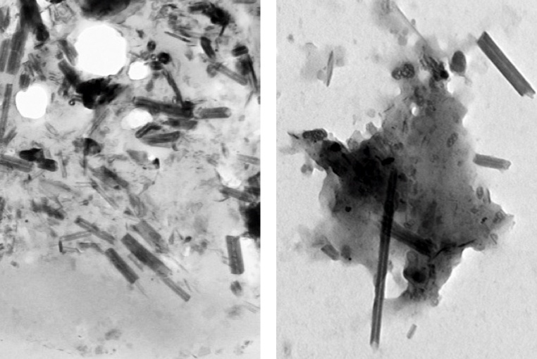
Carbon nanotubes (rods) and nanoparticles (black clumps) found inside a lung cell vacuole (left) are similar to those found in vehicle exhaust in tailpipes of cars in Paris (right) (credit: Fathi Moussa/Paris-Saclay University)

Carbon nanotubes (CNTs) have been found in cells extracted from the airways of Parisian children under routine treatment for asthma, according to a report in the journal EBioMedicine (open access) by scientists in France and at Rice University.

The cells were taken from 69 randomly selected asthma patients aged 2 to 17 who underwent routine fiber-optic bronchoscopies as part of their treatment. The researchers analyzed particulate matter found in the alveolar macrophage cells (also known as dust cells), which help stop foreign materials like particles and bacteria from entering the lungs.

The study partially answers the question of what makes up the black material inside alveolar macrophages, the original focus of the study. The researchers found single-walled and multiwalled carbon nanotubes and amorphous carbon among the cells.

The nanotube aggregates in the cells ranged in size from 10 to 60 nanometers in diameter and up to several hundred nanometers in length, small enough that optical microscopes would not have been able to identify them in samples from former patients. The new study used more sophisticated tools, including high-resolution transmission electron microscopy, X-ray spectroscopy, Raman spectroscopy, and near-infrared fluorescence microscopy to definitively identify them in the cells and in the environmental samples.

“The concentrations of nanotubes are so low in these samples that it’s hard to believe they would cause asthma, but you never know,” said Rice chemist Lon Wilson, a corresponding author of the paper. “What surprised me the most was that carbon nanotubes were the major component of the carbonaceous pollution we found in the samples.”

The study notes but does not make definitive conclusions about the controversial proposition that carbon nanotube fibers may act like asbestos, a proven carcinogen. But the authors did note that “long carbon nanotubes and large aggregates of short ones can induce a granulomatous (inflammation) reaction.”

The researchers also suggested previous studies that link the carbon content of airway macrophages and the decline of lung function should be reconsidered in light of the new findings. The researchers also suggested that the large surface areas of nanotubes and their ability to adhere to substances may make them effective carriers for other pollutants.

Carbon nanotubes from forest fires and cars?

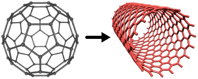
Fullerenes (left) can be converted to carbon nanotubes (right) with a catalytic process, according to Rice chemists (credits: Soroush83/CC and Matías Soto/Rice University)

However, similar nanotubes have been found in samples from the exhaust pipes of Paris vehicles, in dust gathered from various places around the city, in spider webs in India, and even in ice cores, the paper notes.

“We know that carbon nanoparticles are found in nature,” Wilson said, noting that round fullerene (C60) molecules are commonly produced by volcanoes, forest fires, and other combustion of carbon materials. “All you need is a little catalysis to make carbon nanotubes instead of fullerenes.”

A car’s catalytic converter, which turns toxic carbon monoxide into safer emissions, bears at least a passing resemblance to the Rice-invented high-pressure carbon monoxide, or HiPco, process to make carbon nanotubes, he said. “So it is not a big surprise, when you think about it,” Wilson said.

“Based on our discovery of CNTs in tailpipes, we propose that the catalytic converters of the automobiles are manufacturing carbon nanotubes, Wilson told KurzweilAI. “However, we have not actually proven that.”

We are all carbon-nanotube bearers now

For ethical reasons, no cells from healthy patients were analyzed, but because nanotubes were found in all of the samples, the study led the researchers to conclude that carbon nanotubes are likely to be found in everybody.

“It’s kind of ironic. In our laboratory, working with carbon nanotubes, we wear facemasks to prevent exactly what we’re seeing in these samples, yet everyone walking around out there in the world probably has at least a small concentration of carbon nanotubes in their lungs,” he said.

The study followed one released by Rice and Baylor College of Medicine earlier this month with the similar goal of analyzing the black substance found in the lungs of smokers who died of emphysema. That study found carbon black nanoparticles that were the product of the incomplete combustion of such organic material as tobacco.

Co-authors are from Paris-Saclay University, the Paediatric Pulmonology and Allergy Center and the Department of Anatomo-Pathology of the Groupe hospitalier La Roche-Guyon, and Paris Diderot University. The Welch Foundation partially supported the research.


Abstract of Anthropogenic Carbon Nanotubes Found in the Airways of Parisian Children

Compelling evidence shows that fine particulate matters (PM) from air pollution penetrate lower airways and are associated with adverse health effects even within concentrations below those recommended by the WHO. A paper reported a dose-dependent link between carbon content in alveolar macrophages (assessed only by optical microscopy) and the decline in lung function. However, to the best of our knowledge, PM had never been accurately characterized inside human lung cells and the most responsible components of the particulate mix are still unknown. On another hand carbon nanotubes (CNTs) from natural and anthropogenic sources might be an important component of PM in both indoor and outdoor air.

We used high-resolution transmission electron microscopy and energy dispersive X-ray spectroscopy to characterize PM present in broncho-alveolar lavage-fluids (n = 64) and inside lung cells (n = 5 patients) of asthmatic children. We show that inhaled PM mostly consist of CNTs. These CNTs are present in all examined samples and they are similar to those we found in dusts and vehicle exhausts collected in Paris, as well as to those previously characterized in ambient air in the USA, in spider webs in India, and in ice core. These results strongly suggest that humans are routinely exposed to CNTs.

Graphene nano-coils discovered to be powerful natural electromagnets

A nano-coil made of graphene could be an effective solenoid/inductor for electronic applications (credit: Yakobson Research Group/Rice University)

Rice University scientists have discovered that a widely used electronic part called a solenoid could be scaled down to nano-size with macro-scale performance.

The secret: a spiral form of atom-thin graphene that, remarkably, can be found in nature, even in common coal, according to Rice theoretical physicist Boris Yakobson and his colleagues.

The researchers determined that when a voltage is applied to such a “nano-coil,” current will flow around the helical path and produce a magnetic field, as it does in macroscale solenoids. The discovery is detailed in a new paper in the American Chemical Society journal Nano Letters.

“Perhaps this might work in reverse here: An electron current, pumped through by the applied voltage, at certain conditions may just cause the graphene spiral to spin, like a fast little electro-turbine,” Yakobson speculated.

Basic solenoid design. A current flowing through the coil generates a magnetic field, which causes a ferromagnetic plunger to be attracted or repelled. (credit: Society of Robots)

Solenoids are components with wires coiled around a metallic core. They produce a magnetic field when carrying current, turning them into electromagnets. These are widespread in electronic and mechanical devices, from circuit boards to transformers to cars.

They also serve as inductors, which are primary components in electric circuits that regulate current (the lump in power cables that feed electronic devices contains an inductor, which blocks RF interference). In their smallest form, inductors are a part of integrated circuits.

Nano-solenoids with a macro punch

While transistors get steadily smaller, basic inductors in electronics have become relatively bulky, said Fangbo Xu, a Rice alumnus and lead author of the paper. “It’s the same inside the circuits,” he said. “Commercial spiral inductors on silicon occupy excessive area. If realized, graphene nano-solenoids could change that.”

The nano-solenoids analyzed through computer models at Rice should be capable of producing powerful magnetic fields of about 1 tesla, about the same as the coils found in typical loudspeakers, according to Yakobson and his team — and about the same field strength as some MRI machines. They found the magnetic field would be strongest in the hollow, nanometer-wide cavity at the spiral’s center.

The spiral form is attributable to a simple topological trick, he said. Graphene is made of hexagonal arrays of carbon atoms. Malformed hexagons known as dislocations along one edge force the graphene to twist around itself, akin to a continuous nanoribbon that mimics a mathematical construct known as a Riemann surface.

The researchers demonstrated theoretically how energy would flow through the hexagons in nano-solenoids with edges in either armchair or zigzag formations. In one case, they determined the performance of a conventional spiral inductor of 205 microns in diameter could be matched by a nano-solenoid just 70 nanometers wide.

Multiple uses

Because graphene has no energy band gap (which gives a material semiconducting properties), electricity should move through without any barriers. But in fact, the width of the spiral and the configuration of the edges — either armchair or zigzag — influences how the current is distributed, and thus its inductive properties.

The researchers suggested it should be possible to isolate graphene screw dislocations from crystals of graphitic carbon (graphene in bulk form), but enticing graphene sheets to grow in a spiral would allow for better control of its properties, Yakobson said.

Xu suggested nano-solenoids may also be useful as molecular relays or switchable traps for magnetic molecules or radicals in chemical probes.

Co-authors are Rice graduate student Henry Yu and alumnus Arta Sadrzadeh. Yakobson is the Karl F. Hasselmann Professor of Materials Science and NanoEngineering and a professor of chemistry.

The research was supported by the Office of Naval Research’s Multidisciplinary University Research Initiative (MURI), the National Science Foundation and the Air Force Office of Scientific Research MURI.

The strongest known permanent-magnet in the world is at the Los Alamos National Laboratory campus of the National High Magnetic Field Laboratory, at 100 tesla. Does that mean 100 of these nanocoils could equal it? Comment below.


Abstract of Surfaces of Carbon as Graphene Nanosolenoids

Traditional inductors in modern electronics consume excessive areas in the integrated circuits. Carbon nanostructures can offer efficient alternatives if the recognized high electrical conductivity of graphene can be properly organized in space to yield a current-generated magnetic field that is both strong and confined. Here we report on an extraordinary inductor nanostructure naturally occurring as a screw dislocation in graphitic carbons. Its elegant helicoid topology, resembling a Riemann surface, ensures full covalent connectivity of all graphene layers, joined in a single layer wound around the dislocation line. If voltage is applied, electrical currents flow helically and thus give rise to a very large (∼1 T at normal operational voltage) magnetic field and bring about superior (per mass or volume) inductance, both owing to unique winding density. Such a solenoid of small diameter behaves as a quantum conductor whose current distribution between the core and exterior varies with applied voltage, resulting in nonlinear inductance.

FDA approves the first 3D-printed drug product

How ZipDose Technology works. The result is a porous drug product that disintegrates with just a sip of liquid. (credit: Aprecia Pharmaceuticals Company)

The FDA has approved the first 3D-printed drug — Aprecia’s SPRITAM (levetiracetam) for oral use as a prescription adjunctive therapy in the treatment of seizures in adults and children with epilepsy.

SPRITAM manufacturing uses 3D printing to produce a porous formulation that rapidly disintegrates with a sip of liquid, making it easier to swallow.

SPRITAM® levetiracetam, for oral use: 750 mg (foreground) and 1000 mg (background) (credit: Aprecia Pharmaceuticals Company)

The “ZipDose Technology” allows for delivering a high drug load, up to 1,000 mg in a single dose, which is expected to help patients take their medication as prescribed. SPRITAM is expected to be available in the first quarter of 2016.

Nearly three million people in the United States have been diagnosed with active epilepsy, with an estimated 460,000 of those cases occurring in children.