Biocompatible interfaces replace silicon and metal in neural prosthetic devices

Left: collagen; right: matrigel (credit: Wen Shen et al./Microsystems & Nanoengineering)

Researchers at the University of Georgia’s Regenerative Bioscience Center have developed a biocompatible implantable neural prosthetic device to replace silicon and noble metal in neural prosthetic devices. The goal is to avoid immune-system rejection, failures due to tissue strain, neurodegeneration, and decreased fidelity of recorded neural signals.

Implantable neural prosthetic devices in the brain have been around for almost two decades, helping people living with limb loss and spinal cord injury become more independent, for example. They are also used for deep brain stimulation and brain-controlled prosthetic devices. However, existing neural prosthetic devices suffer from immune-system rejection, and most are believed to eventually fail because of a mismatch between the soft brain tissue and the rigid devices.

The researchers used a combination of a two materials as structural support for neural electrodes.

Collagen. Its higher mechanical strength can support initial insertion while softening after implantation. Collagen is an extracellular matrix environment (ECM) protein that is critical in the formation of connective structures in tendons, organs, and basement membranes in the body and features long fibrils and 3D structures with high tensile strengths. The ECM is a collection of molecules secreted by cells that provides structural and biochemical support to surrounding cells.

Matrigel, a gelatinous ECM protein mixture resembling the complex extracellular neuronal environment, used to provide a more neuronal-compatible substrate.

A representative extracellular matrix-based implantable neural electrode device and an enlarged view of the electrode tip (credit: Wen Shen et al./Microsystems & Nanoengineering)

“This is not by any means the device that you’re going to implant into a patient,” said Karumbaiah, an assistant professor of animal and dairy science in the UGA College of Agricultural and Environmental Sciences. “This is proof of concept that extracellular matrix can be used to ensheathe a functioning electrode without the use of any other foreign or synthetic materials.”

The collaboration, led by Wen Shen and Mark Allen of the University of Pennsylvania, found that the extracellular matrix derived electrodes adapted to the mechanical properties of brain tissue and were capable of acquiring neural recordings from the brain cortex.

Currently, one out of every 190 Americans is living with limb loss, according to the National Institutes of Health. There is a significant burden in cost of care and quality of life for people suffering from this disability.

The research is described in an open-access paper in the journal Microsystems & Nanoengineering.


Abstract of Extracellular matrix-based intracortical microelectrodes: Toward a microfabricated neural interface based on natural materials

Extracellular matrix (ECM)-based implantable neural electrodes (NEs) were achieved using a microfabrication strategy on natural-substrate-based organic materials. The ECM-based design minimized the introduction of non-natural products into the brain. Further, it rendered the implants sufficiently rigid for penetration into the target brain region and allowed them subsequently to soften to match the elastic modulus of brain tissue upon exposure to physiological conditions, thereby reducing inflammatory strain fields in the tissue. Preliminary studies suggested that ECM-NEs produce a reduced inflammatory response compared with inorganic rigid and flexible approaches. In vivo intracortical recordings from the rat motor cortex illustrate one mode of use for these ECM-NEs.

MIT designs small, modular, efficient fusion power plant

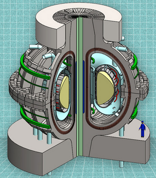
A cutaway view of the proposed ARC reactor (credit: MIT ARC team)

MIT plans to create a new compact version of a tokamak fusion reactor with the goal of producing practical fusion power, which could offer a nearly inexhaustible energy resource in as little as a decade.

Fusion, the nuclear reaction that powers the sun, involves fusing pairs of hydrogen atoms together to form helium, accompanied by enormous releases of energy.

The new fusion reactor, called ARC, would take advantage of new, commercially available superconductors — rare-earth barium copper oxide (REBCO) superconducting tapes (the dark brown areas in the illustration above) — to produce stronger magnetic field coils, according to Dennis Whyte, a professor of Nuclear Science and Engineering and director of MIT’s Plasma Science and Fusion Center.

The stronger magnetic field makes it possible to produce the required magnetic confinement of the superhot plasma — that is, the working material of a fusion reaction — but in a much smaller device than those previously envisioned. The reduction in size, in turn, makes the whole system less expensive and faster to build, and also allows for some ingenious new features in the power plant design.

The proposed reactor is described in a paper in the journal Fusion Engineering and Design, co-authored by Whyte, PhD candidate Brandon Sorbom, and 11 others at MIT.

Power plant prototype

The new reactor is designed for basic research on fusion and also as a potential prototype power plant that could produce 270MW of electrical power. The basic reactor concept and its associated elements are based on well-tested and proven principles developed over decades of research at MIT and around the world, the team says. An experimental tokamak was built at Princeton Plasma Physics Laboratory circa 1980.

The hard part has been confining the superhot plasma — an electrically charged gas — while heating it to temperatures hotter than the cores of stars. This is where the magnetic fields are so important — they effectively trap the heat and particles in the hot center of the device.

While most characteristics of a system tend to vary in proportion to changes in dimensions, the effect of changes in the magnetic field on fusion reactions is much more extreme: The achievable fusion power increases according to the fourth power of the increase in the magnetic field.

Tenfold boost in power

The new superconductors are strong enough to increase fusion power by about a factor of 10 compared to standard superconducting technology, Sorbom says. This dramatic improvement leads to a cascade of potential improvements in reactor design.

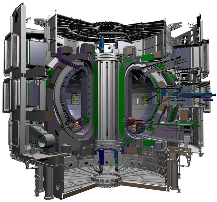
ITER — the world’s largest tokamak — is expected to be completed in 2019, with deuterium-tritium operations in 2027 and 2000–4000MW of fusion power onto the grid in 2040 (credit: ITER Organization)

The world’s most powerful planned fusion reactor, a huge device called ITER that is under construction in France, is expected to cost around $40 billion. Sorbom and the MIT team estimate that the new design, about half the diameter of ITER (which was designed before the new superconductors became available), would produce about the same power at a fraction of the cost, in a shorter construction time, and with the same physics.

Another key advance in the new design is a method for removing the fusion power core from the donut-shaped reactor without having to dismantle the entire device. That makes it especially well-suited for research aimed at further improving the system by using different materials or designs to fine-tune the performance.

In addition, as with ITER, the new superconducting magnets would enable the reactor to operate in a sustained way, producing a steady power output, unlike today’s experimental reactors that can only operate for a few seconds at a time without overheating of copper coils.

Liquid protection

Another key advantage is that most of the solid blanket materials used to surround the fusion chamber in such reactors are replaced by a liquid material that can easily be circulated and replaced, eliminating the need for costly replacement procedures as the materials degrade over time.

Right now, as designed, the reactor should be capable of producing about three times as much electricity as is needed to keep it running, but the design could probably be improved to increase that proportion to about five or six times, Sorbom says. So far, no fusion reactor has produced as much energy as it consumes, so this kind of net energy production would be a major breakthrough in fusion technology, the team says.

The design could produce a reactor that would provide electricity to about 100,000 people, they say. Devices of a similar complexity and size have been built within about five years, they say.

“Fusion energy is certain to be the most important source of electricity on earth in the 22nd century, but we need it much sooner than that to avoid catastrophic global warming,” says David Kingham, CEO of Tokamak Energy Ltd. in the UK, who was not connected with this research. “This paper shows a good way to make quicker progress,” he says.

The MIT research, Kingham says, “shows that going to higher magnetic fields, an MIT specialty, can lead to much smaller (and hence cheaper and quicker-to-build) devices.” The work is of “exceptional quality,” he says; “the next step … would be to refine the design and work out more of the engineering details, but already the work should be catching the attention of policy makers, philanthropists and private investors.”

The research was supported by the U.S. Department of Energy and the National Science Foundation.


Abstract of ARC: A compact, high-field, fusion nuclear science facility and demonstration power plant with demountable magnets

The affordable, robust, compact (ARC) reactor is the product of a conceptual design study aimed at reducing the size, cost, and complexity of a combined fusion nuclear science facility (FNSF) and demonstration fusion Pilot power plant. ARC is a ∼200–250 MWe tokamak reactor with a major radius of 3.3 m, a minor radius of 1.1 m, and an on-axis magnetic field of 9.2 T. ARC has rare earth barium copper oxide (REBCO) superconducting toroidal field coils, which have joints to enable disassembly. This allows the vacuum vessel to be replaced quickly, mitigating first wall survivability concerns, and permits a single device to test many vacuum vessel designs and divertor materials. The design point has a plasma fusion gain of Qp ≈ 13.6, yet is fully non-inductive, with a modest bootstrap fraction of only ∼63%. Thus ARC offers a high power gain with relatively large external control of the current profile. This highly attractive combination is enabled by the ∼23 T peak field on coil achievable with newly available REBCO superconductor technology. External current drive is provided by two innovative inboard RF launchers using 25 MW of lower hybrid and 13.6 MW of ion cyclotron fast wave power. The resulting efficient current drive provides a robust, steady state core plasma far from disruptive limits. ARC uses an all-liquid blanket, consisting of low pressure, slowly flowing fluorine lithium beryllium (FLiBe) molten salt. The liquid blanket is low-risk technology and provides effective neutron moderation and shielding, excellent heat removal, and a tritium breeding ratio ≥ 1.1. The large temperature range over which FLiBe is liquid permits an output blanket temperature of 900 K, single phase fluid cooling, and a high efficiency helium Brayton cycle, which allows for net electricity generation when operating ARC as a Pilot power plant.

Controlling inflammation to reduce chronic disease risk

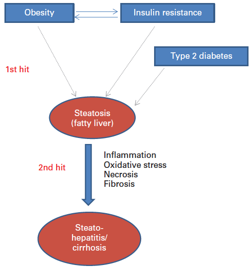
Two-hit model of non-alcoholic fatty liver disease (credit: ILSI Europe)

In an open-access paper in the British Journal of Nutrition, a coalition of 17 experts explain how elevated unresolved chronic inflammation is involved a range of chronic diseases, and how nutrition influences inflammatory processes and helps reduce chronic risk of diseases.

According to the authors, “the nutrition status of the individual with for example a deficiency or excess of certain micronutrients (e.g. folate, vitamin B12, vitamin B6, vitamin 1, vitamin E, zinc) may lead to an ineffective or excessive inflammatory response.

“Studies have showed that high consumption of fat and glucose may induce post-prandial inflammation (manifesting itself after the consumption of a meal), which may have consequences for the development of diabetes and cardiovascular diseases. The Western-style diet, rich in fat and simple sugars but often poor in specific micronutrients, is linked to the increased prevalence of diseases with strong immunogical and autoimmune components, including allergies, food allergies, atopic dermatitis and obesity.”

“Inflammation acts as both a friend and foe, being essential in metabolic regulation, with unresolved low-grade chronic inflammation being a pathological feature of a wide range of chronic conditions including the metabolic syndrome and cardiovascular diseases,” commented a co-author, Prof. Anne Marie Minihane, University of East Anglia (UK).


Abstract of Low-grade inflammation, diet composition and health: current research evidence and its translation

The importance of chronic low-grade inflammation in the pathology of numerous age-related chronic conditions is now clear. An unresolved inflammatory response is likely to be involved from the early stages of disease development. The present position paper is the most recent in a series produced by the International Life Sciences Institute’s European Branch (ILSI Europe). It is co-authored by the speakers from a 2013 workshop led by the Obesity and Diabetes Task Force entitled ‘Low-grade inflammation, a high-grade challenge: biomarkers and modulation by dietary strategies’. The latest research in the areas of acute and chronic inflammation and cardiometabolic, gut and cognitive health is presented along with the cellular and molecular mechanisms underlying inflammation–health/disease associations. The evidence relating diet composition and early-life nutrition to inflammatory status is reviewed. Human epidemiological and intervention data are thus far heavily reliant on the measurement of inflammatory markers in the circulation, and in particular cytokines in the fasting state, which are recognised as an insensitive and highly variable index of tissue inflammation. Potential novel kinetic and integrated approaches to capture inflammatory status in humans are discussed. Such approaches are likely to provide a more discriminating means of quantifying inflammation–health/disease associations, and the ability of diet to positively modulate inflammation and provide the much needed evidence to develop research portfolios that will inform new product development and associated health claims.

Google Glass could bring toxicology specialists to remote emergency rooms

(credit: Google)

Researchers at the University of Massachusetts Medical School have found that Google Glass — presumably the Enterprise Edition — could effectively extend bedside toxicology consults to distant health care facilities such as community and rural hospitals to diagnose and manage poisoned patients, according to a paper in the Journal of Medical Toxicology.

“In the present era of value-based care, a toxicology service using hands-free devices, such as Google Glass, could conceivably expand its coverage area and enhance patient care, while potentially decreasing overall treatment costs,” said Peter R. Chai, MD, toxicology fellow at UMass Medical School. “Our work shows that the data transmitted by Google Glass can be used to supplement traditional telephone consults, validate bedside physical exams, and diagnose and manage patients.”

Traditional telemedicine devices usually consist of large desktop or laptop computers affixed to a big cart that has to be rolled from exam room to exam room. “Glass is positioned perfectly as an emergency medicine telemedical device. Its small, hands free and portable, so you can bring it right to the bedside and have a real-time specialist with you when you need one,” he said.

In the study, emergency medicine residents at UMass Memorial Medical Center performed 18 toxicology consults with Google Glass. ER physicians wearing Google Glass evaluated the patients at bedside while a secure video feed was sent to the toxicology supervising consultant. The supervising consultant then guided the resident through text messages displayed on the Glass. Consultants also obtained static photos of medication bottles, electrocardiograms (EKG) and other pertinent information at the discretion of the supervisor.

As a result of using Google Glass, consulting toxicologists reported being more confident in diagnosing specific toxidromes. Additional data collected showed that the use of Google Glass also changed management of patient care in more than half of the cases seen. Specifically, six of those patients received antidotes they otherwise would not have. Overall, 89 percent of the cases seen with Glass were considered successful by the consulting toxicologist.

Google currently lists several companies involved in the medical field as Glass At Work partners, such as Advanced Medical Applications, which specializes in “solutions in telemedicine, live-surgery demonstrations, and remote medical training.”

According to 9to5Google sources, the Google Glass Enterprise Edition will feature “a robust hinge mechanism that allows the computer and battery modules to fold down like a regular pair of glasses, and a hinge for folding down the left side of the band as well.” It also “includes a larger prism display for a better viewing experience, an Intel Atom processor that brings better performance, moderately improved battery life, and better heat management.”


Abstract of The Feasibility and Acceptability of Google Glass for Teletoxicology Consults

Teletoxicology offers the potential for toxicologists to assist in providing medical care at remote locations, via remote, interactive augmented audiovisual technology. This study examined the feasibility of using Google Glass, a head-mounted device that incorporates a webcam, viewing prism, and wireless connectivity, to assess the poisoned patient by a medical toxicology consult staff. Emergency medicine residents (resident toxicology consultants) rotating on the toxicology service wore Glass during bedside evaluation of poisoned patients; Glass transmitted real-time video of patients’ physical examination findings to toxicology fellows and attendings (supervisory consultants), who reviewed these findings. We evaluated the usability (e.g., quality of connectivity and video feeds) of Glass by supervisory consultants, as well as attitudes towards use of Glass. Resident toxicology consultants and supervisory consultants completed 18 consults through Glass. Toxicologists viewing the video stream found the quality of audio and visual transmission usable in 89 % of cases. Toxicologists reported their management of the patient changed after viewing the patient through Glass in 56 % of cases. Based on findings obtained through Glass, toxicologists recommended specific antidotes in six cases. Head-mounted devices like Google Glass may be effective tools for real-time teletoxicology consultation.

How to create a genius mouse

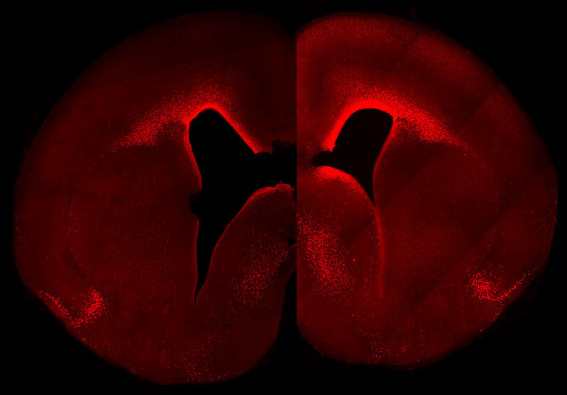
The left-brain hemisphere of a normal mouse shows the normal level and cellular distribution of the Pax6 gene expression in the developing neocortex. The right-brain hemisphere shows a sustained, primate-like Pax6 expression pattern in the neocortex of a double transgenic mouse embryo. These animals have more Pax6-positive progenitor cells and a higher Pax6 expression level in the germinal layer close to the ventricle in the right hemisphere. (credit: © MPI of Molecular Cell Biology & Genetics)

Researchers at the Max Planck Institute of Molecular Cell Biology and Genetics have created a transgenic mouse in which a gene called Pax6, during embryonic development, is highly expressed in a specific group of brain cortical cells called neural progenitor stem cells (the cells that generate all cells that make up the brain).

The resulting mouse brain generated more neurons than normal and exhibited primate-like features — notably those in the top layer, a characteristic feature of an expanded neocortex.

Mouse basal progenitors, in contrast to human, do not express Pax6. In humans, basal progenitors can undergo multiple rounds of cell division, thereby substantially increasing neuron number and ultimately the size of the neocortex.

“The evolutionary expansion of the neocortex is a hallmark of species with higher cognitive functions,” explains Wieland Huttner, the research group leader and director at the MPI-CBG. “Our findings contribute to our understanding of the molecular mechanisms underlying this expansion. While the findings demonstrate how altering the expression of a single key gene can make a big difference to brain development, a future challenge will be to obtain a comprehensive, integrated view of all the molecular changes that made our brains big.”

The study was published in an open-access paper in the journal PLOS Biology.

The paper, reassuringly, did not mention plans to create a transgenic genius cat in case the transgenic mouse gene escaped the laboratory.


Abstract of Sustained Pax6 Expression Generates Primate-like Basal Radial Glia in Developing Mouse Neocortex

The evolutionary expansion of the neocortex in mammals has been linked to enlargement of the subventricular zone (SVZ) and increased proliferative capacity of basal progenitors (BPs), notably basal radial glia (bRG). The transcription factor Pax6 is known to be highly expressed in primate, but not mouse, BPs. Here, we demonstrate that sustaining Pax6 expression selectively in BP-genic apical radial glia (aRG) and their BP progeny of embryonic mouse neocortex suffices to induce primate-like progenitor behaviour. Specifically, we conditionally expressed Pax6 by in utero electroporation using a novel, Tis21–CreERT2 mouse line. This expression altered aRG cleavage plane orientation to promote bRG generation, increased cell-cycle re-entry of BPs, and ultimately increased upper-layer neuron production. Upper-layer neuron production was also increased in double-transgenic mouse embryos with sustained Pax6 expression in the neurogenic lineage. Strikingly, increased BPs existed not only in the SVZ but also in the intermediate zone of the neocortex of these double-transgenic mouse embryos. In mutant mouse embryos lacking functional Pax6, the proportion of bRG among BPs was reduced. Our data identify specific Pax6 effects in BPs and imply that sustaining this Pax6 function in BPs could be a key aspect of SVZ enlargement and, consequently, the evolutionary expansion of the neocortex.

New solid-state memory technology allows for highest-density non-volatile storage

A schematic shows the layered structure of new type of solid-state memory developed at Rice University (credit: Tour Group/Rice University)

Scientists in the Rice University lab of chemist James Tour have created a solid-state memory technology that allows for high-density 162 gigabits nonvolatile storage, much higher than other oxide-based memory systems under investigation by scientists. (Eight bits equal one byte; a 162-gigabit unit would store about 20 gigabytes of information.)

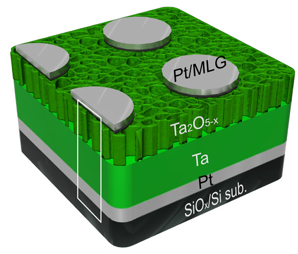
Applying voltage to a 250-nanometer-thick sandwich of graphene, tantalum, nanoporous tantalum oxide (an insulator), and platinum creates addressable bits where the layers meet. Control voltages shift oxygen ions and vacancies to switch the bits between ones and zeroes.

Like the Tour lab’s previous discovery of silicon oxide memories, the new devices require only two electrodes per circuit, making them simpler than present-day flash memories, which use three. “This is a new way to make ultradense, nonvolatile computer memory,” Tour said.

Nonvolatile random-access memories, such as such as flash memory in smartphones and tablets, hold their data even when the power is off, unlike volatile random-access computer memories (in most computers), which lose their contents when the machine is shut down.

Modern memory chips have many requirements: They have to read and write data at high speed and hold as much as possible. They must also be durable and show good retention of that data while using minimal power. These are provided by Rice’s new design, which requires only one hundredth the amount of energy required with present devices, Tour says.

“This tantalum memory is based on two-terminal systems, so it’s all set for 3-D memory stacks,” he said. “And it doesn’t even need diodes or selectors, making it one of the easiest ultradense memories to construct. This will be a real competitor for the growing memory demands in high-definition video storage and server arrays.”

A layered structure of tantalum, tantalum oxide, multilayer graphene, and platinum is the basis for a new type of nonvolatile memory (credit: Tour Group/Rice University)

In making the material, the researchers found the tantalum oxide gradually loses oxygen ions, changing from an oxygen-rich, nanoporous semiconductor at the top to oxygen-poor at the bottom. Where the oxygen disappears completely, it becomes pure tantalum, a metal. The graphene does double duty as a barrier that keeps platinum from migrating into the tantalum oxide and causing a short circuit.*


Rice University | Tantalum oxide memory: Slices taken from a tantalum oxide-based memory developed at Rice University show the partially interconnected and randomly distributed internal pores in the material.

Tour said tantalum oxide memories can be fabricated at room temperature. He noted the control voltage that writes and rewrites the bits is adjustable, which allows a wide range of switching characteristics.

(As the researchers note in a paper in the journal Nano Letters, nonvolatile resistive oxide-based memories can also offer faster switching speed. That suggests that tantalum oxide memories might one day further improve MIT’s “BlueDBM” solution for improved handling of big data by making nonvolatile memory more efficient, as described on KurzweilAI last week.)

The remaining hurdles to commercialization of tantalum oxide memories include the fabrication of a dense enough crossbar device to address individual bits and a way to control the size of the nanopores.

The research is described online in the American Chemical Society Researchers at Korea University-Korea Institute of Science and Technology and University of Massachusetts, Amherst where also involved.

Tour is the T.T. and W.F. Chao Chair in Chemistry as well as a professor of materials science and nanoengineering and of computer science and a member of Rice’s Richard E. Smalley Institute for Nanoscale Science and Technology.


Rice University | Tantalum oxide memory 2

* The researchers determined three related factors give the memories their unique switching ability:

  • The control voltage mediates how electrons pass through a boundary that can flip from an ohmic (current flows in both directions) to a Schottky (current flows one way) contact and back.
  • The boundary’s location can change based on oxygen vacancies. These are “holes” in atomic arrays where oxygen ions should exist, but don’t. The voltage-controlled movement of oxygen vacancies shifts the boundary from the tantalum/tantalum oxide interface to the tantalum oxide/graphene interface. “The exchange of contact barriers causes the bipolar switching,” said Gunuk Wang, lead author of the study and a former postdoctoral researcher at Rice.
  • The flow of current draws oxygen ions from the tantalum oxide nanopores and stabilizes them. These negatively charged ions produce an electric field that effectively serves as a diode to hinder error-causing crosstalk. While researchers already knew the potential value of tantalum oxide for memories, such arrays have been limited to about a kilobyte because denser memories suffer from crosstalk that allows bits to be misread.

Abstract of Three-Dimensional Networked Nanoporous Ta2O5–x Memory System for Ultrahigh Density Storage

Oxide-based resistive memory systems have high near-term promise for use in nonvolatile memory. Here we introduce a memory system employing a three-dimensional (3D) networked nanoporous (NP) Ta2O5–x structure and graphene for ultrahigh density storage. The devices exhibit a self-embedded highly nonlinear I–V switching behavior with an extremely low leakage current (on the order of pA) and good endurance. Calculations indicated that this memory architecture could be scaled up to a ∼162 Gbit crossbar array without the need for selectors or diodes normally used in crossbar arrays. In addition, we demonstrate that the voltage point for a minimum current is systematically controlled by the applied set voltage, thereby offering a broad range of switching characteristics. The potential switching mechanism is suggested based upon the transformation from Schottky to Ohmic-like contacts, and vice versa, depending on the movement of oxygen vacancies at the interfaces induced by the voltage polarity, and the formation of oxygen ions in the pores by the electric field.

How aging cripples the immune system

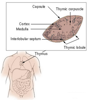
Thymus (credit: Wikimedia Commons)

Aging cripples the production of new immune cells, decreasing the immune system’s response to vaccines and putting the elderly at risk of infection, but antioxidants in the diet may slow this damaging process.

That’s a new finding by scientists from the Florida campus of The Scripps Research Institute (TSRI), published in an open-access paper in the journal Cell Reports.

The problem is focused on an organ called the thymus, which produces T lymphocytes (a type of white blood cell) — critical immune cells that must be continuously replenished so they can respond to new infections.

“The thymus begins to atrophy rapidly in very early adulthood, simultaneously losing its function,” said TSRI Professor Howard Petrie.

“This new study shows for the first time a mechanism for the long-suspected connection between normal immune function and antioxidants.”

How antioxidant enzyme deficiency leads to metabolic damage

Scientists have been hampered in their efforts to develop specific immune therapies for the elderly by a lack of knowledge of the underlying mechanisms of this process.

To explore these mechanisms, Petrie and his team developed a computational approach for analyzing the activity of genes in two major cell types in the thymus — stromal cells and lymphoid cells — in mouse tissues, which are similar to human tissues in terms of function and age-related atrophy.

The team found that stromal cells were specifically deficient in an antioxidant enzyme called catalase. That resulted in elevated levels of the reactive oxygen byproducts of metabolism, which cause accelerated metabolic damage.*

New support for the “free-radical theory” of aging

(Credit: Bethany Christmann)

Taken together, the findings provide support for the “free-radical theory” of aging, which proposes that reactive oxygen species (such as hydrogen peroxide), produced during normal metabolism (and from other sources) cause cellular damage that contributes to aging and age-related diseases.

Free radicals are especially reactive atoms or groups of atoms that have one or more unpaired electrons.  Besides those produced in the body as a by-product of normal metabolism, they can also be introduced from an outside source, such as tobacco smoke or other toxins.

Other studies have suggested that sex hormones, particularly androgens such as testosterone, play a major role in the aging process. But according to the researchers, those studies have failed to answer the key question: why does the thymus atrophy so much more rapidly than other body tissues?

“There’s no question that the thymus is remarkably responsive to androgens,” Petrie noted, “but our study shows that the fundamental mechanism of aging in the thymus, namely accumulated metabolic damage, is the same as in other body tissues. However, the process is accelerated in the thymus by a deficiency in the essential protective effects of catalase, which is found at higher levels in almost all other body tissues.”

It’s complicated

However, lowering free radicals with antioxidants has not always conferred the expected benefits, according to Senior scientist and Buck Institute professor Judith Campisi, PhD.

In a study published August 3 online in Proceedings of the National Academy of Sciences, scientists in her lab bred mice that produced excess free radicals that damaged the mitochondria in their skin. Based on the free-radical theory, the scientists expected to see accelerated aging across the mouse lifespan.

Instead, they saw a surprising benefit in young animals: accelerated wound healing due to increased epidermal (skin) differentiation and re-epithelialization.

However, the mice paid a price over time. Campisi said mitochondrial damage from excess free radicals caused some of the skin cells to go into senescence — they stopped dividing and started accumulating.  Campisi said that over time, the energy available to the epidermal stems cells was depleted — the stem cells simply became too scarce and the mice showed expected signs of aging: thin skin and poor wound healing.

“It may be that nature used free radicals to optimize skin health, but because this process is not deleterious to the organism until later in life, past its reproductive age, there was no [evolutionary benefit from evolving] ways to alter this mechanism,” suggested Michael Velarde, PhD, a postdoctoral fellow in the Campisi lab.

* To confirm the central role of catalase, the scientists increased levels of this enzyme in genetically altered animal models, resulting in preservation of thymus size for a much longer period. In addition, animals that were given two common dietary antioxidants, including vitamin C, were also protected from the effects of aging on the thymus.


Abstract of Metabolic Damage and Premature Thymus Aging Caused by Stromal Catalase Deficiency

T lymphocytes are essential mediators of immunity that are produced by the thymus in proportion to its size. The thymus atrophies rapidly with age, resulting in progressive diminution of new T cell production. This decreased output is compensated by duplication of existing T cells, but it results in gradual dominance by memory T cells and decreased ability to respond to new pathogens or vaccines. Here, we show that accelerated and irreversible thymic atrophy results from stromal deficiency in the reducing enzyme catalase, leading to increased damage by hydrogen peroxide generated by aerobic metabolism. Genetic complementation of catalase in stromal cells diminished atrophy, as did chemical antioxidants, thus providing a mechanistic link between antioxidants, metabolism, and normal immune function. We propose that irreversible thymic atrophy represents a conventional aging process that is accelerated by stromal catalase deficiency in the context of an intensely anabolic (lymphoid) environment.

Abstract of Pleiotropic age-dependent effects of mitochondrial dysfunction on epidermal stem cells

Tissue homeostasis declines with age partly because stem/progenitor cells fail to self-renew or differentiate. Because mitochondrial damage can accelerate aging, we tested the hypothesis that mitochondrial dysfunction impairs stem cell renewal or function. We developed a mouse model, Tg(KRT14-cre/Esr1)20Efu/J × Sod2tm1Smel, that generates mitochondrial oxidative stress in keratin 14-expressing epidermal stem/progenitor cells in a temporally controlled manner owing to deletion of Sod2, a nuclear gene that encodes the mitochondrial antioxidant enzyme superoxide dismutase 2 (Sod2). Epidermal Sod2 loss induced cellular senescence, which irreversibly arrested proliferation in a fraction of keratinocytes. Surprisingly, in young mice, Sod2 deficiency accelerated wound closure, increasing epidermal differentiation and reepithelialization, despite the reduced proliferation. In contrast, at older ages, Sod2 deficiency delayed wound closure and reduced epidermal thickness, accompanied by epidermal stem cell exhaustion. In young mice, Sod2 deficiency accelerated epidermal thinning in response to the tumor promoter 12-O-tetradecanoylphorbol-13-acetate, phenocopying the reduced regeneration of older Sod2-deficient skin. Our results show a surprising beneficial effect of mitochondrial dysfunction at young ages, provide a potential mechanism for the decline in epidermal regeneration at older ages, and identify a previously unidentified age-dependent role for mitochondria in skin quality and wound closure.

Electro-optical modulator is 100 times smaller, consumes 100th of the energy

Colorized electron microscope image of a micro-modulator made of gold. In the slit in the center of the picture, light is converted into plasmon polaritons, modulated, and then re-converted into light pulses (credit: Haffner et al. Nature Photonics)

Researchers at ETH Zurich have developed a modulator that is a 100 times smaller than conventional modulators, so it can now be integrated into electronic circuits. Transmitting large amounts of data via the Internet requires high-performance electro-optic modulators — devices that convert electrical signals (used in computers and cell phones) into light signals (used in fiber-optic cables).

Today, huge amounts of data are sent incredibly fast through fiber-optic cables as light pulses. For that purpose they first have to be converted from electrical signals, which are used by computers and telephones, into optical signals. Today’s electro-optic modulators are more complicated and large, compared with electronic devices that can be as small as a few micrometers.

The plasmon trick

To build the smallest possible modulator they first need to focus a light beam whose intensity they want to modulate into a very small volume. The laws of optics, however, dictate that such a volume cannot be smaller than the wavelength of the light itself. Modern telecommunications use near-infrared laser light with a wavelength of 1500 nanometers (1.5 micrometers), which sets the lower limit for the size of a modulator.

To beat that limit and to make the device even smaller, the light is first turned into surface-plasmon-polaritons. Plasmon-polaritons are a combination of electromagnetic fields and electrons that propagate along a surface of a metal strip. At the end of the strip they are converted back to light once again. The advantage of this detour is that plasmon-polaritons can be confined in a much smaller space than the light they originated from.

The modulator is much smaller than conventional devices so it consumes very little energy — only a few thousandth of a Watt at a data transmission rate of 70 Gigabits per second. This corresponds to about 100th of the energy consumption of commercial models. And that means more data can be transmitted at higher speeds. The device is also cheaper to produce.

The research is described in a paper in the journal Nature Photonics.


Abstract of All-plasmonic Mach–Zehnder modulator enabling optical high-speed communication at the microscale

Optical modulators encode electrical signals to the optical domain and thus constitute a key element in high-capacity communication links. Ideally, they should feature operation at the highest speed with the least power consumption on the smallest footprint, and at low cost. Unfortunately, current technologies fall short of these criteria. Recently, plasmonics has emerged as a solution offering compact and fast devices. Yet, practical implementations have turned out to be rather elusive. Here, we introduce a 70 GHz all-plasmonic Mach–Zehnder modulator that fits into a silicon waveguide of 10 μm length. This dramatic reduction in size by more than two orders of magnitude compared with photonic Mach–Zehnder modulators results in a low energy consumption of 25 fJ per bit up to the highest speeds. The technology suggests a cheap co-integration with electronics.

‘Plasmonic’ material could bring ultrafast all-optical communications

This rendering depicts a new “plasmonic oxide material” that could make possible devices for optical communications that are at least 10 times faster than conventional technologies (credit: Purdue University/Nathaniel Kinsey)

Researchers at Purdue University have created a new “plasmonic oxide material” that could make possible modulator devices for optical communications (fiber optics, used for the Internet and cable television) that are at least 10 times faster than conventional technologies.

The optical material, made of aluminum-doped zinc oxide (AZO) also requires less power than other “all-optical” semiconductor devices. That is essential for the faster operation, which would otherwise generate excessive heat with the increase transmission speed.

The material has been shown to work in the near-infrared range of the spectrum, which is used in optical communications, and it is compatible with the CMOS semiconductor manufacturing process used to construct integrated circuits.

Faster optical transistors replace silicon

The researchers have proposed creating an “all-optical plasmonic modulator using CMOS-compatible materials,” or an optical transistor, which allows for the speedup compared to systems that use silicon chips.

A cycle takes about 350 femtoseconds to complete in the new AZO films, which is roughly 5,000 times faster than crystalline silicon.

The researchers “doped” zinc oxide with aluminum (thus the AZO), meaning the zinc oxide is impregnated with aluminum atoms to alter the material’s optical properties. Doping the zinc oxide causes it to behave like a metal at certain wavelengths and like a dielectric at other wavelengths.

The AZO also makes it possible to “tune” the optical properties of metamaterials.

Findings were detailed in an open-access research paper appearing in July in the journal Optica, published by the Optical Society of America.

The ongoing research is funded by the Air Force Office of Scientific Research, a Marie Curie Outgoing International Fellowship, the National Science Foundation, and the Office of Naval Research.


Abstract of Epsilon-near-zero Al-doped ZnO for ultrafast switching at telecom wavelengths

Transparent conducting oxides have recently gained great attention as CMOS-compatible materials for applications in nanophotonics due to their low optical loss, metal-like behavior, versatile/tailorable optical properties, and established fabrication procedures. In particular, aluminum-doped zinc oxide (AZO) is very attractive because its dielectric per-mittivity can be engineered over a broad range in the near-IR and IR. However, despite all these beneficial features, the slow (>100 ps) electron-hole recombination time typical of these compounds still represents a fundamental limitation impeding ultrafast optical modulation. Here we report the first epsilon-near-zero AZO thin films that simultaneously exhibit ultrafast carrier dynamics (excitation and recombination time below 1 ps) and an outstanding reflectance modulation up to 40% for very low pump fluence levels (<4 mJ∕cm2) at a telecom wavelength of 1.3 μm. The unique properties of the demonstrated AZO thin films are the result of a low-temperature fabrication procedure promoting deep-level defects within the film and an ultrahigh carrier concentration. © 2015 Optical Society of America

Move over, autonomous AI weapons, there’s a new risk in town: ‘gene drives’

Wyss Institute scientists believe that synthetic gene drives, if researched responsibly, might be used in the future to render mosquito populations unable to transmit malaria (credit: CDC)

An international group of 26 experts, including prominent genetic engineers and fruit fly geneticists, has unanimously recommended a series of preemptive measures to safeguard gene drive research from accidental (or intentional) release from laboratories.

RNA-guided gene drives are genetic elements — found naturally in the genomes of most of the world’s organisms — that increase the chance of the gene they carry being passed on to all offspring. So  they can quickly spread through populations if not controlled.

Looking to these natural systems, researchers around the world, including some  scientists, are developing synthetic gene drives that could one day be leveraged by humans to purposefully alter the traits of wild populations of organisms to prevent disease transmission and eradicate invasive species.

What could possibly go wrong?

These synthetic gene drives, designed using an RNA-guided gene editing system called CRISPR, could one day improve human health and the environment by preventing mosquitoes and ticks from spreading diseases such as malaria and Lyme; by promoting sustainable agriculture through control of crop pests without the use of toxic pesticides and herbicides; and by protecting at-risk ecosystems from the spread of destructive, invasive species such as rats or cane toads.

Most genome alterations don’t persist in nature. Only 50 percent of transgenic mosquito offspring (left) will carry the altered gene, so it may persist at low frequency or go instinct. With gene drive (right), using CRISPR/Cas9, all of the offspring will carry the altered gene and will be inherited through the population. (credit: adapted from Wyss Institute video)

However, the development of RNA-guided gene drive technology calls for enhanced safety measures. That’s because its capability to also affect shared ecosystems if organisms containing synthetic gene drives are accidentally or deliberately released from a laboratory. This potential risk is especially relevant with highly mobile species such as fruit flies or mosquitoes.

Guidelines available

“One of the great successes of engineering is the development of safety features, such as the rounding of sharp corners on objects and the invention of airbags for cars, and in biological engineering we want to emulate the process of designing safety features in ways relevant to the technologies we develop,” said Wyss Core Faculty member George Church, Ph.D., who leads the Synthetic Biology Platform at the Wyss Institute. Church is also the Robert Winthrop Professor of Genetics at Harvard Medical School and Professor of Health Sciences and Technology at Harvard and MIT.

At the Wyss Institute, enhanced protocols for safely and securely researching emerging biotechnologies, including RNA–guided gene drives, have already been formally implemented. The safeguards were put in place proactively, step–by–step, in direct parallel with the development of the first RNA-guided gene drives at the Wyss Institute.

The working documents have been made publicly available by the Institute to encourage widespread adoption of multi-tier confinement and risk assessment procedures. Church was instrumental in the design of the enhanced biosafety and biosecurity protocols.

Now, research teams from the Wyss Institute and University of California, San Diego — the only two groups to have published work on RNA-guided CRISPR gene drives — have proactively assembled an international group of 26 experts, including prominent genetic engineers and fruit fly geneticists, to unanimously recommend a series of preemptive measures to safeguard gene drive research.

Open-access research recommended

Led by Wyss Institute Technology Development Fellow, Kevin Esvelt, Ph.D., and UC San Diego Professor of Cell and Developmental Biology Ethan Bier, Ph.D., the 26 authors of this consensus recommendation, which is published online in Science Express journal and includes representatives from every major group known to be working on gene drives, calls for all researchers to use multiple confinement strategies in order to prevent the accidental alteration of wild populations.

The group also provides explicit recommended guidelines for regulatory authorities evaluating proposed new work. And Esvelt and others are hopeful that the field of gene drive research is so nascent that it may be possible to build a community of scientists that share their research with the public throughout the development process.

“This would promote collaboration and avoid needless duplication of efforts among different research groups while allowing diverse voices to help guide the development of a technology that could improve our shared world,” said Esvelt. “And eventually, it might inspire a similar shift towards full transparency in other scientific fields of collective public importance.”

“The scientific community has a responsibility to the public and to the environment to constantly assess how new biotechnologies could potentially impact our world,” said Wyss Institute Founding Director Donald E. Ingber, M.D., Ph.D.

“This proactive consensus recommendation — reached in an extraordinary demonstration of the power of scientific collaboration over competition — provides concrete, useful guidelines for safeguarding our shared ecosystem while ensuring that remarkable breakthroughs, such as synthetic gene drives, can be applied to their full potential for the greater good.”


Wyss Institute at Harvard University | CRISPER-Cas9: Gene Drive

This animation explains how gene drives could one day be used to spread gene alterations through targeted wild populations over many generations, for purposes such as preventing spread of insect-borne disease and controlling invasive plant species. To ensure gene drives have the potential to be used for the greater good in the future, Wyss Institute Technology Development Fellow Kevin Esvelt, Ph.D., has co-led an international consensus of 26 scientists to recommend safeguards to prevent synthetic gene drive research from having any accidental impacts on the world’s shared ecosystems.


Abstract of A mucosal vaccine against Chlamydia trachomatis generates two waves of protective memory T cells

INTRODUCTION: Administering vaccines through nonmucosal routes often leads to poor protection against mucosal pathogens, presumably because such vaccines do not generate memory lymphocytes that migrate to mucosal surfaces. Although mucosal vaccination induces mucosa-tropic memory lymphocytes, few mucosal vaccines are used clinically; live vaccine vectors pose safety risks, whereas killed pathogens or molecular antigens are usually weak immunogens when applied to intact mucosa. Adjuvants can boost immunogenicity; however, most conventional mucosal adjuvants have unfavorable safety profiles. Moreover, the immune mechanisms of protection against many mucosal infections are poorly understood.

RATIONALE: One case in point is Chlamydia trachomatis (Ct), a sexually transmitted intracellular bacterium that infects >100 million people annually. Mucosal Ct infections can cause female infertility and ectopic pregnancies. Ct is also the leading cause of preventable blindness in developing countries and induces pneumonia in infants. No approved vaccines exist to date. Here, we describe a Ct vaccine composed of ultraviolet light–inactivated Ct (UV-Ct) conjugated to charge-switching synthetic adjuvant nanoparticles (cSAPs). After immunizing mice with live Ct, UV-Ct, or UV-Ct–cSAP conjugates, we characterized mucosal immune responses to uterine Ct rechallenge and dissected the underlying cellular mechanisms.

RESULTS: In previously uninfected mice, Ct infection induced protective immunity that depended on CD4 T cells producing the cytokine interferon-γ, whereas uterine exposure to UV-Ct generated tolerogenic Ct-specific regulatory T cells, resulting in exacerbated bacterial burden upon Ct rechallenge. In contrast, mucosal immunization with UV-Ct–cSAP elicited long-lived protection. This differential effect of UV-Ct–cSAP versus UV-Ct was because the former was presented by immunogenic CD11b+CD103 dendritic cells (DCs), whereas the latter was presented by tolerogenic CD11bCD103+ DCs. Intrauterine or intranasal vaccination, but not subcutaneous vaccination, induced genital protection in both conventional and humanized mice. Regardless of vaccination route, UV-Ct–cSAP always evoked a robust systemic memory T cell response. However, only mucosal vaccination induced a wave of effector T cells that seeded the uterine mucosa during the first week after vaccination and established resident memory T cells (TRM cells). Without TRM cells, mice were suboptimally protected, even when circulating memory cells were abundant. Optimal Ct clearance required both early uterine seeding by TRM cells and infection-induced recruitment of a second wave of circulating memory cells.

CONCLUSIONS: Mucosal exposure to both live Ct and inactivated UV-Ct induces antigen-specific CD4 T cell responses. While immunogenic DCs present the former to promote immunity, the latter is instead targeted to tolerogenic DCs that exacerbate host susceptibility to Ct infection. By combining UV-Ct with cSAP nanocarriers, we have redirected noninfectious UV-Ct to immunogenic DCs and achieved long-lived protection. This protective vaccine effect depended on the synergistic action of two memory T cell subsets with distinct differentiation kinetics and migratory properties. The cSAP technology offers a platform for efficient mucosal immunization that may also be applicable to other mucosal pathogens.