Sri Lanka to be first country in the world with universal Internet access

(credit: Google)

Sri Lanka may soon become the first country in the world to have universal Internet access. On July 28, the government of Sri Lanka signed a Memorandum of Understanding with Google to launch Project Loon, according to Sri Lanka Internet newspaper ColumboPage.

Google is providing high-altitude balloons, using the standard telco high-speed 4G LTE protocol, according to Project Loon project lead Mike Cassidy, in a video (below), “so anyone with a smart phone will be able to get Internet access. … “Since launching a handful of balloons in New Zealand at our Project launch in 2013 we’ve flown millions of test kilometers around the world trying to learn what it will take to provide connectivity to everyone, anywhere, with balloons.”

Project Loon began with a pilot test in June 2013, when 30 balloons were launched from New Zealand’s South Island and beamed Internet to a small group of pilot testers, Google notes. … “Looking ahead, Project Loon will continue to expand the pilot, with the goal of establishing a ring of uninterrupted connectivity at latitudes in the Southern Hemisphere.”*

“In a few months we will truly be able to say: Sri Lanka covered,” said Sri Lanka Foreign, Telecommunications and IT Minister Mangala Samaraweera.

Long-time Sri Lanka resident and telecommunications pioneer Arthur C. Clarke would be proud.

* “Project Loon is just one way Google is looking to expand Internet coverage around the globe,” PC Magazine reports. “Last year, it bought satellite maker Skybox for $500 million to help improve its maps, but also Internet access and disaster relief. Earlier in the year, Google also bought Titan Aerospace, a company that makes solar-powered, near-orbital drones that can fly for about five years nonstop.”


Project Loon: Scaling Up

A simulated robot with bacterial brain

Computational Simulation of microbiome-host interactions. (A) A basic gene circuit forms the core of all simulated gene network behavior. (B) Green fluorescent protein (GFP, shown as a green dot) from this circuit is conceptualized to be detected by an onboard miniature, epifluorescent microscope (EFM). (C) A computational simulation of microbiome GFP production based upon an analytical model for the circuit in (A). In a built system, this protein fluorescence signal would be the light detected by the EFM. (D) The conceptualized robot uses onboard electronics to convert the measured light signals into electrical (voltage) signals. (E) Voltage signals meeting specific criteria activate pre-programmed robot motion subroutines. (F) The resulting emergent behavior potentially leads a robot to a carbon fuel depot. Here, robot behavior resulting from a simulation of the circuit in (A) is shown. The robot was programmed with motion subroutines that activate to seek arabinose (synthesized from glucose, orange square) depots following receipt of lactose (cyan triangles). (credit: Keith C. Heyde & Warren C. Ruder/Scientific Reports)

Virginia Tech scientist Warren Ruder, an assistant professor of biological systems engineering, has created an in silico (computer-simulated) model of a biomimetic robot controlled by a bacterial brain.

The study was inspired by real-world experiments where the mating behavior of fruit flies was manipulated using bacteria, and in which mice exhibited signs of lower stress when implanted with probiotics (“healthy” bacteria).

A math model of microbiome-controlled behavior

The deeper motivation for the study was to understand how the microbiome (the bacteria in the human body, thought to number ten times more than human cells) might influence human behavior. For example, some studies show that the gut microbiome influences human eating behavior and dietary choices to favor the survival of the bacteria. (See Do gut bacteria control your mind? for example.)

As explained in an open-access paper published recently in Scientific Reports, Ruder’s study revealed unique decision-making behavior by a bacteria-robot system by coupling and computationally simulating equations that describe three distinct elements: engineered gene circuits in E. coli, microfluid bioreactors, and robot movement.

In the mathematical model, the theoretical robot was equipped with sensors and a miniature microscope to measure the color. The hypothetical robot was designed to read E. coli bacterial gene expression levels (how much protein is created by specific genes), using light sensors in miniature microscopes. The bacteria turned green or red, depending on what they ate.

Bacteria that act like tigers?

Interestingly, the bacteria in the model began to approach a fuel source with “stalk-pause-strike” behavior, characteristic of predators.

Ruder’s modeling study also demonstrates that these sorts of biosynthetic experiments could be done in the future with a minimal amount of funds, opening up the field to a much larger pool of researchers.

Understanding the biochemical sensing between organisms could have far reaching implications in ecology, biology, and robotics, Ruder suggests.

In agriculture, bacteria-robot model systems could enable robust studies that explore the interactions between soil bacteria and livestock. In healthcare, further understanding of bacteria’s role in controlling gut physiology could lead to bacteria-based prescriptions (probiotics) to treat mental and physical illnesses. Ruder also envisions droids that could execute tasks such as deploying bacteria to remediate oil spills.

Bacteria effects on behavior

The findings also add to the ever-growing body of research about bacteria in the human body that are thought to regulate health and mood, and especially the theory that bacteria also affect behavior.

“We hope to help democratize the field of synthetic biology for students and researchers all over the world with this model,” said Ruder. “In the future, rudimentary robots and E. coli that are already commonly used separately in classrooms could be linked with this model to teach students from elementary school through the Ph.D.-level about bacterial relationships with other organisms.”

Ruder plans next to create a real-world version of the experiment, creating mobile robots with bioreactors on board that harbor living colonies of bacteria that direct the robot’s behavior.

The Air Force Office of Scientific Research funded the mathematical modeling of gene circuitry in E. coli, and the Virginia Tech Student Engineers’ Council has provided funding to move these models and resulting mobile robots into the classroom as teaching tools.


Virginia Tech | Scientist shows bacteria could control robots


Abstract of Exploring Host-Microbiome Interactions using an in Silico Model of Biomimetic Robots and Engineered Living Cells

The microbiome’s underlying dynamics play an important role in regulating the behavior and health of its host. In order to explore the details of these interactions, we created anin silico model of a living microbiome, engineered with synthetic biology, that interfaces with a biomimetic, robotic host. By analytically modeling and computationally simulating engineered gene networks in these commensal communities, we reproduced complex behaviors in the host. We observed that robot movements depended upon programmed biochemical network dynamics within the microbiome. These results illustrate the model’s potential utility as a tool for exploring inter-kingdom ecological relationships. These systems could impact fields ranging from synthetic biology and ecology to biophysics and medicine.

The brain’s got rhythm

A snapshot illustration showing how the anterior (blue) and posterior (orange) regions of the frontal cortex sync up to communicate cognitive goals to one another  (credit: Bradley Voytek)

Like a jazz combo, the human brain improvises while its rhythm section keeps up a steady beat. But when it comes to taking on intellectually challenging tasks, groups of neurons tune in to one another for a fraction of a second and harmonize, then go back to improvising, according to new research led by UC Berkeley.

These findings, reported Monday (July 27) in the journal Nature Neuroscience, could pave the way for more targeted treatments for people with brain disorders marked by fast, slow, or chaotic brain waves (neural oscillations) — such as Parkinson’s disease, schizophrenia and autism, which are characterized in part by offbeat brain rhythms.

Keeping the beat

“The human brain has 86 billion or so neurons all trying to talk to each other in this incredibly messy, noisy and electrochemical soup,” said study lead author Bradley Voytek. “Our results help explain the mechanism for how brain networks quickly come together and break apart as needed.”

Working with cognitively healthy epilepsy patients, Voytek and fellow researchers at UC Berkeley’s Helen Wills Neuroscience Institute used electrocorticography (ECoG) — which places electrodes directly on the exposed surface of the brain — to measure neural oscillations as the patients performed cognitively challenging tasks.  This showed how the rhythms control communication between brain regions.

They found that as the mental exercises became more demanding, theta waves at 4–8 Hertz (cycles per second) synchronized within the brain’s frontal lobe, enabling it to connect with brain sub-regions, such as the motor cortex.

“In these brief moments of synchronization, quick communication occurs as the neurons between brain regions lock into these frequencies, and this measure is critical in a variety of disorders,” said Voytek, an assistant professor of cognitive science at UC San Diego who conducted the study as a postdoctoral fellow in neuroscience at UC Berkeley.

There are five types of brain wave frequencies — Gamma, Beta, Alpha, Theta and Delta — and each are thought to play a different role. For example, Theta waves help coordinate neurons as we move around our environment, and thus are key to processing spatial information.

Off-tempo 

In people with autism, the connection between Alpha waves and neural activity has been found to weaken when they process emotional images, according to Voytek. And people with Parkinson’s disease show abnormally strong Beta waves in the motor cortex, locking neurons into the wrong groove and inhibiting movement. Fortunately, electrical deep brain stimulation can disrupt abnormally strong Beta waves in Parkinson’s and alleviate symptoms,

For the study, epilepsy patients viewed shapes of increasing complexity on a computer screen and were tasked with using different fingers (index or middle) to push a button depending on the shape, color or texture of the shape. The exercise started out simply with participants hitting the button with, say, an index finger each time a square flashed on the screen. But it grew progressively more difficult as the shapes became more layered with colors and textures, and their fingers had to keep up.

As the tasks became more demanding, the oscillations kept up, coordinating more parts of the frontal lobe and synchronizing the information passing between those brain regions. “The results revealed a delicate coordination in the brain’s code,” Voytek said. “Our neural orchestra may need no conductor, just brain waves sweeping through to briefly excite neurons, like millions of fans in a stadium doing ‘The Wave.’”

Scientists at Brown University, the Department of Veterans Affairs, UCSF, Johns Hopkins University, and Stanford University were also involved in the research.

UPDATE July 29, 2015: lead author’s correction to UC Berkeley press release: “pre-frontal” in illustration caption changed to “frontal” and  “connect with other brain regions” changed to “connect with brain sub-regions” (H/T to “betaelements” for those catches)


Abstract of Oscillatory dynamics coordinating human frontal networks in support of goal maintenance

Humans have a capacity for hierarchical cognitive control—the ability to simultaneously control immediate actions while holding more abstract goals in mind. Neuropsychological and neuroimaging evidence suggests that hierarchical cognitive control emerges from a frontal architecture whereby prefrontal cortex coordinates neural activity in the motor cortices when abstract rules are needed to govern motor outcomes. We utilized the improved temporal resolution of human intracranial electrocorticography to investigate the mechanisms by which frontal cortical oscillatory networks communicate in support of hierarchical cognitive control. Responding according to progressively more abstract rules resulted in greater frontal network theta phase encoding (4–8 Hz) and increased prefrontal local neuronal population activity (high gamma amplitude, 80–150 Hz), which predicts trial-by-trial response times. Theta phase encoding coupled with high gamma amplitude during inter-regional information encoding, suggesting that inter-regional phase encoding is a mechanism for the dynamic instantiation of complex cognitive functions by frontal cortical subnetworks.

How hybrid solar-cell materials may capture more solar energy

Innovative techniques for reducing solar-cell installation costs by capturing more solar energy per unit area by using hybrid materials have recently been announced by two universities.

Capturing more of the spectrum

Chemists at the University of California, Riverside have found an ingenious way to lower solar cell installation costs by reducing the size of solar collectors (credit: David Monniaux)

The University of California, Riverside strategy for making solar cells more efficient is to use the near-infrared region of the sun’s spectrum, which is not absorbed by current solar cells.

The researchers report in Nano Letters that a hybrid material that combines inorganic materials (cadmium selenide and lead selenide semiconductor nanocrystals) with organic molecules (diphenylanthracene and rubrene) could allow for an increase of solar photovoltaic efficiency by 30 percent or more, according to Christopher Bardeen, a UC Riverside professor of chemistry.

The new material also has wide-ranging applications such as in biological imaging, data storage and organic light-emitting diodes. “The ability to move light energy from one wavelength to [a] more useful region — for example, from red to blue — can impact any technology that involves photons as inputs or outputs,” he said.

The research was supported by grants from the National Science Foundation and the U.S. Army.

Plasmonic nanostructures and metal oxides

Rice researchers selectively filtered high-energy hot electrons from their less-energetic counterparts using a Schottky barrier (left) created with a gold nanowire on a titanium dioxide semiconductor. A second setup (right), which included a thin layer of titanium between the gold and the titanium dioxide, did not filter electrons based on energy level. (credit: B. Zheng/Rice University)

Meanwhile, new research from Rice’s Laboratory for Nanophotonics (LANP) has found a way to boost the efficiency and also reduce the cost of photovoltaic solar cells by using high-efficiency light-gathering plasmonic nanostructures combined with low-cost semiconductors, such as metal oxides.

“We can tune plasmonic structures to capture light across the entire solar spectrum,” claims Rice’s Naomi Halas, co-author of an open-access paper in Nature Communications. “The efficiency of [conventional] semiconductor-based solar cells can never be extended in this way because of the inherent optical properties of the semiconductors.”

The researchers found in an experiment that a solar cell using a “Schottky barrier” device allowed only “hot electrons” (electrons in the metal that have a much higher energy level) to pass from a gold nanowire to the semiconductor, unlike an “Ohmic device,” which let all electrons pass.

Today’s most efficient photovoltaic cells use a combination of semiconductors that are made from rare and expensive elements like gallium and indium, so this finding promises to further reduce the cost of solar cells.


Abstract of Hybrid Molecule–Nanocrystal Photon Upconversion Across the Visible and Near-Infrared

The ability to upconvert two low energy photons into one high energy photon has potential applications in solar energy, biological imaging, and data storage. In this Letter, CdSe and PbSe semiconductor nanocrystals are combined with molecular emitters (diphenylanthracene and rubrene) to upconvert photons in both the visible and the near-infrared spectral regions. Absorption of low energy photons by the nanocrystals is followed by energy transfer to the molecular triplet states, which then undergo triplet–triplet annihilation to create high energy singlet states that emit upconverted light. By using conjugated organic ligands on the CdSe nanocrystals to form an energy cascade, the upconversion process could be enhanced by up to 3 orders of magnitude. The use of different combinations of nanocrystals and emitters shows that this platform has great flexibility in the choice of both excitation and emission wavelengths.

Abstract of Distinguishing between plasmon-induced and photoexcited carriers in a device geometry

The use of surface plasmons, charge density oscillations of conduction electrons of metallic nanostructures, to boost the efficiency of light-harvesting devices through increased light-matter interactions could drastically alter how sunlight is converted into electricity or fuels. These excitations can decay directly into energetic electron–hole pairs, useful for photocurrent generation or photocatalysis. However, the mechanisms behind plasmonic carrier generation remain poorly understood. Here we use nanowire-based hot-carrier devices on a wide-bandgap semiconductor to show that plasmonic carrier generation is proportional to internal field-intensity enhancement and occurs independently of bulk absorption. We also show that plasmon-induced hot electrons have higher energies than carriers generated by direct excitation and that reducing the barrier height allows for the collection of carriers from plasmons and direct photoexcitation. Our results provide a route to increasing the efficiency of plasmonic hot-carrier devices, which could lead to more efficient devices for converting sunlight into usable energy.

AI and robotics researchers call for global ban on autonomous weapons

More than 1,000 leading artificial intelligence (AI) and robotics researchers and others, including Stephen Hawking and Elon Musk, just signed and published an open letter from the Future of Life Institute (FLI) today calling for a ban on offensive autonomous weapons.

FLI defines “autonomous weapons” as those that select and engage targets without human intervention, such as armed quadcopters that can search for and eliminate people meeting certain pre-defined criteria, but do not include cruise missiles or remotely piloted drones for which humans make all targeting decisions.

The researchers believe that AI technology has reached a point where the deployment of such systems is feasible within years, not decades, and that the stakes are high: autonomous weapons have been described as the third revolution in warfare, after gunpowder and nuclear arms.

Only be a matter of time until they appear on the black market

“If any major military power pushes ahead with AI weapon development, a global arms race is virtually inevitable, and the endpoint of this technological trajectory is obvious: autonomous weapons will become the Kalashnikovs of tomorrow. Unlike nuclear weapons, they require no costly or hard-to-obtain raw materials, so they will become ubiquitous and cheap for all significant military powers to mass-produce.

“It will only be a matter of time until they appear on the black market and in the hands of terrorists, dictators wishing to better control their populace, warlords wishing to perpetrate ethnic cleansing, etc. Autonomous weapons are ideal for tasks such as assassinations, destabilizing nations, subduing populations and selectively killing a particular ethnic group. We therefore believe that a military AI arms race would not be beneficial for humanity.”

The proposed ban is similar to the broadly supported international agreements that have successfully prohibited chemical, biological weapons, blinding laser weapons, and space-based nuclear weapons.

“We believe that AI has great potential to benefit humanity in many ways, and that the goal of the field should be to do so. Starting a military AI arms race is a bad idea, and should be prevented by a ban on offensive autonomous weapons beyond meaningful human control,” the letter concludes.

List of signatories

Super-elastic conducting fibers for artificial muscles, sensors, capacitors

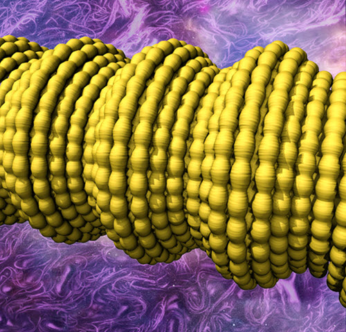
UT Dallas scientists have constructed novel fibers by wrapping sheets of tiny carbon nanotubes to form a sheath around a long rubber core. This illustration shows complex two-dimensional buckling, shown in yellow, of the carbon nanotube sheath/rubber-core fiber. The buckling results in a conductive fiber with super elasticity and novel electronic properties. (credit: UT Dallas Alan G. MacDiarmid Nanotech Institute)

An international research team based at The University of Texas at Dallas has made electrically conducting fibers that can be reversibly stretched to more than 14 times their initial length and whose electrical conductivity increases 200-fold when stretched.

The research team is using the new fibers to make artificial muscles, as well as capacitors with energy storage capacity that increases about tenfold when the fibers are stretched.

Fibers and cables derived from the invention might one day be used as interconnects for super-elastic electronic circuits, robots and exoskeletons having great reach, morphing aircraft, giant-range strain sensors, failure-free pacemaker leads, and super-stretchy charger cords for electronic devices.

Wrapping carbon nanotube sheets into fibers

In a study published in the July 24 issue of the journal Science, the scientists describe how they constructed the fibers by wrapping lighter-than-air, electrically conductive sheets of tiny carbon nanotubes to form a jelly-roll-like sheath around a long rubber core.

The new fibers differ from conventional materials in several ways. For example, when conventional fibers are stretched, the resulting increase in length and decrease in cross-sectional area restricts the flow of electrons through the material. But even a “giant” stretch of the new conducting sheath-core fibers causes little change in their electrical resistance, said Dr. Ray Baughman, senior author of the paper and director of the Alan G. MacDiarmid NanoTech Institute at UT Dallas.

One key to the performance of the new conducting elastic fibers is the introduction of buckling into the carbon nanotube sheets. Because the rubber core is stretched along its length as the sheets are being wrapped around it, when the wrapped rubber relaxes, the carbon nanofibers form a complex buckled structure, which allows for repeated stretching of the fiber.

“Think of the buckling that occurs when an accordion is compressed, which makes the inelastic material of the accordion stretchable,” said Baughman, the Robert A. Welch Distinguished Chair in Chemistry at UT Dallas.

“We make the inelastic carbon nanotube sheaths of our sheath-core fibers super stretchable by modulating large buckles with small buckles, so that the elongation of both buckle types can contribute to elasticity. These amazing fibers maintain the same electrical resistance, even when stretched by giant amounts, because electrons can travel over such a hierarchically buckled sheath as easily as they can traverse a straight sheath.”

Radical electronic and mechanical devices possible

By adding a thin overcoat of rubber to the sheath-core fibers and then another carbon nanotube sheath, the researchers made strain sensors and artificial muscles in which the buckled nanotube sheaths serve as electrodes and the thin rubber layer is a dielectric, resulting in a fiber capacitor. These fiber capacitors exhibited the unrivaled capacitance change of 860 percent when the fiber was stretched 950 percent.

Adding twist to these double-sheath fibers resulted in fast, electrically powered torsional — or rotating — artificial muscles that could be used to rotate mirrors in optical circuits or pump liquids in miniature devices used for chemical analysis. The conducting elastomers can be fabricated in diameters ranging from the very small — about 150 microns, or twice the width of a human hair — to much larger sizes, depending on the size of the rubber core. Individual small fibers also can be combined into large bundles and plied together like yarn or rope,” according to the researchers.

“This technology could be well-suited for rapid commercialization,” said Dr. Raquel Ovalle-Robles MS’06 PhD’08, an author on the paper and chief research and intellectual properties strategist at Lintec of America’s Nano-Science & Technology Center.

“The rubber cores used for these sheath-core fibers are inexpensive and readily available,” she said. “The only exotic component is the carbon nanotube aerogel sheet used for the fiber sheath.”


UT Dallas | UT Dallas Nanotech CNT rubber fiber

In this video, two lab demonstrations show the near invariance of resistance during the stretching of carbon-nanotube-sheathed rubber fibers.


UT Dallas Comets |UTD Nanotech pacemaker lead demo


Abstract of Hierarchically buckled sheath-core fibers for superelastic electronics, sensors, and muscles

Superelastic conducting fibers with improved properties and functionalities are needed for diverse applications. Here we report the fabrication of highly stretchable (up to 1320%) sheath-core conducting fibers created by wrapping carbon nanotube sheets oriented in the fiber direction on stretched rubber fiber cores. The resulting structure exhibited distinct short- and long-period sheath buckling that occurred reversibly out of phase in the axial and belt directions, enabling a resistance change of less than 5% for a 1000% stretch. By including other rubber and carbon nanotube sheath layers, we demonstrated strain sensors generating an 860% capacitance change and electrically powered torsional muscles operating reversibly by a coupled tension-to-torsion actuation mechanism. Using theory, we quantitatively explain the complementary effects of an increase in muscle length and a large positive Poisson’s ratio on torsional actuation and electronic properties.

Novel DNA origami structures

The versatility of the DNA origami 3D wireframe design technique created by Arizona State University Biodesign Institute researcher Hao Yan is demonstrated with the construction of this snub cube model, an Archimedean solid with 60 edges, 24 vertices and 38 faces including 6 squares and 32 equilateral triangles. (credit: TED-43/Wikimedia Commons)

Hao Yan, a researcher at Arizona State University’s Biodesign Institute, has extended DNA origami — which uses combinations of DNA base pairs to create new 2-D and 3-D nanoforms — into imaginative new forms that may one day lead to microelectronics and biomedical innovations.

“Earlier design methods [for DNA origami] used strategies including parallel arrangement of DNA helices to approximate arbitrary shapes, but precise fine-tuning of DNA wireframe architectures that connect vertices in 3D space has required a new approach,” says Yan, the Milton D. Glick Distinguished Chair of Chemistry and Biochemistry at ASU and directs Biodesign’s Center for Molecular Design and Biomimetics.

The new study describes wireframe structures of high complexity and programmability that are fabricated by precise control of branching and curvature, using novel organizational principles for the designs. (Wireframes are skeletal three-dimensional models represented purely through lines and vertices.)

The resulting nanoforms include symmetrical lattice arrays, quasicrystalline structures, curvilinear arrays, and a simple wire art sketch in the 100-nm scale, as well as 3-D objects including a snub cube with 60 edges and 24 vertices and a reconfigurable Archimedean solid that can be controlled to make the unfolding and refolding transitions between 3D and 2D.

The research appears in the advanced online edition of the journal Nature Nanotechnology.


DNA origami: how it works

In previous investigations, the Yan group created subtle architectural forms at an astonishingly minute scale, some measuring only tens of nanometers across — roughly the diameter of a virus particle. These nano-objects include spheres, spirals, flasksMöbius forms, and even an autonomous spider-like robot capable of following a prepared DNA track.
(credit: Fei Zhang et al./Nature Nanotechnology)

The technique of DNA origami capitalizes on the simple base-pairing properties of DNA, a molecule built from the four nucleotides Adenine (A), Thymine (T), Cytosine (C) and Guanine (G). The rules of the game are simple: A’s always pair with T’s and C’s with G’s. Using this abbreviated vocabulary, the myriad body plans of all living organisms are constructed; though duplicating even Nature’s simpler designs has required great ingenuity.

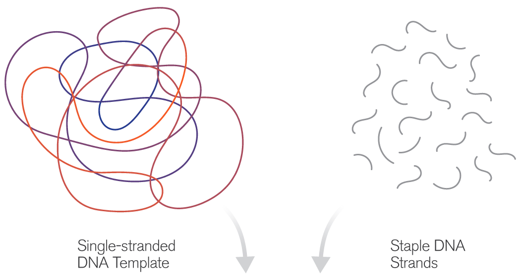
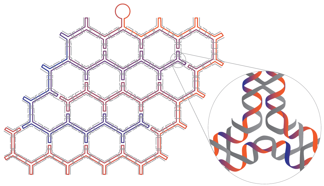
The basic idea of DNA origami is to use a length of single-stranded DNA as a scaffold for the desired shape. Base-pairing of complementary nucleotides causes the form to fold and self-assemble. The process is guided by the addition of shorter “staple strands,” which act to help fold the scaffold and to hold the resulting structure together. Various imaging technologies are used to observe the tiny structures, including fluorescence-, electron- and atomic force microscopy.


DNA origami originally produced nanoarchitectures of purely aesthetic interest, but refinements of the technique have opened the door to a new range of exciting applications including molecular cages for the encapsulation of molecules, enzyme immobilization and catalysis, chemical and biological sensing tools, drug delivery mechanisms, and molecular computing devices.

The technique described in the new study takes this approach a step further, allowing researchers to overcome local symmetry restrictions, creating wireframe architectures with higher order arbitrariness and complexity. Here, each line segment and vertex is individually designed and controlled. The number of arms emanating from each vertex may be varied from 2 to 10 and the precise angles between adjacent arms can be modified.
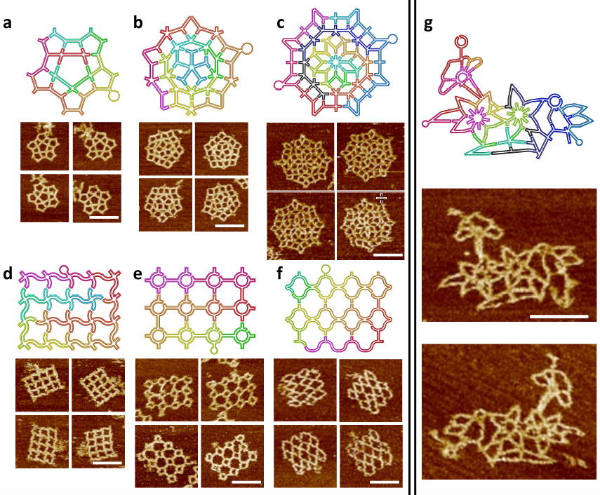
The scaffold folding path and representative AFM images for intricate 2D patterns. (a) A star-shape pattern without translational symmetry. (a) A Penrose tiling. (c) An 8-fold quasicrystalline pattern. (d-f) Three curved structures. (d) A waving grid. (e) A sphere array. (f) A fishnet. (g) A flower-and-bird pattern. All scale bars are 100 nm. Two scaffolds (one colorful and one black-blue) are used in c and g. (credit: Fei Zhang et al./Nature Nanotechnology)

In the current study, the method was first applied to symmetrical, regularly repeating polygonal designs, including hexagonal, square and triangular tiling geometries. Such common designs are known as tessellation patterns. A clever strategy involving a series of bridges and loops was used to properly route the scaffold strand, allowing it to pass through the entire structure, touching all lines of the wireframe once and only once. Staple strands were then applied to complete the designs.

In subsequent stages, the researchers created more complex wireframe structures, without the local translational symmetry found in the tessellation patterns. Three such patterns were made, including a star shape, a 5-fold Penrose tile and an 8-fold quasicrystalline pattern. (Quasicrystals are structures that are highly ordered but non-periodic. Such patterns can continuously fill available space, but are not translationally symmetric.)

Loop structures inserted into staple strands and unpaired nucleotides at the vertex points of the scaffold strands were also used, allowing researchers to perform precision modifications to the angles of junction arms.

The new design rules were next tested with the assembly of increasingly complex nanostructures, involving vertices ranging from 2 to 10 arms, with many different angles and curvatures involved, including a complex pattern of birds and flowers. The accuracy of the design was subsequently confirmed by AFM imaging, proving that the method could successfully yield highly sophisticated wireframe DNA nanostructures.

3D wire-frame Archimedean solid structures. (a) A 3D model of an Archimedean solid cuboctahedron with 12 vertices and 24 edges. Each vertex is a 4 Å~ 4 junction, and each edge is a 14-turn long double DNA duplex. (b) Left: Models showing possible conformations of the structure when deposited on mica surface; Right: the corresponding AFM images. (c) The reconfiguration between 3D and 2D can be realized by strand displacement by adding fuel and set strands. Top: reconfiguration schematics, Bottom: AFM images showing the transition. All scale bars in AFM images are 100 nm. (d) A 3D model of snub cube with 24 vertices and 60 edges. Each vertex is 5 Å~ 4 junction, and each edge is a 5-turn double DNA duplex. (e) Three views of the DNA snub cube from the design model (top of page). (credit: Fei Zhang et al./Nature Nanotechnology)

The method was then adapted to produce a number of 3D structures as well, including a cuboctahedron, and another Archimedian solid known as a snub cube — a structure with 60 edges, 24 vertices and 38 faces, including 6 squares and 32 equilateral triangles.

The authors stress that the new design innovations described can be used to compose and construct any imaginable wireframe nanostructure— a significant advancement for the burgeoning field.

On the horizon, nanoscale structures may one day be marshaled to hunt cancer cells in the body or act as robot assembly lines for the design of new drugs.


Abstract of Complex wireframe DNA origami nanostructures with multi-arm junction vertices

Structural DNA nanotechnology and the DNA origami technique, in particular, have provided a range of spatially addressable two- and three-dimensional nanostructures. These structures are, however, typically formed of tightly packed parallel helices. The development of wireframe structures should allow the creation of novel designs with unique functionalities, but engineering complex wireframe architectures with arbitrarily designed connections between selected vertices in three-dimensional space remains a challenge. Here, we report a design strategy for fabricating finite-size wireframe DNA nanostructures with high complexity and programmability. In our approach, the vertices are represented by n × 4 multi-arm junctions (n = 2–10) with controlled angles, and the lines are represented by antiparallel DNA crossover tiles of variable lengths. Scaffold strands are used to integrate the vertices and lines into fully assembled structures displaying intricate architectures. To demonstrate the versatility of the technique, a series of two-dimensional designs including quasi-crystalline patterns and curvilinear arrays or variable curvatures, and three-dimensional designs including a complex snub cube and a reconfigurable Archimedean solid were constructed.

An anti-inflammatory ‘smart drug’ that activates only in high-inflammation areas

Ben-Gurion University of the Negev (BGU) in Israel and University of Colorado researchers have developed a dynamic anti-inflammatory “smart” drug that can target specific sites in the body and could enhance the body’s natural ability to fight infection while reducing side effects.

This protein molecule, reported in the current issue of Journal of Immunology, has an exceptional property: when injected, it’s non-active. But upon reaching a local site with excessive inflammation, it becomes activated. Most other anti-inflammatory agents have broad effects in the body.

“This development is important because inhibition of inflammation in a non-specific manner reduces the natural ability to fight infections and is a common side effect of anti-inflammatory biologic therapeutics,” says Dr. Peleg Rider of BGU’s Department of Clinical Biochemistry and Pharmacology.

Using such a non-specific agent means any patient who suffers from local inflammation could be exposed to opportunistic infections at distant sites, such as lungs, which could risk tuberculosis, for example. This is especially a concern for immunosuppressed patients, as well as older patients and patients undergoing chemotherapy as part of an anti-cancer treatment course.

Mimicking a natural inflammatory process

The “chimeric IL-1Ra” protein combines an anti-inflammatory protein (right) with a peptide (left) that inactivates the IL-1Ra protein — except when the IL-1Ra portion encounters inflammatory enzymes, resulting in cleaving and releasing anti-inflammatory molecules (credit: Peleg Rider et al./Journal of Immunology)

“The beauty of this invention lies in the use of a known natural biological code,” Rider explains. “We mimicked a natural process that occurs during inflammation.”

Here’s how it works. The protein molecule (known as Chimeric IL-1Ra) is actually a chimera, combining two protein domains — both originating from the potent inflammatory IL-1 (interleukin family) protein (a group of 11 cytokines). (IL-1 normally plays a central role in regulating both immune and inflammatory responses to infections.)

The first part of this chimeric protein (IL-1beta) keeps a potent IL-1 natural infllammatory inhibitor (known as IL-1Ra) inactive. But when the IL-1Ra protein molecule encounters inflammatory enzymes, it springs into action, overriding IL-1beta, and is cleaved (split open), releasing powerful active molecules to reduce inflammation.

Rider, along with BGU’s Dr. Eli Lewis and Prof. Charles Dinarello of the University of Colorado, demonstrated their findings in a mouse model of local inflammation. They showed that leukocytes, which infiltrate inflammatory sites, indeed activate the chimeric protein, which in turn reduces local inflammation. The extent of activation of the protein correlated with the amount of inflammatory stimuli.

“Thus, a point that is highly relevant to clinical practice arises. Upon resolution of inflammation, the activation of the protein is also reduced and side effects are avoided,” Rider explains.

The new chimeric molecule was patented by BGN Technologies, BGU’s technology transfer company, and by the University of Colorado.

The research was supported by the Kamin program of Israel’s Ministry of Economy’s Chief Scientist’s Office.


Abstract of  IL-1 Receptor Antagonist Chimeric Protein: Context-Specific and Inflammation-Restricted Activation

Both IL-1α and IL-1β are highly inflammatory cytokines mediating a wide spectrum of diseases. A recombinant form of the naturally occurring IL-1R antagonist (IL-1Ra), which blocks IL-1R1, is broadly used to treat autoimmune and autoinflammatory diseases; however, blocking IL-1 increases the risk of infection. In this study, we describe the development of a novel form of recombinant IL-1Ra, termed chimeric IL-1Ra. This molecule is a fusion of the N-terminal peptide of IL-1β and IL-1Ra, resulting in inactive IL-1Ra. Because the IL-1β N-terminal peptide contains several protease sites clustered around the caspase-1 site, local proteases at sites of inflammation can cleave chimeric IL-1Ra and turn IL-1Ra active. We demonstrate that chimeric IL-1Ra reduces IL-1–mediated inflammation in vitro and in vivo. This unique approach limits IL-1 receptor blockade to sites of inflammation, while sparing a multitude of desired IL-1–related activities, including host defense against infections and IL-1–mediated repair.

NASA discovers first near-Earth-size planet in the habitable zone around a Sun-like star

This artist’s concept compares Earth (left) to the new planet, called Kepler-452b, which is about 60 percent larger in diameter (credits: NASA/JPL-Caltech/T. Pyle)

NASA’s Kepler mission has discovered the first near-Earth-size planet in the “habitable zone” around a Sun-like star. This discovery joins 11 other new small habitable zone candidate planets, marking another milestone in the journey to find another “Earth.”

The newly discovered Kepler-452b, located 1,400 light-years away in the constellation Cygnus, is the smallest planet to date discovered orbiting in the habitable zone — the area around a star where liquid water could pool on the surface of an orbiting planet — of a G2-type star, like our sun. The confirmation of Kepler-452b brings the total number of confirmed planets to 1,030.

“It’s awe-inspiring to consider that this planet has spent 6 billion years in the habitable zone of its star; longer than Earth,” said Jon Jenkins, Kepler data analysis lead at NASA’s Ames Research Center, who led the team that discovered Kepler-452b. ” That’s substantial opportunity for life to arise, should all the necessary ingredients and conditions for life exist on this planet.”

The CRISPR craze: genome editing technologies poised to revolutionize medicine and industry

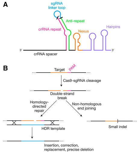
Genome editing by engineered Cas9 systems (credit: Mary Ann Liebert, Inc., publishers)

CRISPR/Cas systems for genome editing have revolutionized biological research over the past three years, and their ability to make targeted changes in DNA sequences in living cells with relative ease and affordability is now being applied to clinical medicine and will have a significant impact on advances in drug and other therapies, agriculture, and food products.

The power and promise of this innovation are presented in the Review article “The Bacterial Origins of the CRISPR Genome-Editing Revolution,” published in a special issue of Human Gene Therapy, a peer-reviewed journal from Mary Ann Liebert, Inc., publishers. The article is available open access until October 15, 2015 on the Human Gene Therapy website.

Erik Sontheimer, University of Massachusetts Medical School, Worcester, and Rodolphe Barrangou, North Carolina State University, Raleigh, describe the origins of this technology, which were derived from DNA sequences found in many bacteria known as clustered, regularly interspaced, short palindromic repeats (CRISPR) regions. These are part of bacteria’s protective immune system.

These regions have been developed into genome editing tools comprised of a “hardware” component (an RNA-guided DNA-targeting system that breaks a DNA strand at a specific site, with the help of the Cas protein), and a “software” component that can be programmed, and re-programmed, to repair or replace a faulty gene.

Impacts beyond academic research

“Although the CRISPR craze has yielded tremendous scientific progress and critical technological advances, it is important to keep in mind that the sgRNA–Cas9 technology is only 3 years old, and that notwithstanding current progress and momentum, we are yet to fully unleash the potential of these tools,” the authors note.

“Beyond academic research and the media, the most significant impact of CRISPR may well turn out to be in industry, with unprecedented levels of interest and investment from multiple distinct business segments, including pharmaceuticals and biotech, as well as covering the food supply chain from agriculture to livestock to other food products.”


Abstract of The Bacterial Origins of the CRISPR Genome-Editing Revolution

Like most of the tools that enable modern life science research, the recent genome-editing revolution has its biological roots in the world of bacteria and archaea. Clustered, regularly interspaced, short palindromic repeats (CRISPR) loci are found in the genomes of many bacteria and most archaea, and underlie an adaptive immune system that protects the host cell against invasive nucleic acids such as viral genomes. In recent years, engineered versions of these systems have enabled efficient DNA targeting in living cells from dozens of species (including humans and other eukaryotes), and the exploitation of the resulting endogenous DNA repair pathways has provided a route to fast, easy, and affordable genome editing. In only three years after RNA-guided DNA cleavage was first harnessed, the ability to edit genomes via simple, user-defined RNA sequences has already revolutionized nearly all areas of biological science. CRISPR-based technologies are now poised to similarly revolutionize many facets of clinical medicine, and even promise to advance the long-term goal of directly editing genomic sequences of patients with inherited disease. In this review, we describe the biological and mechanistic basis for these remarkable immune systems, and how their engineered derivatives are revolutionizing basic and clinical research.